-
Effect of high irradiance and short exposure curing time on the fracture toughness of bulk-fill resin-based composite: an in vitro study
-
Beatriz Ometto Sahadi, Tainah Oliveira Rifane, Carolina Bosso André, Vitaliano Gomes Araújo-Neto, Richard Thomas Bengt Price, Marcelo Giannini
-
Restor Dent Endod 2026;51(2):e23. Published online April 20, 2026
-
DOI: https://doi.org/10.5395/rde.2026.51.e23
-
-
 Abstract Abstract
 PDF PDF PubReader PubReader ePub ePub
- Objectives
This study aimed to determine the effect of high irradiance and short exposure time on the fracture toughness of bulk-fill resin-based composites (RBCs).
Methods
Three RBCs were tested: Tetric PowerFill (TPF; Ivoclar Vivadent), Opus Bulk Fill APS (OBF; FGM Dental Group), and Filtek One Bulk Fill (FOB; Solventum). Sixty single-edge-notched disc specimens were prepared using a fracture toughness mold. Each group consisted of 20 samples, divided into two subgroups (n = 10). The RBCs were lightcured either for 3 seconds in high-irradiance mode (‘3s cure’) or for the manufacturer-recommended times (TPF, 10 seconds; OBF, 30 seconds; FOB, 20 seconds) in ‘high power’ mode using the Bluephase PowerCure (Ivoclar Vivadent). The peak spectral wavelength was measured using a spectrophotometer. Specimens were tested on a universal testing machine, and data were analyzed by two-way analysis of variance and Bonferroni test (α = 0.05).
Results
Radiant exposure values (J/cm²) were 9.5 for the 3-second mode and 12.4, 24.8, and 37.1 for 10, 20, and 30 seconds (high power mode), respectively. FOB (4.22 and 3.79 MPa∙m0.5 for 20 and 3 seconds) had the highest mean fracture toughness, while OBF showed the lowest (2.01 and 2.10 MPa∙m0.5 for 30 and 3 seconds). TPF produced intermediate results (2.72 and 2.70 MPa∙m0.5 for 10 and 3 seconds). Exposure time did not affect TPF and OBF, while the 3-second exposure significantly reduced the fracture toughness for FOB.
Conclusions
The RBCs tested had different fracture toughness values regardless of exposure time. High irradiance and short exposure can reduce fracture toughness depending on the RBC tested.
-
Can different agents reduce the damage caused by bleaching gel to pulp tissue? A systematic review of basic research
-
Letícia Aparecida Silva Batista, Alexandre Henrique dos Reis-Prado, Hebertt Gonzaga dos Santos Chaves, Lara Cancella de Arantes, Luís Fernando Santos Alves Morgan, Carolina Bosso André, Thaís Yumi Suzuki, Francine Benetti
-
Restor Dent Endod 2023;48(4):e39. Published online November 6, 2023
-
DOI: https://doi.org/10.5395/rde.2023.48.e39
-
-
Abstract
PDF
 Supplementary MaterialPubReaderePub Supplementary MaterialPubReaderePub
- Objectives
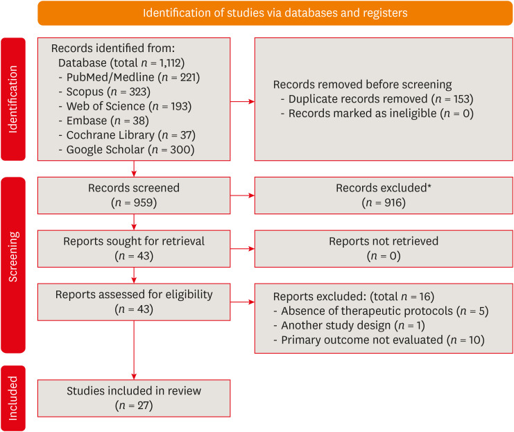
This study aimed to investigate the effectiveness of different topical/systemic agents in reducing the damage caused by bleaching gel to pulp tissue or cells. Materials and MethodsElectronic searches were performed in July 2023. In vivo and in vitro studies evaluating the effects of different topical or systemic agents on pulp inflammation or cytotoxicity after exposure to bleaching agents were included. The risk of bias was assessed. ResultsOut of 1,112 articles, 27 were included. Nine animal studies evaluated remineralizing/anti-inflammatories agents in rat molars subjected to bleaching with 35%–38% hydrogen peroxide (HP). Five of these studies demonstrated a significant reduction in inflammation caused by HP when combined with bioglass or MI Paste Plus (GC America), or following KF-desensitizing or Otosporin treatment (n = 3). However, orally administered drugs did not reduce pulp inflammation (n = 4). Cytotoxicity (n = 17) was primarily assessed using the 3-(4,5-dimethylthiazol-2-yl)-2,5-diphenyltetrazolium bromide assay on human dental pulp cells and mouse dental papilla Cell-23 cells. Certain substances, including sodium ascorbate, butein, manganese chloride, and peroxidase, were found to reduce cytotoxicity, particularly when applied prior to bleaching. The risk of bias was high in animal studies and low in laboratory studies. ConclusionsFew in vivo studies have evaluated agents to reduce the damage caused by bleaching gel to pulp tissue. Within the limitations of these studies, it was found that topical agents were effective in reducing pulp inflammation in animals and cytotoxicity. Further analyses with human pulp are required to substantiate these findings. Trial RegistrationPROSPERO Identifier: CRD42022337192
-
Citations
Citations to this article as recorded by  - 3D-Printed and Bioprinted Scaffolds in Regenerative Endodontics: A Systematic Review
Hebertt Gonzaga dos Santos Chaves, Diana B. Sequeira, Vilton Cardozo Moreira Dias, Alberto Cabrera-Fernández, João Peça, Francine Benetti, João Miguel Marques dos Santos
Applied Sciences.2026; 16(8): 3940. CrossRef - Clinical Study on the Efficacy of 35% Hydrogen Peroxide Gel According to Exposure Time (40 min vs. 20 min) by Spectrophotometry
Trinidad Rincón, Maria Portillo Muñoz, Maria Lobato, Ana María Martín Casado, Laryssa Mylenna Madruga Barbosa, Alessandro Loguercio, Cristina Gómez‐Polo
Journal of Esthetic and Restorative Dentistry.2026;[Epub] CrossRef - Clareamento dental e TikTok: avaliação da qualidade do conteúdo em mídia social
Rafaele T Costa, Thayna Silva do Carmo Tavares, André Walsh-Monteiro
Ciência ET Praxis.2025; 21(36): 111. CrossRef - Synthesis, characterization and evaluation of novel bleaching gels containing bioactive glass and nano-hydroxyapatite on hydrogen peroxide diffusion, bleaching efficacy and enamel protection
Adrieli Burey, Byron Carpio-Salvatierra, Michael Favoretto, María Luján Méndez Bauer, Viviane Hass, Alessandra Reis, Alessandro D. Loguercio, Paulo Vitor Farago
Clinical Oral Investigations.2025;[Epub] CrossRef - Cytotoxicity of Bleaching Products: A Systematic Review
Mireia Montaner, José Luis Sanz, Carmen Llena, María Melo, Clara Puig-Herreros, James Ghilotti
Applied Sciences.2024; 14(9): 3680. CrossRef
-
4,166
View
-
56
Download
-
4
Web of Science
-
5
Crossref
|