
Funded articles
- Page Path
- HOME > Browse articles > Funded articles
Research Articles
- Effect of high irradiance and short exposure curing time on the fracture toughness of bulk-fill resin-based composite: an in vitro study
- Beatriz Ometto Sahadi, Tainah Oliveira Rifane, Carolina Bosso André, Vitaliano Gomes Araújo-Neto, Richard Thomas Bengt Price, Marcelo Giannini
- Restor Dent Endod 2026;51(2):e23. Published online April 20, 2026
- DOI: https://doi.org/10.5395/rde.2026.51.e23
- Funded: National Council for Scientific and Technological Development, São Paulo Research Foundation, Coordenação de Aperfeiçoamento de Pessoal de Nível Superior, Dalhousie University, Faculty of Dentistry
-
 Abstract
 PDF
PDF PubReader
 ePub
ePub - Objectives
This study aimed to determine the effect of high irradiance and short exposure time on the fracture toughness of bulk-fill resin-based composites (RBCs).
Methods
Three RBCs were tested: Tetric PowerFill (TPF; Ivoclar Vivadent), Opus Bulk Fill APS (OBF; FGM Dental Group), and Filtek One Bulk Fill (FOB; Solventum). Sixty single-edge-notched disc specimens were prepared using a fracture toughness mold. Each group consisted of 20 samples, divided into two subgroups (n = 10). The RBCs were lightcured either for 3 seconds in high-irradiance mode (‘3s cure’) or for the manufacturer-recommended times (TPF, 10 seconds; OBF, 30 seconds; FOB, 20 seconds) in ‘high power’ mode using the Bluephase PowerCure (Ivoclar Vivadent). The peak spectral wavelength was measured using a spectrophotometer. Specimens were tested on a universal testing machine, and data were analyzed by two-way analysis of variance and Bonferroni test (α = 0.05).
Results
Radiant exposure values (J/cm²) were 9.5 for the 3-second mode and 12.4, 24.8, and 37.1 for 10, 20, and 30 seconds (high power mode), respectively. FOB (4.22 and 3.79 MPa∙m0.5 for 20 and 3 seconds) had the highest mean fracture toughness, while OBF showed the lowest (2.01 and 2.10 MPa∙m0.5 for 30 and 3 seconds). TPF produced intermediate results (2.72 and 2.70 MPa∙m0.5 for 10 and 3 seconds). Exposure time did not affect TPF and OBF, while the 3-second exposure significantly reduced the fracture toughness for FOB.
Conclusions
The RBCs tested had different fracture toughness values regardless of exposure time. High irradiance and short exposure can reduce fracture toughness depending on the RBC tested.
- 841 View
- 70 Download

- Magnitude of pulp space narrowing over time and contributing factors in teeth with vital pulp therapy: a retrospective cohort study
- Akarapong Boontankun, Papimon Chompu‑inwai, Chanika Manmontri, Nattakan Chaipattanawan, Areerat Nirunsittirat, Phichayut Phinyo, Trasapong Thaiupathump
- Restor Dent Endod 2026;51(2):e24. Published online May 13, 2026
- DOI: https://doi.org/10.5395/rde.2026.51.e24
- Funded: Chiang Mai University
-
Abstract
PDF
 Supplementary MaterialPubReaderePub
 - Objectives
This study aimed to compare the magnitude of pulp space narrowing over time—measured as the change in pulp/tooth proportion from baseline—between mandibular molars treated with different types of vital pulp therapy (VPT) and their contralateral sound molars (controls). This study also investigated factors influencing the magnitude of pulp space narrowing in molars that have undergone VPT.
Methods
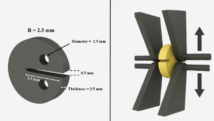
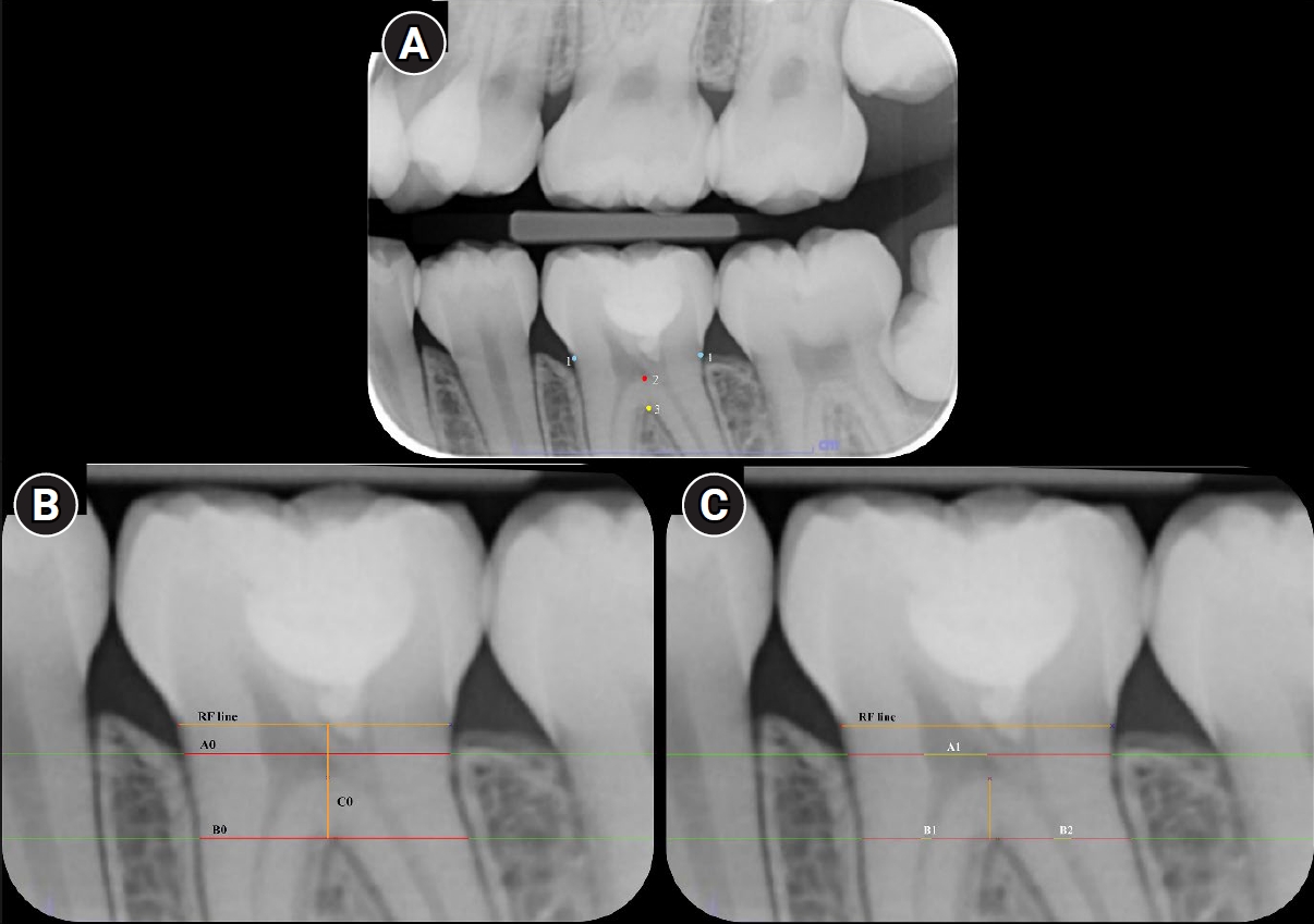
This retrospective cohort study involved the assessment of bitewing radiographs of VPT-treated molars and controls at baseline and follow-up. Using reference points and lines on the radiograph, pulp/tooth proportions were measured by examiners. The intraclass correlation coefficient (ICC) was used to report examiner reliability. The changes in pulp/tooth proportions from baselines were compared between subgroups using multilevel mixed effect linear regression and the Wald test.
Results
A total of 382 bitewing radiographs from 134 teeth were included. The follow-up period ranged from 6 to 84 months (mean, 27.12 ± 17.67 months). ICC values indicated good to excellent examiner reliability. Compared to the controls, changes in pulp/tooth proportion from baselines, indicating pulp space narrowing, were significantly greater in teeth with partial pulpotomy (at pulp chamber width) and coronal pulpotomy (at pulp canal width). Factors affecting the magnitude of pulp space narrowing included the more invasive type of VPT and the more severe preoperative diagnosis.
Conclusions
The magnitude of pulp space narrowing was greater in VPT-treated molars than in controls. The more invasive type of VPT and severe preoperative diagnosis were factors contributing to the magnitude of pulp space narrowing.
- 1,060 View
- 61 Download
- Interplay of hypoxia, angiogenesis, and macrophages in pulp and periapical lesions: an immunohistochemical cross-sectional study
- Puja Chatterjee, Mala Kamboj, Shweta Mittal, Anjali Narwal, Anju Devi
- Restor Dent Endod 2026;51(2):e22. Published online April 23, 2026
- DOI: https://doi.org/10.5395/rde.2026.51.e22
- Funded: Post Graduate Dissertation Support (PGDS-II) Scheme-II, Research Cell, Pt BD Sharma University of Health Sciences, Rohtak, Haryana-124001, India
-
Abstract
PDFPubReaderePub
- Objectives
This study evaluated and correlated the immune expression of hypoxia and angiogenesis with macrophages in periapical granuloma (PG), radicular cyst (RC), and healthy pulp (HP).
Methods
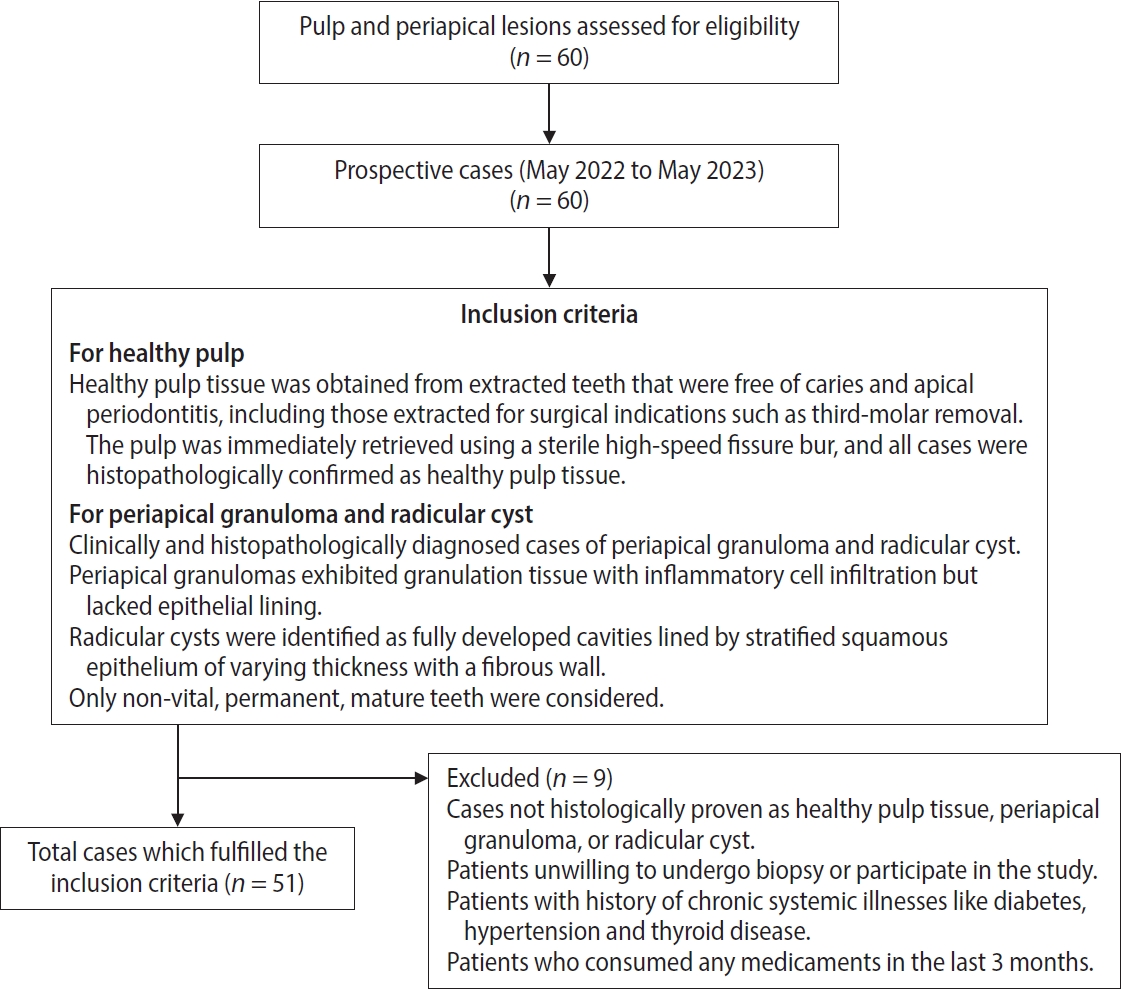
An observational study was performed on 51 tissue blocks equally divided among the groups, stained immunohistochemically for hypoxia-inducible factor (HIF)-1α, vascular endothelial growth factor (VEGF), and CD68, and the mean expression was calculated. Data were analyzed using Kruskal-Wallis, Mann-Whitney, Spearman correlation tests (p < 0.001), and multiple linear regression analysis (p ≤ 0.05).
Results
HIF-1α expression was highest in PG than RC and HP (p < 0.001). Significant differences were found between HP, PG, and RC (both p < 0.001). VEGF expression was highest in RC than in PG and HP (p < 0.001), with significant differences between HP and both PG and RC (p < 0.001); pairwise comparisons were significant between all groups (p < 0.001, p < 0.001, p = 0.018). Correlation analysis showed significant correlations between VEGF and CD68 in HP and PG (p = 0.007 and p = 0.028, respectively). Linear regression showed that study groups were significantly associated with mean scores of HIF-1α, VEGF, and CD68 (p = 0.002, p = 0.001, p < 0.001).
Conclusions
HIF-1α, VEGF, and CD68 showed increased expression in PGs and RCs, suggesting an association between hypoxic conditions, enhanced angiogenic activity, and macrophage presence within the periapical inflammatory microenvironment. Future studies exploring HIF-1α and VEGF inhibitors as potential treatment modalities for periapical lesions are warranted.
- 902 View
- 49 Download
- In vitro assessment of geometric characteristics in canal preparation using nickel-titanium files used for minimal invasiveness: an experimental study
- EunJin Jang, Hyeon-Cheol Kim, WooCheol Lee
- Restor Dent Endod 2026;51(2):e26. Published online May 13, 2026
- DOI: https://doi.org/10.5395/rde.2026.51.e26
- Funded: SNUDH Research Fund
-
Abstract
PDFPubReaderePub
- Objectives
This study aimed to assess geometric characteristics in canal preparation using nickel-titanium (NiTi) files used for minimal invasiveness.
Methods
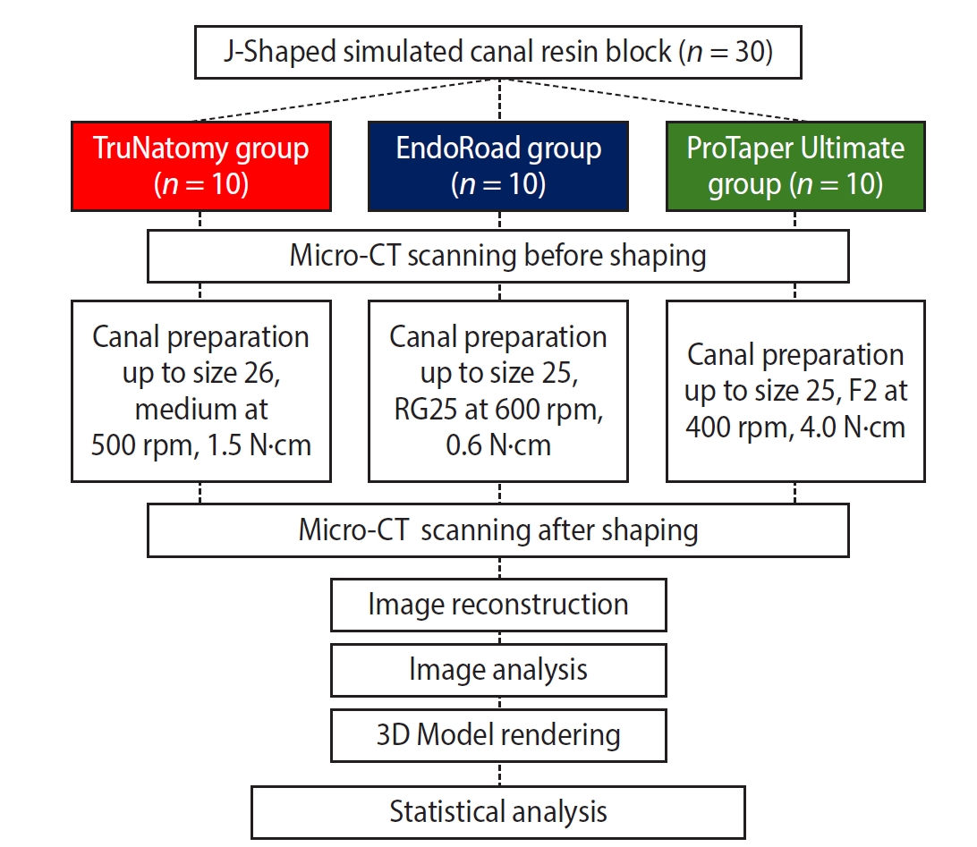
Thirty J-shaped simulated canals in resin blocks were instrumented with either TruNatomy (TR; Dentsply Sirona), EndoRoad (ER; Maruchi), or ProTaper Ultimate (PTU; Dentsply Sirona). The simulated canal blocks were scanned using microcomputed tomography before and after instrumentation. The scanned images were reconstructed, and the canal surface area was measured from 0.5 to 6.5 mm from the apex. Three-dimensional representative models of each group were rendered. The data were statistically analyzed using one-way analysis of variance and Kruskal-Wallis test at 95% significance level.
Results
TR showed a superior ability to maintain the canal’s center. TR demonstrated comparable apical preparation to PTU. ER showed a smaller and limited apical preparation than other systems, with a tendency for canal preparation toward the inner side of the curvature. PTU featured the largest prepared apical size among the file groups and tended to straighten the curvature by preparing the canal more towards the outward side. The surface area instrumented using each NiTi file showed statistically significant differences among the three groups at all levels except 0.5, 2.0, and 3.5 mm from the apex (p < 0.05). There was no statistically significant difference between TR and PTU at a level of 0.5 mm from the apex (p > 0.05).
Conclusions
While PTU is suitable for general canal preparation to facilitate irrigation and intracanal medication, TR and ER excel in preserving canal centering with minimal concern for canal transportation by minimally invasive preparation.
- 707 View
- 36 Download
- Influence of adjacent restorative material and distance on the accuracy of inlay cavity impressions with intraoral scanner: an in vitro study
- So-Yeon Lee, Sung-Ae Son, Jae-Hoon Kim, Deog-Gyu Seo, Jeong-Kil Park
- Restor Dent Endod 2026;51(1):e6. Published online January 23, 2026
- DOI: https://doi.org/10.5395/rde.2026.51.e6
- Funded: Pusan National University
-
Abstract
PDFPubReaderePub
- Objectives
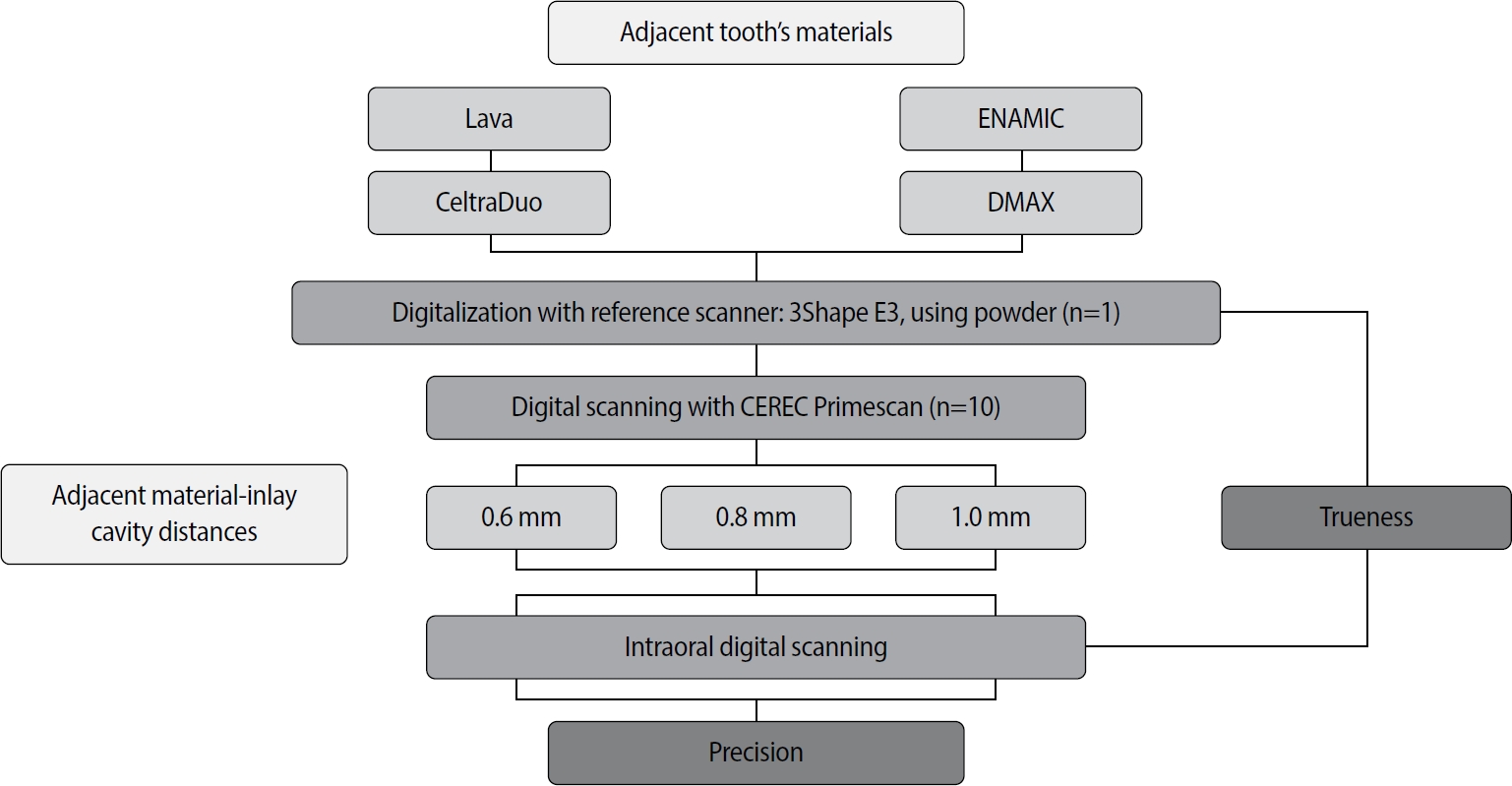
This study aimed to evaluate the influence of adjacent restorative material and interproximal distance on the accuracy of digital impressions of inlay cavities obtained using an intraoral scanner.
Methods
A disto-occlusal inlay cavity was prepared on a mandibular right first molar model, and digital scans were performed using a CEREC Primescan (Dentsply Sirona). The adjacent restorative materials used were Lava (3M ESPE), ENAMIC (VITA Zahnfabrik), Celtra Duo (Dentsply Sirona), and DMAX (DMAX), and the interproximal distances were set to 0.6 mm, 0.8 mm, and 1.0 mm. The obtained scan data were analyzed using GOM Inspect software (GOM GmbH).
Results
Trueness, maximum positive and negative deviations, and precision were significantly influenced by both the adjacent restorative material and the interproximal distance, while their interaction showed a significant effect only on precision. Celtra Duo demonstrated the highest trueness, with mean deviation values decreasing from 7.8 μm at a 0.6 mm interproximal distance to 7.3 μm at 1.0 mm. ENAMIC showed the best precision, presenting mean deviations of 2.6 μm at 0.6 mm, 2.9 μm at 0.8 mm, and 2.4 μm at 1.0 mm. A narrow interproximal distance of 0.6 mm resulted in lower trueness, measured at 8.3 μm, and the highest precision deviation of 3.4 μm. In contrast, an interproximal distance of 1.0 mm yielded improved scan accuracy, with increased trueness and reduced precision variation.
Conclusions
Digital impression accuracy of inlay cavities was influenced by adjacent restorative material and interproximal distance, suggesting clinical consideration is needed in CAD/CAM workflows to optimize restoration fit. -
Citations
Citations to this article as recorded by
- 3D-SCANNING IN PROSTHETIC DENTISTRY: ADVANTAGES, DISADVANTAGES, AND DEVELOPMENT PROSPECTS
V. S. Kuz, O. I. Teslenko, H. M. Kuz, H. M. Balia, Yu. S. Lunkova, O. V. Shemetov, I. M. Martynenko
Bulletin of Problems Biology and Medicine.2026; 1(1): 98. CrossRef
- 3D-SCANNING IN PROSTHETIC DENTISTRY: ADVANTAGES, DISADVANTAGES, AND DEVELOPMENT PROSPECTS
- 2,205 View
- 125 Download
- 1 Crossref
- Comparative evaluation of dentinal tubule occlusion by desensitizing agents after tooth bleaching: an in vitro study
- Dimitrios Dionysopoulos, Petros Mourouzis, Spyros Papageorgiou, Kosmas Tolidis
- Restor Dent Endod 2026;51(1):e8. Published online February 10, 2026
- DOI: https://doi.org/10.5395/rde.2026.51.e8
- Funded: Aristotle University of Thessaloniki
-
Abstract
PDFPubReaderePub
- Objectives
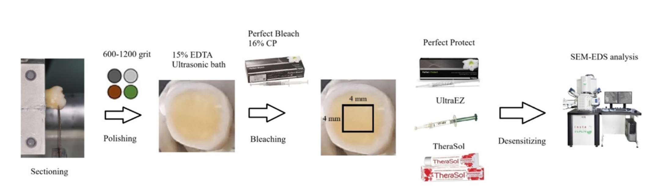
This study aimed to evaluate the efficacy of three commercially available desensitizing agents in occluding dentinal tubules, which may help reduce tooth sensitivity following a bleaching treatment.
Methods
Twenty healthy human third molars were utilized in this investigation. The samples were prepared by transversely sectioning 2.5 mm of the crowns to expose the dentin. They were initially treated with 15% ethylenediaminetetraacetic acid gel for 4 minutes, followed by application of Perfect Bleach (VOCO GmbH) bleaching agent (16% carbamide peroxide) for 2 hours. The samples were randomly allocated into four groups (n = 5), each receiving one of the following treatments: group 1: No treatment (control), group 2: treated with UltraEZ (Ultradent Products Inc.,), containing potassium nitrate and sodium fluoride, group 3: treated with Perfect Protect (VOCO GmbH), also containing potassium nitrate and sodium fluoride and group 4: treated with TheraSol Whitening & Sensitive (ABC Kinitron IKE), containing strontium acetate and sodium monofluorophosphate. Subsequently, the specimens were examined using scanning electron microscopy (SEM) and energy-dispersive X-ray spectroscopy to evaluate dentin tubule occlusion.
Results
SEM observations showed no occlusion of dentin tubules in the control group, whereas groups 2 to 4 exhibited significant occlusion. The most effective treatment was Perfect Protect (p < 0.05), while UltraEZ and TheraSol Whitening & Sensitive demonstrated similar effectiveness, with no statistically significant difference between them (p > 0.05).
Conclusions
The tested desensitizing agents effectively occluded dentin tubules to a considerable extent. Differences in their effectiveness were attributed to variations in their formulations.
- 2,201 View
- 189 Download
- Cone-beam computed tomography analysis of maxillary premolar canal anatomy: Ahmed’s versus Vertucci’s classifications in a Jordanian cohort
- Raidan Ba-Hattab, Muna M. Shaweesh, Nessrin A. Taha, Elham S. Abu Alhaija
- Restor Dent Endod 2026;51(1):e11. Published online February 26, 2026
- DOI: https://doi.org/10.5395/rde.2026.51.e11
- Funded: Jordan University of Science and Technology
-
Abstract
PDFPubReaderePub
- Objectives
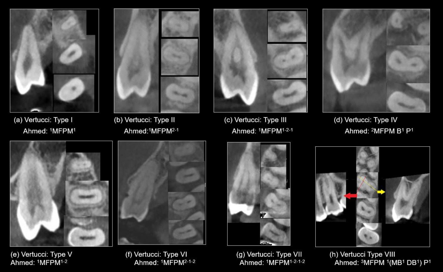
This study analyzed the root and canal configurations of maxillary premolars in a Jordanian subpopulation using cone-beam computed tomography (CBCT) and classified them based on Vertucci’s and Ahmed’s systems.
Methods
Two hundred CBCT scans of 800 maxillary premolars were retrospectively assessed for root morphology, canal configurations, and root canal divergence and merging. Data was statistically analyzed.
Results
The study included 70 males and 130 females. Most right and left maxillary first premolars (RFPM, LFPM) had two roots (59.0% and 58.5%), with a significant association between sex and root number for RFPM and LFPM (p < 0.05). In contrast, the right and left maxillary second premolars (RSPM, LSPM) mostly had a single root (87.5% and 88.5%), with no association with sex. Vertucci’s classification showed type IV as the predominant configuration in first premolars (RFPM, 65.0% and LFPM, 67.0%) and type I in second premolars (RSPM, 44.0% and LSPM, 49.0%). A significant sex association was found only with RSPM. Ahmed’s classification revealed that maxillary premolar with two separated roots and two separated canals (2MP B1 P1) was mostly found in first premolars (RFPM, 58.0% and LFPM, 56.0%), and maxillary premolar with one root and one canal (1MP1) in second premolars (RSPM, 44.0% and LSPM, 49.0%), with a significant sex association for RSPM and LSPM (p < 0.05). Age had no impact, and symmetry was observed between the right and left sides. Three-rooted premolars were identified in four cases. Almost all of Vertucci’s types and numerous codes from Ahmed’s classification were documented.
Conclusions
CBCT revealed diverse anatomical variations in the Jordanian subpopulation, with Ahmed’s classification providing more detailed canal configurations than Vertucci’s, uncovering previously overlooked variations.
- 1,084 View
- 56 Download
- The influence of bioactive glass (BGS-7) on enamel remineralization: an in vitro study
- Chaeyoung Lee, Eunseon Jeong, Kun-Hwa Sung, Su-Jung Park, Yoorina Choi
- Restor Dent Endod 2025;50(4):e33. Published online October 15, 2025
- DOI: https://doi.org/10.5395/rde.2025.50.e33
- Funded: National Research Foundation of Korea
-
Abstract
PDFPubReaderePub
- Objectives
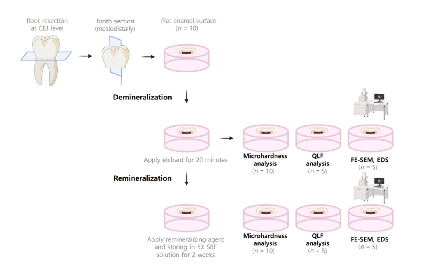
The aim of this study was to compare the remineralizing capacity of bioactive glass (BGS-7, CGBIO) with other agents.
Methods
Twenty caries-free third molars were sectioned and demineralized. Specimens were divided into four groups: (1) control, (2) Clinpro XT varnish (Solventum), (3) 1.23% acidulated phosphate fluoride gel, and (4) a new type of CaO-SiO2-P2O5-B2O3 system of bioactive glass ceramics (BGS-7). Agents were applied and stored in simulated body fluid at 37℃ for 2 weeks. Microhardness was measured using the Vickers hardness testing method. Five specimens per group were analyzed using quantitative light-induced fluorescence (QLF) to assess mineral loss. Field-emission scanning electron microscopy (FE-SEM) and energy-dispersive X-ray spectroscopy (EDS) were used to examine the surface morphology and elemental composition. Data were analyzed using paired t-test and one-way analysis of variance (p < 0.05).
Results
BGS-7 showed the highest microhardness values and the greatest recovery in QLF analysis (p < 0.05). FE-SEM revealed granular precipitates on demineralized enamel in the BGS-7 group. EDS confirmed the presence of newly formed silicon and fluoride layers.
Conclusions
BGS-7 demonstrated superior remineralization capacity compared to other agents, suggesting its potential as an effective remineralizing material. -
Citations
Citations to this article as recorded by- Bacterial ghosts (BGs): A promising approach as candidate vaccine
Helal F. Hetta, Ibraheem M. Mwafey, Noura H. Abd Ellah, Fawaz E. Alanazi, Yasmin N. Ramadan
World Journal of Microbiology and Biotechnology.2026;[Epub] CrossRef
- Bacterial ghosts (BGs): A promising approach as candidate vaccine
- 2,800 View
- 255 Download
- 1 Web of Science
- 1 Crossref
- Comparison of remineralization in caries-affected dentin using calcium silicate, glass ionomer cement, and resin-modified glass ionomer cement: an in vitro study
- Kwanchanok Youcharoen, Onwara Akkaratham, Papichaya Intajak, Pipop Saikaew, Sirichan Chiaraputt
- Restor Dent Endod 2025;50(4):e37. Published online November 14, 2025
- DOI: https://doi.org/10.5395/rde.2025.50.e37
- Funded: Srinakharinwirot University
-
Abstract
PDFPubReaderePub
- Objectives
This study evaluated the ability of calcium silicate cement (CSC) as a remineralizing agent compared with conventional glass ionomer cement (GIC) and resin-modified GIC (RMGIC) to remineralize artificial caries-affected dentin.
Methods
Twenty-five class V cavities were prepared on extracted human third molars. Twenty teeth underwent artificial caries induction. The remaining five teeth with sound dentin serve as the positive control. The twenty demineralized teeth were subdivided into four groups (n = 5): carious dentin without restoration (negative control [NC]), carious dentin restored with CSC (Biodentine, Septodont), carious dentin restored with GI (Fuji IX, GC Corporation), and carious dentin restored with RMGIC (Fuji II LC, GC Corporation). Following restoration, the specimens were stored in artificial saliva for 7 days. The elastic modulus was evaluated by a nanoindentation test. The mineral composition was analyzed by scanning electron microscopy-energy-dispersive X-ray spectroscopy (SEM-EDX), and the mineral composition at the dentin-material interface.
Results
CSC had a higher modulus of elasticity compared to GI, RMGI, and NC groups (p < 0.05). Higher calcium and phosphorus content was observed under CSC restorations, as indicated by SEM-EDX examination, which may lead to better remineralization.
Conclusions
Compared to GI and RMGI, CSC showed the best remineralization and mechanical reinforcement in caries-affected dentin, indicating CSC for use in minimally invasive restorative dentistry. -
Citations
Citations to this article as recorded by- Comparison of mineral precipitation, elemental release, pH change and cytotoxicity of calcium-silicate cements and an experimental resin-modified glass ionomer cement containing bioactive glass
Wisitsin Potiprapanpong , Parichart Naruphontjirakul, Naruporn Monmaturapoj, Siriporn Tanodekaew, Somruethai Channasanon, Arnit Toneluck, Somying Patntirapong, Piyaphong Panpisut
Biomaterial Investigations in Dentistry.2026; 13: 337. CrossRef
- Comparison of mineral precipitation, elemental release, pH change and cytotoxicity of calcium-silicate cements and an experimental resin-modified glass ionomer cement containing bioactive glass
- 3,039 View
- 272 Download
- 1 Crossref
- How protocol, posts, and experience affect fracture detection in multi-rooted teeth using cone-beam computed tomography: an ex vivo experimental study
- Gleica Dal’ Ongaro Savegnago, Gabriela Marzullo de Abreu, Carolina Baumgratz Spiger, Lucas Machado Maracci, Wislem Miranda de Mello, Gabriela Salatino Liedke
- Restor Dent Endod 2025;50(3):e23. Published online July 24, 2025
- DOI: https://doi.org/10.5395/rde.2025.50.e23
- Funded: Coordination for Funding and Support of Tertiary Education
-
Abstract
PDFPubReaderePub
- Objectives
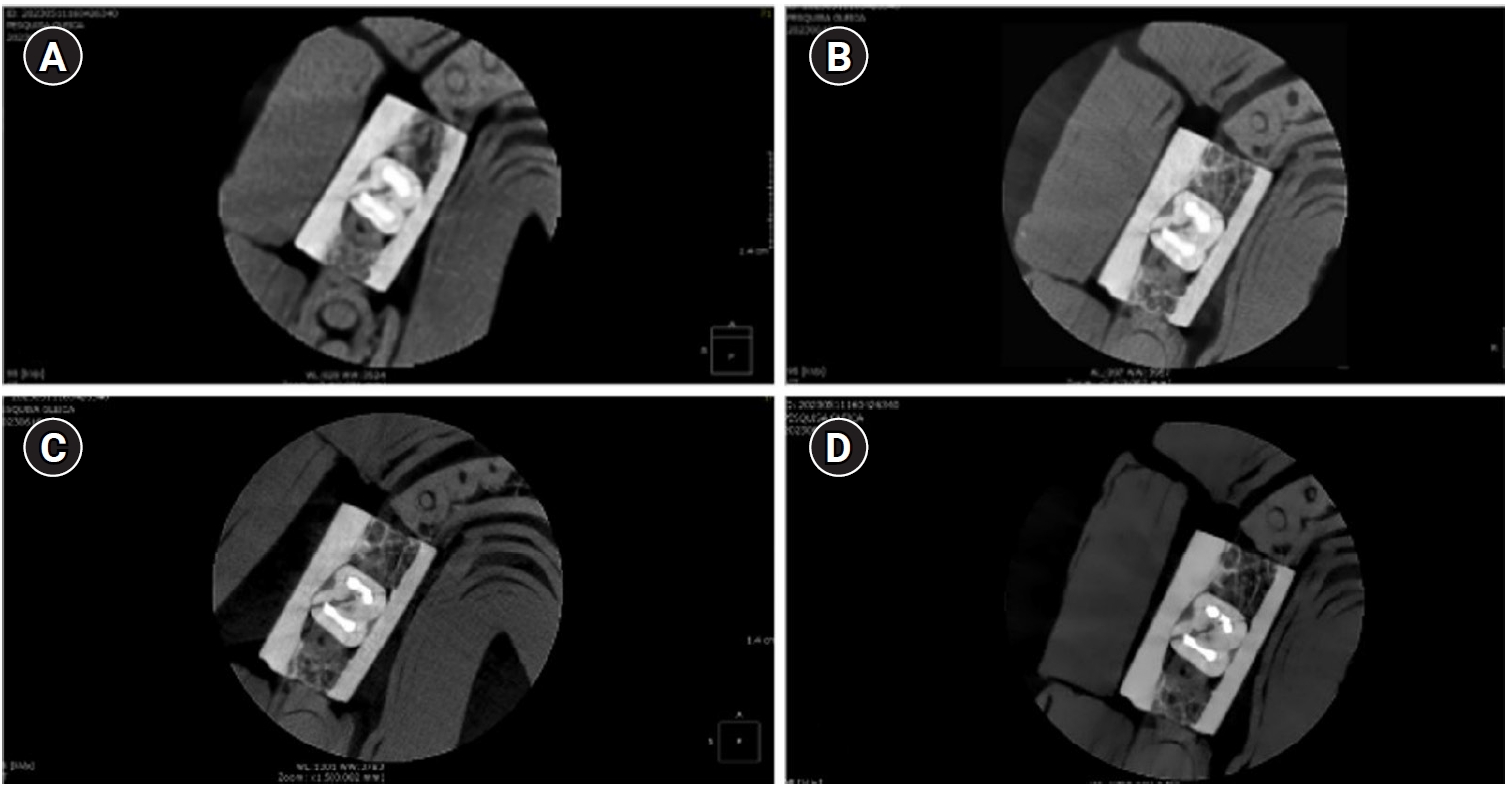
This study aimed to evaluate the influence of cone-beam computed tomography (CBCT) acquisition protocol, the presence of intraradicular metal post, and examiner experience on the detection of complete root fractures in multi-rooted teeth.
Methods
Twenty human molar teeth filled with gutta-percha were placed into artificial alveoli created in bovine ribs. The sample was divided into two groups based on the presence or absence of intraradicular posts in the distal roots. CBCT scans were obtained using four acquisition protocols with varying voxel sizes (0.28, 0.2, 0.125, and 0.80 mm). Following the creation of controlled fractures using a chisel and hammer, CBCT imaging was repeated, resulting in 160 images. Five examiners assessed the images using OnDemand software (KaVo Dental GmbH). Sensitivity, specificity, and accuracy were calculated for each examiner, CBCT protocol, and post-condition. Statistical comparisons were performed using Cochran’s Q test and McNemar test, and a significance level of 5%.
Results
In teeth without metallic posts, sensitivity, specificity, and accuracy values exceeded 0.70, 0.70, and 0.80, respectively. However, the presence of metallic posts significantly reduced diagnostic performance, particularly in low-resolution protocols evaluated by less-experienced examiners.
Conclusions
CBCT acquisition protocols should be selected based on the presence of metallic posts to optimize root fracture detection in multi-rooted teeth. Examiner experience also plays a critical role in diagnostic accuracy.
- 3,212 View
- 110 Download
- Calcium silicate-based sealers remnants in isthmuses of mesial roots of mandibular molars: an in vitro evaluation
- David Saldanha de Brito Alencar, Ana Cristina Padilha Janini, Lauter Eston Pelepenko, Brenda Fornazaro Moraes, Francisco Haiter Neto, Marco Antonio Hungaro Duarte, Marina Angélica Marciano
- Restor Dent Endod 2025;50(3):e25. Published online July 15, 2025
- DOI: https://doi.org/10.5395/rde.2025.50.e25
- Funded: Coordination for the Improvement of Higher Education Personnel, São Paulo Research Foundation
-
Abstract
PDFPubReaderePub
- Objectives
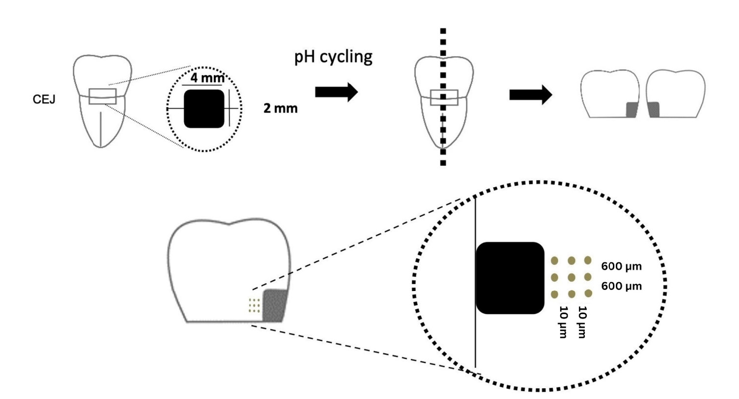
Endodontic retreatment aims to address treatment failure through the removal of root canal filling materials. This in vitro study evaluated the presence of filling material remnants in the mesial root canals, specifically focusing on the isthmuses, of mandibular molars after retreatment.
Methods
One hundred extracted mandibular molar mesial roots with isthmuses were prepared with an R25 file, obturated with one of five calcium silicate-based sealers (BioRoot RCS [Septodont], MTApex [Ultradent Products Inc.], EndoSequence BC Sealer HiFlow [Brasseler USA], Bio-C Sealer [Angelus]) or an epoxy resin-based sealer (AH Plus Jet [Dentsply Maillefer]), all stained with rhodamine B, and stored at 37ºC for 30 days to allow for setting. Retreatment was subsequently performed using R40 and XP-endo Finisher R instruments (FKG Dentaire) with 2.5% sodium hypochlorite irrigation. The presence of remaining filling material was then assessed using confocal microscopy, and setting times were tested per ISO 6876:2012.
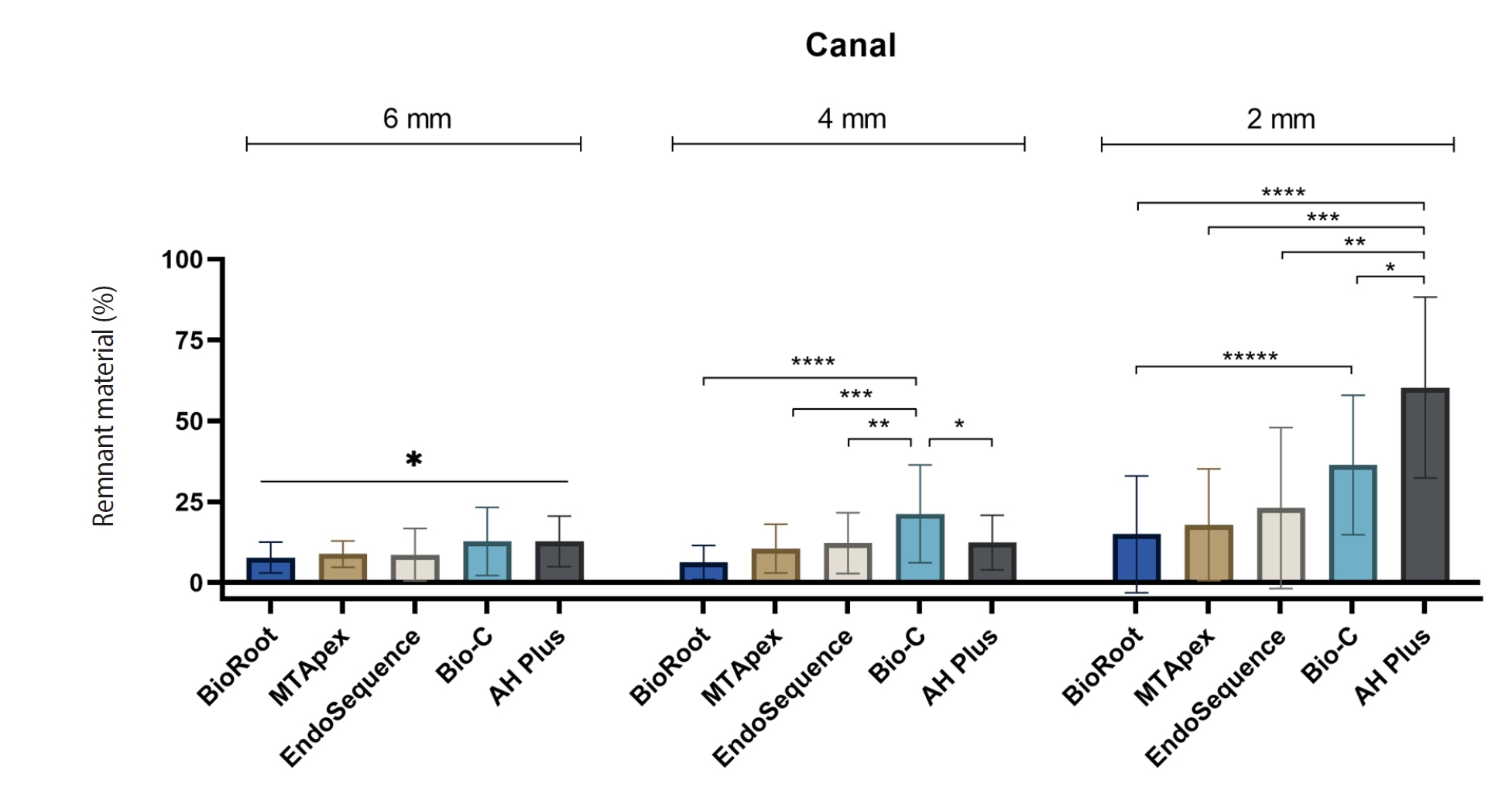
Results
AH Plus Jet showed the most remnants at 2 mm and the longest retreatment time. Calcium silicate-based sealers exhibited prolonged setting times under dry conditions, with EndoSequence BC Sealer HiFlow showing a particularly extended setting period.
Conclusions
Despite retreatment, residues remained in all canals and isthmus regions, particularly Bio-C Sealer and AH Plus Jet in apical areas, emphasizing the difficulty of complete removal and the persistence of filling material. -
Citations
Citations to this article as recorded by- Bonding effects of mechanical removal of bioceramic sealer residues using glycine or glass microparticles abrasion
Jesus Aranda, Julia de Freitas Ceccato, Eduardo Fernández Godoy, João Felipe Besegato, Joissi Ferrari Zaniboni, Regina Guenka Palma-Dibb, Milton Carlos Kuga
International Journal of Adhesion and Adhesives.2026; 148: 104289. CrossRef
- Bonding effects of mechanical removal of bioceramic sealer residues using glycine or glass microparticles abrasion
- 2,866 View
- 130 Download
- 1 Web of Science
- 1 Crossref
- Does the use of different root canal sealers and adhesive resin cements impact the bond strength of glass fiber posts?
- Ália Regina Neves de Paula Porto, Rudá França Moreira, Felipe Gonçalves Belladonna, Victor Talarico Leal Vieira, Emmanuel João Nogueira Leal da Silva
- Restor Dent Endod 2025;50(3):e29. Published online August 29, 2025
- DOI: https://doi.org/10.5395/rde.2025.50.e29
- Funded: CAPES (n.001), CNPq and FAPERJ
-
Abstract
PDFPubReaderePub
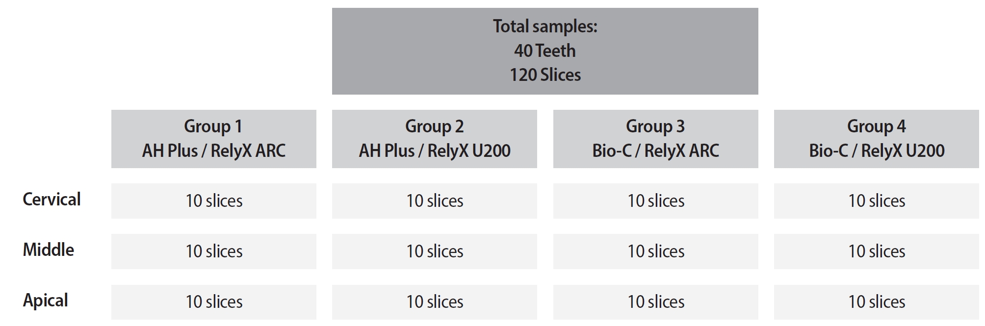
- Objectives
This study aimed to assess the influence of two endodontic sealers on the bond strength of glass fiber posts using conventional and self-adhesive resin cement through a push-out test. Methods: Forty central human incisors were randomly divided into four groups (n = 10) based on sealer (epoxy resin- based or calcium silicate-based) and cement (conventional and self-adhesive resin) types: AH Plus (Dentsply De- Trey)/RelyX ARC (3M ESPE), AH Plus/RelyX U200 (3M ESPE), Bio-C Sealer (Angelus)/RelyX ARC, and Bio-C Sealer/RelyX U200. After canal filling and post cementation, roots were sectioned to obtain one specimen per root third. A pushout test and failure pattern assessment were conducted, with bond strength analyzed using the one-way analysis of variance and Tukey test. Results: AH Plus/RelyX ARC showed the highest bond strength values, with a significant difference in the middle third. The most common failure was mixed (55%), while adhesive failures made up 45%, with 23.5% at the cement/post interface and 21.5% at the cement/dentin interface. Conclusions: AH Plus/RelyX ARC provided the highest bond strength values for glass fiber posts to dentin. -
Citations
Citations to this article as recorded by- Effect of Endodontic Sealers on the Bond Strength of Glass Fibre Posts: A Systematic Review
Thiago Bessa Marconato Antunes, Juliana D. Bronzato, Vanessa Gallego Arias Pecorari, Jennifer Santos Pereira, Talita Tartari, Adriana de Jesus Soares, Brenda P. F. A. Gomes, Marina Angélica Marciano
Australian Endodontic Journal.2026;[Epub] CrossRef
- Effect of Endodontic Sealers on the Bond Strength of Glass Fibre Posts: A Systematic Review
- 2,962 View
- 176 Download
- 1 Web of Science
- 1 Crossref
- Bibliometric analysis of the GentleWave system: trends, collaborations, and research gaps
- Raimundo Sales de Oliveira Neto, Thais de Moraes Souza, João Vitor Oliveira de Amorim, Thaine Oliveira Lima, Guilherme Ferreira da Silva, Rodrigo Ricci Vivan, Murilo Priori Alcalde, Marco Antonio Hungaro Duarte
- Restor Dent Endod 2025;50(2):e17. Published online May 12, 2025
- DOI: https://doi.org/10.5395/rde.2025.50.e17
- Funded: São Paulo Research Foundation
-
Abstract
PDFSupplementary MaterialPubReaderePub
- Objectives
The study aimed to conduct a bibliometric analysis of the GentleWave system (Sonendo, Inc.).
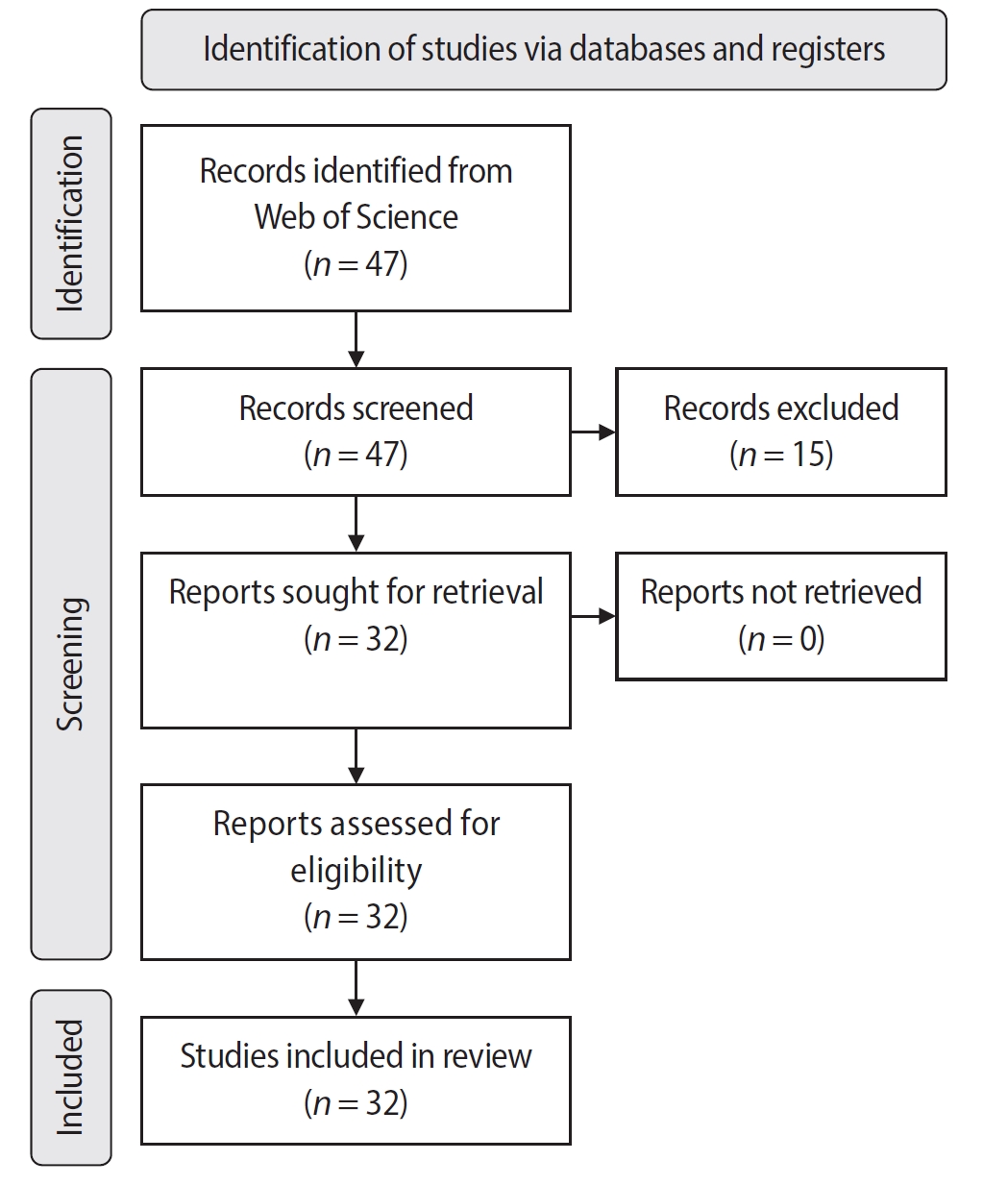
Methods
An electronic search was conducted in June 2024 using the Web of Science Collection database. Two reviewers independently screened publications, extracting data on authorship, publication details, study design, and citation metrics. Statistical analyses were performed in R to assess variable correlations, while the VOSviewer (Visualization of Similarities Viewer) software was used to map author and keyword networks.
Results
The search yielded 47 records, with 32 studies included. Publications spanned 2014 to 2024. The Journal of Endodontics published the highest number of studies (n = 15), and the International Endodontic Journal had the highest impact factor (5.4). The University of British Columbia and Sonendo, Inc. were the most frequent affiliations. Among the 32 articles, 28 were in vitro studies, primarily focusing on microbiology (n = 9). A total of 95 authors were identified, with Haapasalo and Shen being the most cited (n = 229). The articles accumulated 495 citations, demonstrating a strong positive correlation between the number of studies and citation counts (r = 0.98).
Conclusions
The analysis highlights a predominance of in vitro studies. Geographic concentration in the United States and Canada limits diversity, while the strong correlation between study numbers and citations suggests that increased publication volume enhances visibility. -
Citations
Citations to this article as recorded by- Three-year Outcomes of Conventional Versus Minimally Invasive Endodontic Treatment Protocols: A Retrospective Study
Kiavash Hossini, He Liu, Ya Shen, Jolanta Aleksejuniene, Fahda Algahtani, Ahmed Hieawy
Journal of Endodontics.2026; 52(4): 558. CrossRef
- Three-year Outcomes of Conventional Versus Minimally Invasive Endodontic Treatment Protocols: A Retrospective Study
- 4,859 View
- 104 Download
- 1 Web of Science
- 1 Crossref
- Cleaning protocols to enhance bond strength of fiberglass posts on root canals filled with bioceramic sealer: an in vitro comparative study
- Thiago Bessa Marconato Antunes, Juliana Delatorre Bronzato, Joice Graciani, Ana Cristina Padilha Janini, Rocharles Cavalcante Fontenele, Francisco Haiter Neto, Brenda Paula Figueiredo de Almeida Gomes, Marina Angélica Marciano da Silva
- Restor Dent Endod 2025;50(2):e20. Published online May 21, 2025
- DOI: https://doi.org/10.5395/rde.2025.50.e20
- Funded: State of São Paulo Research Foundation, State of São Paulo Research Foundation, Improvement of Higher Education Personnel
-
Abstract
PDFPubReaderePub
- Objectives
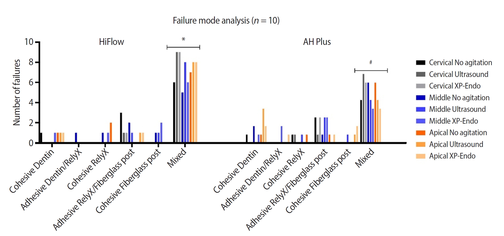
This study aimed to evaluate whether the agitation protocols using ultrasonic inserts or the XP-endo Finisher R file improved the removal of two different endodontic sealer remnants and the bond strength of fiberglass posts to dentin.
Methods
Seventy-two human teeth were selected. The canals were prepared with Reciproc 50 and Easy ProDesign 30/.10 and root filled according to the endodontic sealer groups: AH Plus or EndoSequence BC Sealer HiFlow. The samples were kept at 37ºC and 95% humidity for 28 days. During the post space preparation, the obturation was removed with Largo burs, and the groups were divided according to the irrigant agitation protocols (n = 12): no agitation, agitation with R1-Clearsonic associated with E1-Irrisonic ultrasonic inserts, or agitation with XP-endo Finisher R file. The fiberglass posts were cemented with RelyX ARC. The roots were sectioned into slices and submitted to the push-out test. Micro-computed tomography analysis was used to check the effectiveness of irrigating solution agitation in the elimination of remnants.
Results
The cleaning protocols with agitation were more effective in increasing the bond strength of posts to dentin for both sealer groups compared to non-agitation (p < 0.05). There was no difference between the same cleaning protocols for the different sealers. Among the different thirds, there was no statistical difference for the same sealer in the different cleaning protocols (p > 0.05).
Conclusions
Both agitation protocols effectively clean root-filled canals sealed with resin-based and calcium silicate-based sealers during fiberglass post space preparation. These protocols result in improved bond strength compared to non-agitation methods. -
Citations
Citations to this article as recorded by- Cleaning efficacy and bond interaction of glycine-based air polishing and glass microparticles abrasion on dentin impregnated with premixed bioceramic sealer
Ândresson Aurélio Fernandes Martins, Maria Carolina Sidonio Alves, Bruno Martins Maciel, José Rodolfo Estruc Verbicário, João Felipe Besegato, Wilfredo Gustavo Escalante-Otárola, Milton Carlos Kuga
International Journal of Adhesion and Adhesives.2026; 147: 104277. CrossRef - Effect of Endodontic Sealers on the Bond Strength of Glass Fibre Posts: A Systematic Review
Thiago Bessa Marconato Antunes, Juliana D. Bronzato, Vanessa Gallego Arias Pecorari, Jennifer Santos Pereira, Talita Tartari, Adriana de Jesus Soares, Brenda P. F. A. Gomes, Marina Angélica Marciano
Australian Endodontic Journal.2026;[Epub] CrossRef - Effects of Calcium Silicate-Based Sealer Residues on Adhesive Bonding to Coronal Dentin: An in Vitro Study
Mariana Bena Gelio, Thais Piragine Leandrin, Ana Lídia Pinheiro Silva Sato, Milton Carlos Kuga, Joissi Ferrari Zaniboni
Journal of Dental Research, Dental Clinics, Dental Prospects.2026; 20(1): 25. CrossRef
- Cleaning efficacy and bond interaction of glycine-based air polishing and glass microparticles abrasion on dentin impregnated with premixed bioceramic sealer
- 5,380 View
- 249 Download
- 2 Web of Science
- 3 Crossref
- Stress distribution of restorations in external cervical root resorption under occlusal and traumatic loads: a finite element analysis
- Padmapriya Ramanujam, Paul Kevin Abishek Karthikeyan, Vignesh Srinivasan, Selvakarthikeyan Ulaganathan, Velmurugan Natanasabapathy, Nandini Suresh
- Restor Dent Endod 2025;50(2):e21. Published online May 21, 2025
- DOI: https://doi.org/10.5395/rde.2025.50.e21
- Funded: Meenakshi Academy of Higher Education and Research
-
Abstract
PDFPubReaderePub
- Objectives
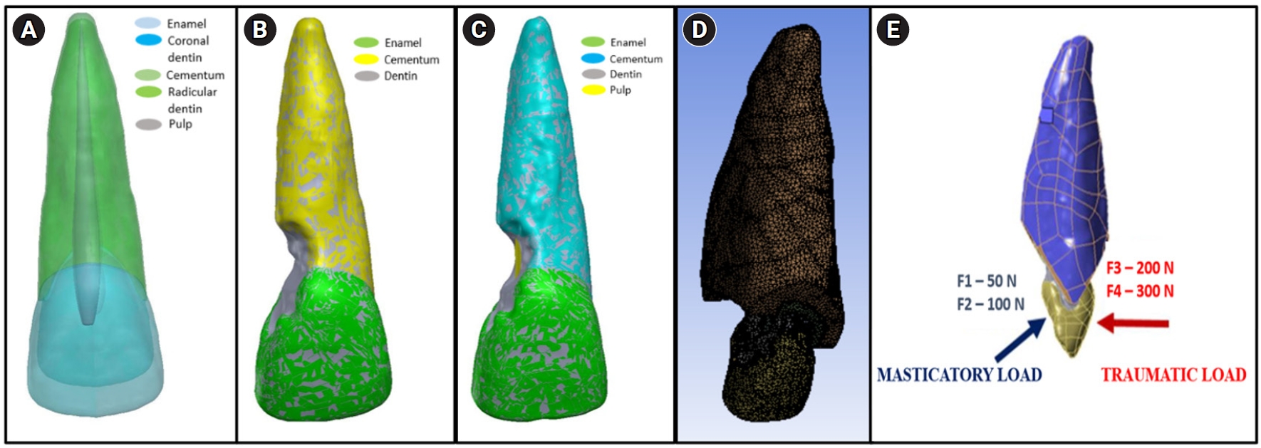
This study analyzed the stress distribution in a maxillary central incisor with external cervical resorptive defect restored with different restorative materials under normal masticatory and traumatic loading conditions using finite element analysis.
Methods
Cone-beam computed tomography of an extracted intact incisor and created resorptive models (Patel’s 3D classification-2Bd and 2Bp) in the maxillary central incisor was performed for finite element models. The 2Bd models were restored either with glass ionomer cement (GIC)/Biodentine (Septodont) or a combination of both with composite resin. 2Bp models were restored externally with a combination technique and internally with root canal treatment. The other model was external restoration with GIC and internal with fiber post. Two masticatory loads were applied at 45˚ to the palatal aspect, and two traumatic loads were applied at 90˚ to the buccal aspect. Maximum von Mises stresses were calculated, and stress distribution patterns were studied.
Results
In 2Bd models, all restorative strategies decreased stress considerably, similar to the control model under all loads. In 2Bp models, the dentin component showed maximum stress at the deepest portion of the resorptive defect, which transfers into the adjacent pulp space. In 2Bp defects, a multilayered restoration externally and root canal treatment internally provides better stress distribution compared to the placement of a fiber post.
Conclusions
Increase in load, proportionally increased von Mises stress, despite the direction or angulation of the load. Multilayered restoration is preferred for 2Bd defects, and using an internal approach of root canal treatment is suggested to restore 2Bp defects.
- 2,952 View
- 167 Download
- Effect of quality of radiographs taken during root canal treatment on technical quality of root canal fillings and endodontic outcome
- Jia Min Ng, Yan Yee Lee, Prashanti Chippagiri, Elaheh Ahanin, Abhishek Parolia
- Restor Dent Endod 2025;50(1):e3. Published online January 7, 2025
- DOI: https://doi.org/10.5395/rde.2025.50.e3
- Funded: International Medical University, Malaysia
-
Abstract
PDFPubReaderePub
- Objectives
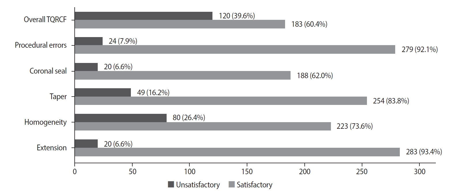
This study evaluated the number and quality of working length (WL) and master cone (MC) radiographs taken during root canal treatment by dental undergraduates, and their associations with the technical quality of root canal fillings (TQRCF) and endodontic outcomes (EO).
Methods
A retrospective evaluation of radiographs from 303 root canal-treated teeth in 231 patients was conducted, with 72 patients attending recall visits to assess EO. The chi-square and one-way analysis of variance tests were performed.
Results
A total of 505 WL and 557 MC radiographs were reviewed, with 72.9% and 75% deemed satisfactory, respectively. Satisfactory TQRCF was achieved in 60.4% of cases. Significant associations were found between the extension of the file in WL and gutta-percha in MC radiographs and TQRCF (p = 0.000). Misinterpretation of these radiographs resulted in poor TQRCF. Furthermore, 64.2% of teeth had satisfactory EO. A significant relationship was noted between the quality of MC radiographs and both TQRCF (p = 0.043) and EO (p = 0.003).
Conclusions
Unsatisfactory MC radiographs were linked to poor TQRCF and unfavorable EO. Regular radiographic training is recommended to enhance EO. -
Citations
Citations to this article as recorded by- Radiographic evaluation of root canal fillings: can undergraduate dental students perform it?
Emine Odabaşı Tezer, Fadi Nahas, Alhabab Shbitah, İrem Dilara Kılıç, Ahmet Bölük, Meltem Öztan
BMC Medical Education.2026;[Epub] CrossRef - Assessment of radiographic errors and repetition rates in undergraduate endodontic education: a retrospective clinical study
Marwa Ameen, Abdul Rahman Saleh, Dunia Alhadi, Manal Almaslamani
The Saudi Dental Journal.2025;[Epub] CrossRef - Application of Periapical Radiography in Root Canal Treatment: A Literature Review
Jennifer Lois Violita Malau, Keizha Allysia Nabila, Widiani Harrista, Regina Amara Ginting, Tassa Kusuma Arya Putri, Jatu Rachel Keshena
Acta Odontologica Indonesia.2025; 1(2): 49. CrossRef
- Radiographic evaluation of root canal fillings: can undergraduate dental students perform it?
- 14,234 View
- 305 Download
- 2 Web of Science
- 3 Crossref
- Surface properties and susceptibility to staining of a resin composite after brushing with different whitening toothpastes
- Aline da Silva Barros, Carolina Meneghin Barbosa, Renata Siqueira Scatolin, Waldemir Francisco Vieira Junior, Laura Nobre Ferraz
- Restor Dent Endod 2025;50(1):e6. Published online February 26, 2025
- DOI: https://doi.org/10.5395/rde.2025.50.e6
- Funded: Research Support of the State of São Paulo
-
Abstract
PDFPubReaderePub
- Objectives
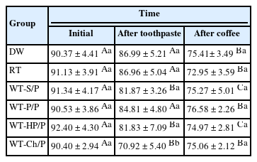
This study investigated the effects of different whitening toothpaste (WT) on the surface properties and staining susceptibility of a resin composite.
Methods
Cylindrical samples were prepared with a micro-hybrid resin composite and were randomized into groups according to the toothpaste (n = 12): distilled water (DW), regular toothpaste (RT), WT with silica + pyrophosphate (WT-S/P), WT with pentaphosphate and pyrophosphate (WT-P/P), WT with hydrogen peroxide and pyrophosphate (WT-HP/P) and WT with charcoal and pyrophosphate (WT-Ch/P). The samples were brushed for 825 cycles in an automatic brushing machine, simulating 30 days of brushing. After that, an immersion in coffee (10 mL/sample) was performed for 30 minutes for 30 days. The analyses of color, surface microhardness (SMH), and surface roughness (Ra) were performed at the initial time, after brushing with toothpaste and after immersion in coffee. The ΔL*, Δa*, Δb*, ΔEab, Δand E00 values were calculated comparing after toothpaste with initial time and after coffee with after toothpaste. Data were analyzed using a mixed linear model for repeated measures (SMH), Kruskal-Wallis, Dunn, Friedman, and Nemenyi tests, with α = 0.05.
Results
For ΔL*, the WT-Ch/P group had the lowest values and differed from the other groups comparing the after toothpaste with the initial time interval (p < 0.001). The WT-Ch/P group had the lowest SMH values in after-toothpaste time (p < 0.001). In after-toothpaste time and after coffee time, the WT-S/P group had the highest Ra values and differed from the groups except the WT-Ch/P group (p < 0.001).
Conclusions
The toothpaste composition affects the surface characteristics and susceptibility to staining of the resin composite. The charcoal-based toothpaste had the worst performance for the color analyses and SMH. -
Citations
Citations to this article as recorded by- Color Stability and Surface Roughness of Esthetic Resin Composites Following Simulated Toothbrushing with Whitening Toothpastes
Ecehan Kaplan , Ayşe Dündar, Çağatay Barutçugil
Odovtos - International Journal of Dental Sciences.2026; 1(1): 595. CrossRef - Influence of commercial mouth rinses with different formulations on enamel properties during at-home bleaching
Thalita Novello Coelho, Ana Júlia Gil, Marcos Roberto Lima Benati, Carolina Meneghin Barbosa, Tatiane Cristina Dotta, Waldemir Francisco Vieira-Junior, Renata Siqueira Scatolin, Laura Nobre Ferraz
Odontology.2026;[Epub] CrossRef
- Color Stability and Surface Roughness of Esthetic Resin Composites Following Simulated Toothbrushing with Whitening Toothpastes
- 6,696 View
- 199 Download
- 1 Web of Science
- 2 Crossref
- Shaping ability and cyclic fatigue resistance between Genius ProFlex, ZenFlex, and TruNatomy rotary systems: an experimental study
- Raimundo Sales de Oliveira Neto, Murilo Priori Alcalde, Pedro Cesar Gomes Titato, Pedro Henrique Souza Calefi, Carlos Alberto Spironelli Ramos, Guilherme Ferreira da Silva, Rodrigo Ricci Vivan, Marco Antonio Hungaro Duarte
- Restor Dent Endod 2025;50(1):e9. Published online February 13, 2025
- DOI: https://doi.org/10.5395/rde.2025.50.e9
- Funded: São Paulo State Research Foundation, National Research Council
-
Abstract
PDFPubReaderePub
- Objectives
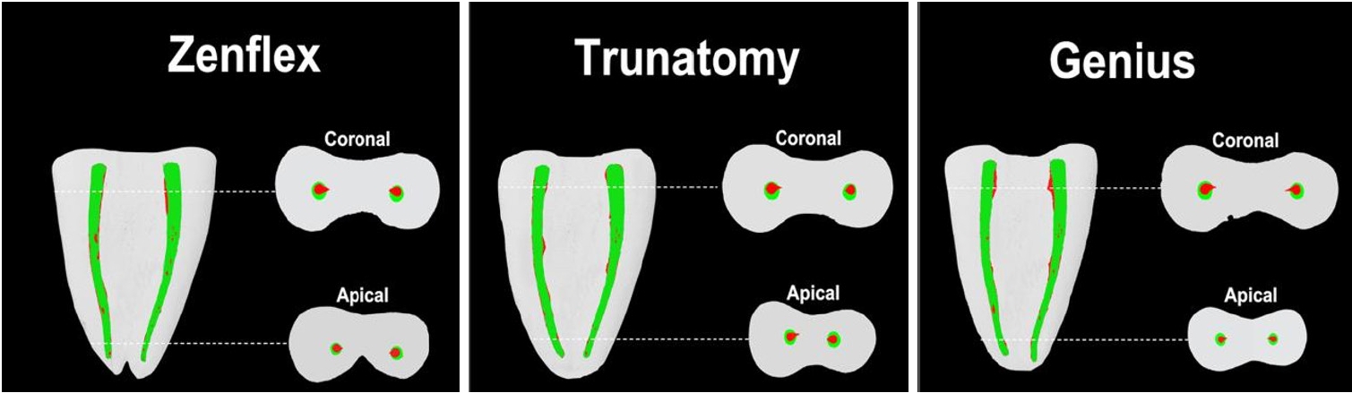
The aim of this study was to investigate the efficacy of three newly introduced rotary endodontic systems: Genius ProFlex (Medidenta), TruNatomy (Dentsply Maillefer), and ZenFlex (Kerr).
Methods
Forty-five mandibular molars with root canal curvatures <5° were utilized. Micro-computed tomography scans were performed pre- and post-preparation to assess apical transportation, centralization, percentage of dentin wear, and canal volume alterations. Eight instruments of each diameter underwent cyclic fatigue testing.
Results
The percentage of dentin wear on mesial and distal walls showed no significant differences among ZenFlex, TruNatomy, and Genius ProFlex at 1, 2, 3, and 4 mm from the apical foramen and root canal orifice (p > 0.05). Centering ability varied in the mesiolingual canal (p < 0.05). No notable differences were observed in transportation (p > 0.05). Genius ProFlex demonstrated lower volumetric changes (p < 0.05). There were significant differences in cyclic fatigue, with higher values for Genius ProFlex and lower values for TruNatomy (p < 0.05).
Conclusions
The three nickel-titanium rotary instruments are safe and efficient for root canal preparation, with Genius ProFlex exhibiting superior cyclic fatigue resistance. -
Citations
Citations to this article as recorded by- Influence of kinematic motion and instrumentation strategy on apical debris extrusion during root canal preparation: An in vitro study
Amira Alghazaly, Jumanah Aljohani, Khadijah Mohabat, Rafah Ghous
Journal of Conservative Dentistry and Endodontics.2026; 29(7): 741. CrossRef - Comparison of Shaping Ability and Apical Debris Extrusion Using 4 Different Nickel–Titanium Single‐File Systems
Siyu Li, Mengzhen Tang, Xi Wang, Jian Yang, Hyun-Do Jung
International Journal of Biomaterials.2025;[Epub] CrossRef
- Influence of kinematic motion and instrumentation strategy on apical debris extrusion during root canal preparation: An in vitro study
- 4,296 View
- 190 Download
- 1 Web of Science
- 2 Crossref
- Success rates comparison of endodontic microsurgery and single implants with comprehensive and explicit criteria: a systematic review and meta-analysis
- Min Jung Ko, Ju Hyun Park, Na Rae Lee, Joon-Ho Yoon, Young-Taek Kim, Sin-Yeon Cho
- Restor Dent Endod 2025;50(1):e8. Published online February 19, 2025
- DOI: https://doi.org/10.5395/rde.2025.50.e8
- Funded: National Evidence-based Healthcare Collaborating Agency
-
Abstract
PDFSupplementary MaterialPubReaderePub
- Objectives
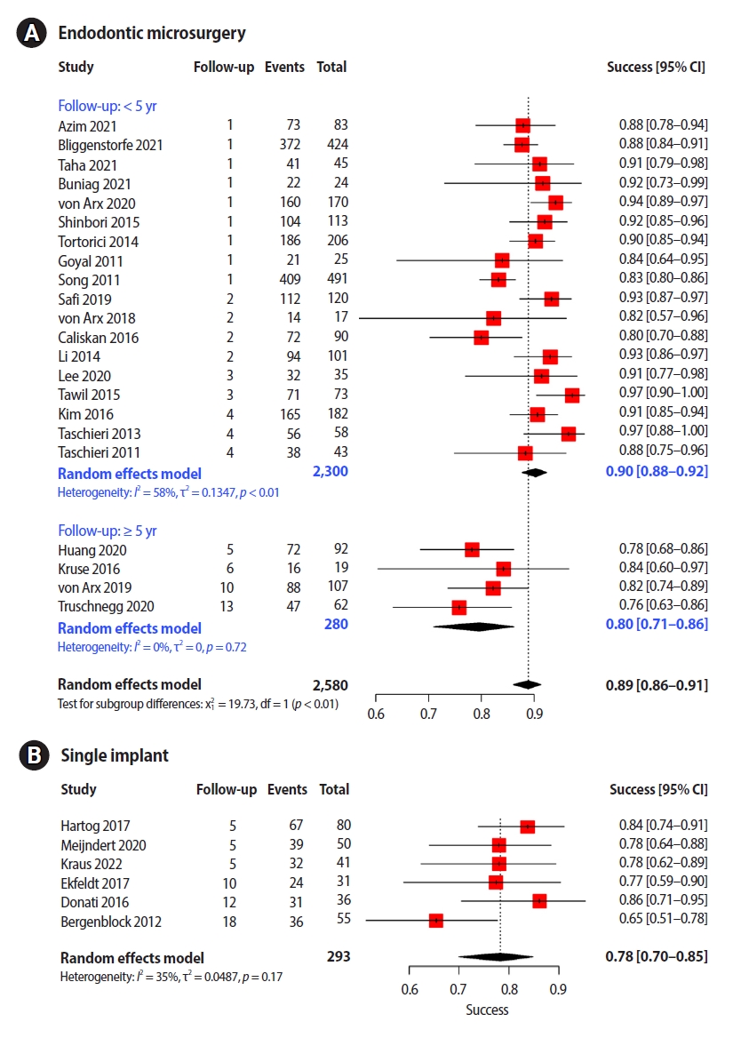
While the success criteria of endodontic microsurgery (EMS) have been consistently defined and widely accepted, the success criteria of dental implants are outdated and focus only on the implant fixture and surrounding bone. This study aimed to evaluate the outcomes of EMS and single implants (SIs) with explicit criteria.
Methods
We searched for articles published from January 2010 to February 2022 and discussed them and consulted with a clinical advisory committee composed of four dental specialists and one epidemiologist during article selection and data extraction.
Results
Twenty-two EMS studies and six SI studies were included in the meta-analysis. Teeth treated using EMS had a pooled success rate of 89% (90% at <5-year follow-up and 80% at ≥5-year follow-up) and the pooled success rate of SI was 78%.
Conclusions
The success rates of the two procedures with similar follow-up periods were comparable. Subgroup analysis found no other variable that significantly influenced study heterogeneity. Considering the treatment sequence and the similar success rates, it would be advantageous to consider EMS, rather than implants, first in a situation where both procedures are applicable. -
Citations
Citations to this article as recorded by- Surgical Management of a Separated Instrument and Radicular Cyst: A Nine-Month Cone Beam Computed Tomography (CBCT) Follow-up
Dipti Chauhan, Hemant Yadav, Anshu Minocha, Vishal Sharma
Cureus.2025;[Epub] CrossRef - Cost-effectiveness of Endodontic Retreatment vs Implants: A 5-year Retrospective Analysis in India
Pramod Kumar, Himanshu Sharma
Journal of Clinical Insights and Research in Dentistry.2025; 1(3): 121. CrossRef
- Surgical Management of a Separated Instrument and Radicular Cyst: A Nine-Month Cone Beam Computed Tomography (CBCT) Follow-up
- 10,924 View
- 227 Download
- 2 Crossref
 First
First Prev
Prev

