
Articles
- Page Path
- HOME > Restor Dent Endod > Volume 50(3); 2025 > Article
- Research Article Calcium silicate-based sealers remnants in isthmuses of mesial roots of mandibular molars: an in vitro evaluation
-
David Saldanha de Brito Alencar1
 , Ana Cristina Padilha Janini1, Lauter Eston Pelepenko1, Brenda Fornazaro Moraes1, Francisco Haiter Neto2, Marco Antonio Hungaro Duarte3, Marina Angélica Marciano1,*
 -
Restor Dent Endod 2025;50(3):e25.
DOI: https://doi.org/10.5395/rde.2025.50.e25
Published online: July 15, 2025
1Department of Restorative Dentistry, Endodontics, Piracicaba Dental School, State University of Campinas, Piracicaba, Brazil
2Department of Oral Diagnosis, Radiology, Piracicaba Dental School, State University of Campinas, Piracicaba, Brazil
3Department of Restorative Dentistry, Dental Materials and Endodontics, Bauru Dental School, University of São Paulo, Bauru, Brazil
- *Correspondence to Marina Angélica Marciano, DDS, MSc Department of Restorative Dentistry, Dental Materials and Endodontics, Bauru Dental School, University of São Paulo, Piracicaba, Avenida Limeira, 901, Piracicaba, SP 13414-903, Brazil Email: marinama@unicamp.br
• Received: March 21, 2025 • Revised: April 12, 2025 • Accepted: April 15, 2025
© 2025 The Korean Academy of Conservative Dentistry
This is an Open-Access article distributed under the terms of the Creative Commons Attribution Non-Commercial License (http://creativecommons.org/licenses/by-nc/4.0) which permits unrestricted non-commercial use, distribution, and reproduction in any medium, provided the original work is properly cited.
Abstract
-
Objectives Endodontic retreatment aims to address treatment failure through the removal of root canal filling materials. This in vitro study evaluated the presence of filling material remnants in the mesial root canals, specifically focusing on the isthmuses, of mandibular molars after retreatment.
-
Methods One hundred extracted mandibular molar mesial roots with isthmuses were prepared with an R25 file, obturated with one of five calcium silicate-based sealers (BioRoot RCS [Septodont], MTApex [Ultradent Products Inc.], EndoSequence BC Sealer HiFlow [Brasseler USA], Bio-C Sealer [Angelus]) or an epoxy resin-based sealer (AH Plus Jet [Dentsply Maillefer]), all stained with rhodamine B, and stored at 37ºC for 30 days to allow for setting. Retreatment was subsequently performed using R40 and XP-endo Finisher R instruments (FKG Dentaire) with 2.5% sodium hypochlorite irrigation. The presence of remaining filling material was then assessed using confocal microscopy, and setting times were tested per ISO 6876:2012.
-
Results AH Plus Jet showed the most remnants at 2 mm and the longest retreatment time. Calcium silicate-based sealers exhibited prolonged setting times under dry conditions, with EndoSequence BC Sealer HiFlow showing a particularly extended setting period.
-
Conclusions Despite retreatment, residues remained in all canals and isthmus regions, particularly Bio-C Sealer and AH Plus Jet in apical areas, emphasizing the difficulty of complete removal and the persistence of filling material.
INTRODUCTION
Endodontic treatment failure can arise from the persistence of residual bacteria following chemical-mechanical preparation, sustained by fluid percolation from the periapex, leading to intra- and extraradicular infections and subsequent periradicular lesions [1]. These microorganisms can resist to the endodontic treatment and remain in regions of anatomical complexities of root canals such as isthmuses, C-shaped canals, lateral canals, ramifications, and dentinal tubules [2,3].
Endodontic sealers, depending on their material characteristic, can influence the patency regaining, tooth retreatability, retreatment time, and the presence of obturation material remnants within the root canals; several previous studies evaluated the effectiveness of removal investigating different types of sealers [4]. Among these endodontic materials, the most evaluated were those based on epoxy resin, calcium silicate-based, and zinc oxide and eugenol-based, but there are some controversial results between these studies. The comparison between the removal of epoxy resin and calcium silicate-based sealers showed similar results between these materials [5–7], while other studies mentioned a greater amount of residue for epoxy resin-based sealers [8,9], whereas there are reports of greater amounts for calcium silicate-based sealers [10]. Besides, there are reports that the calcium silicate-based sealers require a long time for retreatment [8]. There is evidence that the composition of the material is a variable that interferes with root canal retreatment, considering these discrepancies reported.
Complementary endodontic instruments can potentially aid during retreatment in the removal of obturation material. XP-endo Finisher R rotary file (FKG Dentaire, La Chaux-de-Fonds, Switzerland) was designed to be used in regions of anatomical complexities such as root canal isthmuses along with instrumentation during root canal retreatment [11,12]; besides, there are reports of its use in oval root canals [13]. Previous reports also associated the use of the XP-endo Finisher in retreated root canals filled with calcium silicate-based sealers as an improvement for the obturation material removal [5,9]. Therefore, the investigation regarding the use of an additional cleaning instrument is crucial, especially considering anatomical irregularities [4].
The correlation between filling material removal during retreatment and sealer type warrants investigation. Sealers vary in composition, with some requiring mixing to start setting immediately, while “ready-to-use” sealers rely on assumed moisture contact within root canal dentin to initiate setting and hydration [14].
The hydration process of ‘ready-to-use’ sealers in root canals is still not well elucidated, and whether the total setting reaction takes place, because these materials are moisture-dependent. Thus, it is possible that in areas of anatomical complexities, such as isthmuses, dentin moisture is not sufficient for the complete setting of ‘ready-to-use’ sealers, considering the high volume of material in these regions [15]. A delayed setting reaction in sealers is thought to increase solubility and the risk of failure. However, no studies have addressed the effectiveness of retreatment in root canal isthmus regions focusing on pre-mixed calcium silicate-based materials, particularly in narrow areas like those in maxillary premolars and between the mesiolingual and mesiobuccal canals of mandibular molars [16,17]. In isthmuses, these two forms of calcium silicate-based sealers (powder/liquid and ‘ready-to-use’) behavior regarding their hydration reactions and setting deserve investigation.
Based on the potential influence of local humidity on the setting reaction of calcium silicate-based sealers and their subsequent removal, particularly in areas of anatomical complexity such as isthmuses, we hypothesized that the effectiveness of retreatment would be affected. Therefore, the aim of this study was to evaluate the remaining filling material in canals with isthmuses of mesial roots of mandibular molars using five different sealers. Additionally, to evaluate the time of retreatability and the setting time of the investigated sealers.
METHODS
The manuscript of this laboratory study has been written according to Preferred Reporting Items for Laboratory Studies in Endodontology (PRILE) 2021 guidelines (Nagendrababu et al. 2021, doi: 10.1111/iej.13542).
The research project was submitted and approved by the ethics committees of the University of São Paulo and the University of Campinas (CEP 5479358/CEP 5691992).
The sample size for human teeth was determined using G*Power v3.1 (Heinrich-Heine-Universität Düsseldorf, Germany), based on prior studies [13,15]. For analysis of variance (ANOVA) comparison of four groups, a standard deviation of 0.26, a minimum detectable difference of 0.15, a test power of 0.80, and an alpha of 0.05 indicated 20 teeth per group. Similarly, the sample size for sealer setting time analysis was calculated using prior studies [18,19] and a pilot study, with an alpha of 0.05, power of 0.80, and an effect size of 2.45, resulting in six samples per group.
The study included 100 human mandibular molars (first and second) from donors aged 30 to 60 years, stored in 0.9% saline solution. Teeth met the inclusion criteria of no previous endodontic treatment, extensive caries, or incomplete root formation. Isthmuses at 2, 4, and 6 mm from the apex were confirmed, and only Vertucci types II, III, V, VI, and VII canal configurations were selected. Microcomputed tomography (SkyScan 1174; Bruker, Kontich, Belgium) was used to assess canal anatomy and isthmus presence, ensuring consistent mesial canal patterns.
Initially, the coronal access was performed. Afterward, the teeth foramina were sealed with utility wax and inserted into a silicone mold, then filled with colorless acrylic resin up to the enamel-cement junction. After resin hardening, the block with the tooth was removed from the silicone mold. Markings were made on the blocks corresponding to 2, 4, and 6 mm from the apex, and then, cross-sectional cuts were made through these areas using a 0.3-mm-thick diamond disc coupled to an Isomet cutter (Buehler, Lake Bluff, IL, USA), resulting in 2-mm-thick sections. The sections were placed into an ultrasonic bath with distilled water for 7 minutes to remove the debris originating from the cut. The sections were again inserted into the silicone mold and prepared for canal instrumentation up to foraminal patency. The instrumentation protocol started with initial exploration with a #10 C-pilot file (VDW GmbH, Munich, Germany) using exploration movements (introduction of the file with light pressure and 1/4 rotation turn clockwise and counter-clockwise) to check the existence of foraminal patency. In cases where patency was not established after this exploration, reaming movements and light apical-only instrumentation movements were performed.
Then, driven by an electric motor (VDW GmbH), Reciproc Blue R25 system (VDW GmbH) instrumentation was performed with reciprocating motion using the actual length of the tooth as the working length. For irrigation, each tooth was irrigated with 5-mL sodium hypochlorite 2.5% (Asfer Indústria Química Ltda, São Caetano do Sul, Brazil) using a 30-guage Navitip needle (Ultradent Products Inc., South Jordan, UT, USA) inserted at 2 mm short of the working length and remaining in the canals for 30 seconds. A final irrigation protocol was performed using 2-mL 17% ethylenediaminetetraacetic acid (EDTA; Maquira, Maringá, Brazil) using a 30-G Navitip needle. Subsequently, solution activation was performed for 20 seconds with an E1-Irrisonic 20.01 ultrasonic insert (Helse Dental Technology, Santa Rosa de Viterbo, Brazil) at 2 mm short of the working length. This procedure was repeated in triplicate, resulting in a total of 6 mL of 17% EDTA and 60 seconds of activation. Finally, root canals were irrigated with 6 mL of saline solution and dried using 35.04 paper points (Dentsply Maillefer, Ballaigues, Switzerland).
To ensure consistent levels of cleanliness among all root canals and achieve sample standardization, a low-vacuum scanning electron microscope (SEM; PSEM eXpress, Aspex Corp., Delmont, PA, USA) was performed on all teeth prior to obturation without any sample preparation. Subsequently, the teeth underwent randomization and were allocated into five groups (n = 20) based on the experimental design, according to the materials detailed in Table 1.
The experimental groups underwent the root canal obturation process using the single-cone technique. Initially, the sealers were introduced into the canals using a size 30 Lentulo instrument (Dentsply Maillefer), positioned 2 mm short of the apex. Subsequently, gutta-percha cones 25.08 (VDW GmbH) were inserted 1 mm short of the apex along with the respective endodontic sealer. Periapical radiographs were taken simultaneously to assess the quality of obturation.
According to a previous method [15] for confocal microscopy analysis, the sealers were mixed with rhodamine B 0.1% dye before their insertion into the root canals. After completing the obturation, the teeth were placed in containers with moistened gauze and stored in a humid oven at 37ºC for 1 month. In sequence, samples underwent a canal retreatment procedure. For this procedure, all teeth were retreated using R40 (VDW GmbH) and instrumentation supplemented with XP-endo Finisher R, with 2.5% sodium hypochlorite irrigation using a syringe with a 30-G Navitip needle until reestablishing apical patency.
All chemical-mechanical preparation, obturation, and retreatment procedures were performed by the same previously trained professional (DSBA) with the assistance of an operative microscope.
Images of the 2, 4, and 6 mm of the apex were obtained through confocal microscopy (Confocal Microscope Leica TCS SPE; Leica Microsystems GmbH, Wetzlar, Germany; 50× magnification) with the following parameters: objective lens, N Plan 5.0 × 0.12 DRY; laser, 532 nm (visible range), emission bandwidth, 546 to 740 nm; and image resolution, 512 × 512.
The images were analyzed using the Image Tool 3.0 software (The University of Texas Health Science Center at San Antonio, San Antonio, TX, USA), and the calculation of both the total area of remaining endodontic sealers and the sealer in the isthmuses was performed after retreatment and expressed in percentage according to the following equation :
The retreatment time during endodontic retreatment procedures was evaluated by recording time (in seconds), with a digital stopwatch. Time measurement started from the moment the instrument encountered the root canal, and the final time for retreatment was recorded when apical patency was reestablished.
The setting time test was prepared using moist and dry methods, according to the ISO 6876:2012 standards. The moist method used previously manufactured round plaster molds (Durone-IV; Dentsply Maillefer, Ballaigues, Switzerland) measuring 10 × 1 mm, which were kept immersed in distilled water for 24 hours at 37ºC and filled with endodontic sealers (n = 6). For the dry method, the tested endodontic sealers (n = 6) were placed inside stainless steel rings (10 × 2 mm), under a glass plate, and stored at 37°C relative humidity.
The setting of the materials was evaluated by placing a 100-G Gilmore needle with a 2-mm tip vertically into the sample surface initially to determine the initial setting time every 30 minutes and at shorter intervals as the setting reaction progressed. The initial setting time was determined from the beginning of material manipulation until the needle indentation was no longer observed on the material’s surface.
GraphPad Prism 9 software (GraphPad Software, San Diego, CA, USA) was used for statistical analysis. Normal distributions were assessed using Shapiro-Wilk test. Mixed ANOVA of the within-and-between effects of the subjects used a post hoc analysis with the Tukey test for multiple comparisons. All statistical tests were performed at a significance level of 5% (α = 0.05).
RESULTS
The analysis of the remnant endodontic sealers after the retreatment for both the canals and isthmuses of the mesial roots of mandibular molars, in the regions of 6, 4, and 2 mm, is shown in Figures 1 and 2, respectively.
For the analysis of the remnant filling material inside the canals, in the 6-mm section, there was no statistically significant difference between the remnant filling material in any of the endodontic sealers (p > 0.05). In the 4-mm section, the Bio-C Sealer (Angelus, Londrina, Brazil) presented an average of remnant material inside the canals (21.3% ± 15.1%) greater than all other sealers tested (p < 0.05). At the 2-mm section, AH Plus Jet exhibited the highest mean percentage of remnant in the canals (12.4% ± 8.4%) when compared to other materials (p < 0.05).
For the remnant filling material inside the isthmuses, in the 6-mm section, there was also no statistically significant difference between the remaining filling material in the isthmuses between the endodontic sealers evaluated (p > 0.05). In the 4-mm section, AH Plus Jet sealer showed a statistically significant difference in relation to the percentage of remnant filling material (68.0% ± 26.5) when compared to the BioRoot RCS (Septodont, Saint-Maur‑des‑Fossés, France) and MTApex sealers (Ultradent Products Inc.) (p < 0.05). Besides, in the region of 2 mm, AH Plus Jet also presented the highest percentage of remnant filling material (80.4% ± 19.4) than the BioRoot RCS and MTApex materials (p < 0.05). Representative confocal microscopy images of the evaluated samples after the retreatment procedures are shown in Figure 3, indicating a higher degree of remnants within the isthmuses.
The time elapsed for the endodontic retreatments until the reestablishment of the working length patency was obtained is indicated in Table 2. Patency was reestablished in all specimens. AH Plus Jet demanded the longest mean retreatment time compared to all other materials (p < 0.05). There was no statistically significant difference between BioRoot RCS, MTApex, and EndoSequence BC Sealer HiFlow sealers (Brasseler USA, Savannah, GA, USA) (p > 0.05), and Bio-C Sealer required the lowest retreatment time.
Table 3 shows the mean and standard deviation values for the setting times for materials’ setting time in moist and dry methods. Regardless of the method of analysis, EndoSequence BC Sealer HiFlow had the highest average setting time among the evaluated calcium silicate-based materials (p < 0.05). In the moist method, all calcium silicate-based sealers obtained a lower mean setting time when compared with the dry method (p < 0.05). AH Plus Jet was not influenced by the experimental setting time method (p > 0.05).
DISCUSSION
The aim of the study was to evaluate the remaining filling material in canals with isthmuses of mesial roots of mandibular molars. This was achieved by preparing mesial roots of mandibular molars with confirmed isthmuses, obturating them with five different sealers, and performing standardized retreatment procedures. The amount of remaining filling material could be assessed using confocal microscopy, allowing for direct visualization and comparison among the sealers. By focusing specifically on the isthmus areas, the study successfully quantified the remnants left by each material, providing insight into their retreatability and highlighting differences in removal efficiency among the tested sealers.
All endodontic sealers evaluated left remnants of obturation material after the endodontic retreatment procedures. Besides, the percentage of remnant filling material in the root canals directly correlated with the root canal thirds; the closer to the apical region, the greater the amount of remnants. This is the first study analyzing the removal of Bio-C Sealer in a simulated retreatment condition; however, a previous study [20] reported that the use of the XP-endo Finisher R file was more effective in removing Bio-C Sealer cement from oval canals of mandibular premolars when compared to ultrasound-activated irrigation and EndoActivator (Dentsply Maillefer) . However, a differentiation between the main root canal and isthmuses was not performed in their work. Studies [21–23] indicate higher solubility for the ready-to-use Bio-C Sealer, theoretically facilitating its removal during endodontic retreatment. Although the setting behavior of calcium silicate-based sealers was not evaluated during obturation in this study, the expected setting was not observed. This may be related to the influence of isthmuses in mesial roots on local humidity and the possibility of incomplete canal drying before obturation. These factors could have affected the sealer’s setting reaction. Further studies are needed to investigate how anatomical variations and different drying protocols impact the clinical setting behavior of calcium silicate-based sealers.
A previous study [24] found a greater amount of remnant for AH Plus in comparison with the EndoSequence BC Sealer HiFlow in the apical 4-mm section; conversely, here, similar results for these materials were observed at this level. At the 2-mm section of the root canal, AH Plus Jet had the highest percentage of remnants when compared to other materials, corroborating with a previous study [9], but diverging from the findings of others [6], since these report no statistically significant difference between the remnants of AH Plus Jet, BioRoot RCS, and EndoSequence BC Sealer HiFlow when removed by different endodontic instruments. This high permanence of AH Plus Jet remnants in root canals and isthmus regions may be associated both with its composition, as it is a resin-based material, and with its low solubility, which is directly correlated with many other physicochemical properties, when compared to other materials, such as BioRoot RCS, Bio-C Sealer, and EndoSequence BC Sealer [23].
The evaluation of the remnant filling material in root canals is a topic previously discussed [4,6,7,11,18]; however, isthmuses were not regarded in previous studies. This anatomical occurrence deserves attention since it is included as a factor for failure in endodontic procedures due to its insufficient preparation during endodontic instrumentation [19,25]. Here, the isthmus regions of 4- and 2-mm from the apex showed similar results for the remnant filling material. A previous research [26] used microcomputed tomography and passive ultrasonic irrigation with EndoSeal MTA (Maruchi, Wonju, Korea) and AH Plus Jet and showed comparable remnants for both cements, whereas in the present study, which employed XP-endo Finisher R, different results were found, highlighting the influence of instrumentation on results. The XP-endo Finisher R, with its flexibility, tip design, and expansion movement, likely played a key role in improving the removal of filling material, especially in isthmus areas. These mechanical properties are crucial for enhancing cleaning efficiency in complex anatomies, and further development towards this performance to be added to the endodontic armamentarium is highly welcome.
In the present study, ready-to-use materials had similar percentages in relation to the amount of remnant filling material in the isthmus region. No previous works were carried out analyzing canals and isthmuses individually; however, the use of a complementary instrument for cleaning and removing filling material from the root canal system was previously reported [22], mainly focusing on isthmus areas. Individual assessment of root canals and isthmuses is important because these anatomical regions present distinct challenges during retreatment. Evaluating them separately provides a clearer understanding of cleaning effectiveness, highlights persistent difficulties, and helps optimize techniques for more complete filling material removal. The complementation of endodontic instrumentation during retreatment seems to be highly recommended, as reported by previous studies [21,23,24].
The retreatment time analysis showed that canal patency was reestablished in all specimens after endodontic retreatment. Considering the retreatment time, AH Plus Jet had a longer retreatment time when compared to all other evaluated materials, which contrasts with the results of previous studies [8,27]. Although those studies did not relate the retreatment time to the presence or absence of isthmuses, this divergence may lie in their use of single-rooted canals without curvatures.
The association between the composition of endodontic sealers and their physicochemical properties is utterly dependent on a predictable setting of the material [24]. The setting time analysis performed here using two methods is justified by the high variances of the setting reaction of ready-to-use sealers, since the humidity of the dentinal tubules is an essential factor for its hydration and the complete setting reaction [28]. Under in vitro controlled humidity conditions, it is notable that the ambient humidity condition has significant effects on calcium silicate-based materials. The powder and liquid calcium silicate-based sealers, BioRoot RCS and MTApex, did not show a statistically significant difference between them for the moist method, whereas the opposite occurs in the dry method. Corroborating with the findings for BioRoot RCS [29] sealer, but contrasts with MTApex Sealer [19], probably due to different testing conditions between these studies. For EndoSequence BC Sealer HiFlow and Bio-C Sealer, the results presented here corroborated the results from previous studies [30]. In a clinical setting, this moisture-dependent setting reaction makes it unreliable for use, especially in situations of endodontic procedures where there is no certainty about the remaining moisture in the root canal.
The remnant filling material analysis after endodontic retreatment, here performed, potentially poses a methodological limitation, since a full volume remnant analysis using a three-dimensional method would be applicable [31]. However, for the confocal microscope analysis quantification of the remnant filling material 2-mm-thick sections were used, intending to overcome the lack of three-dimensional analysis. Although recent literature highlights that rhodamine B may not be ideal for the tested sealers due to its moisture affinity, potentially leading to overestimated staining, these findings primarily concern moist dentinal tubules. In the present study, the assessment focused on the isthmus, a region intentionally dried with paper cones following each manufacturer’s guidelines, including those for ready-to-use sealers. Nonetheless, despite the dry conditions, the possibility of altered staining behavior remains; thus, this must be taken into consideration while interpreting the results presented here.
CONCLUSIONS
In conclusion, this in vitro evaluation demonstrated that filling material remnants persist in both the canals and isthmus regions of mesial roots of mandibular molars after endodontic retreatment, irrespective of the sealer used. Notably, AH Plus Jet required the longest retreatment time, while Bio-C Sealer exhibited the shortest. Furthermore, the setting time of calcium silicate-based sealers was significantly influenced by humidity conditions, highlighting a potential limitation in clinical applications where moisture control can be challenging. Clinically, this suggests that clinicians should be aware of the potential for residual filling material and the variable retreatability of different sealers when planning endodontic retreatment procedures.
-
CONFLICT OF INTEREST
No potential conflict of interest relevant to this article was reported.
-
FUNDING/SUPPORT
This study was supported by the Coordination for the Improvement of Higher Education Personnel (CAPES, Finance Code 001) and the São Paulo Research Foundation (FAPESP, grant number 2021/03171-0).
-
ACKNOWLEDGMENTS
The authors are grateful for the support provided by the Coordination for the Improvement of Higher Education Personnel and the São Paulo Research Foundation.
-
AUTHOR CONTRIBUTIONS
Conceptualization, Resources: Marciano MA, Alencar DSB. Data curation, Investigation, Methodology, Validation: all authors. Formal analysis: Alencar DSB, Marciano MA, Pelepenko LE. Funding acquisition, Visualization: Alencar DSB, Marciano MA. Project administration: Marciano MA, Duarte MAH, Alencar DSB, Pelepenko LE. Supervision: Marciano MA. Writing - original draft: Alencar DSB, Marciano MA. Writing - review & editing: Moraes BF, Janini ACP, Pelepenko LE. All authors read and approved the final manuscript.
-
DATA SHARING STATEMENT
The datasets are not publicly available but are available from the corresponding author upon reasonable request.
Figure 1.

Representation of the mean and standard deviation values in percentage of remnant filling material in canals at the 6-, 4- and 2-mm levels. Bars linking different materials with asterisks indicate statistically significant differences (*p < 0.05, **p < 0.01, ***p < 0.001). Manufacturer information for each material is provided in Table 1.
Figure 2.

Representation of the mean and standard deviation values in percentage of remnant filling material in isthmuses at the 6-, 4- and 2-mm levels. Bars linking different materials with asterisks indicate statistically significant differences (*p < 0.05, **p < 0.01). Manufacturer information for each material is provided in Table 1.
Figure 3.

Confocal microscopy representative images of the canal and isthmuses at 4 mm after endodontic retreatment procedures with the tested materials. Manufacturer information for each material is provided in Table 1.
Table 1.
Composition and batch number of the root canal sealers used in the analysis
Table 2.
The retreatment time for all tested materials
| Tested material | Retreatment time (sec) |
|---|---|
| BioRoot RCS | 167.3 ± 46.01B |
| MTApex | 161.5 ± 36.46BC |
| EndoSequence BC Sealer HiFlow | 170.2 ± 25.88B |
| Bio-C Sealer | 130.4 ± 11.37C |
| AH Plus Jet | 225.2 ± 33.57A |
Values are presented as mean ± standard deviation.
Different uppercase letters indicate statistically significant differences between materials (p < 0.05).
Manufacturer information for each material is provided in Table 1.
Table 3.
Representation of the values of setting time of the tested materials
Values are presented as mean ± standard deviation.
Different uppercase letters indicate statistically significant differences between materials (p < 0.05). Different lowercase letters indicate statistically significant differences the method for each material (p < 0.05).
Manufacturer information for each material is provided in Table 1.
- 1. Lin LM, Skribner JE, Gaengler P. Factors associated with endodontic treatment failures. J Endod 1992;18:625-627.ArticlePubMed
- 2. Song M, Kim HC, Lee W, Kim E. Analysis of the cause of failure in nonsurgical endodontic treatment by microscopic inspection during endodontic microsurgery. J Endod 2011;37:1516-1519.ArticlePubMed
- 3. Kim Y, Lee D, Kim DV, Kim SY. Analysis of cause of endodontic failure of C-shaped root canals. Scanning 2018;2018:2516832.ArticlePubMedPMCPDF
- 4. Hess D, Solomon E, Spears R, He J. Retreatability of a bioceramic root canal sealing material. J Endod 2011;37:1547-1549.ArticlePubMed
- 5. Aksel H, Küçükkaya Eren S, Askerbeyli Örs S, Serper A, Ocak M, Çelik HH. Micro-CT evaluation of the removal of root fillings using the ProTaper Universal Retreatment system supplemented by the XP-Endo Finisher file. Int Endod J 2019;52:1070-1076.ArticlePubMedPDF
- 6. Romeiro K, de Almeida A, Cassimiro M, Gominho L, Dantas E, Chagas N, et al. Reciproc and Reciproc Blue in the removal of bioceramic and resin-based sealers in retreatment procedures. Clin Oral Investig 2020;24:405-416.ArticlePubMedPDF
- 7. Kim K, Kim DV, Kim SY, Yang S. A micro-computed tomographic study of remaining filling materials of two bioceramic sealers and epoxy resin sealer after retreatment. Restor Dent Endod 2019;44:e18.ArticlePubMedPMCPDF
- 8. Alsubait S, Alhathlol N, Alqedairi A, Alfawaz H. A micro-computed tomographic evaluation of retreatability of BioRoot RCS in comparison with AH Plus. Aust Endod J 2021;47:222-227.ArticlePubMedPDF
- 9. Crozeta BM, Lopes FC, Menezes Silva R, Silva-Sousa YT, Moretti LF, Sousa-Neto MD. Retreatability of BC Sealer and AH Plus root canal sealers using new supplementary instrumentation protocol during non-surgical endodontic retreatment. Clin Oral Investig 2021;25:891-899.ArticlePubMedPDF
- 10. Oltra E, Cox TC, LaCourse MR, Johnson JD, Paranjpe A. Retreatability of two endodontic sealers, EndoSequence BC Sealer and AH Plus: a micro-computed tomographic comparison. Restor Dent Endod 2017;42:19-26.ArticlePubMedPMCPDF
- 11. Alves FR, Marceliano-Alves MF, Sousa JC, Silveira SB, Provenzano JC, Siqueira JF Jr. Removal of root canal fillings in curved canals using either reciprocating single- or rotary multi-instrument systems and a supplementary step with the XP-Endo Finisher. J Endod 2016;42:1114-1119.ArticlePubMed
- 12. Kapasi K, Kesharani P, Kansara P, Patil D, Kansara T, Sheth S. In vitro comparative evaluation of efficiency of XP-endo shaper, XP-endo finisher, and XP-endo finisher-R files in terms of residual root filling material, preservation of root dentin, and time during retreatment procedures in oval canals: a cone-beam computed tomography analysis. J Conserv Dent 2020;23:145-151.ArticlePubMedPMC
- 13. De-Deus G, Belladonna FG, Zuolo AS, Cavalcante DM, Carvalhal JC, Simões-Carvalho M, et al. XP-endo Finisher R instrument optimizes the removal of root filling remnants in oval-shaped canals. Int Endod J 2019;52:899-907.ArticlePubMedPDF
- 14. Camilleri J. Will bioceramics be the future root canal filling materials? Curr Oral Health Rep 2017;4:228-238.ArticlePDF
- 15. Marciano MA, Ordinola-Zapata R, Cunha TV, Duarte MA, Cavenago BC, Garcia RB, et al. Analysis of four gutta-percha techniques used to fill mesial root canals of mandibular molars. Int Endod J 2011;44:321-329.ArticlePubMed
- 16. Vertucci FJ. Root canal morphology and its relationship to endodontic procedures. Endod Top 2005;10:3-29.Article
- 17. Adcock JM, Sidow SJ, Looney SW, Liu Y, McNally K, Lindsey K, et al. Histologic evaluation of canal and isthmus debridement efficacies of two different irrigant delivery techniques in a closed system. J Endod 2011;37:544-548.ArticlePubMed
- 18. Rabello CZ, Kopper PM, Ferri LJ, Signor B, Hashizumi LN, Fontanella VR, et al. Physicochemical properties of three bioceramic cements. Braz Oral Res 2022;36:e069.ArticlePubMed
- 19. Janini AC, Pelepenko LE, Gomes BP, Marciano MA. Physico-chemical properties of calcium silicate-based sealers in powder/liquid and ready-to-use forms. Braz Dent J 2022;33:18-25.ArticlePubMedPMC
- 20. Pinto JC, Torres FF, Santos-Junior AO, Tavares KI, Guerreiro-Tanomaru JM, Tanomaru-Filho M. Influence of sealer and supplementary approach on filling material removal during endodontic retreatment. Braz Oral Res 2024;38:e022.ArticlePubMedPMC
- 21. Rosatto CM, Souza GL, Ferraz DC, Silva MJ, Tanomaru Filho M, Moura CC. Physicochemical properties and osteoclastogenesis for three premixed calcium silicate-based sealers post set. Braz Oral Res 2022;36:e065.ArticlePubMed
- 22. Zordan-Bronzel CL, Esteves Torres FF, Tanomaru-Filho M; Chávez-Andrade GM, Bosso-Martelo R; Guerreiro-Tanomaru JM. Evaluation of physicochemical properties of a new calcium silicate-based sealer, Bio-C Sealer. J Endod 2019;45:1248-1252.ArticlePubMed
- 23. Silva EJ, Cardoso ML, Rodrigues JP, De-Deus G, Fidalgo TK. Solubility of bioceramic- and epoxy resin-based root canal sealers: a systematic review and meta-analysis. Aust Endod J 2021;47:690-702.ArticlePubMedPDF
- 24. Yang R, Tian J, Huang X, Lei S, Cai Y, Xu Z, et al. A comparative study of dentinal tubule penetration and the retreatability of EndoSequence BC Sealer HiFlow, iRoot SP, and AH Plus with different obturation techniques. Clin Oral Investig 2021;25:4163-4173.ArticlePubMedPMCPDF
- 25. Duque JA, Duarte MA, Canali LC, Zancan RF, Vivan RR, Bernardes RA, et al. Comparative effectiveness of new mechanical irrigant agitating devices for debris removal from the canal and isthmus of mesial roots of mandibular molars. J Endod 2017;43:326-331.ArticlePubMed
- 26. Lee T, Kahm SH, Kim K, Yang S. The Retrievability of calcium silicate-based sealer during retreatment and the effectiveness of additional passive ultrasonic irrigation: a microcomputed tomographic study. Scanning 2022;2022:3933305.ArticlePubMed
- 27. Kakoura F, Pantelidou O. Retreatability of root canals filled with Gutta percha and a novel bioceramic sealer: a scanning electron microscopy study. J Conserv Dent 2018;21:632-636.ArticlePubMedPMC
- 28. Marchi V, Scheire J, Simon S. Retreatment of root canals filled with BioRoot RCS: an in vitro experimental study. J Endod 2020;46:858-862.ArticlePubMed
- 29. Setzer F, Harley M, Cheung J, Karabucak B. Possible causes for failure of endodontic surgery: a retrospective series of 20 resurgery cases. Eur Endod J 2021;6:235-241.ArticlePubMedPMC
- 30. Aksel H, Makowka S, Bosaid F, Guardian MG, Sarkar D, Azim AA. Effect of heat application on the physical properties and chemical structure of calcium silicate-based sealers. Clin Oral Investig 2021;25:2717-2725.ArticlePubMedPDF
- 31. Neelakantan P, Grotra D, Sharma S. Retreatability of 2 mineral trioxide aggregate-based root canal sealers: a cone-beam computed tomography analysis. J Endod 2013;39:893-896.ArticlePubMed
REFERENCES
Tables & Figures
REFERENCES
Citations
Citations to this article as recorded by 
- Bonding effects of mechanical removal of bioceramic sealer residues using glycine or glass microparticles abrasion
Jesus Aranda, Julia de Freitas Ceccato, Eduardo Fernández Godoy, João Felipe Besegato, Joissi Ferrari Zaniboni, Regina Guenka Palma-Dibb, Milton Carlos Kuga
International Journal of Adhesion and Adhesives.2026; 148: 104289. CrossRef
 ePub Link
 Cite
CiteCalcium silicate-based sealers remnants in isthmuses of mesial roots of mandibular molars: an in vitro evaluation
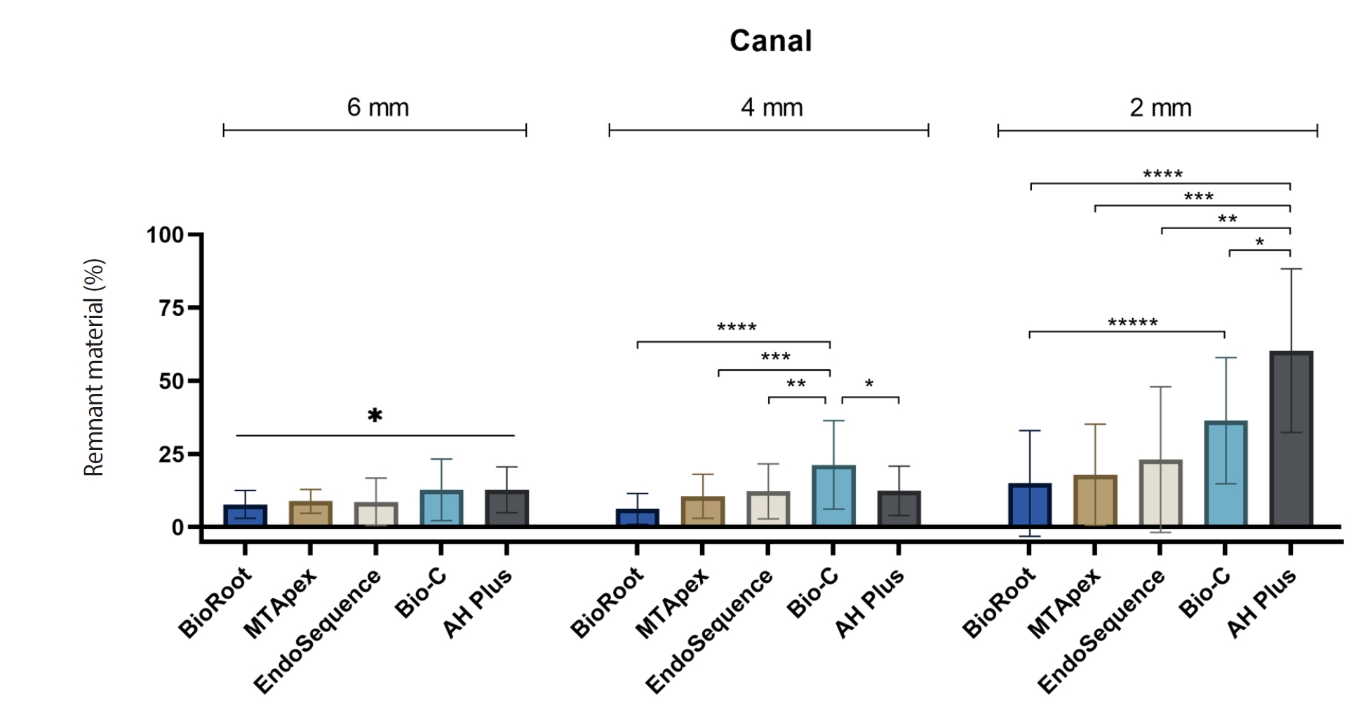
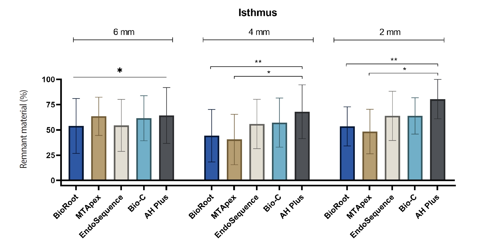
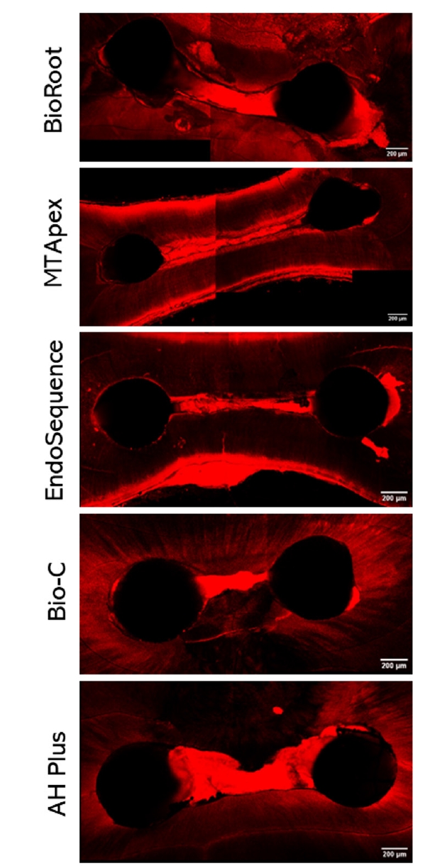
Figure 1. Representation of the mean and standard deviation values in percentage of remnant filling material in canals at the 6-, 4- and 2-mm levels. Bars linking different materials with asterisks indicate statistically significant differences (*p < 0.05, **p < 0.01, ***p < 0.001). Manufacturer information for each material is provided in Table 1.
Figure 2. Representation of the mean and standard deviation values in percentage of remnant filling material in isthmuses at the 6-, 4- and 2-mm levels. Bars linking different materials with asterisks indicate statistically significant differences (*p < 0.05, **p < 0.01). Manufacturer information for each material is provided in Table 1.
Figure 3. Confocal microscopy representative images of the canal and isthmuses at 4 mm after endodontic retreatment procedures with the tested materials. Manufacturer information for each material is provided in Table 1.
Figure 1.
Figure 2.
Figure 3.
Calcium silicate-based sealers remnants in isthmuses of mesial roots of mandibular molars: an in vitro evaluation
| Material | Manufacturer | Batch | Composition |
|---|---|---|---|
| BioRoot RCS | Septodont (Saint-Maur-des-Fossés, France) | Powder B23970 | Tricalcium silicate, zirconium oxide, and povidone |
| Liquid B23099 | Aqueous solution of calcium chloride and polycarboxylate | ||
| MTApex Sealer | Ultradent Products Inc. (South Jordan, UT, USA) | Powder 2019102403 | Tricalcium silicate, tricalcium aluminate, and tantalum oxide |
| Liquid 2020011401 | Water-based gel | ||
| EndoSequence BC Sealer HiFlow | Brasseler USA (Savannah, GA, USA) | 2001SPWF | Tricalcium silicate, dicalcium silicate, calcium hydroxide, zirconium oxide, and fillers |
| Bio-C Sealer | Angelus (Londrina, Brazil) | 60406 | Calcium silicate, calcium aluminate, calcium oxide, zirconium oxide, iron oxide, silicon dioxide and polyethylene glycol |
| AH Plus Jet | Dentsply Maillefer (Ballaigues, Switzerland) | 2204000437 | Epoxide paste: bisphenol-A epoxy resin, bisphenol-F epoxy resin, calcium tungstate, zirconium oxide, aerosol, and pigment |
| Amine paste: 1-adamantane amine N, N’-dibenzyl-5-oxa-nonandiamine-1, 9 TCD-Diamine, calcium tungstate, zirconium oxide, aerosol, and silicone oil |
| Tested material | Retreatment time (sec) |
|---|---|
| BioRoot RCS | 167.3 ± 46.01B |
| MTApex | 161.5 ± 36.46BC |
| EndoSequence BC Sealer HiFlow | 170.2 ± 25.88B |
| Bio-C Sealer | 130.4 ± 11.37C |
| AH Plus Jet | 225.2 ± 33.57A |
| Tested material | Setting time (min) under each condition |
|
|---|---|---|
| Moist | Dry | |
| BioRoot RCS | 251.8 ± 8.98ABCa | 615.8 ± 3.07Ab |
| MTApex | 226.3 ± 40.2Ba | 512.9 ± 18.78Bb |
| EndoSequence BC Sealer HiFlow | 351.6 ± 45.19Ca | 2751 ± 48.67Cb |
| Bio-C Sealer | 65.77 ± 4.71Da | 251.0 ± 52.77Db |
| AH Plus Jet | 615.3 ± 10.29Ea | 675.6 ± 138.7Aa |
Table 1. Composition and batch number of the root canal sealers used in the analysis
Table 2. The retreatment time for all tested materials
Values are presented as mean ± standard deviation. Different uppercase letters indicate statistically significant differences between materials ( Manufacturer information for each material is provided in
Table 3. Representation of the values of setting time of the tested materials
Values are presented as mean ± standard deviation. Different uppercase letters indicate statistically significant differences between materials ( Manufacturer information for each material is provided in

