-
Effects of dentin surface preparations on bonding of self-etching adhesives under simulated pulpal pressure
-
Chantima Siriporananon, Pisol Senawongse, Vanthana Sattabanasuk, Natchalee Srimaneekarn, Hidehiko Sano, Pipop Saikaew
-
Restor Dent Endod 2022;47(1):e4. Published online December 28, 2021
-
DOI: https://doi.org/10.5395/rde.2022.47.e4
-
-
 Abstract  PDF
- Objectives
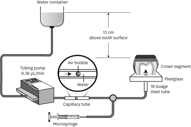
This study evaluated the effects of different smear layer preparations on the dentin permeability and microtensile bond strength (µTBS) of 2 self-etching adhesives (Clearfil SE Bond [CSE] and Clearfil Tri-S Bond Universal [CTS]) under dynamic pulpal pressure. Materials and MethodsHuman third molars were cut into crown segments. The dentin surfaces were prepared using 4 armamentaria: 600-grit SiC paper, coarse diamond burs, superfine diamond burs, and carbide burs. The pulp chamber of each crown segment was connected to a dynamic intra-pulpal pressure simulation apparatus, and the permeability test was done under a pressure of 15 cmH2O. The relative permeability (%P) was evaluated on the smear layer-covered and bonded dentin surfaces. The teeth were bonded to either of the adhesives under pulpal pressure simulation, and cut into sticks after 24 hours water storage for the µTBS test. The resin-dentin interface and nanoleakage observations were performed using a scanning electron microscope. Statistical comparisons were done using analysis of variance and post hoc tests. ResultsOnly the method of surface preparation had a significant effect on permeability (p < 0.05). The smear layers created by the carbide and superfine diamond burs yielded the lowest permeability. CSE demonstrated a higher µTBS, with these values in the superfine diamond and carbide bur groups being the highest. Microscopic evaluation of the resin-dentin interface revealed nanoleakage in the coarse diamond bur and SiC paper groups for both adhesives. ConclusionsSuperfine diamond and carbide burs can be recommended for dentin preparation with the use of 2-step CSE.
-
Citations
Citations to this article as recorded by  - Effect of smear layer pretreatment with EDTA and sodium hypochlorite on the dentin bond durability of universal adhesives
Thanawat Ruaydee, Chantida Pawaputanon Na Mahasarakham, Vanthana Sattabanasuk, Pipop Saikaew
Frontiers in Dental Medicine.2026;[Epub] CrossRef - Determination of marginal permeability of restorations in the cervical region using a universal adhesive system: a randomized controlled open-label laboratory study
Svetlana N. Razumova, Anzhela S. Brago, Oxana R. Ruda, Artur G. Talandis, Lamara M. Khaskhanova, Ruzanna M. Bragunova, Bohdan O. Pecherskyi
Russian Journal of Dentistry.2026; 30(2): 113. CrossRef - Catechol–Phosphonate–Augmented Universal Adhesive for Hydrolysis-Resistant Dentin Bonds: A µTBS and Spectroscopic Study
Rabeia J. Khalil, Suha K. Ibrahim, Athraa H. Madhat, Ali H. Tawfieq
European Journal of Dentistry.2026;[Epub] CrossRef - Critical analysis of the “nanoleakage concept” in endodontics: a narrative review
José Luis Álvarez-Vásquez, Daniel Chavarría-Bolaños
Frontiers in Dental Medicine.2026;[Epub] CrossRef - The effect of different adhesive strategies and diamond burs on dentin bond strength of universal resin cements
Chavakorn Atsavathavornset, Pipop Saikaew, Choltacha Harnirattisai, Hidehiko Sano
Clinical Oral Investigations.2025;[Epub] CrossRef - Universal adhesive systems in dentistry: A narrative review
Svetlana N. Razumova, Anzhela S. Brago, Oxana R. Ruda, Zoya A. Guryeva, Elvira V. Adzhieva
Russian Journal of Dentistry.2024; 28(5): 512. CrossRef - Delayed light activation of resin composite affects the bond strength of adhesives under dynamic simulated pulpal pressure
Nattaporn Sukprasert, Choltacha Harnirattisai, Pisol Senawongse, Hidehiko Sano, Pipop Saikaew
Clinical Oral Investigations.2022; 26(11): 6743. CrossRef
-
5,174
View
-
75
Download
-
5
Web of Science
-
7
Crossref
-
Direct cellular responses of a human periodontal ligament stem cell line to root canal sealers and white mineral trioxide aggregate: an in vitro study
-
Atsushi Tomokiyo, Kenta Tsuchiya, Keita Ipposhi, Salvatore Sauro, Jukka P. Matinlinna, Fumiko Minowa, Hidehiko Sano, Hidefumi Maeda
-
J Korean Acad Conserv Dent ;Published online October 23, 2026
-
DOI: https://doi.org/10.5395/rde.2026.51.e45
[Epub ahead of print]
-
-
Abstract
PDF
 Supplementary Material
- Objectives
Root canal sealers may contact periodontal ligament (PDL) tissues through the apical foramen; therefore, their biological compatibility is critical for periapical healing. This study evaluated the direct cellular responses of human PDL stem cells (PDLSCs) to selected root canal sealers and white mineral trioxide aggregate (WMTA). Methods: Disc-shaped specimens of AH Plus (AH; Dentsply Sirona), GuttaFlow 2 (GF; Coltene), GuttaFlow Bioseal (Bio; Coltene), and ProRoot MTA (WMTA) were directly placed onto cultured clonal human PDLSCs (cell line 2–23). Cell viability, gene expression related to proliferation (CCND1, CCNE1), collagen synthesis (COL1, COL3), and mineralization (BMP2, OPN, CEMP1), collagen deposition, mineralized matrix formation, surface characteristics, and Ca2+ release were evaluated using MTT assay, real-time quantitative polymerase chain reaction, histochemical staining, scanning electron microscopy/ energy-dispersive X-ray spectroscopy, and ion release analysis. Results: AH exhibited marked cytotoxicity, whereas GF, Bio, and WMTA showed higher cytocompatibility. Bio significantly increased proliferation-related gene expression at 7 days. Although early collagen deposition was lower in the Bio and WMTA groups, both materials induced significantly greater mineralized matrix formation after 28 days, accompanied by upregulation of BMP2, OPN, and CEMP1. WMTA showed a rapid initial Ca2+ release, whereas Bio demonstrated a gradual and sustained release profile. Conclusions: The evaluated materials exhibited distinct cellular effects on PDLSCs. The bioceramic silicone-based sealer (Bio) demonstrated favorable cytocompatibility and promoted mineralization-related responses comparable to WMTA, suggesting its potential to support periapical tissue healing. Therefore, Bio and WMTA may enhance PDLSC viability and mineralization, supporting improved periapical healing and long-term treatment outcomes.
|

 KACD