
Search
- Page Path
- HOME > Search
Research Article
- In vitro experimental study comparing continuous and intermittent irrigation protocols: influence of sodium hypochlorite volume and contact time on tissue dissolution
- Alfredo Iandolo, Dina Abdellatif, Davide Mancino, Gwenael Rolin, Camille Coussens, Aurelian Louvrier, Felipe G Belladonna, Edouard Euvrard, Emmanuel João Nogueira Leal da Silva
- Restor Dent Endod 2025;50(4):e36. Published online October 15, 2025
- DOI: https://doi.org/10.5395/rde.2025.50.e36
-
 Abstract
 PDF
PDF PubReader
 ePub
ePub - Objectives
This study aimed to evaluate whether continuous irrigation with larger volumes or allowing sodium hypochlorite (NaOCl) resting time is more critical for pulp tissue dissolution using a controlled artificial root canal system.
Methods
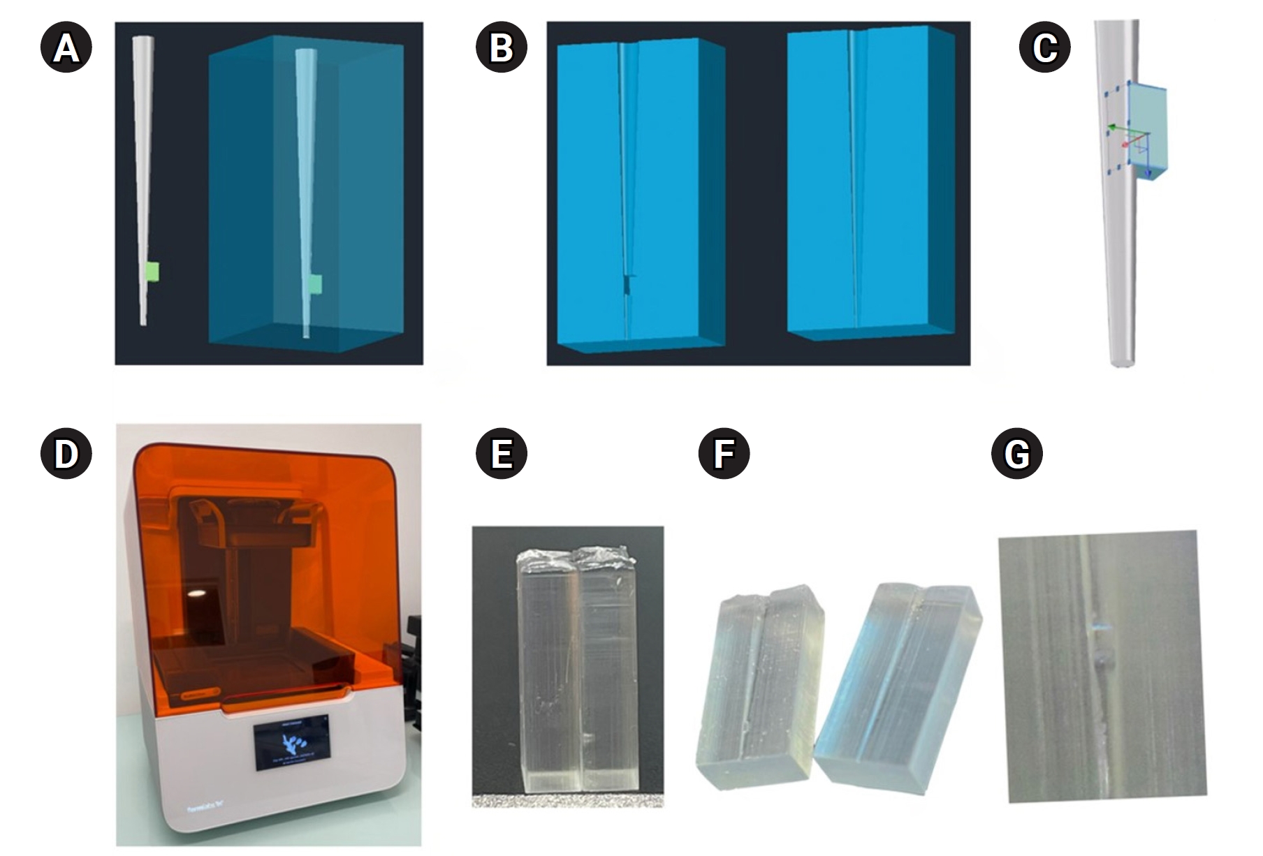
A three-dimensional printed artificial root canal with a lateral canal in the apical third was fabricated. Standardized bovine pulp tissue specimens were inserted, and three irrigation protocols were tested: group A (continuous NaOCl irrigation at 1 mL/min via syringe pump), group B (intermittent NaOCl irrigation with 0.1 mL and a 3-minute resting period), and group C (control, saline irrigation). The time for complete dissolution and the total NaOCl volume were recorded.
Results
Complete dissolution occurred in groups A and B, with significant differences in NaOCl volume and time (p < 0.05). In group A, complete dissolution was consistently observed after the 6th irrigation cycle, corresponding to a total NaOCl volume of 6.0 ± 0.66 mL per test. The average time required for complete dissolution in this group was 6 ± 0.66 minutes. In group B, complete dissolution occurred after the 4th cycle, with a total NaOCl volume of 0.4 ± 0.06 mL per test and a mean dissolution time of 12.6 ± 1.8 minutes.
Conclusions
NaOCl volume and exposure time significantly influence pulp tissue dissolution.
- 2,476 View
- 194 Download

Case Report
- An unusual case of dens invaginatus on a mandibular second molar: a case report
- Davide Mancino, Dina Abdellatif, Alfredo Iandolo, Fabien Bornert, Youssef Haïkel
- Restor Dent Endod 2025;50(1):e2. Published online January 8, 2025
- DOI: https://doi.org/10.5395/rde.2025.50.e2
-
Abstract
PDFPubReaderePub
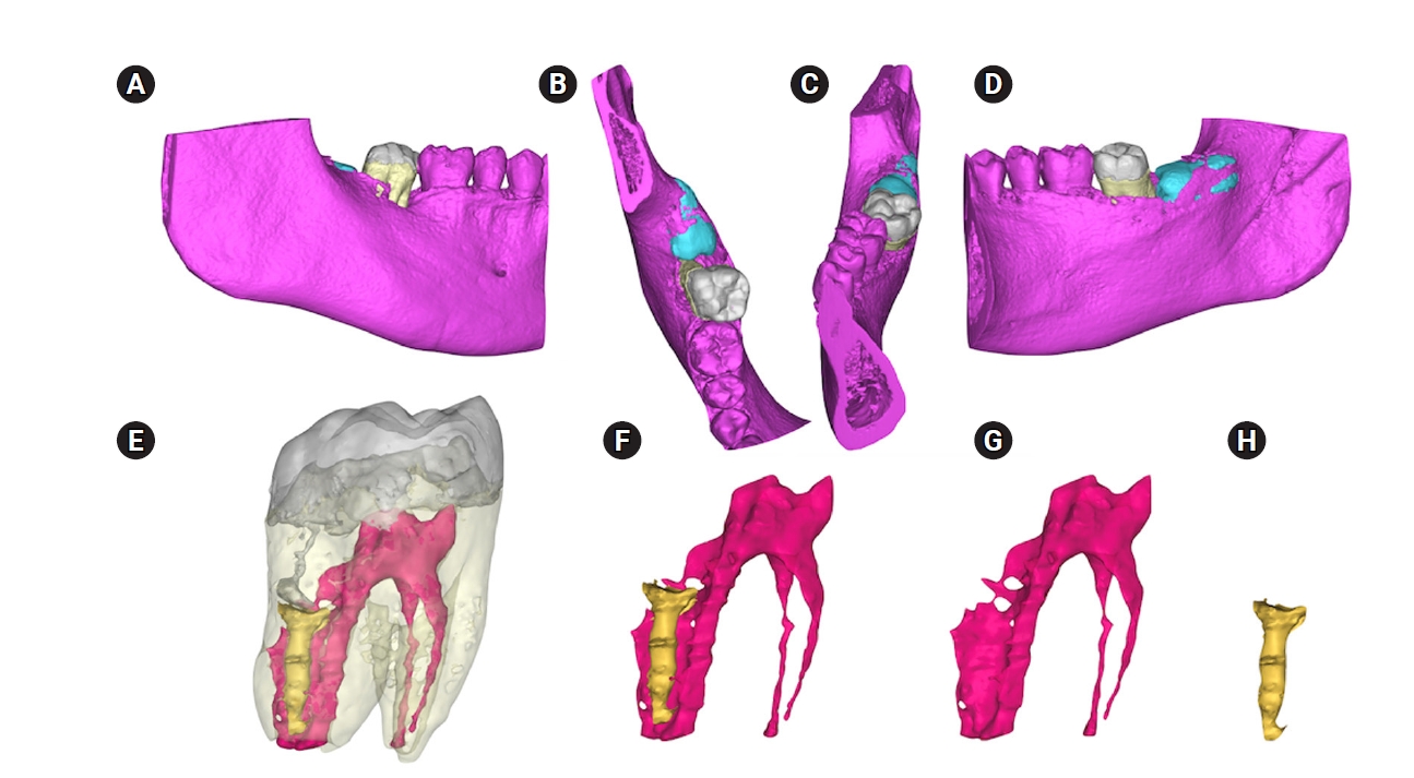
- The present case report describes the endodontic treatment of a type III B dens invaginatus (DI) in a three-rooted mandibular second molar since the invagination invades the root and extends apically. Clinical and cone-beam computed tomography examination of the mandibular second molar showed a broadened coronal morphology, DI, a third root, periapical radiolucency, and compression of a distal root canal by the invagination, which developed an atypical semilunar shape. The tooth was diagnosed with pulpal necrosis, symptomatic apical, and peri-invagination periodontitis. Consequently, three-dimensional virtual reconstruction was conducted to improve anatomical interpretation and case planning and accelerate the intraoperative phase by reducing operator stress and minimizing intraoperative variables. The present case report aims to raise awareness of the existence of DI on the mandibular second molar.
-
Citations
Citations to this article as recorded by
- Dens Invaginatus—Mandibular Second Molar—Case Report
Krystyna Pietrzycka, Natalia Lutomska, Cornelis H. Pameijer, Monika Lukomska-Szymanska
Dentistry Journal.2026; 14(1): 27. CrossRef - Type IIIb dens invaginatus in a maxillary second molar and its microscopic anatomical features: a case report
Mingming Li, Zhiwu Wu, Shaoying Duan, Yuling Zuo
BMC Oral Health.2025;[Epub] CrossRef
- Dens Invaginatus—Mandibular Second Molar—Case Report
- 4,409 View
- 293 Download
- 1 Web of Science
- 2 Crossref
 First
First Prev
Prev

