
Search
- Page Path
- HOME > Search
Research Articles
- Micro-CT evaluation of the removal of root fillings using rotary and reciprocating systems supplemented by XP-Endo Finisher, the Self-Adjusting File, or Er,Cr:YSGG laser
- Gülsen Kiraz, Bulem Üreyen Kaya, Mert Ocak, Muhammet Bora Uzuner, Hakan Hamdi Çelik
- Restor Dent Endod 2023;48(4):e36. Published online October 23, 2023
- DOI: https://doi.org/10.5395/rde.2023.48.e36
-
 Abstract
 PDF
PDF PubReader
 ePub
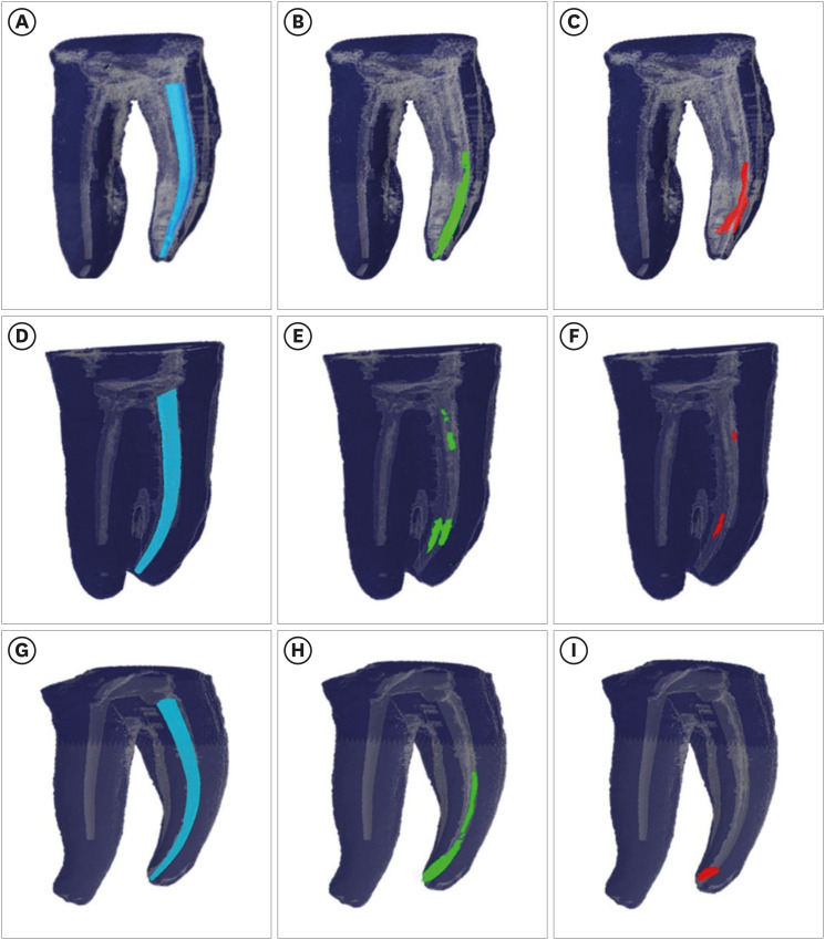
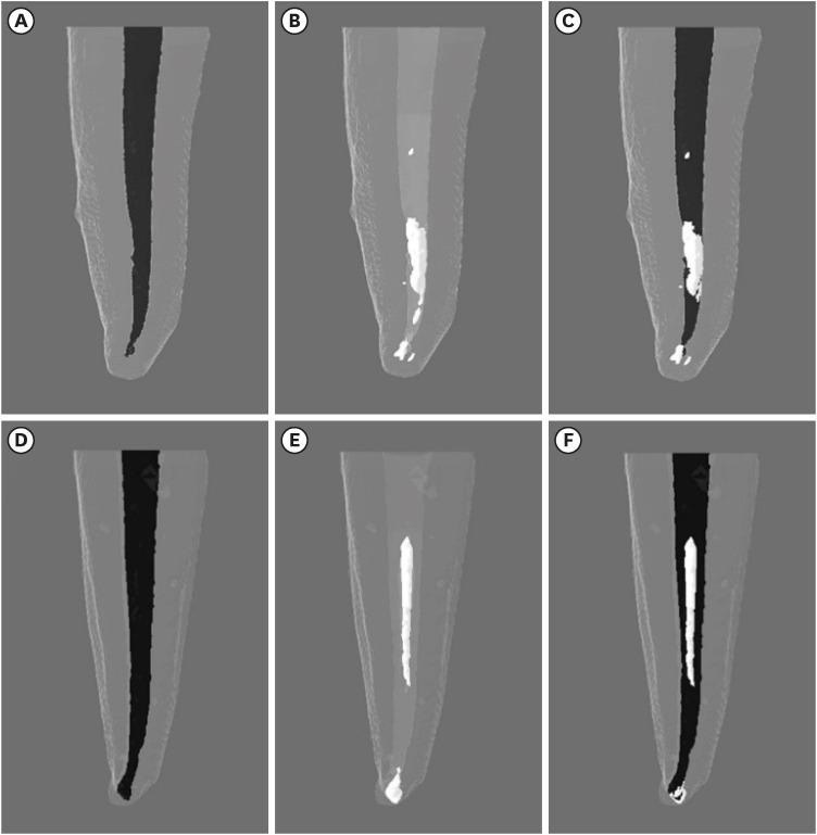
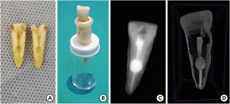
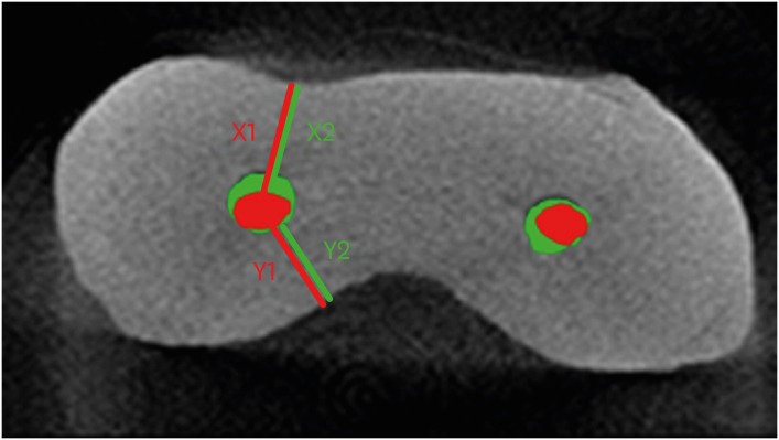
ePub Objectives This study aimed to compare the effectiveness of a single-file reciprocating system (WaveOne Gold, WOG) and a multi-file rotary system (ProTaper Universal Retreatment, PTUR) in removing canal filling from severely curved canals and to evaluate the possible adjunctive effects of XP-Endo Finisher (XPF), the Self-Adjusting File (SAF), and an erbium, chromium: yttrium, scandium, gallium garnet (Er,Cr:YSGG) laser using micro-computed tomography (μCT).
Materials and Methods Sixty-six curved mandibular molars were divided into 2 groups based on the retreatment technique and then into 3 based on the supplementary method. The residual filling volumes and root canals were evaluated with μCT before and after retreatment, and after the supplementary steps. The data were statistically analyzed with the
t -test, Mann-WhitneyU test, analysis of covariance, and factorial analysis of variance (p < 0.05).Results PTUR and WOG showed no significant difference in removing filling materials (
p > 0.05). The supplementary techniques were significantly more effective than reciprocating or rotary systems only (p < 0.01). The supplementary steps showed no significant differences in canal filling removal effectiveness (p > 0.05), but XPF showed less dentin reduction than the SAF and Er,Cr:YSGG laser (p < 0.01).Conclusions The supplementary methods significantly decreased the volume of residual filling materials. XPF caused minimal changes in root canal volume and might be preferred for retreatment in curved root canals. Supplementary approaches after retreatment procedures may improve root canal cleanliness.
-
Citations
Citations to this article as recorded by
- A laboratory study comparing two methods for removing plastic carrier obturators from severely curved root canals
Tania Gancedo-Gancedo, Patricia Pereira-Lores, Venkateshbabu Nagendrababu, Paul MH Dummer, Jenifer Martín-González, Alba Bello-Castro, Inmaculada Tomás, Benjamín Martín-Biedma, Pablo Castelo-Baz
BMC Oral Health.2026;[Epub] CrossRef - Reciproc and XP-endo Shaper Outperform WaveOne Gold in Apical Debris Removal: A Micro-CT Study in 3D-Printed Molars
Yu-juan Zhu, Xue Zhong, Yu-xuan Wu, Shao-lan Li, Qiu-hui Li
Current Medical Science.2026; 46(3): 843. CrossRef - Trends in dentomaxillofacial radiology
Kıvanç Kamburoğlu
World Journal of Radiology.2025;[Epub] CrossRef - Retrieval of AH Plus Bioceramic and Ceraseal Versus AH Plus in Endodontic Retreatment
Eurok Shim, Jee Woo Son, Jiyoung Kwon, Hyun-Jung Kim, Ji-Hyun Jang, Seok Woo Chang, Soram Oh
Journal of Clinical Medicine.2025; 14(6): 1826. CrossRef - Characteristics and Effectiveness of XP‐Endo Files and Systems: A Narrative Review
Sarah M. Alkahtany, Rana Alfadhel, Aseel AlOmair, Sarah Bin Durayhim, Kee Y. Kum
International Journal of Dentistry.2024;[Epub] CrossRef - Effect of the filling technique on the filling removal from oval-shaped canals
Lislaine Valerio, Lisa Yurie Oda, Felipe Andretta Copelli, Clarissa Teles Rodrigues, Everdan Carneiro, Marco Antonio Hungaro Duarte, Bruno Cavalini Cavenago
Clinical Oral Investigations.2024;[Epub] CrossRef
- A laboratory study comparing two methods for removing plastic carrier obturators from severely curved root canals
- 5,282 View
- 127 Download
- 11 Web of Science
- 6 Crossref

- Effectiveness of endodontic retreatment using WaveOne Primary files in reciprocating and rotary motions
- Patricia Marton Costa, Renata Maíra de Souza Leal, Guilherme Hiroshi Yamanari, Bruno Cavalini Cavenago, Marco Antônio Húngaro Duarte
- Restor Dent Endod 2023;48(2):e15. Published online April 25, 2023
- DOI: https://doi.org/10.5395/rde.2023.48.e15
-
Abstract
PDFPubReaderePub
Objectives This study evaluated the efficiency of WaveOne Primary files (Dentsply Sirona) for removing root canal fillings with 2 types of movement: reciprocating (RCP) and continuous counterclockwise rotation (CCR).
Materials and Methods Twenty mandibular incisors were prepared with a RCP instrument (25.08) and filled using the Tagger hybrid obturation technique. The teeth were retreated with a WaveOne Primary file and randomly allocated to 2 experimental retreatment groups (
n = 10) according to movement type: RCP and CCR. The root canals were emptied of filling material in the first 3 steps of insertion, until reaching the working length. The timing of retreatment and procedure errors were recorded for all samples. The specimens were scanned before and after the retreatment procedure with micro-computed tomography to calculate the percentage and volume (mm3) of the residual filling material. The results were statistically evaluated using paired and independentt -tests, with a significance level set at 5%.Results No significant difference was found in the timing of filling removal between the groups, with a mean of 322 seconds (RCP) and 327 seconds (CCR) (
p < 0.05). There were 6 instrument fractures: 1 in a RCP motion file and 5 in continuous rotation files. The volumes of residual filling material were similar (9.94% for RCP and 15.94% for CCR;p > 0.05).Conclusions The WaveOne Primary files used in retreatment performed similarly in both RCP and CCR movements. Neither movement type completely removed the obturation material, but the RCP movement provided greater safety.
-
Citations
Citations to this article as recorded by- Micro-CT evaluation of the removal of root fillings using rotary and reciprocating systems supplemented by XP-Endo Finisher, the Self-Adjusting File, or Er,Cr:YSGG laser
Gülsen Kiraz, Bulem Üreyen Kaya, Mert Ocak, Muhammet Bora Uzuner, Hakan Hamdi Çelik
Restorative Dentistry & Endodontics.2023;[Epub] CrossRef
- Micro-CT evaluation of the removal of root fillings using rotary and reciprocating systems supplemented by XP-Endo Finisher, the Self-Adjusting File, or Er,Cr:YSGG laser
- 3,570 View
- 76 Download
- 1 Crossref
- Morphotypes of the apical constriction of maxillary molars: a micro-computed tomographic evaluation
- Jeffrey Wen-Wei Chang, Kuzhanchinathan Manigandan, Lakshman Samaranayake, Chellapandian NandhaKumar, Pazhamalai AdhityaVasun, Johny Diji, Angambakkam Rajasekharan PradeepKumar
- Restor Dent Endod 2022;47(2):e19. Published online March 24, 2022
- DOI: https://doi.org/10.5395/rde.2022.47.e19
-
Abstract
PDFPubReaderePub
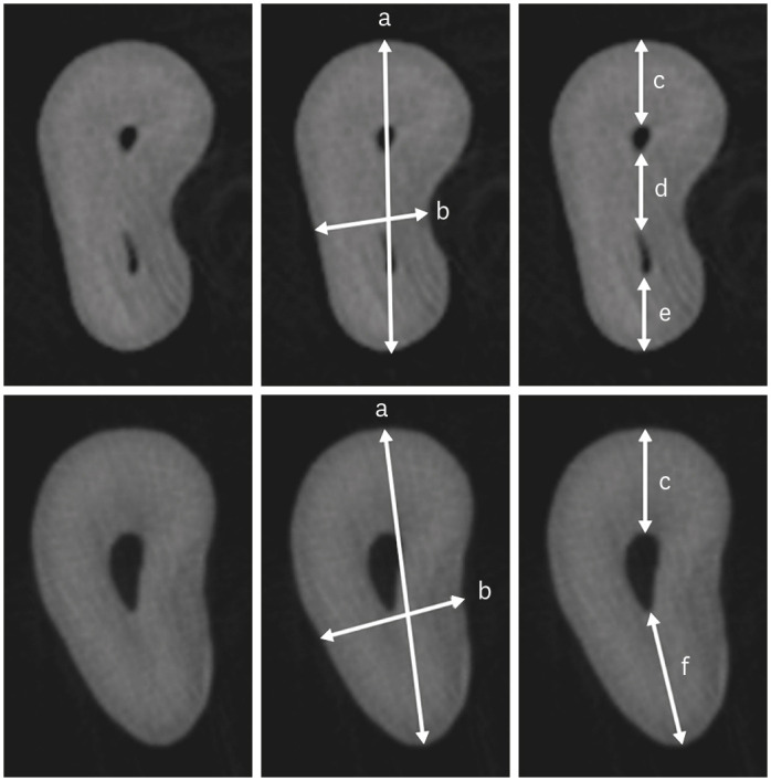
Objectives The aim of this study was to evaluate and compare the apical constriction (AC) and apical canal morphology of maxillary first and second molars, using micro-computed tomography (micro-CT).
Materials and Methods The anatomical features of 313 root canals from 41 maxillary first molars and 57 maxillary second molars of patients with known age and sex were evaluated using micro-CT, with a resolution of 26.7 µm. The factors evaluated were the presence or absence of AC, the morphotypes, bucco-lingual dimension, mesio-distal dimension, and the profile (shape) of AC and the apical root canal. The apical root canal dimensions, location of the apical foramen (AF), AC to AF distance, and presence of accessory canals in the apical 5 mm were also assessed. Descriptive and analytical statistics were used for data evaluation.
Results AC was present in all 313 root canals. Patients’ age and sex did not significantly impact either AC or the apical canal dimensions. The most common AC morphotype detected was the traditional (single) constriction (52%), followed by the parallel (29%) morphotype. The mean AC dimensions in maxillary first molars were not significantly different from those in maxillary second molars. Sixty percent of AF were located within 0.5 mm from the anatomic apex.
Conclusions The most common morphotype of AC detected was the traditional constriction. Neither patients’ age nor sex had a significant impact on the dimensions of the AC or the apical root canal. The majority of AF (60%) were located within 0.5 mm from the anatomic apex.
-
Citations
Citations to this article as recorded by- Evaluation of the accuracy and reliability of two electronic apex locators using micro-computed tomography
Hyoung-Hoon Jo, Kun-Hwa Sung, Tae-Young Park, Jeong-Bum Min, Ho-Keel Hwang
BMC Oral Health.2026;[Epub] CrossRef - In Vivo and In Vitro Accuracy and Precision Evaluations of Mini Electronic Apex Locators
Özlem Kara, Rüstem Kemal Sübay
Australian Endodontic Journal.2025; 51(2): 329. CrossRef - Effect of Coronal Flaring on Initial Apical File Size Estimation in Curved Canals Using Three Distinct Rotary Instruments: A Comparative In Vitro Study
Vinodhini Varatharajan, Muhammed Abdul Rahman Thazhathveedan, Mohammed Salman Kuttikkodan, Ismail Puzhangaraillath Mundanatayil, Amrutha Ravindran Thazhe Mangool, Ashraf Karumbil
Cureus.2024;[Epub] CrossRef - In Vitro Evaluation of the Accuracy of Three Electronic Apex Locators Using Different Sodium Hypochlorite Concentrations
Sanda Ileana Cîmpean, Radu Marcel Chisnoiu, Adela Loredana Colceriu Burtea, Rareș Rotaru, Marius Gheorghe Bud, Ada Gabriela Delean, Ioana-Sofia Pop-Ciutrilă
Medicina.2023; 59(5): 918. CrossRef - Cone beam computed tomography analysis of the root and canal morphology of the maxillary second molars in a Hail province of the Saudi population
Ahmed A. Madfa, Moazzy I. Almansour, Saad M. Al-Zubaidi, Albandari H. Alghurayes, Safanah D. AlDAkhayel, Fatemah I. Alzoori, Taif F. Alshammari, Abrar M. Aldakhil
Heliyon.2023; 9(9): e19477. CrossRef
- Evaluation of the accuracy and reliability of two electronic apex locators using micro-computed tomography
- 3,151 View
- 61 Download
- 7 Web of Science
- 5 Crossref
- Morphological characteristics of the mesiobuccal root in the presence of a second mesiobuccal canal: a micro-CT study
- Lucas P. Lopes Rosado, Matheus Lima Oliveira, Karla Rovaris, Deborah Queiroz Freitas, Frederico Sampaio Neves
- Restor Dent Endod 2022;47(1):e6. Published online January 18, 2022
- DOI: https://doi.org/10.5395/rde.2022.47.e6
-
Abstract
PDFPubReaderePub
Objectives This study investigated the internal morphology of mesiobuccal (MB) roots of maxillary molars with a second mesiobuccal (MB2) canal.
Materials and Methods Forty-seven maxillary first or second molars from Brazilians were scanned using micro-computed tomography. The following measurements were obtained from the MB roots: root thickness, root width, and dentin thickness of the buccal aspect of the first mesiobuccal (MB1) canal, between the MB1 and MB2 canals, and the palatal aspect of the MB2 and MB1 canals at 3 mm from the root apex and in the furcation region. For statistical analysis, the Student’s
t -test and analysis of variance with thepost-hoc Tukey test were used (α = 0.05).Results In maxillary molars with an MB2 canal, MB roots were significantly thicker (
p = 0.0014) and narrower (p = 0.0016) than in maxillary molars without an MB2 canal. The dentin thickness of the palatal aspect of the MB1 canal was also significantly greater than that of MB roots without an MB2 canal at 3 mm from the root apex (p = 0.0007) and in the furcation region (p < 0.0001). In the furcation region of maxillary molars with an MB2 canal, the dentin thickness between the MB1 and MB2 canals was significantly smaller than that in the buccal and palatal aspects (p < 0.0001).Conclusions The internal morphology of MB roots of maxillary molars with an MB2 canal revealed differences in dentin thickness, root diameter, and distance between the canals when compared with maxillary molars without an MB2 canal.
-
Citations
Citations to this article as recorded by- Association between lingual canal detection and buccolingual root width in mandibular anterior teeth: a retrospective CBCT Study
Önder Çam, Melis Oya Ateş, Ali Keleş
BMC Oral Health.2026;[Epub] CrossRef - Effectiveness and safety of three NiTi systems in endodontic retreatment of MB1 and MB2 root canals: a micro-CT and CBCT combined analysis
Airton Oliveira Santos-Junior, Rocharles Cavalcante Fontenele, Karina Ines Medina Carita Tavares, Fernanda Ferrari Esteves Torres, Jáder Camilo Pinto, Pedro Luis Busto Rosim, Andréa Gonçalves, Marco Antonio Hungaro Duarte, Juliane Maria Guerreiro-Tanomaru
Clinical Oral Investigations.2025;[Epub] CrossRef - Cone-beam computed tomography evaluation of root and canal morphology of maxillary molars in a Chinese kazakh population
Shuchun Yang, Chenye Li, Hui Shi, Ming Liu, Xu Wang
BMC Oral Health.2025;[Epub] CrossRef - Can maxillary molar dimensions predict the presence of the second mesiobuccal canal?
Lucas P. Lopes Rosado, Deborah Queiroz Freitas, Karla Rovaris, Matheus L. Oliveira, Frederico Sampaio Neves
Oral Radiology.2023; 39(3): 482. CrossRef - Can the detection of second mesiobuccal canals be enhanced based on the volume of adjacent canals?
Lucas P. Lopes Rosado, Deborah Q. Freitas, Karla Rovaris, Matheus L. Oliveira, Frederico S. Neves
Archives of Oral Biology.2023; 146: 105604. CrossRef - Assessment of the coronal root canal morphology of permanent maxillary first molars using digital 3D-reconstruction technology based on micro-computed tomography data
Mudan Wang, Yuxuan Gao, Qi Deng, Yuan Gao, Dongzhe Song, Dingming Huang
Journal of Dental Sciences.2023; 18(2): 586. CrossRef
- Association between lingual canal detection and buccolingual root width in mandibular anterior teeth: a retrospective CBCT Study
- 2,608 View
- 48 Download
- 7 Web of Science
- 6 Crossref
- A micro-computed tomography evaluation of voids using calcium silicate-based materials in teeth with simulated internal root resorption
- Vildan Tek, Sevinç Aktemur Türker
- Restor Dent Endod 2020;45(1):e5. Published online November 29, 2019
- DOI: https://doi.org/10.5395/rde.2020.45.e5
-
Abstract
PDFPubReaderePub
Objectives The obturation quality of MTA, Biodentine, Total Fill BC root canal sealer (RCS), and warm gutta-percha (WGP) in teeth with simulated internal root resorption (IRR) was evaluated by using micro-computed tomography.
Materials and Methods Standardized IRR cavities were created using 40 extracted maxillary central incisor teeth and randomly assigned into 4 groups (
n = 10). IRR cavities were filled with MTA, Biodentine, Total Fill BC RCS (bulk-fill form) and WGP + Total Fill BC RCS. Percentage of voids between resorptive cavity walls and obturation material (external void), and inside the filling materials (internal voids) were measured.Results Total Fill BC sealer in the bulk-fill form presented significantly highest values of external and internal void percentages (
p < 0.05). Biodentine showed a significantly lowest external void percentage (p < 0.05). WGP + Total Fill BC RCS presented significantly lower values of internal void percentages than all groups (p < 0.05), except Biodentine (p > 0.05).Conclusion None of the filling materials were created void-free obturation in resorption cavities. Biodentine may favor its application in teeth with IRR over Angelus MTA and bulk-fill form of Total Fill BC.
-
Citations
Citations to this article as recorded by- The role of calcium silicate cements in endodontics: from material science to clinical success
Takwa E. Ellakwa, Ayman Ellakwa, Doha El-Sayed Ellakwa
Discover Materials.2026;[Epub] CrossRef - Micro-CT Assessment of Hydraulic Calcium Silicate Cements for Perforating Internal Resorption in 3D-printed Tooth Replicas at Different Root Thirds: An In Vitro Study
Angelo José Sócrates Torres-Carrillo, Jardel Francisco Mazzi-Chaves, Gustavo Creazzo, Rodrigo E. Salazar-Gamarra, Helena Cristina de Assis, Ronald Ordinola-Zapata, Manoel D. Sousa-Neto, Fabiane Carneiro Lopes-Olhê
Journal of Endodontics.2026;[Epub] CrossRef - Removal of AH Plus Bioceramic Sealer from Artificial Internal Resorption Cavities Using Different Irrigation Activation Systems
Mine Büker, Meltem Sümbüllü, Emine Şimşek, Fadime Sena Sezer
Cumhuriyet Dental Journal.2025; 28(3): 383. CrossRef - Functional and Bioactive Performance of Premixed Bioceramic Sealers with Warm Obturation: A Scoping Review
Patryk Wiśniewski, Stanisław Krokosz, Małgorzata Pietruska, Anna Zalewska
Gels.2025; 11(11): 932. CrossRef - Evaluation of the effectiveness of different supplemental cleaning techniques in the retreatment of roots with small simulated internal resorption cavities: an in vitro comparative study
Sine Güngör Us, Özgür Uzun, Nazlı Merve Güngör
BMC Oral Health.2025;[Epub] CrossRef - Evaluation of Different Techniques and Materials for Filling in 3-dimensional Printed Teeth Replicas with Perforating Internal Resorption by Means of Micro–Computed Tomography
Angelo J.S. Torres-Carrillo, Helena C. Assis, Rodrigo E. Salazar-Gamarra, Leonardo Moreira Teodosio, Alice C. Silva-Sousa, Jardel F. Mazzi-Chaves, Priscila B. Ferreira-Soares, Manoel D. Sousa-Neto, Fabiane C. Lopes-Olhê
Journal of Endodontics.2024; 50(2): 205. CrossRef - Three-Dimensional Measurement of Obturation Quality of Bioceramic Materials in Filling Artificial Internal Root Resorption Cavities Using Different Obturation Techniques: An In Vitro Comparative Study
Ammar M. Sharki, Ahmed H. Ali
Journal of Endodontics.2024; 50(7): 997. CrossRef - Evaluation of calcium hydroxide root canal filling materials by cone beam computed tomography and three-dimensional modeling
Asel Usdat Ozturk, Ekin Dogan, Venus Seyedoskuyi, Berk Senguler, Asli Topaloglu-Ak
Folia Medica.2024; 66(2): 250. CrossRef - Clinical applications of calcium silicate‐based materials: a narrative review
S Küçükkaya Eren
Australian Dental Journal.2023;[Epub] CrossRef - A critical analysis of research methods and experimental models to study root canal fillings
Gustavo De‐Deus, Erick Miranda Souza, Emmanuel João Nogueira Leal Silva, Felipe Gonçalves Belladonna, Marco Simões‐Carvalho, Daniele Moreira Cavalcante, Marco Aurélio Versiani
International Endodontic Journal.2022; 55(S2): 384. CrossRef - An Updated Review on Properties and Indications of Calcium Silicate‐Based Cements in Endodontic Therapy
Fateme Eskandari, Alireza Razavian, Rozhina Hamidi, Khadije Yousefi, Susan Borzou, Zohaib Khurshid
International Journal of Dentistry.2022;[Epub] CrossRef - Efficacy Of Calcium Silicate-Based Sealers In Root Canal Treatment: A Systematic Review
Hattan Mohammed Omar Baismail, Mohammed Ghazi Moiser Albalawi, Alaa Mofareh Thoilek Alanazi, Muhannad Atallah Saleem Alatawi, Badr Soliman Alhussain
Annals of Dental Specialty.2021; 9(1): 87. CrossRef
- The role of calcium silicate cements in endodontics: from material science to clinical success
- 3,272 View
- 42 Download
- 12 Crossref
- Root canal volume change and transportation by Vortex Blue, ProTaper Next, and ProTaper Universal in curved root canals
- Hyun-Jin Park, Min-Seock Seo, Young-Mi Moon
- Restor Dent Endod 2018;43(1):e3. Published online December 24, 2017
- DOI: https://doi.org/10.5395/rde.2018.43.e3
-
Abstract
PDFPubReaderePub
Objectives The aim of this study was to compare root canal volume change and canal transportation by Vortex Blue (VB; Dentsply Tulsa Dental Specialties), ProTaper Next (PTN; Dentsply Maillefer), and ProTaper Universal (PTU; Dentsply Maillefer) nickel-titanium rotary files in curved root canals.
Materials and Methods Thirty canals with 20°–45° of curvature from extracted human molars were used. Root canal instrumentation was performed with VB, PTN, and PTU files up to #30.06, X3, and F3, respectively. Changes in root canal volume before and after the instrumentation, and the amount and direction of canal transportation at 1, 3, and 5 mm from the root apex were measured by using micro-computed tomography. Data of canal volume change were statistically analyzed using one-way analysis of variance and Tukey test, while data of amount and direction of transportation were analyzed using Kruskal-Wallis and Mann-Whitney
U test.Results There were no significant differences among 3 groups in terms of canal volume change (
p > 0.05). For the amount of transportation, PTN showed significantly less transportation than PTU at 3 mm level (p = 0.005). VB files showed no significant difference in canal transportation at all 3 levels with either PTN or PTU files. Also, VB files showed unique inward transportation tendency in the apical area.Conclusions Other than PTN produced less amount of transportation than PTU at 3 mm level, all 3 file systems showed similar level of canal volume change and transportation, and VB file system could prepare the curved canals without significant shaping errors.
-
Citations
Citations to this article as recorded by- The effect of nickel-titanium rotary systems on the biomechanical behaviour of mandibular first molars with curved and straight mesial roots: a finite element analysis study
Yaprak Cesur, Sevinc Askerbeyli Örs, Ahmet Serper, Mert Ocak
BMC Oral Health.2025;[Epub] CrossRef - Micro-Computed Tomographic Evaluation of the Shaping Ability of Vortex Blue and TruNatomyTM Ni-Ti Rotary Systems
Batool Alghamdi, Mey Al-Habib, Mona Alsulaiman, Lina Bahanan, Ali Alrahlah, Leonel S. J. Bautista, Sarah Bukhari, Mohammed Howait, Loai Alsofi
Crystals.2024; 14(11): 980. CrossRef - Evaluation of the Centering Ability and Canal Transportation of Rotary File Systems in Different Kinematics Using CBCT
Nupur R Vasava, Shreya H Modi, Chintan Joshi, Mona C Somani, Sweety J Thumar, Aashray A Patel, Anisha D Parmar, Kruti M Jadawala
World Journal of Dentistry.2024; 14(11): 983. CrossRef - Comparative evaluation of nickel titanium rotary instruments on canal transportation and centering ability in curved canals by using cone beam computed tomography: An in vitro study
Krishnaveni Krishnaveni, Nikitha Kalla, Nagalakshmi Reddy, Sharvanan Udayar
Journal of Dental Specialities.2023; 11(2): 105. CrossRef - Comparative Evaluation of Root Canal Centering Ability of Two Heat-treated Single-shaping NiTi Rotary Instruments in Simulated Curved Canals: An In Vitro Study
Preethi Varadan, Chakravarthy Arumugam, Athira Shaji, R R Mathan
World Journal of Dentistry.2023; 14(6): 535. CrossRef - A Comparison of Canal Width Changes in Simulated Curved Canals prepared with Profile and Protaper Rotary Systems
Aisha Faisal, Huma Farid, Robia Ghafoor
Pakistan Journal of Health Sciences.2022; : 55. CrossRef - Evaluation of the Respect of the Root Canal Trajectory by Rotary Niti Instruments (Protaper®Universal): Retrospective Radiographic Study
Salma El Abbassi, Sanaa Chala, Majid Sakout, Faïza Abdallaoui
Integrative Journal of Medical Sciences.2022;[Epub] CrossRef
- The effect of nickel-titanium rotary systems on the biomechanical behaviour of mandibular first molars with curved and straight mesial roots: a finite element analysis study
- 2,543 View
- 13 Download
- 7 Crossref
Review Article
- In-depth morphological study of mesiobuccal root canal systems in maxillary first molars: review
- Seok-Woo Chang, Jong-Ki Lee, Yoon Lee, Kee-Yeon Kum
- Restor Dent Endod 2013;38(1):2-10. Published online February 26, 2013
- DOI: https://doi.org/10.5395/rde.2013.38.1.2
-
Abstract
PDFPubReaderePub
A common failure in endodontic treatment of the permanent maxillary first molars is likely to be caused by an inability to locate, clean, and obturate the second mesiobuccal (MB) canals. Because of the importance of knowledge on these additional canals, there have been numerous studies which investigated the maxillary first molar MB root canal morphology using
in vivo and laboratory methods. In this article, the protocols, advantages and disadvantages of various methodologies for in-depth study of maxillary first molar MB root canal morphology were discussed. Furthermore, newly identified configuration types for the establishment of new classification system were suggested based on two image reformatting techniques of micro-computed tomography, which can be useful as a further 'Gold Standard' method for in-depth morphological study of complex root canal systems.-
Citations
Citations to this article as recorded by- An epidemiological study of extracted mandibular premolars from adolescent patients in Damascus using two classification system analyzed with CBCT and digital periapical radiographs
Yasser Alsayed Tolibah, Mohammed N. Al-Shiekh, Mohammad Tamer Abbara, Marwan Alhaji, Osama Aljabban, Nada Bshara
BMC Oral Health.2025;[Epub] CrossRef - Cone beam computed tomography analysis of the root and canal morphology of the maxillary second molars in a Hail province of the Saudi population
Ahmed A. Madfa, Moazzy I. Almansour, Saad M. Al-Zubaidi, Albandari H. Alghurayes, Safanah D. AlDAkhayel, Fatemah I. Alzoori, Taif F. Alshammari, Abrar M. Aldakhil
Heliyon.2023; 9(9): e19477. CrossRef - Signs of a missed root canal
M. Yu. Pokrovsky, O. A. Aleshina, T. P. Goryacheva, A. M. Pokrovskiy
Endodontics Today.2023; 21(3): 205. CrossRef - Root Canal Morphology of Maxillary First and Second Molars in a Qatari Population: A Cone-Beam Computed Tomography Study
Maryam Mohammed Al-Obaid, Fatima Abdullah Al-Sheeb
European Dental Research and Biomaterials Journal.2021; 2(01): 34. CrossRef - A Study Comparing the Characteristics of Zinc Oxide Eugenol-Based and Mineral Trioxide Aggregate-Based Root Canal Sealers
Seok-Eun Lee, Ja-Won Cho, Hyun-Jun Yoo, Myung-Gu Lee, Yeol-Mae Jeon, Da-Hui Kim, Hye-Won Park
International Journal of Clinical Preventive Dentistry.2021; 17(3): 117. CrossRef - Root Canal Configuration of Burmese (Myanmar) Maxillary First Molar: A Micro-Computed Tomography Study
M. M. Kyaw Moe, H. J. Jo, J. H. Ha, S. K. Kim, Antonino Lo Giudice
International Journal of Dentistry.2021; 2021: 1. CrossRef - Three-Dimensional Analysis of Root Anatomy and Root Canal Curvature in Mandibular Incisors Using Micro-Computed Tomography with Novel Software
JongKi Lee, Shin-Hoon Lee, Jong-Rak Hong, Kee-Yeon Kum, Soram Oh, Adel Saeed Al-Ghamdi, Fawzi Ali Al-Ghamdi, Ayman Omar Mandorah, Ji-Hyun Jang, Seok Woo Chang
Applied Sciences.2020; 10(12): 4385. CrossRef - An investigation into dose optimisation for imaging root canal anatomy using cone beam CT
Margarete B McGuigan, Christie Theodorakou, Henry F Duncan, Jonathan Davies, Anita Sengupta, Keith Horner
Dentomaxillofacial Radiology.2020; 49(7): 20200072. CrossRef - Analysis of Root Canal Anatomy and Variation in Morphology of Maxillary First Molar Using Various Methods: An In Vitro Study
Youssef A Algarni
World Journal of Dentistry.2019; 10(4): 291. CrossRef - Root Canal Morphology of Mandibular Primary Molars: A Micro-CT Study
Meryem ZİYA, Burcu Nihan YÜKSEL, Şaziye SARI
Cumhuriyet Dental Journal.2019; 22(4): 382. CrossRef - Comparison of the implementation of extra root canal treatment before and after fee schedule change in the Taiwan National Health Insurance System
Nien-Chieh Lee, Yen-Hsiang Chang, Hui-Tzu Tu, Chang-Fu Kuo, Kuang-Hui Yu, Lai-Chu See
Journal of Dental Sciences.2018; 13(2): 145. CrossRef - Influence of environment on testing of hydraulic sealers
Mira Kebudi Benezra, Pierre Schembri Wismayer, Josette Camilleri
Scientific Reports.2017;[Epub] CrossRef - CBCT uses in clinical endodontics: the effect of CBCT on the ability to locate MB2 canals in maxillary molars
J. Parker, A. Mol, E. M. Rivera, P. Tawil
International Endodontic Journal.2017; 50(12): 1109. CrossRef - Comparison of Alternative Image Reformatting Techniques in Micro–Computed Tomography and Tooth Clearing for Detailed Canal Morphology
Ki-Wook Lee, Yeun Kim, Hiran Perinpanayagam, Jong-Ki Lee, Yeon-Jee Yoo, Sang-Min Lim, Seok Woo Chang, Byung-Hyun Ha, Qiang Zhu, Kee-Yeon Kum
Journal of Endodontics.2014; 40(3): 417. CrossRef - In Vitro Biocompatibility, Inflammatory Response, and Osteogenic Potential of 4 Root Canal Sealers: Sealapex, Sankin Apatite Root Sealer, MTA Fillapex, and iRoot SP Root Canal Sealer
Seok-Woo Chang, So-Youn Lee, Soo-Kyung Kang, Kee-Yeon Kum, Eun-Cheol Kim
Journal of Endodontics.2014; 40(10): 1642. CrossRef - Análise do preparo de canais radiculares utilizando-se a diafanização
Georje de Martin, Rogério Albuquerque Azeredo
Revista de Odontologia da UNESP.2014; 43(2): 111. CrossRef
- An epidemiological study of extracted mandibular premolars from adolescent patients in Damascus using two classification system analyzed with CBCT and digital periapical radiographs
- 3,178 View
- 21 Download
- 16 Crossref
Original Articles
- The effect of lactic acid concentration and ph of lactic acid buffer solutions on enamel remineralization
- Jung-Won Kwon, Duk-Gyu Suh, Yun-Jung Song, Yun Lee, Chan-Young Lee
- J Korean Acad Conserv Dent 2008;33(6):507-517. Published online November 30, 2008
- DOI: https://doi.org/10.5395/JKACD.2008.33.6.507
-
Abstract
PDFPubReaderePub
There are considerable in vitro and in vivo evidences for remineralization and demineralization occurring simultaneously in incipient enamel caries. In order to "heal"the incipient dental caries, many experiments have been carried out to determine the optimal conditions for remineralization. It was shown that remineralization is affected by different pH, lactic acid concentrations, chemical composition of the enamel, fluoride concentrations, etc.
Eighty specimens from sound permanent teeth without demineralization or cracks, 0.15 mm in thickness, were immersed in lactic acid buffered demineralization solutions for 3 days. Dental caries with a surface zone and subsurface lesion were artificially produced. Groups of 10 specimens were immersed for 10 or 12 days in lactic acid buffered remineralization solutions consisting of pH 4.3 or pH 6.0, and 100, 50, 25, or 10 mM lactic acid. After demineralization and remineralization, images were taken by polarizing microscopy (x100) and micro-computed tomography. The results were obtained by observing images of the specimens and the density of the caries lesions was determined.
As the lactic acid concentration of the remineralization solutions with pH 4.3 was higher, the surface zone of the carious enamel increased and an isotropic zone of the subsurface lesion was found. However, the total decalcification depth increased at the same time.
In the remineralization solutions with pH 6.0, only the surface zone increased slightly but there was no significant change in the total decalcification depth and subsurface zone.
In the lactic acid buffer solutions with the lower pH and higher lactic acid concentration, there were dynamic changes at the deep area of the dental carious lesion.
-
Citations
Citations to this article as recorded by- Effect of fluoride concentration in pH 4.3 and pH 7.0 supersaturated solutions on the crystal growth of hydroxyapatite
Haneol Shin, Sung-Ho Park, Jeong-Won Park, Chan-Young Lee
Restorative Dentistry & Endodontics.2012; 37(1): 16. CrossRef
- Effect of fluoride concentration in pH 4.3 and pH 7.0 supersaturated solutions on the crystal growth of hydroxyapatite
- 2,356 View
- 11 Download
- 1 Crossref
- A comparison of canal centering abilities of four root canal instrument systems using X-ray micro-computed tomography
- Hye-Suk Ko, Heyon-Mee You, Dong-Sung Park
- J Korean Acad Conserv Dent 2007;32(1):61-68. Published online January 31, 2007
- DOI: https://doi.org/10.5395/JKACD.2007.32.1.061
-
Abstract
PDFPubReaderePub
The purpose of this study was to compare the centering abilities of four root canal instrument systems and the amounts of dentin removed after root canal shaping using them.
The mesial canals of twenty extracted mandibular first molars having 10 - 20° curvature were scanned using X-ray micro-computed tomography (XMCT)-scanner before root canals were instrumented. They were divided into four groups (n = 10 per group). In Group 1, root canals were instrumented by the step-back technique with stainless steel K-Flexofile after coronal flaring. The remainders were instrumented by the crown-down technique with Profile (Group 2), ProTaper (Group 3) or K3 system (Group 4). All canals were prepared up to size 25 at the end-point of preparation and scanned again. Scanned images were processed to reconstruct three-dimensional images using three-dimensional image software and the changes of total canal volume were measured. Pre- and post-operative cross-sectional images of 1, 3, 5, and 7 mm from the apical foramen were compared. For each level, centering ratio were calculated using Adobe Photoshop 6.0 and image software program.
ProTaper and K3 systems have a tendency to remove more dentin than the other file systems. In all groups, the lowest value of centering ratio at 3 mm level was observed. And except at 3 mm level, ProTaper system made canals less centered than the other systems (p < 0.05).
- 1,877 View
- 5 Download
 First
First Prev
Prev

