
Articles
- Page Path
- HOME > Restor Dent Endod > Volume 37(2); 2012 > Article
- Case Report Pre-prosthetic minor tooth movement with elastic separating ring & provisional restoration modification: case report
- Haneol Shin, DDS, Byoung-Duck Roh, DDS, MSD, PhD, Yoo-Seok Shin, DDS, MSD, Chan-Young Lee, DDS, MSD, DDSc
-
2012;37(2):-118.
DOI: https://doi.org/10.5395/rde.2012.37.2.114
Published online: May 18, 2012
Department of Conservative Dentistry, Yonsei University College of Dentistry, Seoul, Korea.
- Correspondence to Chan-Young Lee, DDS, MSD, DDSc. Professor, Department of Conservative Dentistry, Yonsei University College of Dentistry, 50 Yonsei-ro, Seodaemun-gu, Seoul, Korea 120-752. TEL, +82-2-2228-8701; FAX, +82-2-313-7575; chanyoungl@yuhs.ac
• Received: December 14, 2011 • Revised: February 16, 2012 • Accepted: February 17, 2012
©Copyights 2012. The Korean Academy of Conservative Dentistry.
- 1,990 Views
- 20 Download
- 1 Crossref
Abstract
- Proximal caries or coronal defect in posterior teeth may result in the loss of proximal space and drifting of neighboring teeth, which makes restoration difficult. Inability to restore proper contours and to align tooth axis properly are commonly encountered problems when planning tooth restoration. Moreover, tilted teeth aggravate periodontal tissue breakdown, such as pseudo-pocket, and angular osseous defect. The purpose of this case presentation is to describe a simple technique for inducing minor tooth movement with orthodontic separating ring and provisional restoration modification. This method was used to create crown placement space on mesially tilted molar. This method is easy, simple and efficient technique which could be used in interproximal space gaining in selected situation.
- 1. Diedrich P. Preprosthetic orthodontics. J Orofac Orthop. 1996;57: 102-116.PubMed
- 2. Mihram WL, Nemetz H. The tilted molar: a prosthetic and periodontal dilemma. Oral Health. 1991;81: 11-15.PubMed
- 3. Keesee SM, Baty DL, Cameron SM, Lefler TB, Morris WJ. A technique for achieving prerestorative minor tooth movement with orthodontic separators. J Prosthet Dent. 2002;88: 544-547.ArticlePubMed
- 4. Smidt A, Venezia E. Gaining adequate interdental space with modified elastic separating rings: rationale and technique. Quintessence Int. 2002;33: 409-414.PubMed
- 5. VanderWeele RA, Broome JC, Ramer JP. Regaining space by using elastic orthodontic separators. Gen Dent. 1998;46: 454-456.PubMed
- 6. Davidovitch M, Papanicolaou S, Vardimon AD, Brosh T. Duration of elastomeric separation and effect on interproximal contact point characteristics. Am J Orthod Dentofacial Orthop. 2008;133: 414-422.ArticlePubMed
- 7. Bondemark L, Fredriksson K, Ilros S. Separation effect and perception of pain and discomfort from two types of orthodontic separators. World J Orthod. 2004;5: 172-176.PubMed
- 8. Natali AN. Dental Biomechanics. 2003;London, UK and New York, USA: Taylor and Francis Group; 196-197.
- 9. Wehrbein H, Diedrich P. Mesio-marginal findings at tilted molars. A histological-histomorphometric study. Eur J Orthod. 2001;23: 663-670.ArticlePubMed
- 10. Kraal JH, Digiancinto JJ, Dail RA, Lemmerman K, Peden JW. Periodontal conditions in patients after molar uprighting. J Prosthet Dent. 1980;43: 156-162.ArticlePubMed
- 11. Lundgren D, Kurol J, Thorstensson B, Hugoson A. Periodontal conditions around tipped and upright molars in adults. An intra-individual retrospective study. Eur J Orthod. 1992;14: 449-455.ArticlePubMed
REFERENCES


Tables & Figures
REFERENCES
Citations
Citations to this article as recorded by 
- Diagnosis and treatment of teeth with primary endodontic lesions mimicking periodontal disease: three cases with long-term follow ups
Jae-Hyung Lim, Ji-Hyun Lee, Su-Jung Shin
Restorative Dentistry & Endodontics.2014; 39(1): 56. CrossRef
 ePub Link
 Cite
CitePre-prosthetic minor tooth movement with elastic separating ring & provisional restoration modification: case report


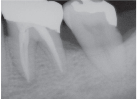
Figure 1
Preoperative periapical x-ray.
Figure 2
Periapical x-ray after root canal treatment.
Figure 3
Initial crown preparation prior to tooth movement.
Figure 4
Elastic ring inserted on #36, 37 interproximal area.
Figure 5
Final crown preparation after finishing tooth movement.
Figure 6
Die adaptation of cast gold crown. Note the recovery of proper distal proximal contour.
Figure 7
Postoperative periapical x-ray.
Figure 8
Postoperative clinical photo.
Figure 9
Estimate the amount of space gaining by measuring change in MD diameter of provisional crown. MD, mesio-distal.
Figure 1
Figure 2
Figure 3
Figure 4
Figure 5
Figure 6
Figure 7
Figure 8
Figure 9
Pre-prosthetic minor tooth movement with elastic separating ring & provisional restoration modification: case report

