Skip Navigation
Skip to contents

Restor Dent Endod : Restorative Dentistry & Endodontics

OPEN ACCESS

Articles

Page Path
HOME > Restor Dent Endod > Volume 37(2); 2012 > Article
  • rupiahtoto
  • rupiahtoto
  • rupiahtoto
  • rupiahtoto
  • rupiahtoto
  • rupiahtoto
  • slot deposit pulsa
  • bo slot gacor
  • toto togel
  • slot gacor
  • login rupiahtoto
  • rupiahtoto
  • rupiahtoto
  • rupiahtoto
  • rupiahtoto
  • Research Article An evaluation of the accuracy of Root ZX according to the conditions of major apical foramen
    Shin-Young Park, DDS, Dong-Kyun Lee, DDS, Ho-Keel Hwang, DDS, PhD
    2012;37(2):-73.
    DOI: https://doi.org/10.5395/rde.2012.37.2.68
    Published online: May 18, 2012

    Department of Conservative Dentistry, Chosun University School of Dentistry, Gwangju, Korea.

    Correspondence to Ho-Keel Hwang, DDS, PhD. Professor, Department of Conservative Dentistry, Chosun University School of Dentistry, 375 Seosuk-dong, Dong-gu, Gwangju, Korea 501-825. TEL, +82-62-220-3840; FAX, +82-62-223-9064; rootcanal@hanmail.net
    • Received: October 14, 2011   • Revised: November 7, 2011   • Accepted: November 16, 2011

    ©Copyights 2012. The Korean Academy of Conservative Dentistry.

    • 407 Views
    • 4 Download
    • 1 Crossref
    prev next
    • Objectives
      The purpose of this study was to assess the accuracy of Root ZX (J. Morita Corp.) according to the location of major foramen and open apex.
    • Materials and Methods
      81 mandibular premolars with mature apices were selected. After access preparation, 27 teeth were instrumented to simulate open apices. 54 teeth were classified according to location of major foramen under surgical microscope (×16). The file was fixed at the location of apical constriction by Root ZX using glass ionomer cement. The apical 4 mm of the apex was exposed and photo was taken and the distance from file tip to the major foramen was measured by calibrating metal ruler on graph paper. The results were statistically analyzed using ANOVA and Scheffe test at p < 0.05 level.
    • Results
      Mean distance from file tip to major foramen was 0.308 mm in Tip foramen group (I), 0.519 mm in Lateral foramen group (II) and 0.932 mm in open apex group (III). Root ZX located apical constriction accurately within ± 0.5 mm in group I of 85.71%, in group II of 59.09%, and in group III of 33.33%. There was a statistically significant difference between group I and III (p < 0.05).
    • Conclusion
      Root ZX located apical constriction accurately regardless of location of major foramen. However, Root ZX couldn't find it in open apex. Clinicians have to use a combination of methods to determine an appropriate working length at open apex. It may be more successful than relying on just electronic apex locator.
    Figure 1
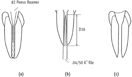
    An open apex model. (a) The canals were instrumented with a #2 Peeso reamer to the actual length; (b) A divergent open apex was prepared by retrograde apical preparation with a .04/50 K3 file inserted to the length of the cutting blade; (c) Simulated open apex model.
    rde-37-68-g001.jpg
    Figure 2
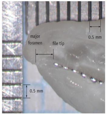
    Tip foramen (group I), in which major foramen is located at the tip along the main axis of root. File was fixed in the position and the distance from file tip to major foramen was measured (×25).
    rde-37-68-g002.jpg
    Figure 3
    Lateral foramen (group II), in which major foramen deviates from the main axis of root. File was fixed in the position and the distance from file tip to major foramen was measured (×25).
    rde-37-68-g003.jpg
    Figure 4
    Simulated open apex (group III). File was fixed in the position and the distance from file tip to major foramen was measured (×25).
    rde-37-68-g004.jpg
    Table 1
    Distance from file tip to major foramen (mm)

    *A significant difference was found between group I and group III (p < 0.05).

    SD, standard deviation.

    rde-37-68-i001.jpg
    Table 2
    Position of file tip relative to major foramen

    *Negative value indicates file tip short from major foramen.

    d, distance from file tip to major foramen.

    rde-37-68-i002.jpg

    No potential conflict of interest relevant to this article was reported.

    Figure 1
    An open apex model. (a) The canals were instrumented with a #2 Peeso reamer to the actual length; (b) A divergent open apex was prepared by retrograde apical preparation with a .04/50 K3 file inserted to the length of the cutting blade; (c) Simulated open apex model.
    rde-37-68-g001.jpg
    Figure 2
    Tip foramen (group I), in which major foramen is located at the tip along the main axis of root. File was fixed in the position and the distance from file tip to major foramen was measured (×25).
    rde-37-68-g002.jpg
    Figure 3
    Lateral foramen (group II), in which major foramen deviates from the main axis of root. File was fixed in the position and the distance from file tip to major foramen was measured (×25).
    rde-37-68-g003.jpg
    Figure 4
    Simulated open apex (group III). File was fixed in the position and the distance from file tip to major foramen was measured (×25).
    rde-37-68-g004.jpg
    Table 1
    Distance from file tip to major foramen (mm)

    *A significant difference was found between group I and group III (p < 0.05).

    SD, standard deviation.

    rde-37-68-i001.jpg
    Table 2
    Position of file tip relative to major foramen

    *Negative value indicates file tip short from major foramen.

    d, distance from file tip to major foramen.

    rde-37-68-i002.jpg
    • 1. Ding J, Gutmann JL, Fan B, Lu Y, Chen H. Investigation of apex locators and related morphological factors. J Endod. 2010;36: 1399-1403.ArticlePubMed
    • 2. Ricucci D, Langeland K. Apical limit of root canal instrumentation and obturation, part 2. a histological study. Int Endod J. 1998;31: 394-409.ArticlePubMed
    • 3. Suzuki K. Experimental study on iontophoresis. J Jpn Stomatol. 1942;16: 411-417.
    • 4. Sunada I. New method for measuring the length of the root canal. J Dent Res. 1962;41: 375-387.ArticlePDF
    • 5. Kobayashi C, Suda H. New electronic canal measuring device based on the ratio method. J Endod. 1994;20: 111-114.ArticlePubMed
    • 6. Kobayashi C. Electronic canal length measurement. Oral Surg Oral Med Oral Pathol Oral Radiol Endod. 1995;79: 226-231.ArticlePubMed
    • 7. Green D. Stereomicroscopic study of 700 root apices of maxillary and mandibular posterior teeth. Oral Surg Oral Med Oral Pathol. 1960;13: 728-733.ArticlePubMed
    • 8. Dummer PM, McGinn JH, Rees DG. The position and topography of the apical canal constriction and apical foramen. Int Endod J. 1984;17: 192-198.ArticlePubMed
    • 9. Kuttler Y. Microscopic investigation of root apexes. J Am Dent Assoc. 1955;50: 544-552.ArticlePubMed
    • 10. Torabinejad M, Walton RE. Endodontics: principles and practice. 2009;4th ed. Missouri: Saunders Elsevier; 29-34.
    • 11. Herrera M, Abalos C, Planas AJ, Llamas R. Influence of apical constriction diameter on Root ZX apex locator precision. J Endod. 2007;33: 995-998.ArticlePubMed
    • 12. Stein TJ, Corcoran JF, Zillich RM. Influence of the major and minor foramen diameters on apical electronic probe measurements. J Endod. 1990;16: 520-522.PubMed
    • 13. Fouad AF, Rivera EM, Krell KV. Accuracy of the Endex with variations in canal irrigants and foramen size. J Endod. 1993;19: 63-67.PubMed
    • 14. Huang L. An experimental study of the principle of electronic root canal measurement. J Endod. 1987;13: 60-64.ArticlePubMed
    • 15. Hachmeister DR, Schindler WG, Walker WA 3rd, Thomas DD. The sealing ability and retention characteristics of mineral trioxide aggregate in a model of apexification. J Endod. 2002;28: 386-390.ArticlePubMed
    • 16. Cho JH, Kum KY, Lee SJ. In vitro evaluation of accuracy and consistency of four different electronic apex locators. J Korean Acad Conserv Dent. 2006;31: 390-397.Article
    • 17. Ponce EH, Vilar Fernández JA. The cemento-dentino-canal junction, the apical foramen, and the apical constriction: evaluation by optical microscopy. J Endod. 2003;29: 214-219.ArticlePubMed
    • 18. Sjogren U, Hägglund B, Sundqvist G, Wing K. Factors affecting the long-term results of endodontic treatment. J Endod. 1990;16: 498-504.ArticlePubMed
    • 19. Pagavino G, Pace R, Baccetti T. A SEM study of in vivo accuracy of the Root ZX electronic apex locator. J Endod. 1998;24: 438-441.ArticlePubMed
    • 20. Goldberg F, De Silvio AC, Manfré S, Nastri N. In vitro measurement accuracy of an electronic apex locator in teeth with simulated apical root resorption. J Endod. 2002;28: 461-463.ArticlePubMed
    • 21. Nguyen HQ, Kaufman AY, Komorowski RC, Friedman S. Electronic length measurement using small and large files in enlarged canals. Int Endod J. 1996;29: 359-364.ArticlePubMed
    • 22. Kim BH, Lee YK, Kim YS. A study on the accuracy of the ROOT-ZX in the canal with mechanically formed constriction. J Korean Acad Conserv Dent. 1999;24: 628-632.
    • 23. Czerw RJ, Fulkerson MS, Donnelly JC. An in vitro test of a simplified model to demonstrate the operation of electronic root canal measuring devices. J Endod. 1994;20: 605-606.ArticlePubMed
    • 24. Cho YL, Son WH, Hwang HK. An accuracy of the several electronic apex locators on the mesial root canal of the mandibular molar. J Korean Acad Conserv Dent. 2005;30: 477-485.Article
    • 25. Lee SJ, Nam KC, Kim YJ, Kim DW. Clinical accuracy of a new apex locator with an automatic compensation circuit. J Endod. 2002;28: 706-709.ArticlePubMed
    • 26. Ounsi HF, Naaman A. In vitro evaluation of the reliability of the Root ZX electronic apex locator. Int Endod J. 1999;32: 120-123.ArticlePubMed
    • 27. Mayeda DL, Simon JH, Aimar DF, Finley K. In vivo measurement accuracy in vital and necrotic canals with the Endex apex locator. J Endod. 1993;19: 545-548.ArticlePubMed
    • 28. Herrera M, Abalos C, Planas AJ, Llamas R. Influence of apical constriction diameter on Root ZX apex locator precision. J Endod. 2007;33: 995-998.ArticlePubMed
    • 29. Shabahang S, Goon WW, Gluskin AH. An in vivo evaluation of Root ZX electronic apex locator. J Endod. 1996;22: 616-618.ArticlePubMed
    • 30. Tselnik M, Baumgartner JC, Marshall JG. An evaluation of root ZX and elements diagnostic apex locators. J Endod. 2005;31: 507-509.ArticlePubMed
    • 31. Meredith N, Gulabivala K. Electrical impedance measurements of root canal length. Endod Dent Traumatol. 1997;13: 126-131.ArticlePubMed
    • 32. Nekoofar MH, Ghandi MM, Hayes SJ, Dummer PM. The fundamental operating principles of electronic root canal length measurement devices. Int Endod J. 2006;39: 595-609.ArticlePubMed
    • 33. Berman LH, Fleischman SB. Evaluation of the accuracy of the Neosono-D electronic apex locator. J Endod. 1984;10: 164-167.ArticlePubMed
    • 34. Saito T, Yamashita Y. Electronic determination of root canal length by newly developed measuring device. Influences of the diameter of apical foramen, the size of K-file and the root canal irrigants. Dent Jpn (Tokyo). 1990;27: 65-72.PubMed
    • 35. Huang L. An experimental study of the principle of electronic root canal measurement. J Endod. 1987;13: 60-64.ArticlePubMed
    • 36. ElAyouti A, Weiger R, Löst C. Frequency of overinstrumentation with an acceptable radiographic working length. J Endod. 2001;27: 49-52.ArticlePubMed

    Tables & Figures

    Figure 1
    An open apex model. (a) The canals were instrumented with a #2 Peeso reamer to the actual length; (b) A divergent open apex was prepared by retrograde apical preparation with a .04/50 K3 file inserted to the length of the cutting blade; (c) Simulated open apex model.
    rde-37-68-g001.jpg
    Figure 2
    Tip foramen (group I), in which major foramen is located at the tip along the main axis of root. File was fixed in the position and the distance from file tip to major foramen was measured (×25).
    rde-37-68-g002.jpg
    Figure 3
    Lateral foramen (group II), in which major foramen deviates from the main axis of root. File was fixed in the position and the distance from file tip to major foramen was measured (×25).
    rde-37-68-g003.jpg
    Figure 4
    Simulated open apex (group III). File was fixed in the position and the distance from file tip to major foramen was measured (×25).
    rde-37-68-g004.jpg
    Table 1
    Distance from file tip to major foramen (mm)

    *A significant difference was found between group I and group III (p < 0.05).

    SD, standard deviation.

    rde-37-68-i001.jpg
    Table 2
    Position of file tip relative to major foramen

    *Negative value indicates file tip short from major foramen.

    d, distance from file tip to major foramen.

    rde-37-68-i002.jpg

    REFERENCES

    • 1. Ding J, Gutmann JL, Fan B, Lu Y, Chen H. Investigation of apex locators and related morphological factors. J Endod. 2010;36: 1399-1403.ArticlePubMed
    • 2. Ricucci D, Langeland K. Apical limit of root canal instrumentation and obturation, part 2. a histological study. Int Endod J. 1998;31: 394-409.ArticlePubMed
    • 3. Suzuki K. Experimental study on iontophoresis. J Jpn Stomatol. 1942;16: 411-417.
    • 4. Sunada I. New method for measuring the length of the root canal. J Dent Res. 1962;41: 375-387.ArticlePDF
    • 5. Kobayashi C, Suda H. New electronic canal measuring device based on the ratio method. J Endod. 1994;20: 111-114.ArticlePubMed
    • 6. Kobayashi C. Electronic canal length measurement. Oral Surg Oral Med Oral Pathol Oral Radiol Endod. 1995;79: 226-231.ArticlePubMed
    • 7. Green D. Stereomicroscopic study of 700 root apices of maxillary and mandibular posterior teeth. Oral Surg Oral Med Oral Pathol. 1960;13: 728-733.ArticlePubMed
    • 8. Dummer PM, McGinn JH, Rees DG. The position and topography of the apical canal constriction and apical foramen. Int Endod J. 1984;17: 192-198.ArticlePubMed
    • 9. Kuttler Y. Microscopic investigation of root apexes. J Am Dent Assoc. 1955;50: 544-552.ArticlePubMed
    • 10. Torabinejad M, Walton RE. Endodontics: principles and practice. 2009;4th ed. Missouri: Saunders Elsevier; 29-34.
    • 11. Herrera M, Abalos C, Planas AJ, Llamas R. Influence of apical constriction diameter on Root ZX apex locator precision. J Endod. 2007;33: 995-998.ArticlePubMed
    • 12. Stein TJ, Corcoran JF, Zillich RM. Influence of the major and minor foramen diameters on apical electronic probe measurements. J Endod. 1990;16: 520-522.PubMed
    • 13. Fouad AF, Rivera EM, Krell KV. Accuracy of the Endex with variations in canal irrigants and foramen size. J Endod. 1993;19: 63-67.PubMed
    • 14. Huang L. An experimental study of the principle of electronic root canal measurement. J Endod. 1987;13: 60-64.ArticlePubMed
    • 15. Hachmeister DR, Schindler WG, Walker WA 3rd, Thomas DD. The sealing ability and retention characteristics of mineral trioxide aggregate in a model of apexification. J Endod. 2002;28: 386-390.ArticlePubMed
    • 16. Cho JH, Kum KY, Lee SJ. In vitro evaluation of accuracy and consistency of four different electronic apex locators. J Korean Acad Conserv Dent. 2006;31: 390-397.Article
    • 17. Ponce EH, Vilar Fernández JA. The cemento-dentino-canal junction, the apical foramen, and the apical constriction: evaluation by optical microscopy. J Endod. 2003;29: 214-219.ArticlePubMed
    • 18. Sjogren U, Hägglund B, Sundqvist G, Wing K. Factors affecting the long-term results of endodontic treatment. J Endod. 1990;16: 498-504.ArticlePubMed
    • 19. Pagavino G, Pace R, Baccetti T. A SEM study of in vivo accuracy of the Root ZX electronic apex locator. J Endod. 1998;24: 438-441.ArticlePubMed
    • 20. Goldberg F, De Silvio AC, Manfré S, Nastri N. In vitro measurement accuracy of an electronic apex locator in teeth with simulated apical root resorption. J Endod. 2002;28: 461-463.ArticlePubMed
    • 21. Nguyen HQ, Kaufman AY, Komorowski RC, Friedman S. Electronic length measurement using small and large files in enlarged canals. Int Endod J. 1996;29: 359-364.ArticlePubMed
    • 22. Kim BH, Lee YK, Kim YS. A study on the accuracy of the ROOT-ZX in the canal with mechanically formed constriction. J Korean Acad Conserv Dent. 1999;24: 628-632.
    • 23. Czerw RJ, Fulkerson MS, Donnelly JC. An in vitro test of a simplified model to demonstrate the operation of electronic root canal measuring devices. J Endod. 1994;20: 605-606.ArticlePubMed
    • 24. Cho YL, Son WH, Hwang HK. An accuracy of the several electronic apex locators on the mesial root canal of the mandibular molar. J Korean Acad Conserv Dent. 2005;30: 477-485.Article
    • 25. Lee SJ, Nam KC, Kim YJ, Kim DW. Clinical accuracy of a new apex locator with an automatic compensation circuit. J Endod. 2002;28: 706-709.ArticlePubMed
    • 26. Ounsi HF, Naaman A. In vitro evaluation of the reliability of the Root ZX electronic apex locator. Int Endod J. 1999;32: 120-123.ArticlePubMed
    • 27. Mayeda DL, Simon JH, Aimar DF, Finley K. In vivo measurement accuracy in vital and necrotic canals with the Endex apex locator. J Endod. 1993;19: 545-548.ArticlePubMed
    • 28. Herrera M, Abalos C, Planas AJ, Llamas R. Influence of apical constriction diameter on Root ZX apex locator precision. J Endod. 2007;33: 995-998.ArticlePubMed
    • 29. Shabahang S, Goon WW, Gluskin AH. An in vivo evaluation of Root ZX electronic apex locator. J Endod. 1996;22: 616-618.ArticlePubMed
    • 30. Tselnik M, Baumgartner JC, Marshall JG. An evaluation of root ZX and elements diagnostic apex locators. J Endod. 2005;31: 507-509.ArticlePubMed
    • 31. Meredith N, Gulabivala K. Electrical impedance measurements of root canal length. Endod Dent Traumatol. 1997;13: 126-131.ArticlePubMed
    • 32. Nekoofar MH, Ghandi MM, Hayes SJ, Dummer PM. The fundamental operating principles of electronic root canal length measurement devices. Int Endod J. 2006;39: 595-609.ArticlePubMed
    • 33. Berman LH, Fleischman SB. Evaluation of the accuracy of the Neosono-D electronic apex locator. J Endod. 1984;10: 164-167.ArticlePubMed
    • 34. Saito T, Yamashita Y. Electronic determination of root canal length by newly developed measuring device. Influences of the diameter of apical foramen, the size of K-file and the root canal irrigants. Dent Jpn (Tokyo). 1990;27: 65-72.PubMed
    • 35. Huang L. An experimental study of the principle of electronic root canal measurement. J Endod. 1987;13: 60-64.ArticlePubMed
    • 36. ElAyouti A, Weiger R, Löst C. Frequency of overinstrumentation with an acceptable radiographic working length. J Endod. 2001;27: 49-52.ArticlePubMed

    Citations

    Citations to this article as recorded by  
    • Effect of file size in measuring electronic working length of teeth with open apex
      Ryeon Jin, Ju-Hee Jeong, A-Ra Cho, Hyoung-Hoon Jo
      Oral Biology Research.2022; 46(1): 36.     CrossRef
    CanvasJS.com
    CanvasJS.com
    CanvasJS.com

    • ePub LinkePub Link
    • Cite
      CITE
      export Copy Download
      Close
      Download Citation
      Download a citation file in RIS format that can be imported by all major citation management software, including EndNote, ProCite, RefWorks, and Reference Manager.

      Format:
      • RIS — For EndNote, ProCite, RefWorks, and most other reference management software
      • BibTeX — For JabRef, BibDesk, and other BibTeX-specific software
      Include:
      • Citation for the content below
      An evaluation of the accuracy of Root ZX according to the conditions of major apical foramen
      Restor Dent Endod. 2012;37(2):68-73.   Published online May 18, 2012
      Close
    • XML DownloadXML Download
    Figure
    • 0
    An evaluation of the accuracy of Root ZX according to the conditions of major apical foramen
    Image Image Image Image
    Figure 1 An open apex model. (a) The canals were instrumented with a #2 Peeso reamer to the actual length; (b) A divergent open apex was prepared by retrograde apical preparation with a .04/50 K3 file inserted to the length of the cutting blade; (c) Simulated open apex model.
    Figure 2 Tip foramen (group I), in which major foramen is located at the tip along the main axis of root. File was fixed in the position and the distance from file tip to major foramen was measured (×25).
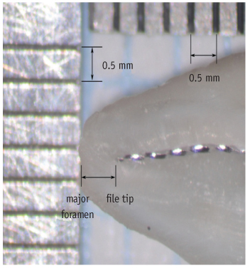
    Figure 3 Lateral foramen (group II), in which major foramen deviates from the main axis of root. File was fixed in the position and the distance from file tip to major foramen was measured (×25).
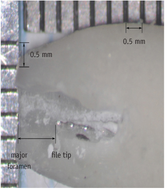
    Figure 4 Simulated open apex (group III). File was fixed in the position and the distance from file tip to major foramen was measured (×25).
    An evaluation of the accuracy of Root ZX according to the conditions of major apical foramen

    Distance from file tip to major foramen (mm)

    *A significant difference was found between group I and group III (p < 0.05).

    SD, standard deviation.

    Position of file tip relative to major foramen

    *Negative value indicates file tip short from major foramen.

    d, distance from file tip to major foramen.

    Table 1 Distance from file tip to major foramen (mm)

    *A significant difference was found between group I and group III (p < 0.05).

    SD, standard deviation.

    Table 2 Position of file tip relative to major foramen

    *Negative value indicates file tip short from major foramen.

    d, distance from file tip to major foramen.


    Restor Dent Endod : Restorative Dentistry & Endodontics
    Close layer
    TOP Mpgyi