
Articles
- Page Path
- HOME > Restor Dent Endod > Volume 37(2); 2012 > Article
- Research Article Color and hardness changes in artificial white spot lesions after resin infiltration
- Ji-Hoon Kim1, Ho-Hyun Son, DDS, PhD2, Juhea Chang, DDS, MSD, PhD3
-
2012;37(2):-95.
DOI: https://doi.org/10.5395/rde.2012.37.2.90
Published online: May 18, 2012
1Department of Dentistry, Seoul National University School of Dentistry, Seoul, Korea.
2Department of Conservative Dentistry, Seoul National University School of Dentistry, Seoul, Korea.
3Clinic for Persons with Disabilities, Seoul National University Dental Hospital, Seoul, Korea.
- Correspondence to Juhea Chang, DDS, MSD, PhD. Clinical assistant professor, Clinic for Persons with Disabilities, Seoul National University Dental Hospital, 101 Daehag-ro, Jongro-gu, Seoul, Korea. TEL, +82-2-2072-3831; FAX, +82-2-2072-2854; juhchang@snu.ac.kr
• Received: December 30, 2011 • Revised: February 24, 2012 • Accepted: February 24, 2012
©Copyights 2012. The Korean Academy of Conservative Dentistry.
- 2,208 Views
- 14 Download
- 10 Crossref
Abstract
-
Objectives The purpose of this study was to determine the effect of resin infiltration technique on color and surface hardness of white spot lesion (WSL) with various degrees of demineralization.
-
Materials and Methods Ten human upper premolars were cut and divided into quarters with a 3 × 4 mm window on the enamel surface. Each specimens were separated into four groups (n = 10) and immersed in demineralization solution to create WSL: control, no treatment (baseline); 12 h, 12 hr demineralization; 24 h, 24 hr demineralization; 48 h, 48 hr demineralization. Resin infiltration was performed to the specimens using Icon (DMG). CIEL*a*b* color parameters of the enamel-dentin complex were determined using a spectroradiometer at baseline, after caries formation and after resin infiltration. Surface hardness was measured by Vickers Micro Hardness Tester (Shimadzu, HMV-2). The differences in color and hardness among the groups were analyzed with ANOVA followed by Tukey test.
-
Results Resin infiltration induced color changes and increased the hardness of demineralized enamel. After resin infiltration, there was no difference in color change (ΔE*) or microhardness among the groups (p < 0.05).
-
Conclusion There was no difference in the effect of resin infiltration on color and hardness among groups with different extents of demineralization.
- 1. Kielbassa AM, Muller J, Gernhardt CR. Closing the gap between oral hygiene and minimally invasive dentistry: a review on the resin infiltration technique of incipient (proximal) enamel lesions. Quintessence Int. 2009;40: 663-681.PubMed
- 2. Featherstone JD. Prevention and reversal of dental caries: role of low level fluoride. Community Dent Oral Epidemiol. 1999;27: 31-40.ArticlePubMed
- 3. Fontana M, Young DA, Wolff MS, Pitts NB, Longbottom C. Defining dental caries for 2010 and beyond. Dent Clin North Am. 2010;54: 423-440.ArticlePubMed
- 4. Kim ME, Jung IY, Kum KY, Lee CY, Roh BD. In vivo quantitative analysis of remineralization effect of remineralization solution 'R' of incipient enamel dental caries. J Korean Acad Conserv Dent. 2002;27: 175-182.Article
- 5. Kim S, Kim EY, Jeong TS, Kim JW. The evaluation of resin infiltration for masking labial enamel white spot lesions. Int J Paediatr Dent. 2011;21: 241-248.ArticlePubMed
- 6. Tüfekçi E, Merrill TE, Pintado MR, Beyer JP, Brantley WA. Enamel loss associated with orthodontic adhesive removal on teeth with white spot lesions: an in vitro study. Am J Orthod Dentofacial Orthop. 2004;125: 733-739.ArticlePubMed
- 7. Gwinnett AJ, Ceen RF. Plaque distribution on bonded brackets: a scanning microscope study. Am J Orthod. 1979;75: 667-677.ArticlePubMed
- 8. Rocha Gomes Torres C, Borges AB, Torres LM, Gomes IS, de Oliveira RS. Effect of caries infiltration technique and fluoride therapy on the colour masking of white spot lesions. J Dent. 2011;39: 202-207.ArticlePubMed
- 9. Meyer-Lueckel H, Paris S, Kielbassa AM. Surface layer erosion of natural caries lesions with phosphoric and hydrochloric acid gels in preparation for resin infiltration. Caries Res. 2007;41: 223-230.ArticlePubMedPDF
- 10. ten Cate JM, Buijs MJ, Miller CC, Exterkate RA. Elevated fluoride products enhance remineralization of advanced enamel lesions. J Dent Res. 2008;87: 943-947.ArticlePubMedPDF
- 11. Son HJ, Kim WC, Jun SH, Kim YS, Ju SW, Ahn JS. Influence of dentin porcelain thickness on layered all-ceramic restoration color. J Dent. 2010;38: Suppl 2. e71-e77.ArticlePubMed
- 12. Zandoná AF, Zero DT. Diagnostic tools for early caries detection. J Am Dent Assoc. 2006;137: 1675-1684.ArticlePubMed
- 13. Bishara SE, Ostby AW. White Spot Lesions: formation, prevention, and treatment. Semin Orthod. 2008;14: 174-182.Article
- 14. Cochrane NJ, Cai F, Huq NL, Burrow MF, Reynolds EC. New approaches to enhanced remineralization of tooth enamel. J Dent Res. 2010;89: 1187-1197.ArticlePubMedPDF
- 15. Son JH, Hur B, Kim HC, Park JK. Management of white spots: resin infiltration technique and microabrasion. J Korean Acad Conserv Dent. 2011;36: 66-71.Article
- 16. Joiner A, Hopkinson I, Deng Y, Westland S. A review of tooth colour and whiteness. J Dent. 2008;36: Suppl 1. S2-S7.ArticlePubMed
- 17. Ko CC, Tantbirojn D, Wang T, Douglas WH. Optical scattering power for characterization of mineral loss. J Dent Res. 2000;79: 1584-1589.ArticlePubMedPDF
- 18. Iwami Y, Hayashi N, Takeshige F, Ebisu S. Relationship between the color of carious dentin with varying lesion activity, and bacterial detection. J Dent. 2008;36: 143-151.ArticlePubMed
- 19. Lim HN, Yu B, Lee YK. Spectroradiometric and spectrophotometric translucency of ceramic materials. J Prosthet Dent. 2010;104: 239-246.ArticlePubMed
- 20. Lim HN, Yu B, Lim JI, Lee YK. Correlations between spectroradiometric and spectrophotometric colors of all-ceramic materials. Dent Mater. 2010;26: 1052-1058.ArticlePubMed
- 21. Douglas RD, Steinhauer TJ, Wee AG. Intraoral determination of the tolerance of dentists for perceptibility and acceptability of shade mismatch. J Prosthet Dent. 2007;97: 200-208.ArticlePubMed
- 22. Kim Y, Son HH, Yi K, Kim HY, Ahn J, Chang J. The color change in artificial white spot lesions measured using a spectroradiometer. Clin Oral Investig. 2012 01 27 (Epub ahead of print).ArticlePDF
- 23. Knösel M, Attin R, Becker K, Attin T. External bleaching effect on the color and luminosity of inactive white-spot lesions after fixed orthodontic appliances. Angle Orthod. 2007;77: 646-652.ArticlePubMedPDF
REFERENCES
Figure 1
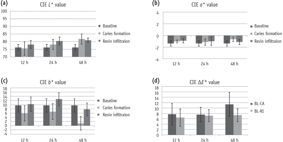
(a) Mean values of CIE L* for three groups (12 h, 24 h, and 48 h demineralization time) measured at baseline, after careis formation, and after resin infiltration; (b) Mean values of CIE a* for three groups; (c) Mean values of CIE b* for three groups; (d) Mean values of color change (CIE ΔE*) after caries formation (BL-CA) and after resin infiltration (BL-RI) for three groups.
Table 1
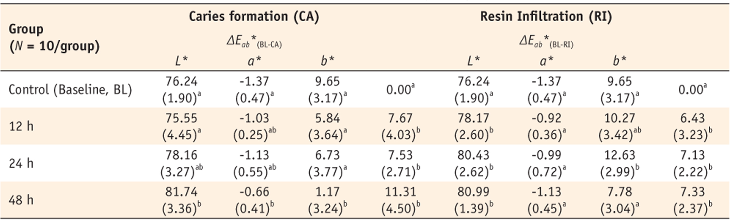
The difference in the mean values (SD) of CIE L*, a*, b*, and ΔE* among the control and the experimental groups after caries formation for three different demineralization times and corresponding resin infiltration
Different superscript letters denote the values that are significantly different from one another in each column (p < 0.05).
Tables & Figures
REFERENCES
Citations
Citations to this article as recorded by 
- In Vitro Evaluation of the Effect of Microabrasion and Resin Infiltration Materials on Enamel Microhardness and Penetration Depth
Elif Ercan Devrimci, İdil Gönüllü, Hande Kemaloğlu, Murat Türkün, Ayşegül Demirbaş
Journal of Functional Biomaterials.2026; 17(2): 67. CrossRef - Evaluation of the color stability of materials used in the resin infiltration technique for artificial white spot lesions
Kkot-Byeol Bae, Tae-geon Kim, Su-Jung Park, Yun-Chan Hwang, In-Nam Hwang
Journal of Dental Rehabilitation and Applied Science.2026; 42(2): 90. CrossRef - Resin infiltration revolution in minimal invasive treatment – A review
Akancha Kumari, Sonal Gupta, Ashima Varshney, Neha Lal
Journal of Global Oral Health.2025; 8: 124. CrossRef - Effect of CPP-ACPF, resin infiltration, and colloidal silica infiltration on surface microhardness of artificial white spot lesions in primary teeth: An in vitro study
ArantaAvinash Chindane, AnilT Patil, B Sandhyarani
Dental Research Journal.2022; 19(1): 52. CrossRef - IMMEDIATE RESULT OF ICON INFILTRATION IN WHITE SPOT LESIONS CAUSED BY FLUOROSIS: A CASE REPORT
Al-Sadi Abdulaziz Nasser Zain, P.M. Skrypnykov, V.I. Shynkevych, O.A. Pysarenko
Ukrainian Dental Almanac.2022; (2): 34. CrossRef - A comparative evaluation of penetration depth and surface microhardness of Resin Infiltrant, CPP-ACPF and Novamin on enamel demineralization after banding: an in vitro study
Nishita Rana, Namita Singh, Shaila, Abi. M. Thomas, Rajan Jairath
Biomaterial Investigations in Dentistry.2021; 8(1): 64. CrossRef - Spectrophotometric Evaluation of Color Change in Tooth Enamel Defects Using Resin Infiltrate: An In Vivo Study
Anil Gupta, Shikha Dogra, Sakshi Joshi, Vimanyu Kataria, Jyotika Saini, Monika Nagpal, Payal Narula
International Journal of Clinical Pediatric Dentistry.2020; 13(2): 150. CrossRef - Effect of Casein Phosphopeptide–amorphous Calcium Phosphate and Calcium Sodium Phosphosilicate on Artificial Carious Lesions: Anin vitroStudy
Iqra Chaudhary, Abhay M Tripathi
International Journal of Clinical Pediatric Dentistry.2017; 10(3): 261. CrossRef - Effect of Resin Infiltration on Artificial Caries: Anin vitroEvaluation of Resin Penetration and Microhardness
Deepesh Prajapati, Rashmi Nayak, Deepika Pai, Nagraj Upadhya, Vipin K Bhaskar, Pujan Kamath
International Journal of Clinical Pediatric Dentistry.2017; 10(3): 250. CrossRef - Application of quantitative light-induced fluorescence to determine the depth of demineralization of dental fluorosis in enamel microabrasion: a case report
Tae-Young Park, Han-Sol Choi, Hee-Won Ku, Hyun-Su Kim, Yoo-Jin Lee, Jeong-Bum Min
Restorative Dentistry & Endodontics.2016; 41(3): 225. CrossRef
 ePub Link
 Cite
CiteColor and hardness changes in artificial white spot lesions after resin infiltration

Figure 1
(a) Mean values of CIE L* for three groups (12 h, 24 h, and 48 h demineralization time) measured at baseline, after careis formation, and after resin infiltration; (b) Mean values of CIE a* for three groups; (c) Mean values of CIE b* for three groups; (d) Mean values of color change (CIE ΔE*) after caries formation (BL-CA) and after resin infiltration (BL-RI) for three groups.
Figure 1
Color and hardness changes in artificial white spot lesions after resin infiltration
The difference in the mean values (SD) of CIE L*, a*, b*, and ΔE* among the control and the experimental groups after caries formation for three different demineralization times and corresponding resin infiltration
Different superscript letters denote the values that are significantly different from one another in each column (p < 0.05).
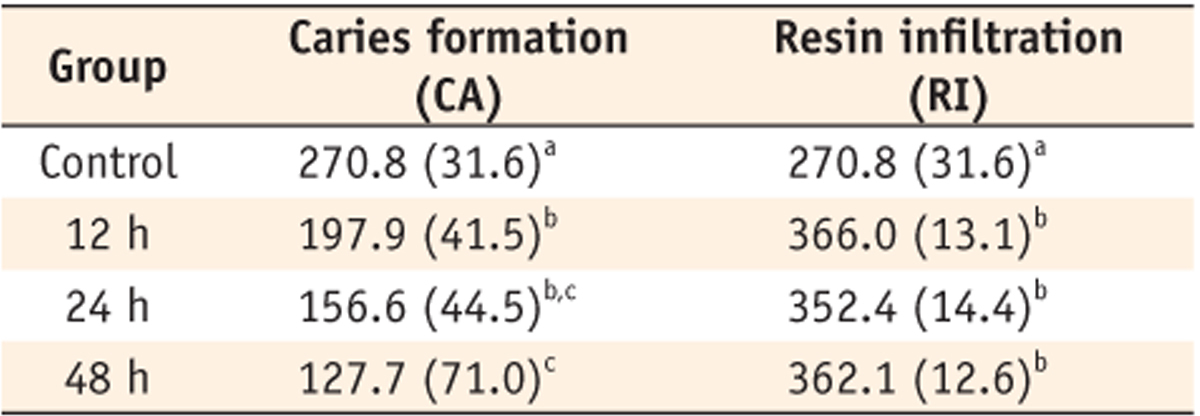
The mean values (SD) of Vickers hardness after caries formation for three different demineralization times and corresponding resin infiltration
Different superscript letters denote the values that are significantly different from one another in each column (p < 0.05).
Table 1
The difference in the mean values (SD) of CIE L*, a*, b*, and ΔE* among the control and the experimental groups after caries formation for three different demineralization times and corresponding resin infiltration
Different superscript letters denote the values that are significantly different from one another in each column (
Table 2
The mean values (SD) of Vickers hardness after caries formation for three different demineralization times and corresponding resin infiltration
Different superscript letters denote the values that are significantly different from one another in each column (

