
Articles
- Page Path
- HOME > Restor Dent Endod > Volume 32(5); 2007 > Article
- Original Article Influence of the labial surface irregularity on the measurement of the tooth color by spectrometer
- Yong-Jin Choi, Su-Jung Park, Hyun-Gu Cho, Yun-Chan Hwang, Won-Mann Oh, Byung-Ju Park1, In-Nam Hwang
-
2007;32(5):-418.
DOI: https://doi.org/10.5395/JKACD.2007.32.5.411
Published online: September 30, 2007
Department of Conservative Dentistry, School of Dentistry, DSRI, Chonnam National University, Korea.
1Department of Oral Biochemistry, School of Dentistry, DSRI, Chonnam National University, Korea.
- Corresponding Author: In-Nam Hwang. Department of Conservative Dentistry, School of Dentistry, Chonnam National University 5 Hak-dong, Dong-gu, Gwangju, 501-757, Korea. Tel: 82-62-220-4443, Fax: 82-62-225-8387, hinso@jnu.ac.kr
• Received: June 16, 2007 • Revised: July 7, 2007 • Accepted: July 31, 2007
Copyright © 2007 Korean Academy of Conservative Dentistry
- 1,617 Views
- 2 Download
- 1 Crossref


Tables & Figures
REFERENCES
Citations
Citations to this article as recorded by 
- A Study on Digital Color Reproduction for Recording Color Appearance of Cultural Heritage
Hyeong Rok Song, Young Hoon Jo
Journal of Conservation Science.2022; 38(2): 154. CrossRef
 ePub Link
 Cite
CiteInfluence of the labial surface irregularity on the measurement of the tooth color by spectrometer
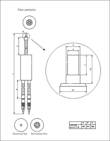


Figure 1
Diagram of the Duplex-Fiber bundle.
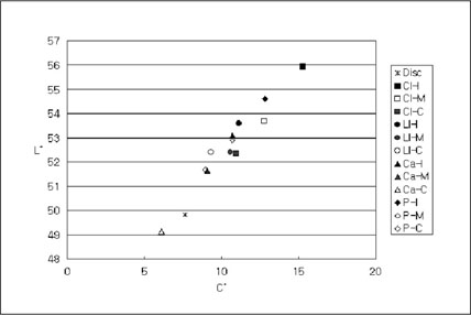
Figure 2
L* and C* value of the tested disc samples and tooth models.
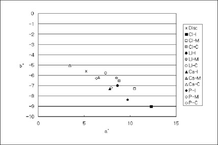
CI : Central incisor, LI : Laterial Incisor, Ca : Canine, P : Premolar, I : Incisal, M : Middle, C : Cervical
Figure 3
a* and b* value of the tested disc samples and tooth models.
Figure 1
Figure 2
Figure 3
Influence of the labial surface irregularity on the measurement of the tooth color by spectrometer
CIE L*a*b* and C* values of disc samples and tooth models
Standard deviations are in parentheses.
Statistical analysis between tooth and measured area
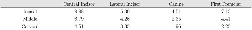
Color difference (ΔE*) between disc sample and tooth models
Color difference among measured area
*There are statistically significant differences among groups (p < 0.01).
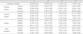
Table 1
CIE L*a*b* and C* values of disc samples and tooth models
Standard deviations are in parentheses.
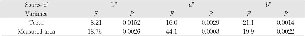
Table 2
Statistical analysis between tooth and measured area
Table 3
Color difference (ΔE*) between disc sample and tooth models
Table 4
Color difference among measured area
*There are statistically significant differences among groups (p < 0.01).

