Skip Navigation
Skip to contents

Restor Dent Endod : Restorative Dentistry & Endodontics

OPEN ACCESS

Articles

Page Path
HOME > Restor Dent Endod > Volume 33(2); 2008 > Article
  • rupiahtoto
  • rupiahtoto
  • rupiahtoto
  • rupiahtoto
  • rupiahtoto
  • rupiahtoto
  • slot deposit pulsa
  • bo slot gacor
  • toto togel
  • slot gacor
  • login rupiahtoto
  • rupiahtoto
  • rupiahtoto
  • rupiahtoto
  • rupiahtoto
  • Original Article Polymerization shrinkage, hygroscopic expansion and microleakage of resin-based temporary filling materials
    Nak Yeon Cho, In-Bog Lee
    2008;33(2):-124.
    DOI: https://doi.org/10.5395/JKACD.2008.33.2.115
    Published online: March 31, 2008

    Department of Consevative Dentistry, School of Dentistry, Seoul National University, Korea.

    Corresponding Author: In-Bog Lee. Department of Conservative Dentistry, School of Dentistry, Seoul National University, 275-1 Yeongeon-Dong, Jongno-Gu, Seoul 110-768, Korea. Tel: 82-2-2072-3953, Fax: 82-2-2072-3859, inboglee@snu.ac.kr
    • Received: January 28, 2008   • Revised: March 5, 2008   • Accepted: March 6, 2008

    Copyright © 2008 Korean Academy of Conservative Dentistry

    • 335 Views
    • 3 Download
    • 7 Crossref
    prev next
    • The purpose of this study was to measure the polymerization shrinkage and hygroscopic expansion of resin-based temporary filling materials and to evaluate microleakage at the interface between the materials and cavity wall.
      Five resin-based temporary filing materials were investigated: Fermit (Vivadent), Quicks (Dentkist), Provifil (Promedica), Spacer (Vericom), Clip (Voco). Caviton (GC) was also included for comparison. Polymerization shrinkage of five resin-based temporary filling materials was measured using the bonded disc method. For the measurement of hygroscopic expansion, the discs of six cured temporary filling materials were immersed in saline and a LVDT displacement sensor was used to measure the expansion for 7 days. For estimating of microleakage, Class I cavities were prepared on 120 extracted human molars and randomly assigned to 6 groups of 20 each. The cavities in each group were filled with six temporary filling materials. All specimens were submitted to 1000 thermo-cycles, with temperature varying from 5℃/55℃. Microleakage was determined using a dye penetration test.
      The results were as follows:
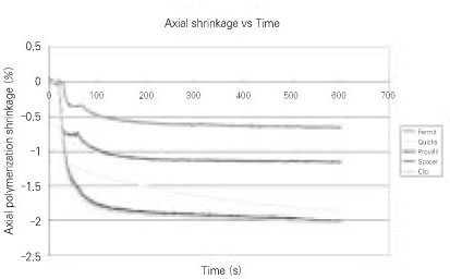
      1. Fermit had significantly less polymerization shrinkage than the other resin-based temporary filling materials. Fermit (0.22 %) < Spacer (0.38 %) < Quicks (0.64 %), Provifil (0.67 %), Clip (0.67 %)

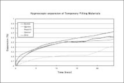
      2. Resin-based temporary filling materials showed 0.43 - 1.1 % expansion in 7 days.

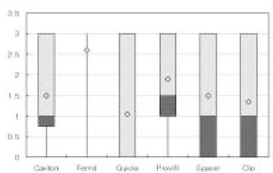
      3. Fermit showed the greatest leakage, while Quicks exhibited the least leakage.

      4. There are no correlation between polymerization shrinkage or hygroscopic expansion and microleakage of resin-based temporary filling materials.

    Figure 1
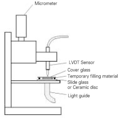
    Structure of an instrument to measure axial polymerization shrinkage.
    jkacd-33-115-g001.jpg
    Figure 2
    Representative curves of axial polymerization shrinkage of temporary filling materials as a function of time.
    jkacd-33-115-g002.jpg
    Figure 3-a
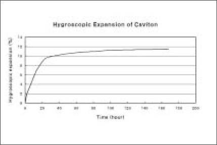
    Hygroscopic expansion of Caviton as a function of time.
    jkacd-33-115-g003.jpg
    Figure 3-b
    Initial hygroscopic expansion of temporary resin materials as a function of time.
    jkacd-33-115-g004.jpg
    Figure 4
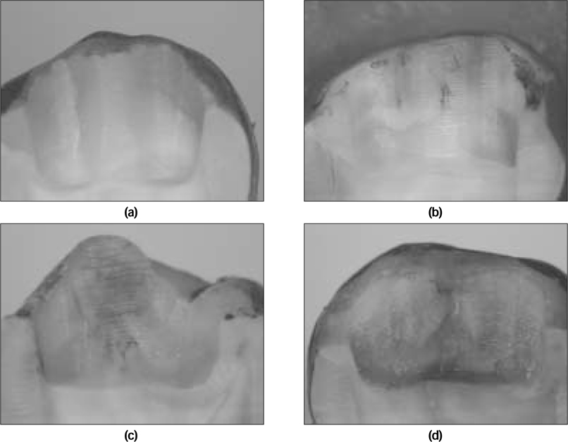
    The representative pictures for different microleakage patterns.
    (a) Score 0: no leakage
    (b) Score 1: leakage up to half of the cavity depth
    (c) Score 2: leakage in the entire cavity depth without reaching the pulpal wall
    (d) Score 3: leakage in the entire cavity depth even reaching the pulpal wall
    jkacd-33-115-g005.jpg
    Figure 5
    Boxplot display of mean and median leakage score for each group.
    The plot illustrates a summary of the microleakage scores based on the median, quartiles, and extreme values. The box represents the interquartile range which contains the 50% of values, the whiskers represent the highest and lowest microleakage values and the bold black line across the box indicates the median microleakage scores. The mark ◇ represents the mean microleakge scores.
    jkacd-33-115-g006.jpg
    Table 1
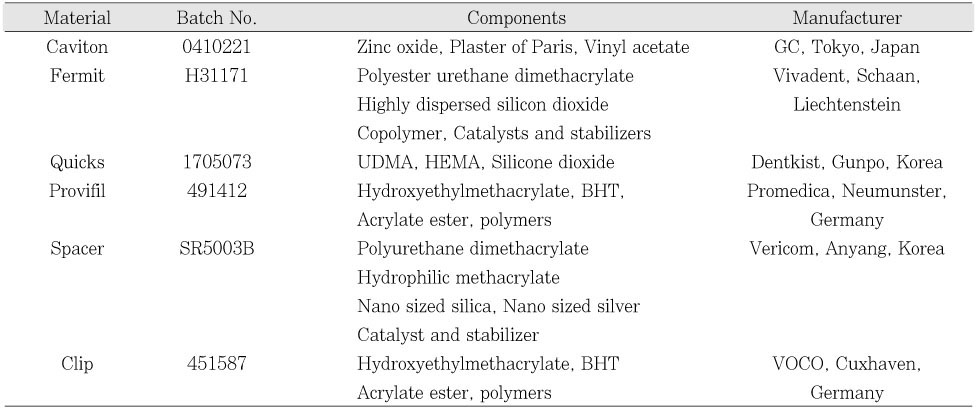
    Materials used in this study
    jkacd-33-115-i001.jpg
    Table 2
    Calculated linear polymerization shrinkage (%) of temporary filling materials

    The numbers in parenthesis are S.D.

    Same superscript letters mean that there is no statistical difference.

    Calculated linear shrinkage = measured axial shrinkage ×(1/3)

    jkacd-33-115-i002.jpg
    Table 3
    Hygroscopic expansion (%) of temporary filling materials at 24 hr and 7 days

    The numbers in parenthesis are S.D.

    Same superscript letters mean that there is no statistical difference.

    jkacd-33-115-i003.jpg
    Table 4
    Number of specimens in each score and mean microleakage score
    jkacd-33-115-i004.jpg
    • 1. Paul SJ, Scharer P. Effect of provisional cements on the bond strength of various adhesive bonding systems on dentine. J Oral Rehabil. 1997;24: 8-14.PubMed
    • 2. Garber DA, Goldstein RE. Porcelain and composite inlays & onlays. 1994;Quintessence Publishing Co, Inc; 57-61.
    • 3. Tulunoglu O, Uctasli MB, Ozdemir S. Coronal microleakage of temporary restorations in previously restored teeth with amalgam and composite. Oper Dent. 2005;30(3):331-337.PubMed
    • 4. Deveaux E, Hildelbert P, Neut C, Romond C. Bacterial microleakage of Cavit, IRM, TERM, and Fermit : a 21-day in vitro study. J Endod. 1999;25(10):653-659.ArticlePubMed
    • 5. Mayer T, Eickholz P. Microleakage of temporary restorations after thermocycling and mechanical loading. J Endod. 1997;23(5):320-322.ArticlePubMed
    • 6. Anderson RW, Powell BJ, Pashley DH. Microleakage of three temporary endodontic restorations. J Endod. 1988;14(10):497-501.PubMed
    • 7. Bobotis HG, Anderson RW, Pashley DH, Pantera EA. A microleakage study of temporary restorative materials used in endodontics. J Endod. 1989;15(12):569-572.ArticlePubMed
    • 8. Uctasli MB, Tinaz AC. Microleakage of different types of termporary restorative materials used in endodontics. J Oral Sci. 2000;42(2):63-67.PubMed
    • 9. Hosoya N, Cox CF, Arai T, Nakamura J. The walking bleach procedure: an in vitro study to measure microleakage of five temporary sealing agents. J Endod. 2000;26(12):716-718.ArticlePubMed
    • 10. Noguera AP, McDonald NJ. A comparative in vitro coronal microleakage study of new endodontic restorative materials. J Endod. 1990;6(11):523-527.
    • 11. Watts DC, Cash AJ. Determination of polymerization shrinkage kinetics in visible-light-cured materials: methods development. Dent Mater. 1991;281-287.ArticlePubMed
    • 12. Lee IB, Cho BH, Son HH, Um CM. The effect of consistency, specimen geometry and adhesion on the linear polymerization shrinkage measurement of light cured composites. Dent Mater. 2006;2(11):1071-1077.
    • 13. Campos PEGA, Sampaio Filho HR, Barceleir MO. Occlusal loading evaluation in the cervical intergrity of class II cavities filled with composite. Oper Dent. 2005;(6):727-732.
    • 14. de Gee AJ, Feilzer AJ, Davidson CL. True linear polymerization shrinkage of unfilled resins and composite determined with a linometer. Dent Mater. 1995;9: 11-14.
    • 15. Davidson CL, Feilzer AJ. Polymerization shrinkage and polymerization shrinkage stress in polymer-based restoratives. J Dent. 1997;25: 435-440.ArticlePubMed
    • 16. Scotti R, Ciocca L, Baldissara P. Microleakage of temporary endodontic restorations in overdenture tooth abutments. Int J Prosthodont. 2002;15: 479-482.PubMed
    • 17. Jacquot BM, Panighi MM, Steinmetz P, G'sell C. Evaluation of temporary restorations' microleakage by means of electrochemical impedance measurements. J Endod. 1996;22(11):586-589.ArticlePubMed
    • 18. Teplitsky PE, Meimaris IT. Sealing ability of Cavit and TERM as intermediate restorative materials. J Endod. 1988;14(6):278-282.PubMed
    • 19. Lee YC, Yang SF, Hwang YF, Chueh LH, Chung KH. Microleakage of endodontic temporary restorative materials. J Endod. 1993;19: 516-520.ArticlePubMed
    • 20. Widerman FH, Eames WB, Serene TP. The physical and biological properties of Cavit. JADA. 1971;82: 378-382.PubMed
    • 21. Webber RT, del Rio CE, Brady JM, Segall RO. Sealing quality of a temporary filling material. Oral Surg. 1978;46: 123-130.ArticlePubMed
    • 22. Uranga A, Blum JY, Esber S, Parahy E, Prado C. A comparative study of four coronal obturation materials in endodontic treatment. J Endod. 1999;25(3):178-180.PubMed
    • 23. Qvist V. The effect of mastication on marginal adaptation of composite restorations in vivo. J Dent Res. 1983;62: 904-906.ArticlePubMedPDF

    Tables & Figures

    Figure 1
    Structure of an instrument to measure axial polymerization shrinkage.
    jkacd-33-115-g001.jpg
    Figure 2
    Representative curves of axial polymerization shrinkage of temporary filling materials as a function of time.
    jkacd-33-115-g002.jpg
    Figure 3-a
    Hygroscopic expansion of Caviton as a function of time.
    jkacd-33-115-g003.jpg
    Figure 3-b
    Initial hygroscopic expansion of temporary resin materials as a function of time.
    jkacd-33-115-g004.jpg
    Figure 4
    The representative pictures for different microleakage patterns.
    (a) Score 0: no leakage
    (b) Score 1: leakage up to half of the cavity depth
    (c) Score 2: leakage in the entire cavity depth without reaching the pulpal wall
    (d) Score 3: leakage in the entire cavity depth even reaching the pulpal wall
    jkacd-33-115-g005.jpg
    Figure 5
    Boxplot display of mean and median leakage score for each group.
    The plot illustrates a summary of the microleakage scores based on the median, quartiles, and extreme values. The box represents the interquartile range which contains the 50% of values, the whiskers represent the highest and lowest microleakage values and the bold black line across the box indicates the median microleakage scores. The mark ◇ represents the mean microleakge scores.
    jkacd-33-115-g006.jpg
    Table 1
    Materials used in this study
    jkacd-33-115-i001.jpg
    Table 2
    Calculated linear polymerization shrinkage (%) of temporary filling materials

    The numbers in parenthesis are S.D.

    Same superscript letters mean that there is no statistical difference.

    Calculated linear shrinkage = measured axial shrinkage ×(1/3)

    jkacd-33-115-i002.jpg
    Table 3
    Hygroscopic expansion (%) of temporary filling materials at 24 hr and 7 days

    The numbers in parenthesis are S.D.

    Same superscript letters mean that there is no statistical difference.

    jkacd-33-115-i003.jpg
    Table 4
    Number of specimens in each score and mean microleakage score
    jkacd-33-115-i004.jpg

    REFERENCES

    • 1. Paul SJ, Scharer P. Effect of provisional cements on the bond strength of various adhesive bonding systems on dentine. J Oral Rehabil. 1997;24: 8-14.PubMed
    • 2. Garber DA, Goldstein RE. Porcelain and composite inlays & onlays. 1994;Quintessence Publishing Co, Inc; 57-61.
    • 3. Tulunoglu O, Uctasli MB, Ozdemir S. Coronal microleakage of temporary restorations in previously restored teeth with amalgam and composite. Oper Dent. 2005;30(3):331-337.PubMed
    • 4. Deveaux E, Hildelbert P, Neut C, Romond C. Bacterial microleakage of Cavit, IRM, TERM, and Fermit : a 21-day in vitro study. J Endod. 1999;25(10):653-659.ArticlePubMed
    • 5. Mayer T, Eickholz P. Microleakage of temporary restorations after thermocycling and mechanical loading. J Endod. 1997;23(5):320-322.ArticlePubMed
    • 6. Anderson RW, Powell BJ, Pashley DH. Microleakage of three temporary endodontic restorations. J Endod. 1988;14(10):497-501.PubMed
    • 7. Bobotis HG, Anderson RW, Pashley DH, Pantera EA. A microleakage study of temporary restorative materials used in endodontics. J Endod. 1989;15(12):569-572.ArticlePubMed
    • 8. Uctasli MB, Tinaz AC. Microleakage of different types of termporary restorative materials used in endodontics. J Oral Sci. 2000;42(2):63-67.PubMed
    • 9. Hosoya N, Cox CF, Arai T, Nakamura J. The walking bleach procedure: an in vitro study to measure microleakage of five temporary sealing agents. J Endod. 2000;26(12):716-718.ArticlePubMed
    • 10. Noguera AP, McDonald NJ. A comparative in vitro coronal microleakage study of new endodontic restorative materials. J Endod. 1990;6(11):523-527.
    • 11. Watts DC, Cash AJ. Determination of polymerization shrinkage kinetics in visible-light-cured materials: methods development. Dent Mater. 1991;281-287.ArticlePubMed
    • 12. Lee IB, Cho BH, Son HH, Um CM. The effect of consistency, specimen geometry and adhesion on the linear polymerization shrinkage measurement of light cured composites. Dent Mater. 2006;2(11):1071-1077.
    • 13. Campos PEGA, Sampaio Filho HR, Barceleir MO. Occlusal loading evaluation in the cervical intergrity of class II cavities filled with composite. Oper Dent. 2005;(6):727-732.
    • 14. de Gee AJ, Feilzer AJ, Davidson CL. True linear polymerization shrinkage of unfilled resins and composite determined with a linometer. Dent Mater. 1995;9: 11-14.
    • 15. Davidson CL, Feilzer AJ. Polymerization shrinkage and polymerization shrinkage stress in polymer-based restoratives. J Dent. 1997;25: 435-440.ArticlePubMed
    • 16. Scotti R, Ciocca L, Baldissara P. Microleakage of temporary endodontic restorations in overdenture tooth abutments. Int J Prosthodont. 2002;15: 479-482.PubMed
    • 17. Jacquot BM, Panighi MM, Steinmetz P, G'sell C. Evaluation of temporary restorations' microleakage by means of electrochemical impedance measurements. J Endod. 1996;22(11):586-589.ArticlePubMed
    • 18. Teplitsky PE, Meimaris IT. Sealing ability of Cavit and TERM as intermediate restorative materials. J Endod. 1988;14(6):278-282.PubMed
    • 19. Lee YC, Yang SF, Hwang YF, Chueh LH, Chung KH. Microleakage of endodontic temporary restorative materials. J Endod. 1993;19: 516-520.ArticlePubMed
    • 20. Widerman FH, Eames WB, Serene TP. The physical and biological properties of Cavit. JADA. 1971;82: 378-382.PubMed
    • 21. Webber RT, del Rio CE, Brady JM, Segall RO. Sealing quality of a temporary filling material. Oral Surg. 1978;46: 123-130.ArticlePubMed
    • 22. Uranga A, Blum JY, Esber S, Parahy E, Prado C. A comparative study of four coronal obturation materials in endodontic treatment. J Endod. 1999;25(3):178-180.PubMed
    • 23. Qvist V. The effect of mastication on marginal adaptation of composite restorations in vivo. J Dent Res. 1983;62: 904-906.ArticlePubMedPDF

    Citations

    Citations to this article as recorded by  
    • Comparison of color stability, gloss, mechanical and physical properties according to dental temporary filling materials type
      Ji-Won Choi, You-Young Shin, Song-Yi Yang
      Korean Journal of Dental Materials.2022; 49(3): 97.     CrossRef
    • Comparative analysis of strain according to two wavelengths of light source and constant temperature bath deposition in ultraviolet-curing resin for dental three-dimensional printing
      Dong-Yeon Kim, Gwang-Young Lee, Hoo-Won Kang, Cheon-Seung Yang
      Journal of Korean Acedemy of Dental Technology.2020; 42(3): 208.     CrossRef
    • Effect of cavity disinfectants on antibacterial activity and microtensile bond strength in class I cavity
      Bo-Ram KIM, Man-Hwan OH, Dong-Hoon SHIN
      Dental Materials Journal.2017; 36(3): 368.     CrossRef
    • Shear bond strength of a self-adhesive resin cement to resin-coated dentin
      Jee-Youn Hong, Cheol-Woo Park, Jeong-Uk Heo, Min-Ki Bang, Jae-Jun Ryu
      The Journal of Korean Academy of Prosthodontics.2013; 51(1): 27.     CrossRef
    • Coronal microleakage of four temporary restorative materials in Class II-type endodontic access preparations
      Sang-Mi Yun, Lorena Karanxha, Hee-Jin Kim, Sung-Ho Jung, Su-Jung Park, Kyung-San Min
      Restorative Dentistry & Endodontics.2012; 37(1): 29.     CrossRef
    • Microtensile bond strength of resin inlay bonded to dentin treated with various temporary filling materials
      Tae-Woo Kim, Bin-Na Lee, Young-Jung Choi, So-Young Yang, Hoon-Sang Chang, Yun-Chan Hwang, In-Nam Hwang, Won-Mann Oh
      Journal of Korean Academy of Conservative Dentistry.2011; 36(5): 419.     CrossRef
    • The Effect of Temporary Filling Materials on The Adhesion between Dentin Adhesive-coated Surface and Resin Inlay
      Tae-Gun Kim, Kwang-Won Lee, Mi-Kyung Yu
      Journal of Korean Academy of Conservative Dentistry.2008; 33(6): 553.     CrossRef
    CanvasJS.com
    CanvasJS.com
    CanvasJS.com

    • ePub LinkePub Link
    • Cite
      CITE
      export Copy Download
      Close
      Download Citation
      Download a citation file in RIS format that can be imported by all major citation management software, including EndNote, ProCite, RefWorks, and Reference Manager.

      Format:
      • RIS — For EndNote, ProCite, RefWorks, and most other reference management software
      • BibTeX — For JabRef, BibDesk, and other BibTeX-specific software
      Include:
      • Citation for the content below
      Polymerization shrinkage, hygroscopic expansion and microleakage of resin-based temporary filling materials
      J Korean Acad Conserv Dent. 2008;33(2):115-124.   Published online March 31, 2008
      Close
    • XML DownloadXML Download
    Figure
    • 0
    Polymerization shrinkage, hygroscopic expansion and microleakage of resin-based temporary filling materials
    Image Image Image Image Image Image
    Figure 1 Structure of an instrument to measure axial polymerization shrinkage.
    Figure 2 Representative curves of axial polymerization shrinkage of temporary filling materials as a function of time.
    Figure 3-a Hygroscopic expansion of Caviton as a function of time.
    Figure 3-b Initial hygroscopic expansion of temporary resin materials as a function of time.
    Figure 4 The representative pictures for different microleakage patterns. (a) Score 0: no leakage (b) Score 1: leakage up to half of the cavity depth (c) Score 2: leakage in the entire cavity depth without reaching the pulpal wall (d) Score 3: leakage in the entire cavity depth even reaching the pulpal wall
    Figure 5 Boxplot display of mean and median leakage score for each group. The plot illustrates a summary of the microleakage scores based on the median, quartiles, and extreme values. The box represents the interquartile range which contains the 50% of values, the whiskers represent the highest and lowest microleakage values and the bold black line across the box indicates the median microleakage scores. The mark ◇ represents the mean microleakge scores.
    Polymerization shrinkage, hygroscopic expansion and microleakage of resin-based temporary filling materials

    Materials used in this study

    Calculated linear polymerization shrinkage (%) of temporary filling materials

    The numbers in parenthesis are S.D.

    Same superscript letters mean that there is no statistical difference.

    Calculated linear shrinkage = measured axial shrinkage ×(1/3)

    Hygroscopic expansion (%) of temporary filling materials at 24 hr and 7 days

    The numbers in parenthesis are S.D.

    Same superscript letters mean that there is no statistical difference.

    Number of specimens in each score and mean microleakage score

    Table 1 Materials used in this study

    Table 2 Calculated linear polymerization shrinkage (%) of temporary filling materials

    The numbers in parenthesis are S.D.

    Same superscript letters mean that there is no statistical difference.

    Calculated linear shrinkage = measured axial shrinkage ×(1/3)

    Table 3 Hygroscopic expansion (%) of temporary filling materials at 24 hr and 7 days

    The numbers in parenthesis are S.D.

    Same superscript letters mean that there is no statistical difference.

    Table 4 Number of specimens in each score and mean microleakage score


    Restor Dent Endod : Restorative Dentistry & Endodontics
    Close layer
    TOP Mpgyi