
Articles
- Page Path
- HOME > Restor Dent Endod > Volume 34(6); 2009 > Article
- Original Article Real-time measurement of dentinal tubular fluid flow during and after amalgam and composite restorations
- Sun-Young Kim1, Byeong-Hoon Cho1, Seung-Ho Baek1, Bum-Sun Lim2, In-Bog Lee1
-
2009;34(6):-476.
DOI: https://doi.org/10.5395/JKACD.2009.34.6.467
Published online: November 30, 2009
1Department of Conservative Dentistry, School of Dentistry, Seoul National University, Korea.
2Department of Dental Biomaterials, School of Dentistry, Seoul National University, Korea.
- Corresponding Author: In-Bog Lee. Department of Conservative Dentistry, School of Dentistry, Seoul National University, 275-1 Yeongeon-Dong, Jongno-Gu, Seoul 110-768, Korea. Tel: 82-2-2072-3953, Fax: 82-2-2072-3859, inboglee@snu.ac.kr
• Received: June 29, 2009 • Revised: August 31, 2009 • Accepted: September 1, 2009
Copyright © 2009 The Korean Academy of Conservative Dentistry
- 1,590 Views
- 4 Download
- 1 Crossref

Tables & Figures
REFERENCES
Citations
Citations to this article as recorded by 
- Real-time measurement of dentinal fluid flow during desensitizing agent application
Sun-Young Kim, Eun-Joo Kim, In-Bog Lee
Journal of Korean Academy of Conservative Dentistry.2010; 35(5): 313. CrossRef
 ePub Link
 Cite
CiteReal-time measurement of dentinal tubular fluid flow during and after amalgam and composite restorations


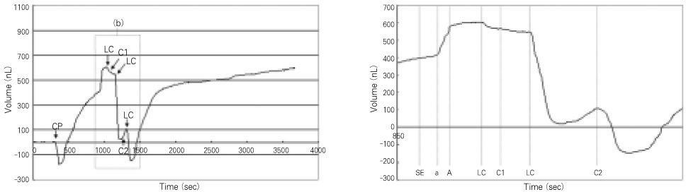

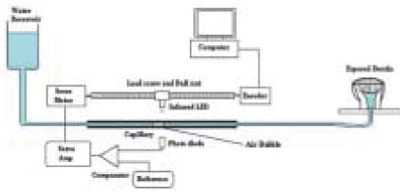
Figure 1
Schematic diagram of the dentinal tubular fluid flow measurement system.
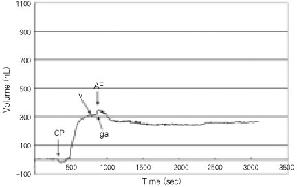
Figure 2
Consecutive DFF during amalgam restoration. Upward movement (+) vs time on graph indicates outward DFF, whereas downward movement (-) indicates inward DFF. CP: cavity preparation; V: varnish application; ga: gentle air stream; AF: amalgam filling.
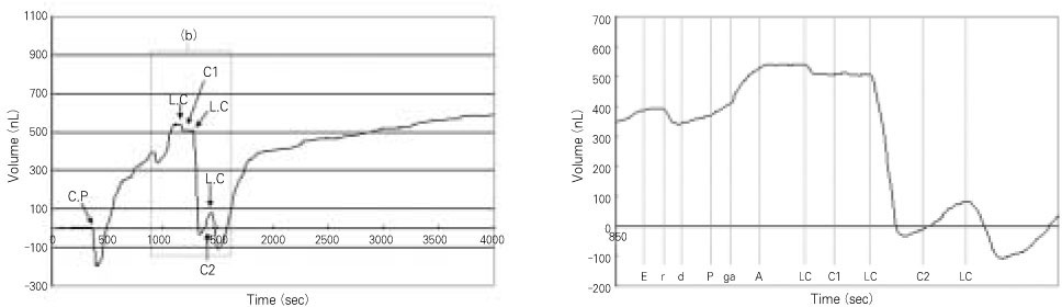
Figure 3
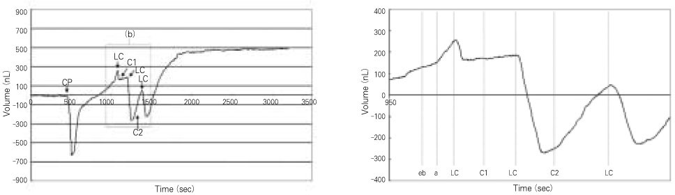
(a) Consecutive DFF during composite restoration with MP. (b) Magnified view of consecutive DFF during bonding procedure of MP. Upward movement (+) vs time on graph indicates outward DFF, whereas downward movement (-) indicates inward DFF. CP: cavity preparation; LC: light curing; C1: first layer filling of composite; C2: second layer filling of composite; E: acid-etching; r: rinse; d: blot-dry; P: primer application; ga: gentle air; A: adhesive application.
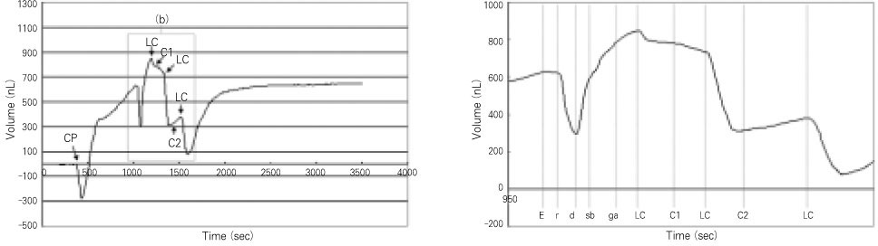
Figure 4
(a) Consecutive DFF during composite restoration with SB. (b) Magnified view of consecutive DFF during bonding procedure of SB. Upward movement (+) vs time on graph indicates outward DFF, whereas downward movement (-) indicates inward DFF. CP: cavity preparation; LC: light curing; C1: first layer filling of composite; C2: second layer filling of composite; E: acid-etching; r: rinse; d: blot-dry; sb: Single Bond application; ga: gentle air.
Figure 5
(a) Consecutive DFF during composite restoration with CE. (b) Magnified view of consecutive DFF during bonding procedure of CE. Upward movement (+) vs time on graph indicates outward DFF, whereas downward movement (-) indicates inward DFF. CP: cavity preparation; LC: light curing; C1: first layer filling of composite; C2: second layer filling of composite; SE: self-etching primer application; a: air-dry; A: adhesive application.
Figure 6
(a) Consecutive DFF during composite restoration with EB. (b) Magnified view of consecutive DFF during bonding procedure of EB. Upward movement (+) vs time on graph indicates outward DFF, whereas downward movement (-) indicates inward DFF. CP: cavity preparation; LC: light curing; C1: first layer filling of composite; C2: second layer filling of composite; eb: Easy Bond application; a: air-dry.
Figure 1
Figure 2
Figure 3
Figure 4
Figure 5
Figure 6
Real-time measurement of dentinal tubular fluid flow during and after amalgam and composite restorations
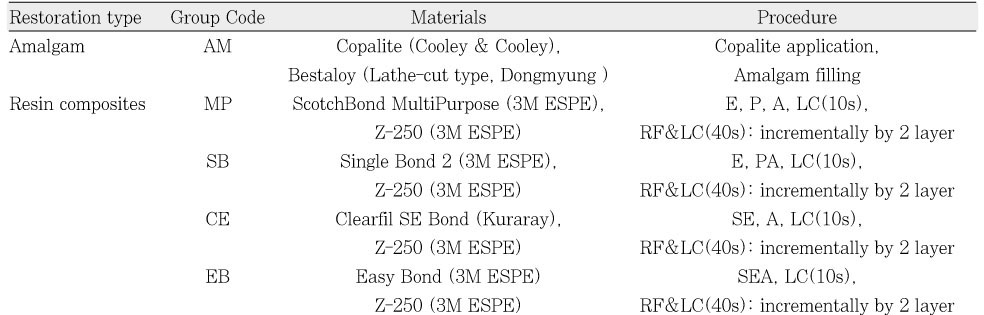
The restorative materials and procedures for each group.
Abbreviation: E, acid etching for 15 sec; P, Primer application; A, Adhesive application; PA, application of mixed agent with primer and adhesive; SE, self-etching primer application; SEA, application of self-etching adhesive; LC, light-curing; RF, resin composite filling.
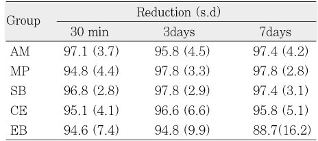
Flow rate reduction (%) of each group at 30 minutes, 3 days, and 7days after restoration with respect to baseline (n=10).
There was no statistically significant difference among groups for each measurement time. There was no statistically significant difference according to the measurement time for each material. s.d=standard deviation.
Table 1
The restorative materials and procedures for each group.
Abbreviation: E, acid etching for 15 sec; P, Primer application; A, Adhesive application; PA, application of mixed agent with primer and adhesive; SE, self-etching primer application; SEA, application of self-etching adhesive; LC, light-curing; RF, resin composite filling.
Table 2
Flow rate reduction (%) of each group at 30 minutes, 3 days, and 7days after restoration with respect to baseline (n=10).
There was no statistically significant difference among groups for each measurement time. There was no statistically significant difference according to the measurement time for each material. s.d=standard deviation.

