
Articles
- Page Path
- HOME > Restor Dent Endod > Volume 35(1); 2010 > Article
- Basic Research Pulp response of beagle dog to direct pulp capping materials: Histological study
-
Ji-Hyun Bae1, Young-Gyun Kim2, Pil-Young Yoon2, Byeong-Hoon Cho3
 , Yong-Hoon Choi1
 -
2010;35(1):-12.
DOI: https://doi.org/10.5395/JKACD.2010.35.1.005
Published online: January 31, 2010
1Department of Conservative Dentistry, Seoul National University Bundang Hospital, Korea.
2Department of Oral Maxillofacial Surgery, Seoul National University Bundang Hospital, Korea.
3Department of Conservative Dentistry, School of Dentistry, Seoul National University, Korea.
- Corresponding Author: Yong-Hoon Choi. Department of Conservative Dentistry, Seoul National University Bundang Hospital, 300 Gumi-dong, Bundang-gu, Seongnam-si Gyeonggi-do, 463-707, Korea. Tel: 82-31-787-2780, Fax: 82-31-787-4068, yhchoi@snubh.org
• Received: September 8, 2009 • Revised: October 1, 2009 • Accepted: October 23, 2009
Copyright © 2010 Korean Academy of Conservative Dentistry
- 1,964 Views
- 13 Download
- 4 Crossref

Tables & Figures
REFERENCES
Citations
Citations to this article as recorded by 
- Experimental Study of Pulp Capping Using Xenogenic Demineralized Dentin Paste
Ji-Young Yun, Yong-Hoon Choi, Young-Kyun Kim, In-Woong Um, Joo-Cheol Park, Ji-Yoon Kim
Journal of Hard Tissue Biology.2016; 25(3): 321. CrossRef - Comparison of gene expression profiles of human dental pulp cells treated with mineral trioxide aggregate and calcium hydroxide
Yong-Beom Kim, Won-Jun Shon, Woocheol Lee, Kee-Yeon Kum, Seung-Ho Baek, Kwang-Shik Bae
Journal of Korean Academy of Conservative Dentistry.2011; 36(5): 397. CrossRef - Gene expression profiling in human dental pulp cells treated with mineral trioxide aggregate
Yong-Beom Kim, Won-Jun Shon, WooCheol Lee, Kee-Yeon Kum, Seung-Ho Baek, Kwang-Shik Bae
Journal of Korean Academy of Conservative Dentistry.2010; 35(3): 152. CrossRef - Histology of dental pulp healing after tooth replantation in rats
Eun-Jin Go, Han-Seong Jung, Eui-Seong Kim, Il-Young Jung, Seung-Jong Lee
Journal of Korean Academy of Conservative Dentistry.2010; 35(4): 273. CrossRef
 ePub Link
 Cite
CitePulp response of beagle dog to direct pulp capping materials: Histological study


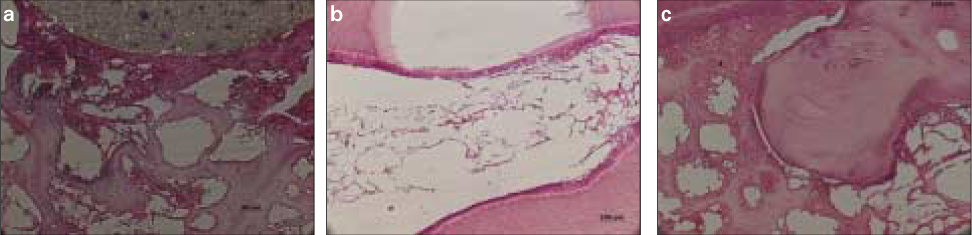

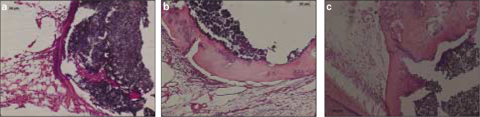
Figure 1
a. Pulp capping with MTA at 7 days. Inflammatory cells infiltrated around the capping material under odontoblast-like cell. b. Pulp capping with MTA at 30 days. Newly formed dentinal bridge was observed. c. Pulp capping with MTA at 90 days. The thickness of hard tissue increased, compared with that of 30 days.
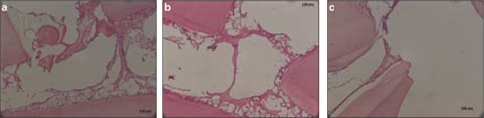
Figure 2
a. Pulp capping with SE Bond at 7 days. Inflammatory cells infiltrated accompanied by soft tissue disturbances. b. Pulp capping with SE Bond at 30 days. Coronal pulp tissue necrosis beneath the exposure site was observed. c. Pulp capping with SE Bond at 90 days. Severe pulp tissue necrosis beneath the exposure site was seen.
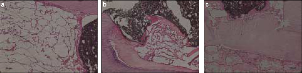
Figure 3
a. Pulp capping with Ultra-blend at 7 days. Inflammatory cell infiltration and hemorrhage was observed. b. Pulp capping with Ultra-blend at 30 days. Odontoblast-like cells arranged beneath exposure site. Pulp degeneration and partial pulp necrosis was seen. There was no hard tissue formation. c. Pulp capping with Ultra-blend at 90 days. Complete dentinal bridge protected the exposure site.
Figure 4
a. Pulp capping with Dycal at 7 days. Pulp tissue degeneration was seen. b. Pulp capping with Dycal at 30 days. Partial pulp tissue degeneration and hard tissue formation were observed. c. Pulp capping with Dycal at 90 days. Complete hard tissue formation and normal soft tissue organization beneath the exposure site.
Figure 1
Figure 2
Figure 3
Figure 4
Pulp response of beagle dog to direct pulp capping materials: Histological study
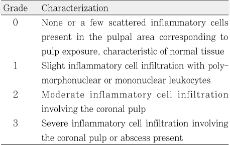
Criteria for grading inflammatory cell response
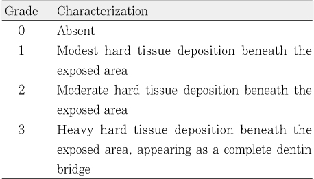
Criteria for grading hard tissue formation
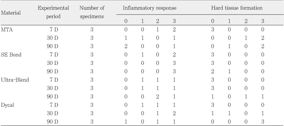
Inflammatory responses and hard tissue formation resulted with 4 pulp capping materials
Table 1
Criteria for grading inflammatory cell response
Table 2
Criteria for grading hard tissue formation
Table 3
Inflammatory responses and hard tissue formation resulted with 4 pulp capping materials

