
Articles
- Page Path
- HOME > Restor Dent Endod > Volume 36(5); 2011 > Article
- Basic Research Effect of Er:YAG lasing on the dentin bonding strength of two-step adhesives
- Byeong-Choon Song, DDS, PhD1, Young-Gon Cho, DDS, MSD, PhD1, Myung-Seon Lee, RDH2
-
2011;36(5):-418.
DOI: https://doi.org/10.5395/JKACD.2011.36.5.409
Published online: September 30, 2011
1Department of Conservative Dentistry, Chosun University School of Dentistry, Gwangju, Korea.
2Department of Dental Hygiene, SeoYeong University, Gwangju, Korea.
- Correspondence to Young-Gon Cho, DDS, MSD, PhD. Professor, Department of Conservative Dentistry, Chosun University School of Dentistry, 421 Seosuk-dong Dong-gu, Gwangju, Korea 501-825. TEL, +82-62-220-3845; FAX, +82-62-223-9064; ygcho@mail.chosun.ac.kr
• Received: July 29, 2011 • Revised: August 29, 2011 • Accepted: August 29, 2011
Copyright © 2011 Korean Academy of Conservative Dentistry
- 1,566 Views
- 2 Download
- 2 Crossref


Tables & Figures
REFERENCES
Citations
Citations to this article as recorded by 
- Effect of Acid or Laser Treatment on Degradation of Dentin Matrix
Aslihan Usumez, Tugrul Sari, Roda Seseogullari Dirihan, Mehmet Esad Guven, Serra Oguz Ahmet, Norbert Gutknecht, Arzu Tezvergil Mutluay
Lasers in Dental Science.2022; 6(2): 99. CrossRef - Ablation of carious dental tissue using an ultrashort pulsed laser (USPL) system
Christoph Engelbach, Claudia Dehn, Christoph Bourauel, Jörg Meister, Matthias Frentzen
Lasers in Medical Science.2015; 30(5): 1427. CrossRef
 ePub Link
 Cite
CiteEffect of Er:YAG lasing on the dentin bonding strength of two-step adhesives


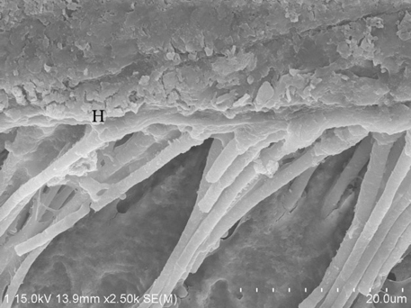
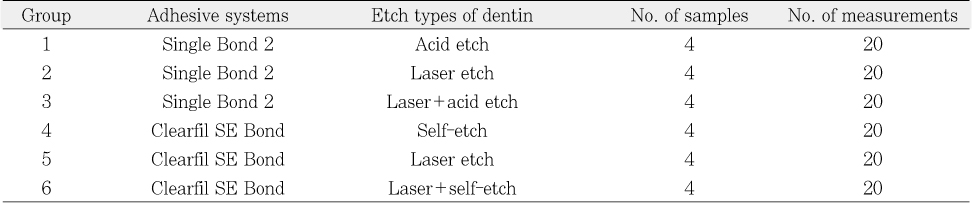
Figure 1
Scanning electron microscophic photograph showing the hybrid layer (H) and many taper resin tags with lateral branches (LB) in Group 1.
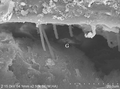
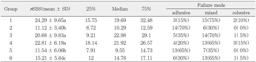
Figure 2
Scanning electron microscophic photograph showing the wide gap (G) and few short and slender resin tags in Group 2.
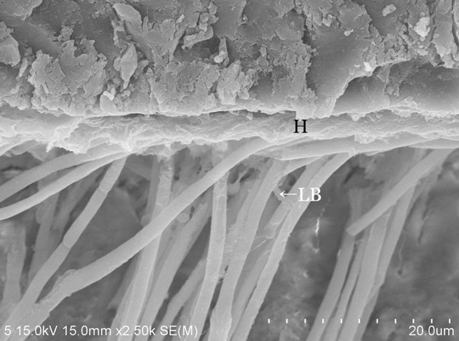
Figure 3
Scanning electron microscophic photograph showing the hybrid layer (H), slender and long rod shaped resin tags with lateral branches (LB) in Group 3.
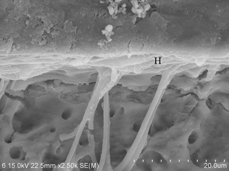
Figure 4
Scanning electron microscophic photograph showing the hybrid layer (H) and many long rod shaped resin tags with lateral branches (LB) in Group 4.
Figure 5
Scanning electron microscophic photograph showing the small gap (G) at the resin-dentin interface and short and slender resin tags in Group 5.
Figure 6
Scanning electron microscophic photograph showingthe hybrid layer (H) and few slender and long resin tags in Group 6.
Figure 1
Figure 2
Figure 3
Figure 4
Figure 5
Figure 6
Effect of Er:YAG lasing on the dentin bonding strength of two-step adhesives
Adhesive systems and resin composite
Group classification by adhesive systems and etch types of dentin
Mean microshear bond strength (MPa) to dentin and failure mode
G1, G4, Acid etch; G2, G5, Laser etch; G3, G6, Laser etch + acid etch.
G1, G2 and G3 used Single Bond 2. G4, G5 and G6 used Clearfil SE Bond.
Different superscripts indicate values of statistically significant difference by Kruskal-Wallis test, median test and Mann-Whitney test.
Table 1
Adhesive systems and resin composite
Table 2
Group classification by adhesive systems and etch types of dentin
Table 3
Mean microshear bond strength (MPa) to dentin and failure mode
G1, G4, Acid etch; G2, G5, Laser etch; G3, G6, Laser etch + acid etch. G1, G2 and G3 used Single Bond 2. G4, G5 and G6 used Clearfil SE Bond. Different superscripts indicate values of statistically significant difference by Kruskal-Wallis test, median test and Mann-Whitney test.

