
Articles
- Page Path
- HOME > Restor Dent Endod > Volume 27(6); 2002 > Article
- Original Article Scanning electron microscopic study on the efficacy of root canal wall debridement of rotary Ni-Ti instruments with different cutting angle
- In-soo Jeon, Kee-yeon Kum, Seong-ho Park, Tai-cheol Yoon
-
2002;27(6):-586.
DOI: https://doi.org/10.5395/JKACD.2002.27.6.577
Published online: November 30, 2002
Department of Conservative Dentistry, Yonsei University, Korea.
Copyright © 2002 Korean Academy of Conservative Dentistry
- 1,660 Views
- 0 Download
- 3 Crossref


Tables & Figures
REFERENCES
Citations
Citations to this article as recorded by 
- Comparative evaluation of dentin volume removal and centralization of the root canal after shaping with the ProTaper Universal, ProTaper Gold, and One-Curve instruments using micro-CT
Hatice Yalniz, Mehrdad Koohnavard, Aysenur Oncu, Berkan Celikten, Ayse Isil Orhan, Kaan Orhan
Journal of Dental Research, Dental Clinics, Dental Prospects.2021; 15(1): 47. CrossRef - Microorganism penetration in dentinal tubules of instrumented and retreated root canal walls.In vitroSEM study
Saad Al-Nazhan, Alaa Al-Sulaiman, Fellwa Al-Rasheed, Fatimah Alnajjar, Bander Al-Abdulwahab, Abdulhakeem Al-Badah
Restorative Dentistry & Endodontics.2014; 39(4): 258. CrossRef - Shaping characteristics of two different motions nickel titanium file: a preliminary comparative study of surface profile and dentin chip
So-Ra Park, Se-Hee Park, Kyung-Mo Cho, Jin-Woo Kim
Journal of Dental Rehabilitation and Applied Science.2014; 30(2): 121. CrossRef
 ePub Link
 Cite
CiteScanning electron microscopic study on the efficacy of root canal wall debridement of rotary Ni-Ti instruments with different cutting angle


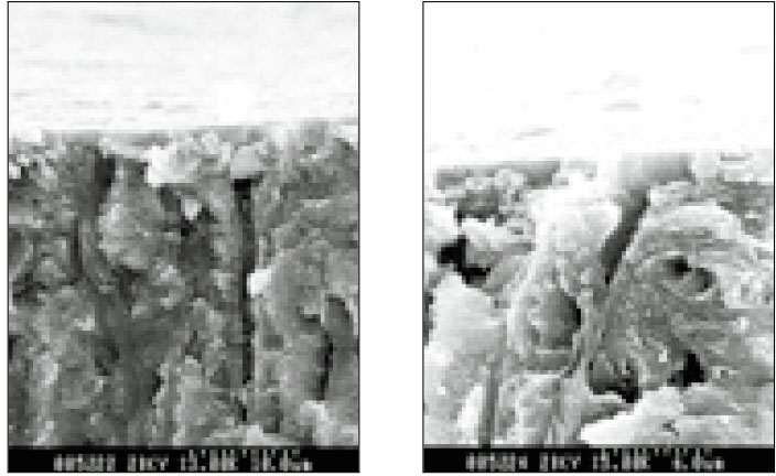
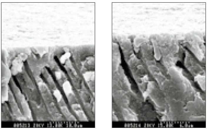


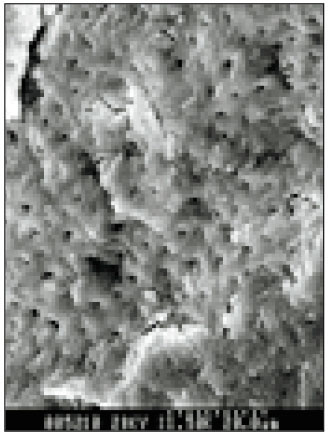
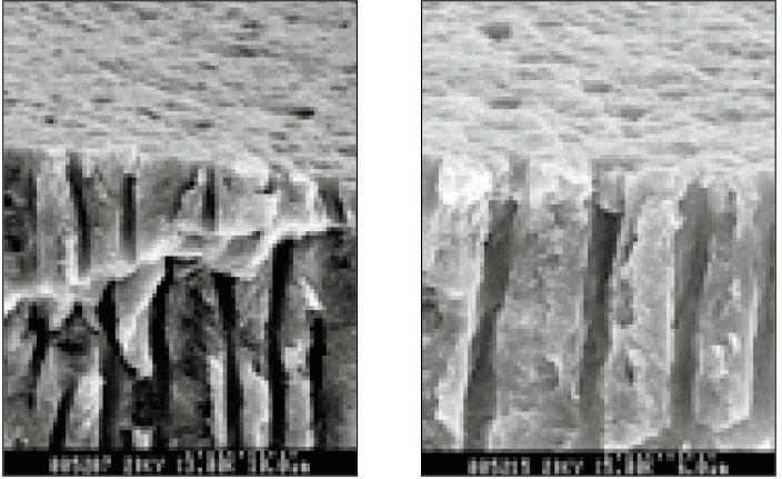
Fig. 1
Scanning electron micrograph of canal wall prepared with Ni-Ti HERO 642(×1,500).
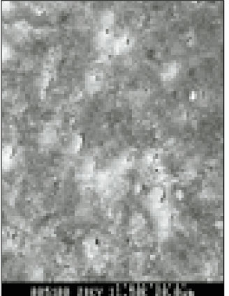
Fig. 2
Scanning electron micrograph of canal wall prepared with Ni-Ti ProFile(×1,500).
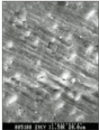
Fig. 3
Scanning electron micrograph of canal wall prepared with stainless-steel engine reamer(×1,500).
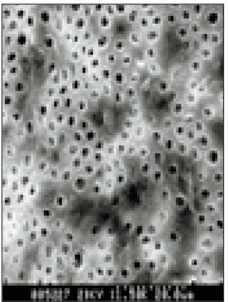
Fig. 4
Scanning electron micrograph of canal wall extirpated with barbed broach but not instrumented(×1,500).
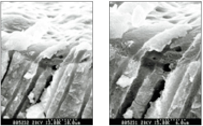
Fig. 5
the penetration depth of smear layer into dentinal tubules was observed on canal wall prepared with Ni-Ti HERO 642(×3,000, ×5,000).
Fig. 6
the penetration depth of smear layer into dentinal tubules was observed on canal wall prepared with Ni-Ti ProFile(×3,000, ×5,000).
Fig. 7
the penetration depth of smear layer into dentinal tubules was observed on canal wall prepared with stainless-steel engine reamer(×3,000, ×5,000).
Fig. 8
the penetration depth of smear layer into dentinal tubules was observed on canal wall extirpated with barbed broach but not instrumented (×3,000, ×5,000).
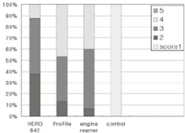
Fig. 9
Total scores of smear layer at apical third in each groups
Fig. 1
Fig. 2
Fig. 3
Fig. 4
Fig. 5
Fig. 6
Fig. 7
Fig. 8
Fig. 9
Scanning electron microscopic study on the efficacy of root canal wall debridement of rotary Ni-Ti instruments with different cutting angle
Comparison of smear layer score
Table 1
Comparison of smear layer score

