
Articles
- Page Path
- HOME > Restor Dent Endod > Volume 27(1); 2002 > Article
- Original Article Color changes in composites according to various light curing sources
- Young-Gon Cho, Myung-Cho Kim
-
2002;27(1):-94.
DOI: https://doi.org/10.5395/JKACD.2002.27.1.087
Published online: January 31, 2002
Department of Conservative Dentistry, College of dentistry, Chosun University, Korea.
Copyright © 2002 Korean Academy of Conservative Dentistry
- 1,661 Views
- 2 Download
- 3 Crossref


Tables & Figures
REFERENCES
Citations
Citations to this article as recorded by 
- Effects of the color components of light-cured composite resin before and after polymerization on degree of conversion and flexural strength
Ji-A Yoo, Byeong-Hoon Cho
Journal of Korean Academy of Conservative Dentistry.2011; 36(4): 324. CrossRef - Effect of the difference in spectral outputs of the single and dual-peak LEDs on the microhardness and the color stability of resin composites
Hye-Jung Park, Sung-Ae Son, Bock Hur, Hyeon-Cheol Kim, Yong-Hoon Kwon, Jeong-Kil Park
Journal of Korean Academy of Conservative Dentistry.2011; 36(2): 108. CrossRef - Color changes in composite resins exposed to xenon lamp
Young-Gon Cho, Jeong-Il Seo, Soo-Mee Kim, Jin-Ho Jeong, Young-Gon Lee
Journal of Korean Academy of Conservative Dentistry.2003; 28(3): 195. CrossRef
 ePub Link
 Cite
CiteColor changes in composites according to various light curing sources
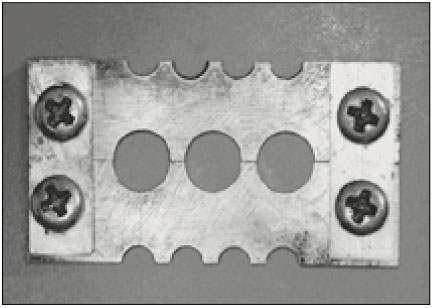
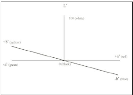


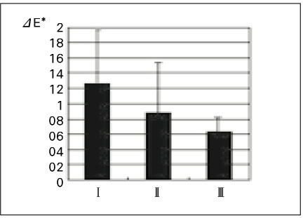
Fig. 1
Separable cylindrical metal mold for sample fabrication
Fig. 2
Diagram of the CIE L*, a*, b* color space
Fig. 3
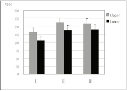
Graphic representating of mean microhardness values(VHN) of each group
I: Apollo 95E, II: XL 3000, III: Spectrum 800
Fig. 4
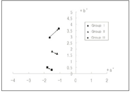
Graphic representation of the chromatic color changes of group I, II and III produced by storing for 30 days in distilled water at 60℃
Fig. 5
Total color difference(ΔE*) in group I, II and III
I: Apollo 95E, II: XL 3000, III: Spectrum 800
Fig. 1
Fig. 2
Fig. 3
Fig. 4
Fig. 5
Color changes in composites according to various light curing sources
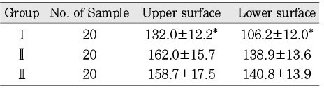
Light curing units used in this study
Microhardness values(VHN) of upper surfaces and lower surfaces of each group(Mean±SD)
I: Apollo 95E , II: XL 3000, III: Spectrum 800
*: Statistically significant difference between groups (p < 0.05)
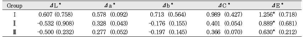
Result of color changes of group I, II and III after storing for 30 days in distilled water at 60℃ expressed as means
ΔL*, Δa*, Δb*, ΔC*: color difference, ΔE*: total color difference.
I: Apollo 95E, II: XL 3000, III: Spectrum 800
Standard deviations are in parentheses. *: significant differences (p<0.05)
Table 1
Light curing units used in this study
Table 2
Microhardness values(VHN) of upper surfaces and lower surfaces of each group(Mean±SD)
I: Apollo 95E , II: XL 3000, III: Spectrum 800 *: Statistically significant difference between groups (p < 0.05)
Table 3
Result of color changes of group I, II and III after storing for 30 days in distilled water at 60℃ expressed as means
ΔL*, Δa*, Δb*, ΔC*: color difference, ΔE*: total color difference. I: Apollo 95E, II: XL 3000, III: Spectrum 800 Standard deviations are in parentheses. *: significant differences (p<0.05)

