
Articles
- Page Path
- HOME > Restor Dent Endod > Volume 42(2); 2017 > Article
- Research Article Bonding of the silane containing multi-mode universal adhesive for lithium disilicate ceramics
- Hyun-Young Lee1, Geum-Jun Han2, Juhea Chang3, Ho-Hyun Son1
-
2017;42(2):-104.
DOI: https://doi.org/10.5395/rde.2017.42.2.95
Published online: January 25, 2017
1Department of Conservative Dentistry, Seoul National University School of Dentistry and Dental Research Institute, Seoul, Korea.
2Department of Dental Biomaterials Science, Seoul National University School of Dentistry and Dental Research Institute, Seoul, Korea.
3Special Care Clinic, Seoul National University Dental Hospital, Seoul, Korea.
- Correspondence to Ho-Hyun Son, DDS, MSD, PhD. Professor, Department of Conservative Dentistry, Seoul National University Dental Hospital, Seoul National University School of Dentistry and Dental Research Institute, 101 Daehak-ro, Jongrogu, Seoul, Korea 03080. TEL, +82-2-2072-2651; FAX, +82-2-2072-3859; hhson@snu.ac.kr
• Received: July 11, 2016 • Accepted: December 6, 2016
©Copyrights 2017. The Korean Academy of Conservative Dentistry.
This is an Open Access article distributed under the terms of the Creative Commons Attribution Non-Commercial License (http://creativecommons.org/licenses/by-nc/3.0/) which permits unrestricted non-commercial use, distribution, and reproduction in any medium, provided the original work is properly cited.
- 5,034 Views
- 29 Download
- 28 Crossref


Tables & Figures
REFERENCES
Citations
Citations to this article as recorded by 
- Clinical Roles of Nanoparticles in Orthodontic Bonding Materials
Maria Arampatzi, Ellas Spyratou, Iosif Sifakakis, Efstathios P. Efstathopoulos
Applied Sciences.2026; 16(4): 1996. CrossRef - Influence of silane-containing universal adhesives and separate silanization on lithium disilicate bond strength
Laís Giacomini Bernardi, Michel Wendlinger, Romina Ñaupari-Villasante, Mauricio Aguirre-Balseca, Alessandro Dourado Loguercio, João Carlos Gomes
The Journal of Prosthetic Dentistry.2026;[Epub] CrossRef - The influence of different factors on the bond strength of lithium disilicate-reinforced glass–ceramics to Resin: a machine learning analysis
Jiawen Liu, Suqing Tu, Mingjuan Wang, Du Chen, Chen Chen, Haifeng Xie
BMC Oral Health.2025;[Epub] CrossRef - Influence of different primers and adhesive system combinations on the durability of resin bonding to lithium disilicate
Christine Yazigi, Shila Alawi, Sebastian Wille, Matthias Kern
The Journal of Prosthetic Dentistry.2025; 134(3): 749. CrossRef - Shear Bond Strength and Finite Element Stress Analysis of Composite Repair Using Various Adhesive Strategies With and Without Silane Application
Elif Ercan Devrimci, Hande Kemaloglu, Cem Peskersoy, Tijen Pamir, Murat Turkun
Applied Sciences.2025; 15(15): 8159. CrossRef - Effect of multiple firings on mechanical and optical properties of CAD/CAM lithium disilicate-based glass ceramics
Chawal Padunglappisit, Pitsucha Charoensakthanakul, Sintwo Wongthongdee, Kan Wongkamhaeng
BMC Oral Health.2025;[Epub] CrossRef - Effect of universal adhesives and self-etch ceramic primers on bond strength to glass-ceramics: A systematic review and meta-analysis of in vitro studies
Renally Bezerra Wanderley Lima, Isis de Araújo Ferreira Muniz, Débora e Silva Campos, Fabián Murillo-Gómez, Ana Karina Maciel de Andrade, Rosângela Marques Duarte, Grace Mendonça de Souza
The Journal of Prosthetic Dentistry.2024; 131(3): 392. CrossRef - Effect of the difference water amounts and hydrolysis times of silane coupling agent on the shear bond strength between lithium disilicate glass ceramic and composite resin
Pimchanok OSOTPRASIT, Sasipin LAUVAHUTANON, Yosnarong SIRIMETHAWONG, Patcharanun CHAIAMORNSUP, Pornpot JIANGKONGKHO
Dental Materials Journal.2024; 43(3): 375. CrossRef - Is additional silane application necessary for a new silane‐containing universal adhesive to bond to glass ceramics?
Priscila Luciane da Silva, Hélio Radke Bittencourt, Luiz Henrique Burnett, Ana Maria Spohr
Journal of Esthetic and Restorative Dentistry.2024; 36(10): 1452. CrossRef - The Effect of Various Lasers on the Bond Strength Between Orthodontic Brackets and Dental Ceramics: A Systematic Review and Meta-Analysis
Seyed Ali Mosaddad, Jaafar Abduo, Mehrnaz Zakizade, Hamid Tebyaniyan, Ahmed Hussain
Photobiomodulation, Photomedicine, and Laser Surgery.2024; 42(1): 20. CrossRef - Long-Term Bonding Performance of One-Bottle vs. Two-Bottle Bonding Agents to Lithium Disilicate Ceramics
Masao Irie, Masahiro Okada, Yukinori Maruo, Goro Nishigawa, Takuya Matsumoto
Polymers.2024; 16(16): 2266. CrossRef - Bond strength to different CAD/CAM lithium disilicate reinforced ceramics
Mona Alhomuod, Jin‐Ho Phark, Sillas Duarte
Journal of Esthetic and Restorative Dentistry.2023; 35(1): 129. CrossRef - Surface Treatment Effect on Shear Bond Strength between Lithium Disilicate Glass-Ceramic and Resin Cement
Siripan Simasetha, Awiruth Klaisiri, Tool Sriamporn, Kraisorn Sappayatosok, Niyom Thamrongananskul
European Journal of Dentistry.2022; 16(02): 373. CrossRef - Bonding of Clear Aligner Composite Attachments to Ceramic Materials: An In Vitro Study
Bashair A. Alsaud, Maher S. Hajjaj, Ahmad I. Masoud, Ensanya A. Abou Neel, Dalia A. Abuelenain, Amal I. Linjawi
Materials.2022; 15(12): 4145. CrossRef - Bonding of different resin luting materials to composite, polymer-infiltrated and feldspathic ceramic CAD/CAM blocks
Burcu Dikici, Esra Can Say
Journal of Adhesion Science and Technology.2022; 36(14): 1572. CrossRef - Influence of mechanical and chemical pre-treatments on the repair of a hybrid ceramic
Sascha Niklas Jung, Stefan Rüttermann
Dental Materials.2022; 38(7): 1140. CrossRef - Effect of Silane-Containing Universal Adhesives on the Bonding Strength of Lithium Disilicate
Yu-Ri Kim, Jae-Hoon Kim, Sung-Ae Son, Jeong-Kil Park
Materials.2021; 14(14): 3976. CrossRef - Ceramics in dentistry: which material is appropriate for the anterior or posterior Dentition? Part 1: materials science
Loo Chien Win, Peter Sands, Stephen J Bonsor, FJ Trevor Burke
Dental Update.2021; 48(8): 680. CrossRef - The effect of different ceramic surface treatments on the repair bond strength of resin composite to lithium disilicate ceramic
Nanako UEDA, Tomohiro TAKAGAKI, Toru NIKAIDO, Rena TAKAHASHI, Masaomi IKEDA, Junji TAGAMI
Dental Materials Journal.2021; 40(5): 1073. CrossRef - Bonding Strength of Universal Adhesives to Indirect Substrates: A Meta‐Analysis of in Vitro Studies
Carlos Enrique Cuevas‐Suárez, Wellington Luiz de Oliveira da Rosa, Rafael Pino Vitti, Adriana Fernandes da Silva, Evandro Piva
Journal of Prosthodontics.2020; 29(4): 298. CrossRef - Effect of different surface treatments and multimode adhesive application on the Weibull characteristics, wettability, surface topography and adhesion to CAD/CAM lithium disilicate ceramic
Karina Barbosa Souza, Dayanne Monielle Duarte Moura, Sarah Emille Gomes da Silva, Gabriela Monteiro de Araújo, Rafael de Almeida Spinelli Pinto, Fabíola Pessôa Pereira Leite, Mutlu Özcan, Rodrigo Othávio de Assunção e Souza
Journal of Applied Oral Science.2020;[Epub] CrossRef - Effects of the ratio of silane to 10-methacryloyloxydecyl dihydrogenphosphate (MDP) in primer on bonding performance of silica-based and zirconia ceramics
Minkhant Koko, Tomohiro Takagaki, Ahmed Abdou, Masanao Inokoshi, Masaomi Ikeda, Takahiro Wada, Motohiro Uo, Toru Nikaido, Junji Tagami
Journal of the Mechanical Behavior of Biomedical Materials.2020; 112: 104026. CrossRef - Influence of surface treatments and repair materials on the shear bond strength of CAD/CAM provisional restorations
Ki-Won Jeong, Sung-Hun Kim
The Journal of Advanced Prosthodontics.2019; 11(2): 95. CrossRef - Microtensile bond strengths of adhesively bonded polymer-based CAD/CAM materials to dentin
Nuray CAPA, Esra CAN SAY, Cansin CELEBI, Ayca CASUR
Dental Materials Journal.2019; 38(1): 75. CrossRef - Simplified Surface Treatments for Ceramic Cementation: Use of Universal Adhesive and Self-Etching Ceramic Primer
Heloísa A. B. Guimarães, Paula C. Cardoso, Rafael A. Decurcio, Lúcio J. E. Monteiro, Letícia N. de Almeida, Wellington F. Martins, Ana Paula R. Magalhães
International Journal of Biomaterials.2018; 2018: 1. CrossRef - Effects of surface treatments on repair bond strength of a new CAD/CAM ZLS glass ceramic and two different types of CAD/CAM ceramics
Ayse Seda Ataol, Gulfem Ergun
Journal of Oral Science.2018; 60(2): 201. CrossRef - An in vitro evaluation of fracture load of implant‐supported zirconia‐based prostheses fabricated with different veneer materials
Hiroki Takata, Futoshi Komine, Junichi Honda, Markus B. Blatz, Hideo Matsumura
Clinical Oral Implants Research.2018; 29(4): 396. CrossRef - Effects of multiple firings on mechanical properties and resin bonding of lithium disilicate glass-ceramic
Hongliang Meng, Haifeng Xie, Lu Yang, Bingzhuo Chen, Ying Chen, Huaiqin Zhang, Chen Chen
Journal of the Mechanical Behavior of Biomedical Materials.2018; 88: 362. CrossRef
 ePub Link
 Cite
CiteBonding of the silane containing multi-mode universal adhesive for lithium disilicate ceramics
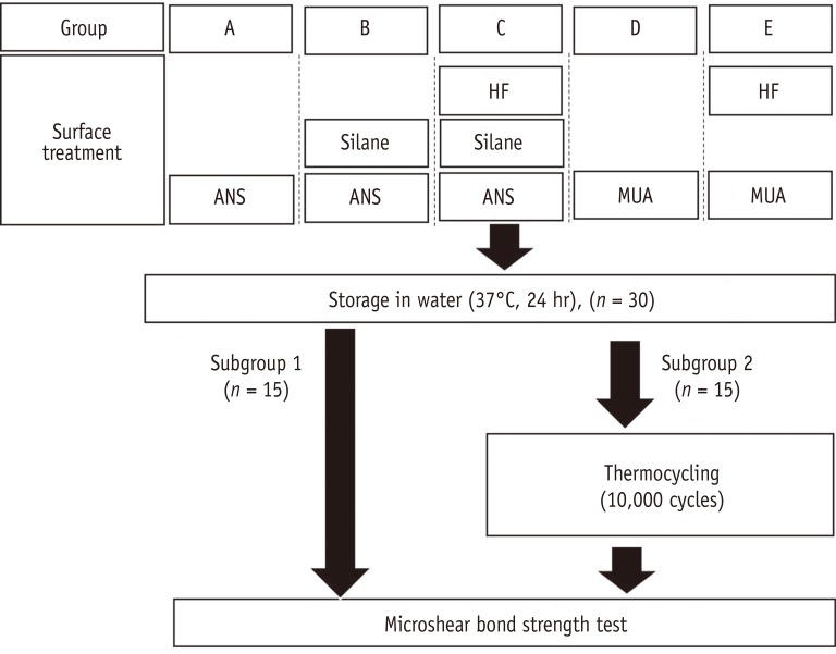
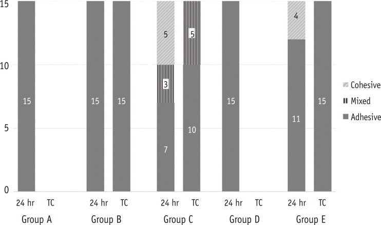

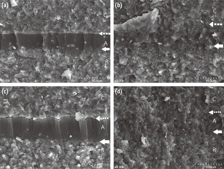
Figure 1 Experimental design of the study. HF, Hydrofluoric acid; ANS, adhesive that does not contain silane (Porcelain Bonding Resin, Bisco); MUA, Multi-mode universal adhesive (Single Bond Universal, 3M EPSE).
Figure 2 Failure mode distribution after microshear bond strength testing. TC, thermocycling.
Figure 3 Representative SEM photomicrographs of fractured ceramic surfaces after microshear bond strength testing showing (a) adhesive failure; (b) mixed failure; (c) cohesive failure at ×100 magnification. The arrow shows the fracture origin and the direction of the arrow represents that of shear force. In Figure (c), the resin cement remained on the loading point side. C, ceramic; A, adhesive; R, resin cement.
Figure 4 SEM micrographs of the fractured surfaces comparing the adaptation between the adhesive and the ceramic surfaces treated with different procedures: (a) Group B (silane, adhesive that did not contain silane [ANS], and resin cement) before thermocycling. The surface of the lithium disilicate ceramic was flat, and there was no micro-undercut, because hydrofluoric acid (HF) had not been applied. The adhesive and resin cement layers can be discriminated. There were some filler particles in the adhesive layer; (b) Group C (HF, silane, ANS, and resin cement) before thermocycling. The borders of each material were not easily distinguishable because the adhesive had infiltrated the micro-undercut and the fillers were distributed throughout the full thickness of the adhesive; (c) Group E (HF, multi-mode universal adhesive [MUA], and resin cement) before thermocycling. The etched ceramic surface had micro-undercuts and MUA had infiltrated the undercuts. However, there was a gap between the adhesive and the ceramic surface; (d) Group C (HF, silane, ANS, and resin cement) after thermocycling. This had a similar morphology to Figure 4b. Dashed arrow, the interface of the ceramic and adhesive; hollow arrow, the interface of the adhesive and resin cement. C, ceramic; A, adhesive; R, resin cement.
Figure 1
Figure 2
Figure 3
Figure 4
Bonding of the silane containing multi-mode universal adhesive for lithium disilicate ceramics
Materials used in the study
| Product | Manufacturer | Main components* |
|---|---|---|
| IPS e.max CAD | Ivoclar Vivadent | Lithium disilicate |
| Ceramic etching gel | Ivoclar Vivadent | 5% hydrofluoric acid |
| Bis-Silane | Bisco | Ethanol, silane |
| Porcelain Bonding Resin | Bisco | Bis-GMA, UDMA, TEGDMA |
| Single Bond Universal | 3M ESPE | Organophosphate monomer (MDP), Bis-GMA, HEMA, Vitrebond copolymer, filler, ethanol, water, initiators, silane |
| NX3 | Kerr | 7,7,9-trimethyl-4,13-dioxo-3,14-dioxa-5,12-diazahexadecane-1,16-diyl bismethacrylate, TEGDMA, HEMA |
| Filtek Z250 | 3M EPSE | Bis-GMA, UDMA, Bis-EMA, PEGDMA, TEGDMA, silane-treated ceramic |
Bis-GMA, bisphenol A diglycidyl ether dimethacrylate; UDMA, urethane dimethacrylate; TEGDMA, triethylene glycol dimethacrylate; MDP, 10-methacryloyloxydecyl dihydrogen phosphate; HEMA, 2-hydroxyethyl methacrylate; Bis-EMA, bisphenol A ethoxylated dimethacrylate; PEGDMA, polyethylene glycol dimethacrylate.
*As provided by the manufacturers.
Microshear bond strength after different surface treatments on lithium disilicate
| Group | Treatment | Bond strength (MPa) | Reduction rate of bond strength (%) | |
|---|---|---|---|---|
| 24 hr | Thermocycling | |||
| A | ANS | 1.35 ± 1.12aA | 0.00 ± 0.00aB | 100.0 |
| B | S + ANS | 8.66 ± 2.83bA | 2.68 ± 1.43bB | 69.1 |
| C | HF + S + ANS | 27.14 ± 6.85cA | 13.08 ± 3.80cB | 51.8 |
| D | MUA | 1.53 ± 0.61aA | 0.00 ± 0.00aB | 100.0 |
| E | HF + MUA | 21.37 ± 5.08dA | 3.13 ± 1.82bB | 85.4 |
Different superscript lowercase letters in the same column indicate significant differences; different superscript capitalized letters in the same row indicate a significant difference.
Reduction rate of bond strength (%) = ([bond strength after water storage for 24 hours − bond strength after thermocycling]/bond strength after water storage for 24 hours) × 100
ANS, adhesive that did not contain silane; S, silane; HF, hydrofluoric acid; MUA, Multi-mode universal adhesive.
Composition of adhesives according to the material safety data sheets provided by the manufacturers
| Porcelain Bonding Resin (wt%) | Single Bond Universal (wt%) | All-Bond Universal (%) | Clearfil Universal Bond (%) | Adhese Universal (%) | |
|---|---|---|---|---|---|
| Bis-GMA | < 40 | 15 - 20 | 20 - 50 | 15 - 35 | 20 - < 25† |
| Urethane dimethacrylate | < 40 | ||||
| TEGDMA | < 30 | ||||
| HEMA | 15 - 20 | 5 - 25 | 10 - 35 | 20 - < 25† | |
| DGDMA | 5 -15 | ||||
| Ethanol | 10 - 15 | 30 - 50 | < 20 | 10 - 13 | |
| Water | 10 - 15 | * | |||
| MDP | * | 5 - 25 | * | ||
| Silane-treated silica | 5 - 15 | ||||
| 2-propenoic acid, 2-methyl-, reaction products with 1,10-decanediol and phosphorous oxide | 1 - 10 | ||||
| Copolymer of acrylic and itaconic acid | 1 - 5 | ||||
| Dimethylaminobenzoate | < 2 | ||||
| (Dimethylamino) ethyl methacrylate | < 2 | ||||
| Methyl ethyl ketone | < 0.5 | ||||
| Colloidal silica, silane etc. |
Bis-GMA, bisphenol A glycidyl methacrylate; TEGDMA, triethylene glycol dimethacrylate; HEMA, 2-hydroxyethyl methacrylate; DGDMA, decamethylene glycol demethacrylate; MDP, 10-methacryloyloxydecyl dihydrogen phosphate.
*present, but composition information was not provided.
†from 20 or more to less than 25.
Table 1 Materials used in the study
Bis-GMA, bisphenol A diglycidyl ether dimethacrylate; UDMA, urethane dimethacrylate; TEGDMA, triethylene glycol dimethacrylate; MDP, 10-methacryloyloxydecyl dihydrogen phosphate; HEMA, 2-hydroxyethyl methacrylate; Bis-EMA, bisphenol A ethoxylated dimethacrylate; PEGDMA, polyethylene glycol dimethacrylate. *As provided by the manufacturers.
Table 2 Microshear bond strength after different surface treatments on lithium disilicate
Different superscript lowercase letters in the same column indicate significant differences; different superscript capitalized letters in the same row indicate a significant difference. Reduction rate of bond strength (%) = ([bond strength after water storage for 24 hours − bond strength after thermocycling]/bond strength after water storage for 24 hours) × 100 ANS, adhesive that did not contain silane; S, silane; HF, hydrofluoric acid; MUA, Multi-mode universal adhesive.
Table 3 Composition of adhesives according to the material safety data sheets provided by the manufacturers
Bis-GMA, bisphenol A glycidyl methacrylate; TEGDMA, triethylene glycol dimethacrylate; HEMA, 2-hydroxyethyl methacrylate;
DGDMA, decamethylene glycol demethacrylate; MDP, 10-methacryloyloxydecyl dihydrogen phosphate. *present, but composition information was not provided. †from 20 or more to less than 25.

