
Search
- Page Path
- HOME > Search
Research Articles
- Effect of sugar and sweetener on the bleachability of coffee and tea-induced stains on composites: an in vitro experimental study
- Nilay Bayraktar, Osman Kerim Arda Karaca, Yunus Ekşılı, Mustafa Furkan Yıldırım, Osman Tolga Harorli
- Restor Dent Endod 2026;51(2):e16. Published online April 1, 2026
- DOI: https://doi.org/10.5395/rde.2026.51.e16
-
 Abstract
 PDF
PDF PubReader
 ePub
ePub - Objectives
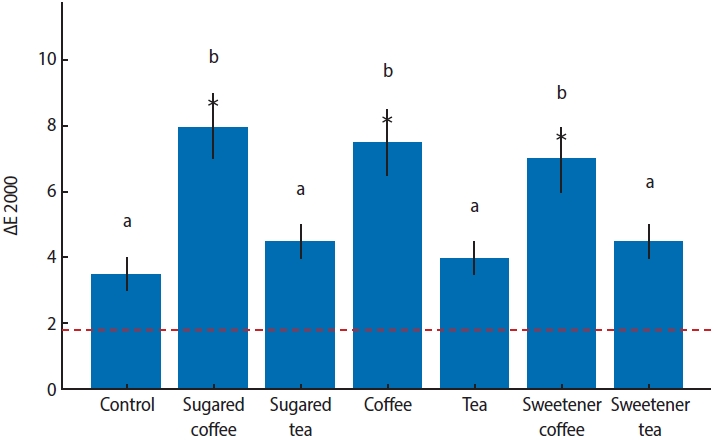
This in vitro study evaluated the effects of various sugary and non-sugary beverages on the color change of a dental composite and the subsequent bleaching efficacy.
Methods
Forty-nine disc-shaped composite samples (Neo Spectra ST, Dentsply Sirona) were split into seven groups at random (n = 7). Distilled water was used to hydrate each sample for 24 hours at 37°C. After 24 hours, the first color measurements (T0) were made by using a clinical spectrophotometer (VITA Easyshade Compact; VITA Zahnfabrik). Color measurements were repeated after 7 days (T1) and 14 days (T2) of immersion in distilled water (control), tea, coffee, sugary tea, sugary coffee, tea with sweetener added, and coffee with sweetener added. After staining for 2 weeks, the specimens were bleached for 6 hours a day for a week using 16% carbamide peroxide (Opalescence Ultradent Products). Color measurements were taken again after bleaching (T3). Using CIEDE2000, color differences (ΔE) were computed. Analysis of variance (ANOVA) and repeated measures ANOVA with a Tukey post hoc test were used to evaluate the data.
Results
After 1 week, coffee-containing solutions produced significantly greater discoloration than the control (p < 0.001). By 2 weeks, tea groups exhibited similar discoloration to coffee groups (p < 0.001). The addition of sugar or sweetener had no significant effect (p > 0.05). Post-bleaching, coffee groups showed lower Whiteness Index values than the control, without statistical significance (p > 0.05).
Conclusions
Coffee and tea markedly stain resin composites, with discoloration persisting post-bleaching, while sugar or sweetener additions exert no significant effect.
- 1,192 View
- 95 Download

- Neuropeptide Y regulation of dental pulp neurogenic inflammation provoked by tooth bleaching agents: a descriptive comparative clinical study
- Javier Caviedes-Bucheli, Néstor Ríos-Osorio, Mario Pérez-Villota, Karolina Aucú-Miño, Diana Escobar-Mafla, Hernán Darío Muñoz-Alvear, José Francisco Gomez-Sosa, Luis Diaz-Barrera, Edgar Güiza – Cristancho, Hugo Roberto Munoz
- Restor Dent Endod 2026;51(1):e10. Published online February 13, 2026
- DOI: https://doi.org/10.5395/rde.2026.51.e10
-
Abstract
PDFPubReaderePub
- Objectives
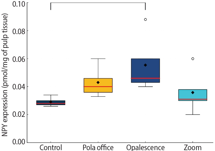
This study aimed to assess the expression of neuropeptide Y (NPY) in human dental pulp after tooth bleaching with three in-office hydrogen peroxide (H2O2)-based systems.
Methods
Forty pulps were collected from premolars scheduled for extraction and divided into four groups (n = 10): Control (no bleaching; basal NPY values); Pola Office (35% H2O2, 8 minutes); Opalescence Boost (40% H2O2, 20 minutes); and Zoom (25% H2O2 + cold blue light, 15 minutes). After extraction, pulps were fixed in 4% formaldehyde and processed. NPY levels were quantified using enzyme-linked immunosorbent assay. Data distribution was assessed with the Shapiro-Wilk test. One-way analysis of variance and Tukey post-hoc test with Bonferroni correction were applied (p < 0.05).
Results
NPY expression differed significantly among groups (p = 0.0097). The control group showed the lowest mean expression (0.026 ± 0.002 pmol/mg of pulp tissue), followed by Zoom (0.031 ± 0.005 pmol/mg), Pola Office (0.040 ± 0.004 pmol/mg), and Opalescence Boost, which exhibited the highest NPY expression (0.044 ± 0.004 pmol/mg). Post-hoc analysis revealed a statistically significant difference between the control and Opalescence Boost groups (p = 0.0122).
Conclusions
The increase in NPY expression—particularly with Opalescence Boost—indicates that in-office bleaching agents can elicit measurable neurobiological responses in pulp tissue after a single application. The significant difference between the control and Opalescence Boost groups suggests a possible H2O2 concentration- or formulation-dependent effect on pulpal neuropeptide activity, underscoring the need for further research on the biological impact of bleaching treatments.
- 1,595 View
- 90 Download
- Comparative evaluation of dentinal tubule occlusion by desensitizing agents after tooth bleaching: an in vitro study
- Dimitrios Dionysopoulos, Petros Mourouzis, Spyros Papageorgiou, Kosmas Tolidis
- Restor Dent Endod 2026;51(1):e8. Published online February 10, 2026
- DOI: https://doi.org/10.5395/rde.2026.51.e8
-
Abstract
PDFPubReaderePub
- Objectives
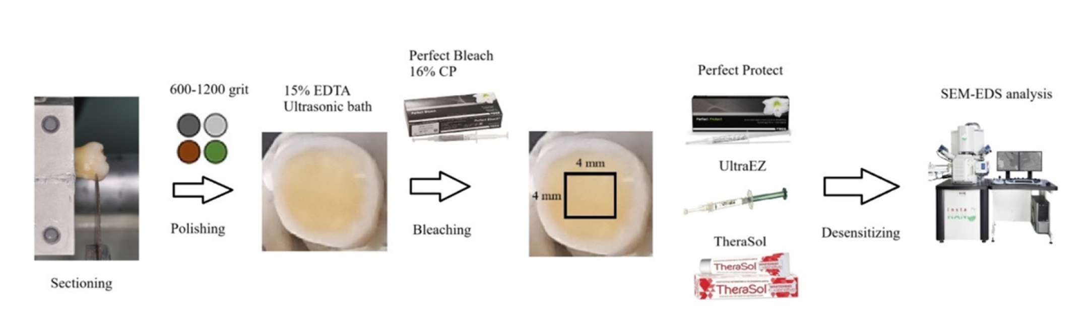
This study aimed to evaluate the efficacy of three commercially available desensitizing agents in occluding dentinal tubules, which may help reduce tooth sensitivity following a bleaching treatment.
Methods
Twenty healthy human third molars were utilized in this investigation. The samples were prepared by transversely sectioning 2.5 mm of the crowns to expose the dentin. They were initially treated with 15% ethylenediaminetetraacetic acid gel for 4 minutes, followed by application of Perfect Bleach (VOCO GmbH) bleaching agent (16% carbamide peroxide) for 2 hours. The samples were randomly allocated into four groups (n = 5), each receiving one of the following treatments: group 1: No treatment (control), group 2: treated with UltraEZ (Ultradent Products Inc.,), containing potassium nitrate and sodium fluoride, group 3: treated with Perfect Protect (VOCO GmbH), also containing potassium nitrate and sodium fluoride and group 4: treated with TheraSol Whitening & Sensitive (ABC Kinitron IKE), containing strontium acetate and sodium monofluorophosphate. Subsequently, the specimens were examined using scanning electron microscopy (SEM) and energy-dispersive X-ray spectroscopy to evaluate dentin tubule occlusion.
Results
SEM observations showed no occlusion of dentin tubules in the control group, whereas groups 2 to 4 exhibited significant occlusion. The most effective treatment was Perfect Protect (p < 0.05), while UltraEZ and TheraSol Whitening & Sensitive demonstrated similar effectiveness, with no statistically significant difference between them (p > 0.05).
Conclusions
The tested desensitizing agents effectively occluded dentin tubules to a considerable extent. Differences in their effectiveness were attributed to variations in their formulations.
- 2,191 View
- 188 Download
- Comparative study of the effectiveness of different bleaching agents on blood-colored extracted teeth and investigation of recoloring after bleaching: an in vitro experimental study
- Gülşen Arslan, Akın Aladağ, Ayşegül Demirbaş, Murat Türkün
- Restor Dent Endod 2025;50(3):e22. Published online July 9, 2025
- DOI: https://doi.org/10.5395/rde.2025.50.e22
-
Abstract
PDFPubReaderePub
- Objectives
This study evaluated the efficacy of three distinct bleaching agents over time on blood-stained, devitalized teeth. Furthermore, the recoloring subsequent to bleaching will be monitored.
Methods
The study was conducted on 60 caries-free, unfilled, upper human incisors. The Freccia and Peters blood staining technique was employed, and four groups (n = 15) were identified: control, 35% hydrogen peroxide-treated, 37% carbamide peroxide-treated, and sodium perborate-treated groups. Color differences were measured using ΔE00, ΔWID, L*, a*, and b* values. To investigate tooth discoloration after bleaching, 10 unbleached teeth with three groups of 10 bleached teeth were compared by vine staining. The group of bleached teeth was restored immediately, another group waited one week, and the third group had sodium ascorbate applied and analyzed using one-way analysis of variance tests (p < 0.05).
Results
Among the groups, carbamide peroxide exhibited the most significant whitening during the 6-day bleaching process, followed by hydrogen peroxide and sodium perborate. Subsequent examination of the wine recoloring of post-bleaching samples demonstrated that bleached teeth exhibited a heightened propensity for recoloration in contrast to unbleached teeth. Notably, sodium ascorbate treatments for hydrogen peroxide neutralization and the wait-and-restore approach were not statistically significant in terms of preventing recoloration.
Conclusions
Sodium perborate is less effective and more time-consuming than hydrogen peroxide or carbamide peroxide for bleaching purposes. Carbamide peroxide is the most effective bleaching agent. The sodium ascorbate treatment and the wait-and-restore approach are ineffective in preventing recoloring. Bleached teeth have more discoloration than unbleached teeth. -
Citations
Citations to this article as recorded by
- The Effect of Adhesive Systems on Shade Matching of Composite Veneer
Fadak Al Marar, Raghad Aljarboua, Fatimah M. Alatiyyah, Shahad AlGhamdi, Faraz Ahmed Farooqi, Lama Almuhanna, Rasha AlSheikh, Abdul Samad Khan
Dentistry Journal.2026; 14(2): 85. CrossRef
- The Effect of Adhesive Systems on Shade Matching of Composite Veneer
- 4,518 View
- 282 Download
- 1 Web of Science
- 1 Crossref
- Can discolored dental composites be bleached in depth?
- Luca Giachetti, Daniele Scaminaci Russo, Michele Nieri, Francesca Cinelli
- Restor Dent Endod 2024;49(3):e23. Published online June 11, 2024
- DOI: https://doi.org/10.5395/rde.2024.49.e23
-
Abstract
PDFPubReaderePub
Objectives Previous
in vitro studies determined the whitening effects of bleaching products on stained resin composite surfaces. Thisin vitro study aimed to verify the effectiveness of a whitening system on composite resin previously subjected to pigmentation, specifically examining the depth of whitening effectiveness within the material structure.Materials and Methods A commercially available nano-filled composite resin was used. Specimens were stained using a coffee-based solution and a 10% carbamide peroxide-based gel was employed as the whitening agent. The pigment’s penetration and the effect of the bleaching gel were evaluated by measuring color (CieLab values) from the outer edge to the inner part of the specimens. Color measurements were taken at 14 points, starting from 0.1 mm from the external perimeter up to 3.0 mm.
Results Analysis of variance tests showed a statistically significant difference between the Control Group (CG), Pigmentation Group, and Whitening Group. The whitening agent was effective up to 1.5 mm in depth, with Whiteness index (W) values not statistically different from those of CG up to 0.5 mm in depth.
Conclusions Whitening agents on nano-filled resin composite previously pigmented appear effective in restoring the W to values similar to the original, particularly in the superficial layers of the sample.
-
Citations
Citations to this article as recorded by- Color Stability of Tooth-Colored Restorative Materials After Exposure to Arabic Coffee and Black Tea: A Systematic Review
Abdulrhman Y Alenezi, Abdulwahab M AlEyada, Yousef H Aldhafiri, Mohammed S Alsubaie, Mohammed S Alshahrani, Mahesh Shenoy
Cureus.2025;[Epub] CrossRef - Comparative evaluation to composite resin bleaching using ozone-enhanced low-concentration hydrogen peroxide
Mahmoud K. AL-Omiri, Dania Sa’ed Hussam Abuherra, Khaled M. AL-Omiri, Ali Y. Alsaeed, Mohammad Alamri, Ali M. Alqahtani, Saleh Ali Alqahtani, Ghadeer Saleh Alwadai, Naif Abogazalah, Edward Lynch
Scientific Reports.2025;[Epub] CrossRef - The effects of mechanical and chemical degradation on the surface roughness, gloss, and color stability of bulk-fill resin composites
Merve Nezir, Hanife Altınışık, Esra Özyurt, Naz Bayar, Mediha Büyükgöze Dindar
BMC Oral Health.2025;[Epub] CrossRef
- Color Stability of Tooth-Colored Restorative Materials After Exposure to Arabic Coffee and Black Tea: A Systematic Review
- 5,125 View
- 158 Download
- 2 Web of Science
- 3 Crossref
Review Article
- Can carbamide peroxide be as effective as hydrogen peroxide for in-office tooth bleaching and cause less sensitivity? A systematic review
- Patrick Wesley Marques de Boa, Kaiza de Sousa Santos, Francisca Jennifer Duarte de Oliveira, Boniek Castillo Dutra Borges
- Restor Dent Endod 2024;49(2):e14. Published online March 20, 2024
- DOI: https://doi.org/10.5395/rde.2024.49.e14
-
Abstract
PDFPubReaderePub
This study aimed to answer the question through a systematic review: Can carbamide peroxide be as effective as hydrogen peroxide and cause less in-office bleaching sensitivity? A literature survey was performed in PubMed/MEDLINE, Embase, Scopus, ISI Web of Science, and gray literature. Primary clinical trials that compared the efficacy or the in-office bleaching sensitivity between carbamide and hydrogen peroxides were included. The risk of bias was evaluated using the RoB2. The certainty of the evidence was assessed using the GRADE approach. DPI training significantly improved the mean scores of the dental undergraduates from 7.53 in the pre-DPI-training test to 9.01 in the post-DPI-training test (
p < 0.001). After 6 weeks, the mean scores decreased marginally to 8.87 in the retention test (p = 0.563). DPI training increased their confidence level from 5.68 pre-DPI training to 7.09 post-DPI training. The limited evidence suggests that the 37% carbamide peroxide may be similarly effective to the 35% hydrogen peroxide for bleaching teeth in-office and causes less bleaching sensitivity. However, more well-designed split-mouth clinical trials are necessary to strengthen the evidence.-
Citations
Citations to this article as recorded by- Impact of nanostructured additives in tooth bleaching agents on enhancing color change and reducing side effects: a scoping review
Patrick Wesley Marques de Boa, Kaiza de Sousa Santos, Aleph Matthews da Silva Souza, Arnóbio Antônio da Silva-Júnior, Boniek Castillo Dutra Borges
Clinical Oral Investigations.2025;[Epub] CrossRef - Quantitative and Qualitative Assessment of Enamel Surface Roughness Following High-Concentration Peroxide Bleaching: A Comparative In Vitro Study
Mamnoon Ghafir, Nida Mehmood, Leeza Bharati, Shreya Bhukal, Ritika Sethi, Aanchal Chaudhary, Seema Gupta
Cureus.2025;[Epub] CrossRef - Using violet light during in-office tooth bleaching to enhance the efficacy of carbamide peroxide without increasing bleaching sensitivity: a systematic review and meta-analysis
Mariana Silva de Bessa, Kaiza de Sousa Santos, Patrick Wesley Marques de Boa, Francisca Jennifer Duarte de Oliveira, Bárbara Faria de Sá Barbosa, Boniek Castillo Dutra Borges
Lasers in Medical Science.2025;[Epub] CrossRef - Influence of Different Light-Activated Bleaching Gels on Pulp Chamber Temperature: An In Vitro Study
Mandana Karimi, Elmira Ataee, Ladan Ranjbar Omrani, Mahdi Abbasi, Elham Ahmadi
Avicenna Journal of Dental Research.2024; 16(4): 225. CrossRef
- Impact of nanostructured additives in tooth bleaching agents on enhancing color change and reducing side effects: a scoping review
- 13,594 View
- 211 Download
- 1 Web of Science
- 4 Crossref
Research Articles
- Can different agents reduce the damage caused by bleaching gel to pulp tissue? A systematic review of basic research
- Letícia Aparecida Silva Batista, Alexandre Henrique dos Reis-Prado, Hebertt Gonzaga dos Santos Chaves, Lara Cancella de Arantes, Luís Fernando Santos Alves Morgan, Carolina Bosso André, Thaís Yumi Suzuki, Francine Benetti
- Restor Dent Endod 2023;48(4):e39. Published online November 6, 2023
- DOI: https://doi.org/10.5395/rde.2023.48.e39
-
Abstract
PDF
 Supplementary MaterialPubReaderePub
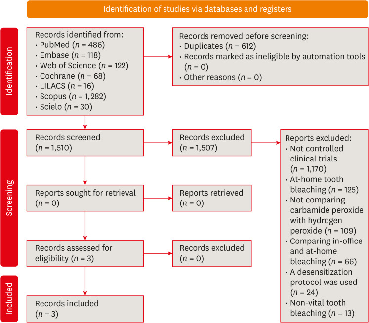
 Objectives This study aimed to investigate the effectiveness of different topical/systemic agents in reducing the damage caused by bleaching gel to pulp tissue or cells.
Materials and Methods Electronic searches were performed in July 2023.
In vivo andin vitro studies evaluating the effects of different topical or systemic agents on pulp inflammation or cytotoxicity after exposure to bleaching agents were included. The risk of bias was assessed.Results Out of 1,112 articles, 27 were included. Nine animal studies evaluated remineralizing/anti-inflammatories agents in rat molars subjected to bleaching with 35%–38% hydrogen peroxide (HP). Five of these studies demonstrated a significant reduction in inflammation caused by HP when combined with bioglass or MI Paste Plus (GC America), or following KF-desensitizing or Otosporin treatment (
n = 3). However, orally administered drugs did not reduce pulp inflammation (n = 4). Cytotoxicity (n = 17) was primarily assessed using the 3-(4,5-dimethylthiazol-2-yl)-2,5-diphenyltetrazolium bromide assay on human dental pulp cells and mouse dental papilla Cell-23 cells. Certain substances, including sodium ascorbate, butein, manganese chloride, and peroxidase, were found to reduce cytotoxicity, particularly when applied prior to bleaching. The risk of bias was high in animal studies and low in laboratory studies.Conclusions Few
in vivo studies have evaluated agents to reduce the damage caused by bleaching gel to pulp tissue. Within the limitations of these studies, it was found that topical agents were effective in reducing pulp inflammation in animals and cytotoxicity. Further analyses with human pulp are required to substantiate these findings.Trial Registration PROSPERO Identifier:
CRD42022337192 -
Citations
Citations to this article as recorded by- 3D-Printed and Bioprinted Scaffolds in Regenerative Endodontics: A Systematic Review
Hebertt Gonzaga dos Santos Chaves, Diana B. Sequeira, Vilton Cardozo Moreira Dias, Alberto Cabrera-Fernández, João Peça, Francine Benetti, João Miguel Marques dos Santos
Applied Sciences.2026; 16(8): 3940. CrossRef - Clinical Study on the Efficacy of 35% Hydrogen Peroxide Gel According to Exposure Time (40 min vs. 20 min) by Spectrophotometry
Trinidad Rincón, Maria Portillo Muñoz, Maria Lobato, Ana María Martín Casado, Laryssa Mylenna Madruga Barbosa, Alessandro Loguercio, Cristina Gómez‐Polo
Journal of Esthetic and Restorative Dentistry.2026;[Epub] CrossRef - Clareamento dental e TikTok: avaliação da qualidade do conteúdo em mídia social
Rafaele T Costa, Thayna Silva do Carmo Tavares, André Walsh-Monteiro
Ciência ET Praxis.2025; 21(36): 111. CrossRef - Synthesis, characterization and evaluation of novel bleaching gels containing bioactive glass and nano-hydroxyapatite on hydrogen peroxide diffusion, bleaching efficacy and enamel protection
Adrieli Burey, Byron Carpio-Salvatierra, Michael Favoretto, María Luján Méndez Bauer, Viviane Hass, Alessandra Reis, Alessandro D. Loguercio, Paulo Vitor Farago
Clinical Oral Investigations.2025;[Epub] CrossRef - Cytotoxicity of Bleaching Products: A Systematic Review
Mireia Montaner, José Luis Sanz, Carmen Llena, María Melo, Clara Puig-Herreros, James Ghilotti
Applied Sciences.2024; 14(9): 3680. CrossRef
- 3D-Printed and Bioprinted Scaffolds in Regenerative Endodontics: A Systematic Review
- 4,441 View
- 60 Download
- 4 Web of Science
- 5 Crossref
- Evaluation of at-home bleaching protocol with application on different surfaces: bleaching efficacy and hydrogen peroxide permeability
- Heloisa Forville, Michael Willian Favoreto, Michel Wendlinger, Roberta Micheten Dias, Christiane Philippini Ferreira Borges, Alessandra Reis, Alessandro D. Loguercio
- Restor Dent Endod 2023;48(4):e33. Published online October 6, 2023
- DOI: https://doi.org/10.5395/rde.2023.48.e33
-
Abstract
PDFPubReaderePub
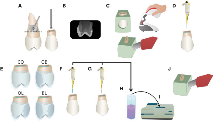
Objectives This study aimed to evaluate the bleaching efficacy and hydrogen peroxide permeability in the pulp chamber by the at-home bleaching gel in protocols applied on different dental surfaces.
Materials and Methods Forty premolars were randomly into 4 groups: control group no bleaching, only application on the buccal surface (OB), only application on the lingual surface (OL) and application in buccal and lingual surfaces, simultaneously (BL). At-home bleaching gel (White Class 7.5%) was used for the procedure. The bleaching efficacy was evaluated with a digital spectrophotometer (color change in CIELAB [Δ
E ab] and CIEDE 2000 [ΔE 00] systems and Whitening Index for Dentistry [ΔWID]). The hydrogen peroxide permeability in the pulp chamber (µg/mL) was assessed using UV-Vis spectrophotometry and data were analyzed for a 1-way analysis of variance and Tukey’s test (α = 0.05).Results All groups submitted to bleaching procedure showed bleaching efficacy when measured with Δ
E ab and ΔE 00 (p > 0.05). Therefore, when analyzed by ΔWID, a higher bleaching efficacy were observed for the application on the groups OB and BL (p = 0.00003). Similar hydrogen peroxide permeability was found in the pulp chambers of the teeth undergoing different protocols (p > 0.05).Conclusions The application of bleaching gel exclusively on the OB is sufficient to achieve bleaching efficacy, when compared to BL. Although the OL protocol demonstrated lower bleaching efficacy based on the ΔWID values, it may still be of interest and relevant in certain clinical scenarios based on individual needs, requiring clinical trials to better understand its specificities.
-
Citations
Citations to this article as recorded by- Effect of whitening pens on hydrogen peroxide permeability in the pulp chamber, color change and surface morphology
Laryssa Mylenna Madruga Barbosa, Gabrielle Gomes Centenaro, Deisy Cristina Ferreira Cordeiro, Maria Alice de Matos Rodrigues, Letícia Condolo, Michael Willian Favoreto, Alessandra Reis, Alessandro D. Loguercio
Journal of Dentistry.2025; 154: 105595. CrossRef - Evaluation of bleaching efficiency of carbamide peroxide applied on different dental surfaces: An in vitro study
R. Gokulnath, R. S. Mohan Kumar, A. Jayasenthil, R. Anjana, G. Sree Vidya
Journal of Conservative Dentistry and Endodontics.2025; 28(4): 366. CrossRef - Characterization and effects on enamel of low-concentration bleaching gels containing hyaluronic acid, NF_TiO2 nanoparticles and irradiated with violet LED light
Marcos Roberto Lima Benati, Matheus Kury, Priscila Borges Gobbo de Melo, Iago César Ribeiro Teles Matos, Roberta Tarkany Basting, Rosanna Tarkany Basting, Fernando Luis Esteban Florez, Vanessa Cavalli
Clinical Oral Investigations.2025;[Epub] CrossRef - Impact of bleaching on white spot lesions: hydrogen peroxide permeability and color alteration
Laryssa Mylenna Madruga Barbosa, Bruno Baracco, Taynara S. Carneiro, Michael Willian Favoreto, Michel Wendlinger, Daniel Jiménez-Díez, Laura Ceballos, Alessandro D. Loguercio
Clinical Oral Investigations.2025;[Epub] CrossRef - Efficacy of a buccal and lingual at‐home bleaching protocol—A randomized, split‐mouth, single‐blind controlled trial
Heloisa Forville, Laís Giacomini Bernardi, Michael Willian Favoreto, Felipe Coppla, Taynara de Souza Carneiro, Fabiana Madalozzo Coppla, Alessandro D. Loguercio, Alessandra Reis
Journal of Esthetic and Restorative Dentistry.2024; 36(9): 1301. CrossRef - REANATOMIZAÇÃO DE DENTE CONOIDE ASSOCIADA A ESTÉTICA VERMELHA: RELATO DE CASO
Ana Karolayne Sousa de Morais, Daniele Fernanda Sousa Barros, Daniel Messias Limeira, Rhana Leticia de Oliveira Faria, Roberta Furtado Carvalho, Sandna Nolêto de Araújo, Laura Barbosa Santos Di Milhomem
Revista Contemporânea.2024; 4(10): e6299. CrossRef - Effect of the reduction in the exposure time to at-home bleaching gel on color change and tooth sensitivity: A systematic review and meta-analysis
Priscila Borges Gobbo de Melo, Letícia Vasconcelos Silva Souza, Lucianne Cople Maia, Guido Artemio Marañón-Vásquez, Matheus Kury, Vanessa Cavalli
Clinical Oral Investigations.2024;[Epub] CrossRef
- Effect of whitening pens on hydrogen peroxide permeability in the pulp chamber, color change and surface morphology
- 5,345 View
- 91 Download
- 5 Web of Science
- 7 Crossref
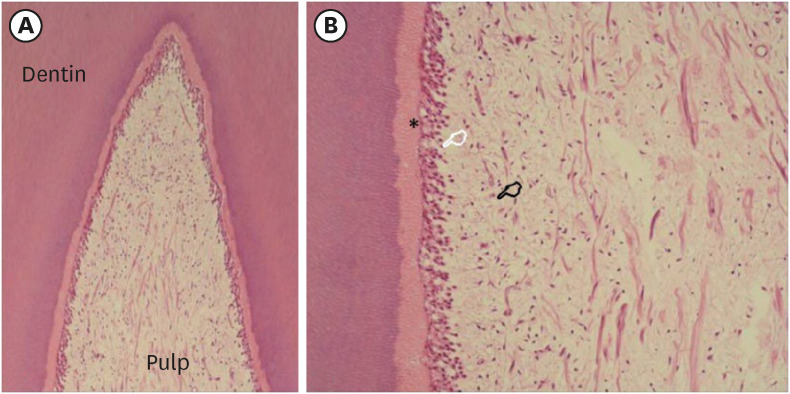
- Effect of medium or high concentrations of in-office dental bleaching gel on the human pulp response in the mandibular incisors
- Douglas Augusto Roderjan, Rodrigo Stanislawczuk, Diana Gabriela Soares, Carlos Alberto de Souza Costa, Michael Willian Favoreto, Alessandra Reis, Alessandro D. Loguercio
- Restor Dent Endod 2023;48(2):e12. Published online March 8, 2023
- DOI: https://doi.org/10.5395/rde.2023.48.e12
-
Abstract
PDFPubReaderePub
Objectives The present study evaluated the pulp response of human mandibular incisors subjected to in-office dental bleaching using gels with medium or high concentrations of hydrogen peroxide (HP).
Materials and Methods The following groups were compared: 35% HP (HP35;
n = 5) or 20% HP (HP20;n = 4). In the control group (CONT;n = 2), no dental bleaching was performed. The color change (CC) was registered at baseline and after 2 days using the Vita Classical shade guide. Tooth sensitivity (TS) was also recorded for 2 days post-bleaching. The teeth were extracted 2 days after the clinical procedure and subjected to histological analysis. The CC and overall scores for histological evaluation were evaluated by the Kruskal-Wallis and Mann-Whitney tests. The percentage of patients with TS was evaluated by the Fisher exact test (α = 0.05).Results The CC and TS of the HP35 group were significantly higher than those of the CONT group (
p < 0.05) and the HP20 group showed an intermediate response, without significant differences from either the HP35 or CONT group (p > 0.05). In both experimental groups, the coronal pulp tissue exhibited partial necrosis associated with tertiary dentin deposition. Overall, the subjacent pulp tissue exhibited a mild inflammatory response.Conclusions In-office bleaching therapies using bleaching gels with 20% or 35% HP caused similar pulp damage to the mandibular incisors, characterized by partial necrosis, tertiary dentin deposition, and mild inflammation.
-
Citations
Citations to this article as recorded by- DENTA: A Dual Enzymatic Nanoagent for Self‐Activating Tooth Whitening and Biofilm Disruption
Junseok Kim, Dai‐Hwan Kim, Priyannth R. Sundharbaabu, Chae Yeon Lee, Jina Bae, Jiyu Hyun, Young‐Ju Jang, Haeni Kim, Min‐Ho Hong, Juewen Liu, Tobias Fey, Suk Ho Bhang, Jun Hyuk Heo, Jung Heon Lee
Advanced Functional Materials.2026;[Epub] CrossRef - Effects of a bioactive desensitizing material on in-office bleaching–induced tooth sensitivity: A randomized double-blind controlled trial
Ghada A. Maghaireh, Hanan Alzraikat, Majd Y. Altarazi
Journal of Dentistry.2026; 166: 106326. CrossRef - Application of the Er:YAG laser in pulpotomy for mature permanent teeth with pulpitis: An animal study
Zeqi Li, Pengfei Xin, Siyao Yang, Xiaoxing Hao, Jun Wang, Kuanshou Zhang, Qingmei Liu, Mohmed Isaqali Karobari
PLOS One.2026; 21(1): e0341017. CrossRef - Can pigments of different natures interfere with the cytotoxicity from in-office bleaching?
Rafael Antonio de Oliveira Ribeiro, Beatriz Voss Martins, Marlon Ferreira Dias, Victória Peruchi, Caroline Anselmi, Igor Paulino Mendes Soares, Josimeri Hebling, Vanessa Cavalli, Carlos Alberto de Souza Costa
Odontology.2025; 113(4): 1447. CrossRef - Does Patient Age Impact In-Office Tooth Bleaching Outcomes? A Parallel Clinical Trial
JL Martins, IS Araújo, JF Rabelo, CJ Soares, AL Faria-e-Silva, AD Loguercio, PCFS Filho, HL Carlo, GR da Silva
Operative Dentistry.2025; 50(3): 251. CrossRef - The pH of Bleaching Gels on the Structural and Biological Response of Dental Tissues: A Scoping Review
Jamile Menezes de Souza, Maria Olimpia Paz Alvarenga, Ana Luisa Cassiano Alves Bezerra, Gabriela Queiroz de Melo Monteiro
Journal of Esthetic and Restorative Dentistry.2025; 37(10): 2193. CrossRef - Efficacy of 35 % self-mixed hydrogen peroxide In-office bleaching with reduced application time: A single-blind randomized controlled trial
Gabrielle Gomes Centenaro, Deisy Cristina Ferreira Cordeiro, Maria Alice de Matos Rodrigues, Mariah Maluf Lenhani, Roberta Micheten Dias, Cristina Gómez Polo, Alessandra Reis, Alessandro D. Loguercio
Journal of Dentistry.2025; 163: 106178. CrossRef - Evaluation of cytotoxicity and bleaching efficacy of gels with calcium polyphosphate and violet LED
Larissa de Jesus Gomes, Rafael Antonio de Oliveira Ribeiro, Mariangela Ivette Guanipa Ortiz, Klaus Rischka, Carlos Alberto de Souza Costa, Débora Alves Nunes Leite Lima
Brazilian Dental Journal.2025;[Epub] CrossRef - Combined catalytic strategies applied to in-office tooth bleaching: whitening efficacy, cytotoxicity, and gene expression of human dental pulp cells in a 3D culture model
Rafael Antonio de Oliveira Ribeiro, Victória Peruchi, Igor Paulino Mendes Soares, Filipe Koon Wu Mon, Diana Gabriela Soares, Josimeri Hebling, Carlos Alberto de Souza Costa
Clinical Oral Investigations.2024;[Epub] CrossRef - Low and high hydrogen peroxide concentrations of in-office dental bleaching associated with violet light: an in vitro study
Isabela Souza Vardasca, Michael Willian Favoreto, Mylena de Araujo Regis, Taynara de Souza Carneiro, Emanuel Adriano Hul, Christiane Philippini Ferreira Borges, Alessandra Reis, Alessandro D. Loguercio, Carlos Francci
Clinical Oral Investigations.2024;[Epub] CrossRef - Evaluation of hydrogen peroxide permeability, color change, and physical–chemical properties on the in‐office dental bleaching with different mixing tip
Michael Willian Favoreto, Sibelli Olivieri Parreiras, Michel Wendlinger, Taynara De Souza Carneiro, Mariah Ignez Lenhani, Christiane Phillipini Ferreira Borges, Alessandra Reis, Alessandro D. Loguercio
Journal of Esthetic and Restorative Dentistry.2024; 36(3): 460. CrossRef - Catalysis-based approaches with biopolymers and violet LED to improve in-office dental bleaching
Rafael Antonio de Oliveira Ribeiro, Beatriz Voss Martins, Marlon Ferreira Dias, Victória Peruchi, Igor Paulino Mendes Soares, Caroline Anselmi, Josimeri Hebling, Carlos Alberto de Souza Costa
Lasers in Medical Science.2024;[Epub] CrossRef - Feasibility and Safety of Adopting a New Approach in Delivering a 450 nm Blue Laser with a Flattop Beam Profile in Vital Tooth Whitening. A Clinical Case Series with an 8-Month Follow-Up
Reem Hanna, Ioana Cristina Miron, Stefano Benedicenti
Journal of Clinical Medicine.2024; 13(2): 491. CrossRef - Hydrogen Peroxide in the Pulp Chamber and Color Change in Maxillary Anterior Teeth After In-Office Bleaching
Alexandra Mena-Serrano, Sandra Sanchez, María G. Granda-Albuja, Michael Willian Favoreto, Taynara de Souza Carneiro, Deisy Cristina Ferreira Cordeiro, Alessandro D. Loguercio, Alessandra Reis
Brazilian Dental Journal.2024;[Epub] CrossRef - Influence of coating dental enamel with a TiF4-loaded polymeric primer on the adverse effects caused by a bleaching gel with 35% H2O2
Victória Peruchi, Rafael Antonio de Oliveira Ribeiro, Igor Paulino Mendes Soares, Lídia de Oliveira Fernandes, Juliana Rios de Oliveira, Maria Luiza Barucci Araújo Pires, Josimeri Hebling, Diana Gabriela Soares, Carlos Alberto de Souza Costa
Journal of the Mechanical Behavior of Biomedical Materials.2024; 153: 106497. CrossRef
- DENTA: A Dual Enzymatic Nanoagent for Self‐Activating Tooth Whitening and Biofilm Disruption
- 4,771 View
- 114 Download
- 14 Web of Science
- 15 Crossref
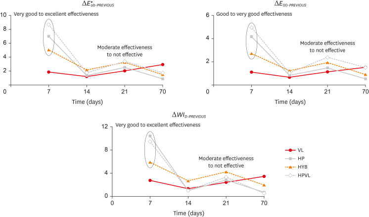
- In-office dental bleaching with violet light emitting diode: bleaching efficacy and pulpal temperature rise
- Brunna Katyuscia de Almeida Guanaes, Talyta Neves Duarte, Gisele Maria Correr, Marina da Rosa Kaizer, Carla Castiglia Gonzaga
- Restor Dent Endod 2022;47(1):e7. Published online February 3, 2022
- DOI: https://doi.org/10.5395/rde.2022.47.e7
-
Abstract
PDFPubReaderePub
Objectives This study evaluated the bleaching efficacy of different in-office protocols associated with violet light emitting diode (V-LED), and measured the pulpal temperature rise caused by V-LED with or without gel application.
Materials and Methods Bovine incisors were distributed in 4 groups (
n = 10): VL – V-LED; HP – 35% hydrogen peroxide (control); HYB – hybrid protocol, V-LED applied without gel for 10 irradiation cycles followed by V-LED applied with gel for another 10 irradiation cycles; and HPVL – gel and V-LED applied for 20 irradiation cycles. Three bleaching sessions were performed with 7-day intervals. Bleaching efficacy was evaluated withE 00 and ΔWID . Data were recorded at baseline, 7, 14, 21 and 70 days. For pulpal temperature rise, thermocouples were placed inside the pulp chamber of human incisors. To determine intrapulpal temperature, the teeth were irradiated with V-LED with or without application of bleaching gel. Color difference data were analyzed by 2-way repeated measures ANOVA and Tukey’s test. Pulpal temperature was analyzed byt -test (α = 5%).Results VL exhibited lower color (
E 00) and whiteness changes (ΔWID ) than the other groups. HPVL presented higher color change values than HYB. HYB and HPVL showed not different ΔWID values; and HP showed the highest whiteness changes at all times. There were significant differences comparing ΔT with gel (8.9°C) and without gel application (7.2°C).Conclusions HPLV was more efficient than HYB. The 2 protocols with VL showed similar results to control. Gel application combined with VL promoted higher pulpal temperature than to the no gel group.
-
Citations
Citations to this article as recorded by- Inverse Heat Conduction Estimation of Heat Flux in Human Dentin from Dental Curing Lights Using the Conjugate Gradient Method
Ahmad Soori, Farshad Kowsary, Shadab Safarzadeh Khosroshahi, Mohammad Vahedi
International Journal of Thermophysics.2026;[Epub] CrossRef - Illuminating the evidence: A comprehensive review of light-assisted in-office tooth bleaching
Márcia V.G.B. Queiroz, Rafael Dascanio, Vinicius H. Hutemma, Diogo A. Chiovetto, Adriano F. Lima, Jorge R. Soto-Montero, Matheus Kury
Journal of Dentistry.2026; 171: 106707. CrossRef - Spectrophotometric Evaluation of Laser-Assisted Dental Bleaching Using Erbium-Doped Yttrium Aluminum Garnet (Er:YAG) and Diode Lasers at Different Wavelengths: An In Vitro Study
Esraa Ihssan Alshibli, Omar H. Hamadah, Mohammad Y. Hajeer
Cureus.2026;[Epub] CrossRef - Effect of antioxidant on tooth sensitivity after bleaching
Mohamed Nabil, Mostafa Mohamed Hasan, Eman Abd Elghany Shebl
Journal of Esthetic and Restorative Dentistry.2024; 36(3): 429. CrossRef - In-office Bleaching Activated With Violet LED: Effect on Pulpal and Tooth Temperature and Pulp Viability
NR Carlos, RT Basting, KR Kantovitz, ES Bronze-Uhle, PN Lisboa Filho, V Cavalli, RT Basting
Operative Dentistry.2024; 49(3): 262. CrossRef - Low and high hydrogen peroxide concentrations of in-office dental bleaching associated with violet light: an in vitro study
Isabela Souza Vardasca, Michael Willian Favoreto, Mylena de Araujo Regis, Taynara de Souza Carneiro, Emanuel Adriano Hul, Christiane Philippini Ferreira Borges, Alessandra Reis, Alessandro D. Loguercio, Carlos Francci
Clinical Oral Investigations.2024;[Epub] CrossRef - Bleaching efficacy of in-office bleaching with violet light using low-concentration hydrogen peroxide nanoparticulate photocatalyst gel: A randomized controlled trial
Gustavo Garcia Castro, Palena Araújo Pinto, Michael Willian Favoreto, Alessandra Reis, Maria Viviana-Mora, Rita de Cássia Mendonça de Miranda, Andres Felipe Milan Cardenas, Alessandro D. Loguercio, Rudys Rodolfo de Jesus Tavarez
Photodiagnosis and Photodynamic Therapy.2024; 50: 104410. CrossRef - Influence of Different Light-Activated Bleaching Gels on Pulp Chamber Temperature: An In Vitro Study
Mandana Karimi, Elmira Ataee, Ladan Ranjbar Omrani, Mahdi Abbasi, Elham Ahmadi
Avicenna Journal of Dental Research.2024; 16(4): 225. CrossRef - Continuous vs fractionated violet LED light protocols for dental bleaching: Evaluations of color change and temperature of the dental pulp and buccal surface
Mayanna Pacheco Trindade Najar, Luciana Hilel Rangel Barbosa, Natália Russo Carlos, Fabiana Mantovani Gomes França, Cecilia Pedroso Turssi, Waldemir Francisco Vieira-Junior, Roberta Tarkany Basting
Photodiagnosis and Photodynamic Therapy.2023; 42: 103631. CrossRef - Improved esthetic efficacy and reduced cytotoxicity are achieved with a violet LED irradiation of manganese oxide-enriched bleaching gels
Marlon Ferreira Dias, Beatriz Voss Martins, Rafael Antonio de Oliveira Ribeiro, Josimeri Hebling, Carlos Alberto de Souza Costa
Lasers in Medical Science.2022;[Epub] CrossRef
- Inverse Heat Conduction Estimation of Heat Flux in Human Dentin from Dental Curing Lights Using the Conjugate Gradient Method
- 4,481 View
- 46 Download
- 10 Web of Science
- 10 Crossref
- Laboratory model to evaluate efficacy of an experimental titanium oxide nanofibers bleaching agent
- Clayton Tran, Ellin Choi, Brittany Watu, Udochukwu Oyoyo, Christopher Perry, So Ran Kwon
- Restor Dent Endod 2021;46(4):e47. Published online September 2, 2021
- DOI: https://doi.org/10.5395/rde.2021.46.e47
-
Abstract
PDFPubReaderePub
Objectives This study aimed to use a laboratory model to evaluate the efficacy of an experimental bleaching agent.
Materials and Methods The model used human extracted molars that were treated and measured for bleaching efficacy. Teeth (
n = 50) were distributed into 5 groups: Negative control (NC): immersion in water for 8 hours; Nanofibers (NFs): Experimental titanium dioxide nanofibers with stirring and light activation for 8 hours; Whitestrips (WS): Crest 3D White Glamorous White Whitestrips, 2 applications daily for 30 minutes, 14 days; 1% hydrogen peroxide (HP) standard: 1% hydrogen peroxide for 8 hours; and 30% HP standard: 30% hydrogen peroxide for 8 hours. Instrumental measurements were performed using a spectrophotometer. Results were recorded at baseline, 1-day post-bleaching, and 1-week post-bleaching. Kruskal-Wallis procedure was used to determine differences in color change. Pearson correlation was used to evaluate the relationship between visual and instrumental measurements. Tests of hypotheses were 2-sided with alpha = 0.05.Results There was no significant difference in color parameters (L1, a1, b1, and shade guide units [SGU]) at baseline (
p > 0.05). There was a significant difference among the groups for overall color change (ΔE*ab) and change in shade guide units (ΔSGU) at 1-day and 1-week post-bleaching (p < 0.05). The higher the HP concentration, the higher the color change as expressed in ΔSGU and ΔE*ab. The negative control exceeded the perceptibility threshold of ΔE* = 1.2 regardless of time point. NFs showed a decrease in chroma, but were not statistically different compared to the negative control.Conclusions The laboratory model was successful in screening an experimental bleaching agent.
-
Citations
Citations to this article as recorded by- Evaluating the Efficacy of Titanium Dioxide Nanoparticles in Combination with Commonly Used Bleaching Agents: An In Vitro Study
Rajasekhar Vemareddy, Sudhakar Naidu, Bala Raju Korrai, Shanmukha Nagadevara, Someshwar Battu, Jyotsnanjali Thati, Sivaji Kavuri
World Journal of Dentistry.2024; 15(5): 377. CrossRef
- Evaluating the Efficacy of Titanium Dioxide Nanoparticles in Combination with Commonly Used Bleaching Agents: An In Vitro Study
- 2,708 View
- 17 Download
- 1 Crossref
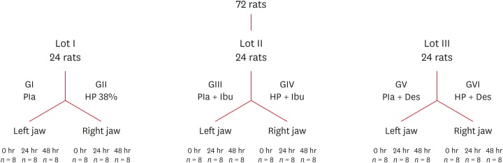
- Influence of pain-relieving therapies on inflammation and the expression of proinflammatory neuropeptides after dental bleaching treatment
- Livia Maria Alves Valentim da Silva, Luciano Tavares Angelo Cintra, Marjorie de Oliveira Gallinari, Francine Benetti, Vanessa Rahal, Edilson Ervolino, Sibele de Alcântara, André Luiz Fraga Briso
- Restor Dent Endod 2020;45(2):e20. Published online February 28, 2020
- DOI: https://doi.org/10.5395/rde.2020.45.e20
-
Abstract
PDFPubReaderePub
Objectives To minimize the tooth sensitivity caused by in-office bleaching, many dentists use non-steroidal anti-inflammatory drugs and topical desensitizing gels containing potassium nitrate and sodium fluoride. This study aimed to evaluate the influence of these substances on inflammation and the expression of substance P and calcitonin gene-related peptide in pulp nerve fibers.
Materials and Methods Seventy-two rats were divided into 6 groups as follows: GI, control; GII, only dental bleaching; GIII, only ibuprofen; GIV, ibuprofen administered 30 minutes before and after the bleaching treatment and every 12 hours until the analysis; GV, only topical application of a desensitizing agent; and GVI, topical application of a desensitizing agent before dental bleaching. Placebo gel was applied to the upper left jaw and the bleaching agent was applied to the upper right jaw in all groups. Subsequently, the groups were divided into 3 subgroups based on the time of analysis: 0, 24, and 48 hours after bleaching (
n = 8). The rats were euthanized and the maxillae were processed and evaluated by histopathological and immunohistochemical analyses. The data were analyzed using the Kruskal-Wallis test, followed by the Dunn test (p < 0.05).Results In the bleaching groups, the inflammatory process and expression of neuropeptides decreased over time. The animals in which a desensitizing agent was applied showed better results within 24 hours.
Conclusions The use of a desensitizing agent had positive effects on inflammation and pain-related neuropeptide expression, minimizing the painful effects of dental bleaching treatment.
-
Citations
Citations to this article as recorded by- Effectiveness of Analgesics in Dental Whitening Pain Management: A Systematic Review and Meta-Analysis
Gabriella Alves Julião Costa, Caio Ferreira Freire Caetano, Ravy Jucá Farias, Diana Araújo Cunha, Dayrine Silveira de Paula, Edson Luiz Cetira Filho, Paulo Goberlânio de Barros Silva
Expert Opinion on Pharmacotherapy.2025; 26(5): 639. CrossRef - The Use of Ozone Therapy in Combination with a Desensitizing Agent for Dentinal Tubules Occlusion: An In Vitro Study
Banna Alnufaiy
The Open Dentistry Journal.2025;[Epub] CrossRef - The role of neurogenic inflammation in pulp repair and the techniques used for its assessment (narrative review)
Muna Sh. Ahmed, Anas F. Mahdee
Frontiers in Dental Medicine.2025;[Epub] CrossRef - Influence of dental bleaching on the pulp tissue: A systematic review of in vivo studies
Mariana Viana Donato, Alexandre Henrique dos Reis‐Prado, Lucas Guimarães Abreu, Lara Cancella de Arantes, Juliana Goto, Hebertt Gonzaga dos Santos Chaves, Luciano Tavares Angelo Cintra, André Luiz Fraga Briso, Isabella Faria da Cunha Peixoto, Francine Ben
International Endodontic Journal.2024; 57(6): 630. CrossRef - Role of induced nitric oxide synthases in orofacial nociception/discomfort after dental tooth bleaching with hydrogen peroxide
Marcílio Rodrigues Pinto, Kirlya Isabel da Silva Medeiros, Letícia Menezes Maia, Antonio Alexandre Coelho, Ana Paula Negreiros Nunes Alves, Caio Ferreira Freire Caetano, Karine Cestaro Mesquita, Paulo Goberlânio de Barros Silva, Fabricio Bitu Sousa
Archives of Oral Biology.2024; 161: 105937. CrossRef - Can different agents reduce the damage caused by bleaching gel to pulp tissue? A systematic review of basic research
Letícia Aparecida Silva Batista, Alexandre Henrique dos Reis-Prado, Hebertt Gonzaga dos Santos Chaves, Lara Cancella de Arantes, Luís Fernando Santos Alves Morgan, Carolina Bosso André, Thaís Yumi Suzuki, Francine Benetti
Restorative Dentistry & Endodontics.2023;[Epub] CrossRef - The Effects of Different Drugs with Anti-Inflamatory Potential in Prevention of Pulp Damage During the Teeth Bleaching
Miona Glisic, Andjela Milojevic, Milica Milinkovic, Marina Rankovic
Experimental and Applied Biomedical Research (EABR).2023;[Epub] CrossRef - Bleaching gel volume influences hydrogen peroxide diffusion, inflammation, and the presence of nitric oxide in the pulp tissue: in vitro and in vivo model
Sibele de ALCÂNTARA, Francine BENETTI, Lívia Maria Alves Valentim da SILVA, Nathália Evelyn da Silva MACHADO, Isabela Joane Prado SILVA, Lara Maria Bueno ESTEVES, Edilson ERVOLINO, Luciano Tavares Angelo CINTRA, André Luiz Fraga BRISO
Journal of Applied Oral Science.2023;[Epub] CrossRef - Design of a thermosensitive ibuprofen-loaded nanogel as smart material applied as anti-inflammatory in tooth bleaching: An in vivo study
Samara K.S.C.F. Moura, Milena L.V. dos Santos, Lucas A. do Nascimento, Mariana F.A. da Silva, Glória M. de França, Lucas M. da Costa, Aldo C. Medeiros, Raimundo F. Araújo-Júnior, Aurigena A. de Araújo, Cláudia N. Oliveira, André L. Dorini, Rejane A. de Ca
Journal of Drug Delivery Science and Technology.2022; 68: 103123. CrossRef - Topical application of Otosporin® before in-office bleaching: a split mouth, triple-blind, multicenter randomized clinical trial
Michael Willian Favoreto, Laína Vochikovski, Renata Maria Oleniki Terra, Veridiana Silva Campos, Mariana Evangelista Santos, Sônia Saeger Meireles, Alessandra Reis, Alessandro D. Loguercio
Clinical Oral Investigations.2022; 26(3): 2555. CrossRef - A novel tooth bleaching gel based on peroxymonosulfate/polyphosphates advanced oxidation process: Effective whitening avoiding pulp damage and sensitivity
Su Yang, Baiyan Sui, Xin Liu, Jiao Sun, Jun Wang
Chemical Engineering Journal.2022; 429: 132525. CrossRef - Effectiveness of Violet LED alone or in association with bleaching gel during dental photobleaching: A Systematic Review
Bianca Rossi, Susana Morimoto, Tamara Kerber Tedesco, Sandra Ribeiro Cunha, Anna Carolina Ratto Tempestini Horliana, Karen Müller Ramalho
Photodiagnosis and Photodynamic Therapy.2022; 38: 102813. CrossRef
- Effectiveness of Analgesics in Dental Whitening Pain Management: A Systematic Review and Meta-Analysis
- 2,608 View
- 14 Download
- 12 Crossref
- Effect of dental bleaching on the microhardness and surface roughness of sealed composite resins
- Renan Aparecido Fernandes, Henrico Badaoui Strazzi-Sahyon, Thaís Yumi Umeda Suzuki, André Luiz Fraga Briso, Paulo Henrique dos Santos
- Restor Dent Endod 2020;45(1):e12. Published online January 10, 2020
- DOI: https://doi.org/10.5395/rde.2020.45.e12
-
Abstract
PDFPubReaderePub
Objectives The aim of this
in vitro study was to evaluate the microhardness and surface roughness of composite resins before and after tooth bleaching procedures.Materials and Methods Sixty specimens were prepared of each composite resin (Filtek Supreme XT and Opallis), and BisCover LV surface sealant was applied to half of the specimens. Thirty enamel samples were obtained from the buccal and lingual surfaces of human molars for use as the control group. The surface roughness and microhardness were measured before and after bleaching procedures with 35% hydrogen peroxide or 16% carbamide (
n = 10). Data were analyzed using 1-way analysis of variance and the Fisher test (α = 0.05).Results Neither hydrogen peroxide nor carbamide peroxide treatment significantly altered the hardness of the composite resins, regardless of surface sealant application; however, both treatments significantly decreased the hardness of the tooth samples (
p < 0.05). The bleaching did not cause any change in surface roughness, with the exception of the unsealed Opallis composite resin and dental enamel, both of which displayed an increase in surface roughness after bleaching with carbamide peroxide (p < 0.05).Conclusions The microhardness and surface roughness of enamel and Opallis composite resin were influenced by bleaching procedures.
-
Citations
Citations to this article as recorded by- Property changes in resin composite exposed to mouth rinses during 10% carbamide peroxide bleaching
Mariana Ferreira da Silva, Giovana Contin Germinari, Carolina Meneghin Barbosa, Tatiane Cristina Dotta, Renata Siqueira Scatolin, Waldemir Francisco Vieira Júnior, Laura Nobre Ferraz
Brazilian Journal of Oral Sciences.2026; 25: e260366. CrossRef - Effect of Bleaching Protocols on the Microhardness and Surface Roughness of Composite Resins with Different Filler Architectures: An In Vitro Study
Flor de Maria Celis-Jacinto , Fredy Hugo Cruzado-Oliva, Jorge Wilfredo Vera-Alvarado
Odovtos - International Journal of Dental Sciences.2026;[Epub] CrossRef - Effect of Bleaching on Surface Roughness and Color Parameters of Coffee-Stained Nanohybrid Dental Composites with Different Viscosities
Hetaf S. Redwan, Mohamed A. Hussein, Mohamed M. Abdul-Monem
European Journal of General Dentistry.2025; 14(01): 027. CrossRef - Effect of Staining and External Bleaching on the Color Stability and Surface Roughness of Universal-Shade Resin-Based Composite
AlHanouf AlHabdan, Amal Alsuhaibani, Lama Alomran, Lulwah Almutib
Clinical, Cosmetic and Investigational Dentistry.2025; Volume 17: 1. CrossRef - Comparative Analysis Between Strip and Gels Indicated for at Home Bleaching: Analysis of Color Alteration, Roughness and Microhardness of Dental Enamel
K. M. S. Aidar, L. T. A. Cintra, M. C. B. Ferreira, T. C. Fagundes, L. M. B. Esteves, J. Goto, A. Catelan, A. L. F. Briso
Journal of Esthetic and Restorative Dentistry.2025; 37(6): 1504. CrossRef - Surface properties and susceptibility to staining of a resin composite after brushing with different whitening toothpastes
Aline da Silva Barros, Carolina Meneghin Barbosa, Renata Siqueira Scatolin, Waldemir Francisco Vieira Junior, Laura Nobre Ferraz
Restorative Dentistry & Endodontics.2025; 50(1): e6. CrossRef - Degradation Resistance of Next-Generation Dental Composites Under Bleaching and Immersion: A Multiscale Investigation
Syed Zubairuddin Ahmed, Shahad Al-Qahtani, Naif H. Al-Qahtani, Hussah Al-Mulhim, Maha Al-Qahtani, Ali Albalushi, Sultan Akhtar
Prosthesis.2025; 7(3): 57. CrossRef - Effect of Over-the-Counter Whitening Dentifrices on the Color Stability and Microhardness of Composite Resins
Xinnuo Yu, Maria Pilar Melo, Sofia Folguera, Carmen Llena
Journal of Composites Science.2025; 9(7): 324. CrossRef - From Microstructure to Shade Shift: Confocal and Spectrophotometric Evaluation of Peroxide-Induced Dental Bleaching
Berivan Laura Rebeca Buzatu, Magda Mihaela Luca, Atena Galuscan, Adrian Ovidiu Vaduva, Aurora Doris Fratila, Ramona Dumitrescu, Ruxandra Sava-Rosianu, Octavia Balean, Roxana Buzatu, Daniela Jumanca
Journal of Clinical Medicine.2025; 14(13): 4642. CrossRef - In Vitro Evaluation of Chemical and Microhardness Alterations in Human Enamel Induced by Three Commercial In-Office Bleaching Agents
Berivan Laura Rebeca Buzatu, Atena Galuscan, Ramona Dumitrescu, Roxana Buzatu, Magda Mihaela Luca, Octavia Balean, Gabriela Vlase, Titus Vlase, Iasmina-Mădălina Anghel, Carmen Opris, Bianca Ioana Todor, Mihaela Adina Dumitrache, Daniela Jumanca
Dentistry Journal.2025; 13(8): 357. CrossRef - Effect of Hydrogen Peroxide Bleaching on Color Stability and Microhardness of Alkasite Restorative Materials: An In Vitro Study
Souad A Alfouzan, Rahaf A Alolayan, Asma Munir Khan
Cureus.2025;[Epub] CrossRef - Evaluation of Color Stability and Surface Roughness of Nanohybrid Resin Composites with Different Photoinitiator Systems After Staining and Home/Office Bleaching: An In Vitro Study
Fatma Yılmaz, Buse Kesgin
Meandros Medical And Dental Journal.2025; 26(3): 240. CrossRef - The Effect of Hydrogen Peroxide With Different Concentration on the Color and Surface Microhardness of the Resin Bracket
Song‐Yi Yang
Clinical and Experimental Dental Research.2025;[Epub] CrossRef - Comparative evaluation of different bleaching agents on the color stability, hardness and surface roughness of indirect esthetic restorative materials with different manufacturing methods
Ayse Atay, Defne Canpolat, Soner Sismanoglu, Aslihan Usumez
BMC Oral Health.2025;[Epub] CrossRef - Comparison of Microhardness and Surface Roughness of New Nanofiber Filled Flowable Composite
Rumeysa Hatice ENGINLER OZLEN, Zumrut Ceren OZDUMAN, Burcu OGLAKCI OZKOC, Evrim ELIGUZELOGLU DALKILIC
Bezmialem Science.2024; 12(4): 406. CrossRef - Effect of Bleaching Agents on Composite Resins with and without Bis-GMA: An In Vitro Study
María Melo, Bianca Dumitrache, James Ghilotti, José Luis Sanz, Carmen Llena
Journal of Functional Biomaterials.2024; 15(6): 144. CrossRef - Changes in physical properties of universal composites and CAD/CAM materials after bleaching and antioxidant applications: Scanning electron microscope and atomic force microscope evaluation
Oguz Kaan Tuysuz, Merve Gurses
Microscopy Research and Technique.2024; 87(5): 977. CrossRef - The Effects of Home and Over-The-Counter Whitening Agents on Surface Roughness and Microhardness of High Aesthetic Composites
Elif İpek KILIÇ DÖNMEZ, İhsan HUBBEZOĞLU
Cumhuriyet Dental Journal.2024; 27(1): 30. CrossRef - Effect of carbamide peroxide treatment on the ion release of different dental restorative materials
Merve Nur Yilmaz, Pinar Gul
BMC Oral Health.2024;[Epub] CrossRef - Inorganic Phosphate Effect in a Hydrogen Peroxide-based Bleaching Agent: Physicochemical, Mechanical, and Morphological Properties of Dental Enamel
KG Garcia, GP Nunes, ACB Delbem, PH dos Santos, GLP Fernandes, HF Robles, PBB Lemos, M Danelon
Operative Dentistry.2024; 49(4): 465. CrossRef - Effect of bleaching and repolishing on whiteness change and staining susceptibility of resin-based materials
Sultan Aktuğ Karademir, Samet Atasoy, Beyza Yılmaz
BMC Oral Health.2024;[Epub] CrossRef - Influence of Low pH on the Microhardness and Roughness Surface of Dental Composite—A Preliminary Study
Leszek Szalewski, Dorota Wójcik, Monika Sowa, Vladyslav Vivcharenko, Krzysztof Pałka
Materials.2024; 17(14): 3443. CrossRef - In Vitro Evaluation of the Effectiveness and pH Variation of Dental Bleaching Gels and Their Effect on Enamel Surface Roughness
Federica Veneri, Francesco Cavani, Giovanni Bolelli, Vittorio Checchi, Alessia Bizzi, Giacomo Setti, Luigi Generali
Dentistry Journal.2024; 12(12): 415. CrossRef - Does the combination of whitening toothpaste and hydrogen peroxide bleaching increase the surface roughness and change the morphology of a nanofilled composite?
Cecília Pereira da Silva Braga Tenório, Matheus Kury, Geyse Maria dos Santos Muniz Mota, Cecília Pedroso Turssi, Flávia Lucisano Botelho do Amaral, Vanessa Cavalli
Brazilian Journal of Oral Sciences.2024; 23: e241938. CrossRef - Effect of peroxide‐free and peroxide‐based in‐office bleaching on the surface and mechanical properties of CAD/CAM esthetic restorative materials
Majed M. Alsarani, Aftab Ahmed Khan, Leonel S. J. Bautista, Hanan Alsunbul, Jukka P. Matinlinna
European Journal of Oral Sciences.2024;[Epub] CrossRef - Effect of Repolishing on Color Stability, Translucency, and Surface Roughness of Aged Monochromatic Dental Composites
Mohamed M. Abdul-Monem, Mohamed A. Hussein, Mona G. Abdelrehim
European Journal of General Dentistry.2024; 13(03): 240. CrossRef - Color changes of nanofiller composite resin after glycerin application immersed in turmeric extract
Sukaton, Galih Sampoerno, Widyajeng Ayu Laksmi, Daradhasih Bestari Santiaji
Conservative Dentistry Journal.2023; 13(1): 37. CrossRef - Effects of Dental Bleaching Agents on the Surface Roughness of Dental Restoration Materials
Alexandru Dan Popescu, Mihaela Jana Tuculina, Oana Andreea Diaconu, Lelia Mihaela Gheorghiță, Claudiu Nicolicescu, Cristian Niky Cumpătă, Cristiana Petcu, Jaqueline Abdul-Razzak, Ana Maria Rîcă, Ruxandra Voinea-Georgescu
Medicina.2023; 59(6): 1067. CrossRef - Effect of Bleaching on the Microhardness and Modulus of Elasticity of ACTIVA BioACTIVE – RESTORATIVE: An In Vitro Study
Sushritha Sricharan, Swaroop Hegde, Narmada J., Indiresha H. Narayana, Chatura Mohan, Nithin K. Shetty
Journal of Advanced Oral Research.2023; 14(2): 190. CrossRef - The effect of bleaching on surface roughness and gloss of different CAD/CAM ceramic and hybrid ceramic materials
Ruwaida Z Alshali, Mohammed A AlQahtani, Dalea M Bukhary, Mlak A Alzahrani, Shatha S Alsoraihi, Majed A Alqahtani
Journal of Applied Biomaterials & Functional Materials.2023;[Epub] CrossRef - Effect of bleaching with 15% carbamide peroxide on color stability of microhybrid, nanohybrid, and nanofilled resin composites, each in 3 staining solutions (coffee, cola, red grape juice): A 3-phase study
Azadeh Ghaemi, Sanaz Sharifishoshtari, Mohsen Shahmoradi, Hossein Akbari, Parisa Boostanifard, Sepideh Bagheri, Mohammadreza Shokuhifar, Negin Ashoori, Vahid Rakhshan
Dental Research Journal.2023;[Epub] CrossRef - Micro-Hardness and Surface Roughness of Bulk-Fill Composite Resin: Effect of Surface Sealant Application and Two Bleaching Regimens
Reham Mohamad Attia, Eman Mohamed Sobhy, Mona El Said Abd El Hameed Essa
European Journal of General Dentistry.2023; 12(03): 169. CrossRef - Shear bond strength after using sealant before bonding: a systematic review and meta-analysis of in vitro studies
Jennifer Hoppe, Thomas Lehmann, Christoph-Ludwig Hennig, Ulrike Schulze-Späte, Collin Jacobs
Clinical Oral Investigations.2022; 26(1): 1. CrossRef - Effect of 16% Carbamide Peroxide and Activated-Charcoal-Based Whitening Toothpaste on Enamel Surface Roughness in Bovine Teeth: An In Vitro Study
Jorge Zamudio-Santiago, Marysela Ladera-Castañeda, Flor Santander-Rengifo, Carlos López-Gurreonero, Alberto Cornejo-Pinto, Ali Echavarría-Gálvez, Luis Cervantes-Ganoza, César Cayo-Rojas
Biomedicines.2022; 11(1): 22. CrossRef - Direct dentin bleaching: Would it be possible?
Camila Ferro Clemente, Sibele de Alcântara, Lívia Maria Alves Valentim da Silva, Lara Maria Bueno Esteves, Anderson Catelan, Karen Milaré Seiscento Aidar, Ticiane Cestari Fagundes, André Luiz Fraga Briso
Photodiagnosis and Photodynamic Therapy.2022; 40: 103121. CrossRef - EFFECT OF İN-OFFİCE BLEACHİNG ON THE SURFACE ROUGHNESS OF DİFFERENT COMPOSİTE RESİNS
Seher KAYA, Ozden OZEL BEKTAS
Cumhuriyet Dental Journal.2022; 25(Supplement): 78. CrossRef - Effect of Polishing on the Surface Microhardness of Nanohybrid Composite Resins Subjected to 35% Hydrogen Peroxide
Giovanna Gisella Ramírez-Vargas, Julia Elbia Medina y Mendoza, Ana Sixtina Aliaga-Mariñas, Marysela Irene Ladera-Castañeda, Luis Adolfo Cervantes-Ganoza, César Félix Cayo-Rojas
Journal of International Society of Preventive and Community Dentistry.2021; 11(2): 216. CrossRef - Intrapulpal Concentration of Hydrogen Peroxide of Teeth Restored With Bulk Fill and Conventional Bioactive Composites
DP Silva, BA Resende, M Kury, CB André, CPM Tabchoury, M Giannini, V Cavalli
Operative Dentistry.2021; 46(3): E158. CrossRef - An Environmental Scanning Electron Microscopy Evaluation on Comparison of Three Different Bleaching Agents using the Laser Activated in-Office Bleaching at Different Wavelengths
Shachi Goenka, Sushil Kumar Cirigiri, Kanika Poplai, Baig Mirza Aslam, Shalini Singh, Shweta Gangavane
Journal of Pharmacy and Bioallied Sciences.2021; 13(Suppl 2): S1478. CrossRef - Effects of Artificial Staining and Bleaching Protocols on the Surface Roughness, Color, and Whiteness Changes of an Aged Nanofilled Composite
Geyse Maria dos Santos Muniz Mota, Matheus Kury, Cecília Pereira da Silva Braga Tenório, Flávia Lucisano Botelho do Amaral, Cecília Pedroso Turssi, Vanessa Cavalli
Frontiers in Dental Medicine.2020;[Epub] CrossRef
- Property changes in resin composite exposed to mouth rinses during 10% carbamide peroxide bleaching
- 4,441 View
- 48 Download
- 40 Crossref
- Evaluation of the effects of whitening mouth rinses combined with conventional tooth bleaching treatments
- Jaqueline Costa Favaro, Omar Geha, Ricardo Danil Guiraldo, Murilo Baena Lopes, Andreza Maria Fábio Aranha, Sandrine Bittencourt Berger
- Restor Dent Endod 2019;44(1):e6. Published online January 30, 2019
- DOI: https://doi.org/10.5395/rde.2019.44.e6
-
Abstract
PDFPubReaderePub
Objectives The aim of the present study was to evaluate the effect of whitening mouth rinses alone and in combination with conventional whitening treatments on color, microhardness, and surface roughness changes in enamel specimens.
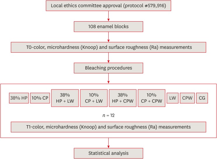
Materials and Methods A total of 108 enamel specimens were collected from human third molars and divided into 9 groups (
n = 12): 38% hydrogen peroxide (HP), 10% carbamide peroxide (CP), 38% HP + Listerine Whitening (LW), 10% CP + LW, 38% HP + Colgate Plax Whitening (CPW), 10% CP + CPW, LW, CPW, and the control group (CG). The initial color of the specimens was measured, followed by microhardness and roughness tests. Next, the samples were bleached, and their color, microhardness, and roughness were assessed. Data were analyzed through 2-way analysis of variance (ANOVA; microhardness and roughness) and 1-way ANOVA (color change), followed by the Tukeypost hoc test. The Dunnett test was used to compare the roughness and microhardness data of the CG to those of the treated groups.Results Statistically significant color change was observed in all groups compared to the CG. All groups, except the LW group, showed statistically significant decreases in microhardness. Roughness showed a statistically significant increase after the treatments, except for the 38% HP group.
Conclusions Whitening mouth rinses led to a whitening effect when they were used after conventional treatments; however, this process caused major changes on the surface of the enamel specimens.
-
Citations
Citations to this article as recorded by- Property changes in resin composite exposed to mouth rinses during 10% carbamide peroxide bleaching
Mariana Ferreira da Silva, Giovana Contin Germinari, Carolina Meneghin Barbosa, Tatiane Cristina Dotta, Renata Siqueira Scatolin, Waldemir Francisco Vieira Júnior, Laura Nobre Ferraz
Brazilian Journal of Oral Sciences.2026; 25: e260366. CrossRef - Effect of Over-the-counter Whitening Products on Postbleaching Enamel Surface Roughness and Shade Recovery: An In Vitro Study
Rocío Llancari-Alonzo, Jorge Manrique-Guzmán, Jorge Manrique-Chávez, Leonor Castro-Ramirez, Carlos López-Gurreonero, Alberto Cornejo-Pinto, César Cayo-Rojas
The Journal of Contemporary Dental Practice.2026; 27(2): 148. CrossRef - Influence of commercial mouth rinses with different formulations on enamel properties during at-home bleaching
Thalita Novello Coelho, Ana Júlia Gil, Marcos Roberto Lima Benati, Carolina Meneghin Barbosa, Tatiane Cristina Dotta, Waldemir Francisco Vieira-Junior, Renata Siqueira Scatolin, Laura Nobre Ferraz
Odontology.2026;[Epub] CrossRef - Which Whitening Mouthwash With Different Ingredients Is More Effective on Color and Bond Strength of Enamel?
Elif Varli Tekingur, Fatih Bedir, Muhammet Karadas, Rahime Zeynep Erdem
Journal of Esthetic and Restorative Dentistry.2025; 37(4): 960. CrossRef - Do Different Tooth Bleaching–Remineralizing Regimens Affect the Bleaching Effectiveness and Enamel Microhardness In Vitro?
Hamideh Sadat Mohammadipour, Parnian Shokrollahi, Sima Gholami, Hosein Bagheri, Fatemeh Namdar, Salehe Sekandari, Cesar Rogério Pucci
International Journal of Dentistry.2024;[Epub] CrossRef - Effect of hydrogen peroxide versus charcoal-based whitening mouthwashes on color, surface roughness, and color stability of enamel
Mayada S. Sultan
BMC Oral Health.2024;[Epub] CrossRef - Effects of online marketplace-sourced over-the-counter tooth whitening products on the colour, microhardness, and surface topography of enamel: an in vitro study
Radhika Agarwal, Nikki Vasani, Urmila Sachin Mense, Niharika Prasad, Aditya Shetty, Srikant Natarajan, Arindam Dutta, Manuel S. Thomas
BDJ Open.2024;[Epub] CrossRef - Effect of Whitening Mouthwashes on Color Change and Enamel Mineralization: An In Vitro Study
Rosa Josefina Roncal Espinoza, José Alberto Castañeda Vía, Alexandra Mena-Serrano, Lidia Yileng Tay
World Journal of Dentistry.2023; 14(9): 739. CrossRef - Effectiveness and Adverse Effects of Over-the-Counter Whitening Products on Dental Tissues
Maiara Rodrigues de Freitas, Marynara Mathias de Carvalho, Priscila Christiane Suzy Liporoni, Ana Clara Borges Fort, Rodrigo de Morais e Moura, Rayssa Ferreira Zanatta
Frontiers in Dental Medicine.2021;[Epub] CrossRef - Renklendirilmiş kompozit rezinin renk değişimine ve yüzey pürüzlülüğüne beyazlatıcı ağız gargarasının etkisi
Şeref Nur MUTLU, Makbule Tuğba TUNCDEMIR
Selcuk Dental Journal.2020; 7(3): 435. CrossRef
- Property changes in resin composite exposed to mouth rinses during 10% carbamide peroxide bleaching
- 2,406 View
- 17 Download
- 10 Crossref
- Effect of various bleaching treatments on shear bond strength of different universal adhesives and application modes
- Fatma Dilsad Oz, Zeynep Bilge Kutuk
- Restor Dent Endod 2018;43(2):e20. Published online April 16, 2018
- DOI: https://doi.org/10.5395/rde.2018.43.e20
-
Abstract
PDFPubReaderePub
Objectives The aim of this
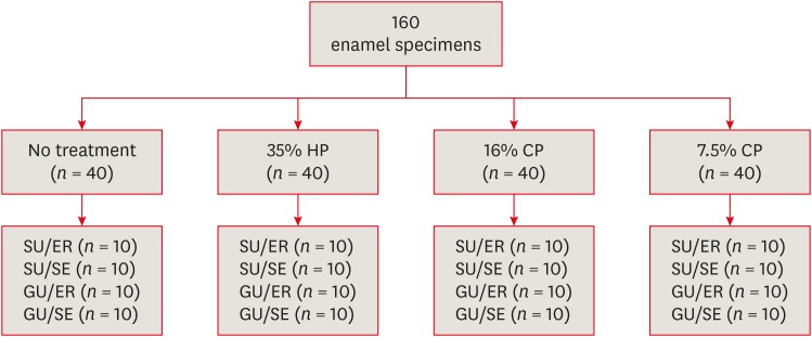
in vitro study was to evaluate the bond strength of 2 universal adhesives used in different application modes to bleached enamel.Materials and Methods Extracted 160 sound human incisors were used for the study. Teeth were divided into 4 treatment groups: No treatment, 35% hydrogen peroxide, 16% carbamid peroxide, 7.5% carbamid peroxide. After bleaching treatments, groups were divided into subgroups according to the adhesive systems used and application modes (
n = 10): 1) Single Bond Universal, etch and rinse mode; 2) Single Bond Universal, self-etch mode; 3) Gluma Universal, etch and rinse mode; 4) Gluma Universal, self-etch mode. After adhesive procedures nanohybrid composite resin cylinders were bonded to the enamel surfaces. All specimens were subjected to shear bond strength (SBS) test after thermocycling. Data were analyzed using a 3-way analysis of variance (ANOVA) and Tukeypost hoc test.Results No significant difference were found among bleaching groups (35% hydrogen peroxide, 16% carbamid peroxide, 7.5% carbamid peroxide, and no treatment groups) in the mean SBS values. There was also no difference in SBS values between Single Bond Universal and Gluma Universal at same application modes, whereas self-etch mode showed significantly lower SBS values than etch and rinse mode (
p < 0.05).Conclusions The bonding performance of the universal adhesives was enhanced with the etch and rinse mode application to bleached enamel and non-bleached enamel.
-
Citations
Citations to this article as recorded by- Antioxidant effect on shear bond strength of resin composite to in-office versus home bleached enamel surface
Maha Mosaad Mohamed, Magda E. -A. Shalaby, Eman A. E. -G. Shebl
Tanta Dental Journal.2025; 22(3): 409. CrossRef - Effects of Time-Elapsed Bleaching on the Surface and Mechanical Properties of Dentin Substrate Using Hydrogen Peroxide-Free Nanohydroxyapatite Gel
Aftab Khan, Abdulaziz AlKhureif, Manal Almutairi, Abrar Nooh, Saeed Hassan, Yasser Alqahtani
International Journal of Nanomedicine.2024; Volume 19: 10307. CrossRef - Effect of sodium ascorbate on the shear bond strength of orthodontic brackets to bleached enamel using universal dental adhesive
Saeid Sadeghian, Kamyar Fathpour, Mahshid Biglari
Dental Research Journal.2023;[Epub] CrossRef - Quantitative Measurements of the Depth of Enamel Demineralization before and after Bleach: An In Vitro Study
Sara Naim, Gianrico Spagnuolo, Essam Osman, Syed Sarosh Mahdi, Gopi Battineni, Syed Saad B. Qasim, Mariangela Cernera, Hasna Rifai, Nada Jaafar, Elie Maalouf, Carina Mehanna Zogheib, Konstantinos Michalakis
BioMed Research International.2022;[Epub] CrossRef - DİŞ BEYAZLATMA İŞLEMİNİN LİTYUM DİSİLİKAT SERAMİĞİN BAĞLANMA DAYANIMINA ETKİSİ
Merve YILDIRAK, Rıfat GÖZNELİ
Atatürk Üniversitesi Diş Hekimliği Fakültesi Dergisi.2020; : 1. CrossRef - The Effect of Different Bleaching Protocols, Used with and without Sodium Ascorbate, on Bond Strength between Composite and Enamel
Maroun Ghaleb, Giovanna Orsini, Angelo Putignano, Sarah Dabbagh, Georges Haber, Louis Hardan
Materials.2020; 13(12): 2710. CrossRef - Influence of phototherapy on adhesive strength and microleakage of bleached enamel bonded to orthodontic brackets: An in-vitro study
Erum Khan, Ibrahim Alshahrani, Muhammad Abdullah Kamran, Abdulaziz Samran, Ali Alqerban, Saad Abdul Rehman
Photodiagnosis and Photodynamic Therapy.2019; 25: 344. CrossRef - Effect of Er: YAG Laser on Microtensile Bond Strength of Bleached Dentin to Composite
Mohsen Rezaei, Elham Aliasghar, Mohammad Bagher Rezvani, Nasim Chiniforush, Zohreh Moradi
Journal of Lasers in Medical Sciences.2019; 10(2): 117. CrossRef
- Antioxidant effect on shear bond strength of resin composite to in-office versus home bleached enamel surface
- 3,127 View
- 31 Download
- 8 Crossref
- Effect of three nanobiomaterials on microhardness of bleached enamel
- Maryam Khoroushi, Farinaz Shirban, Sara Kaveh, Samaneh Doustfateme
- Restor Dent Endod 2016;41(3):196-201. Published online July 14, 2016
- DOI: https://doi.org/10.5395/rde.2016.41.3.196
-
Abstract
PDFPubReaderePub
Objectives The aim of this
in vitro study was to evaluate the effect of incorporating three different nanobiomaterials into bleaching material on microhardness of bleached enamel.Materials and Methods The crowns of 24 extracted sound human molars were sectioned. Sixty enamel specimens (2 × 3 × 4 mm) were selected and divided into five groups (
n = 12): Group 1 received no bleaching procedure (control); Group 2 underwent bleaching with a 40% hydrogen peroxide (HP) gel; Groups 3, 4, and 5 were bleached with a 40% HP gel modified by incorporation of bioactive glass (BAG), amorphous calcium phosphate (ACP) and hydroxyapatite (HA), respectively. The enamel microhardness was evaluated. The differences in Knoop microhardness data of each group were analyzed by one-way ANOVA, followed bypost hoc Tukey tests.Results Significant differences were observed between the study groups. The enamel microhardness changes in Groups 1, 3, 4, and 5 were significantly lower than that of Group 2 (
p < 0.001).Conclusions Within the limitations of this study, it can be concluded that incorporation of each one of the three tested biomaterials as remineralizing agents might be effective in decreasing enamel microhardness changes subsequent to in-office bleaching.
-
Citations
Citations to this article as recorded by- Effects of bleaching agents containing nano-hydroxyapatite on sound and demineralized enamel: an in vitro study
Merve Haberal, Çiğdem Çelik
Odontology.2026;[Epub] CrossRef - Influence of 10% propolis extract on morphology, microhardness, chemical composition, and color of bleached enamel
Roberta Pimentel de Oliveira, Cíntia de Melo Silva, Elma Vieira Takeuchi, Geovanni Pereira Mitre, Lindalva Maria de Meneses Costa Ferreira, Jesuina Lamartine Nogueira Araújo, Hilton Túlio Costi, Maria Sueli da Silva Kataoka, Roseane Maria Ribeiro Costa, C
Odontology.2026;[Epub] CrossRef - Protective role of calcium-based agents in dental bleaching gels: insights from a systematic review and meta-analysis of clinical and laboratory evidence
Gabriel Pereira Nunes, Renata de Oliveira Alves, Geórgia Rondó Peres, Matheus Henrique Faccioli Ragghianti, Priscila Toninatto Alves de Toledo, Alexandre Henrique dos Reis Prado, Carla Ferreira-Baptista, Alberto Carlos Botazzo Delbem
Clinical Oral Investigations.2025;[Epub] CrossRef - Analysis of Enamel Surface and Shear Bond Strength with Orthodontic Bracket at Different Time Intervals After Bleaching Treatment
Yu-Jin Lee, Ji-Yeon Hong, Hye-Min Ku, Song-Yi Yang
Journal of Dental Hygiene Science.2025; 25(2): 130. CrossRef - Effect of strontium fluorophosphate bioactive glass on color, microhardness and surface roughness of bleached enamel
Shiza Yezdani, Monisha Khatri, Sampath Vidhya, Sekar Mahalaxmi
Technology and Health Care.2024; 32(1): 285. CrossRef - In vitro evaluation of the effect of addition of biomaterials to carbamide peroxide on the bleaching efficacy and microhardness of enamel
Sowmya Kavoor, M. A. Ranjini, Naval Abdul Aziz, H. K. Ashok, Roopa R. Nadig
Journal of Conservative Dentistry and Endodontics.2024; 27(3): 310. CrossRef - Effect of hydrogen peroxide and its combination with nano-hydroxyapatite or nano-bioactive glass on the enamel demineralization and tooth color: An in vitro study
Elham Kheradmand, Alirea Daneshkazemi, Abdolrahim Davari, Maede Kave, Solmaz Ghanbarnejad
Dental Research Journal.2023;[Epub] CrossRef - Over‐the‐counter bleaching agents can help with tooth whitening maintenance
Olívia Santana Jorge, Carolina Noronha Ferraz de Arruda, Rafaella Tonani Torrieri, Rocio Geng Vivanco, Fernanda de Carvalho Panzeri Pires‐de‐Souza
Journal of Esthetic and Restorative Dentistry.2022; 34(2): 328. CrossRef - Effect of Indigenously Developed Nano-Hydroxyapatite Crystals from Chicken Egg Shell on the Surface Hardness of Bleached Human Enamel
Divya Kunam, Vidhya Sampath, Sujatha Manimaran, Mahalaxmi Sekar
Contemporary Clinical Dentistry.2019; 10(3): 489. CrossRef - Influence of Time Intervals between Bleaching Procedures on Enamel Microhardness and Surface Roughness
Roberta Pimentel de Oliveira, Juliana Costa Pereira Baia, Mara Eliane Soares Ribeiro, Mario Honorato da Silva e Souza Junior, Sandro Cordeiro Loretto
The Open Dentistry Journal.2018; 12(1): 555. CrossRef
- Effects of bleaching agents containing nano-hydroxyapatite on sound and demineralized enamel: an in vitro study
- 2,351 View
- 8 Download
- 10 Crossref
- Antioxidant therapy enhances pulpal healing in bleached teeth
- Adriano Fonseca Lima, Marcelo Rocha Marques, Diana Gabriela Soares, Josimeri Hebling, Giselle Maria Marchi, Carlos Alberto de Souza Costa
- Restor Dent Endod 2016;41(1):44-54. Published online February 1, 2016
- DOI: https://doi.org/10.5395/rde.2016.41.1.44
-
Abstract
PDFPubReaderePub
Objectives The purpose of this study was to evaluate the histopathological effects of an antioxidant therapy on the pulp tissue of rat teeth exposed to a bleaching gel with 35% hydrogen peroxide.
Materials and Methods Forty rats were subjected to oral ingestion by gavage of distilled water (DW) or ascorbic acid (AA) 90 min before the bleaching therapy. For the bleaching treatment, the agent was applied twice for 5 min each to buccal surfaces of the first right mandibular molars. Then, the animals were sacrificed at 6 hr, 24 hr, 3 day, or 7 day post-bleaching, and the teeth were processed for microscopic evaluation of the pulp tissue.
Results At 6 hr, the pulp tissue showed moderate inflammatory reactions in all teeth of both groups. In the DW and AA groups, 100% and 80% of teeth exhibited pulp tissue with significant necrosis and intense tissue disorganization, respectively. At 24 hr, the AA-treated group demonstrated a greater regenerative capability than the DW group, with less intense inflammatory reaction and new odontoblast layer formation in 60% of the teeth. For up to the 7 day period, the areas of pulpal necrosis were replaced by viable connective tissue, and the dentin was underlined by differentiated odontoblast-like cells in most teeth of both groups.
Conclusions A slight reduction in initial pulpal damage during post-bleaching was promoted by AA therapy. However, the pulp tissue of AA-treated animals featured faster regenerative potential over time.
-
Citations
Citations to this article as recorded by- Clinical Study on the Efficacy of 35% Hydrogen Peroxide Gel According to Exposure Time (40 min vs. 20 min) by Spectrophotometry
Trinidad Rincón, Maria Portillo Muñoz, Maria Lobato, Ana María Martín Casado, Laryssa Mylenna Madruga Barbosa, Alessandro Loguercio, Cristina Gómez‐Polo
Journal of Esthetic and Restorative Dentistry.2026;[Epub] CrossRef - Influence of dental bleaching on the pulp tissue: A systematic review of in vivo studies
Mariana Viana Donato, Alexandre Henrique dos Reis‐Prado, Lucas Guimarães Abreu, Lara Cancella de Arantes, Juliana Goto, Hebertt Gonzaga dos Santos Chaves, Luciano Tavares Angelo Cintra, André Luiz Fraga Briso, Isabella Faria da Cunha Peixoto, Francine Ben
International Endodontic Journal.2024; 57(6): 630. CrossRef - ANALYSIS OF THE INFLUENCE OF THE TEETH WHITENING PROCEDURE ON THE GUM AND CYTOKINE PROFILE OF ORAL FLUID (LITERATURE REVIEW)
S. S. Bozhik, N. V. Hasyuk, V. B. Radchuk
Bulletin of Problems Biology and Medicine.2024; 1(3): 17. CrossRef - Assessing the Viability of Laser-Activated Dental Bleaching Compared to Conventional In-Office Bleaching Methods: A Systematic Review of Clinical and In Vitro Studies
Eugenia Anagnostaki, Valina Mylona, Steven Parker, Mark Cronshaw, Martin Grootveld
Applied Sciences.2023; 13(22): 12459. CrossRef - Effects of the application of sodium ascorbate after in-office bleaching on the penetration of hydrogen peroxide, color change, and microtensile bond strength
Alexandra Mena-Serrano, María G. Granda-Albuja, Jenny Naranjo, Eliana Aldás Fierro, Michael Willian Favoreto, Alessandro D. Loguercio, Alessandra Reis
Brazilian Dental Journal.2023; 34(5): 87. CrossRef - Can different agents reduce the damage caused by bleaching gel to pulp tissue? A systematic review of basic research
Letícia Aparecida Silva Batista, Alexandre Henrique dos Reis-Prado, Hebertt Gonzaga dos Santos Chaves, Lara Cancella de Arantes, Luís Fernando Santos Alves Morgan, Carolina Bosso André, Thaís Yumi Suzuki, Francine Benetti
Restorative Dentistry & Endodontics.2023;[Epub] CrossRef - Design of a thermosensitive ibuprofen-loaded nanogel as smart material applied as anti-inflammatory in tooth bleaching: An in vivo study
Samara K.S.C.F. Moura, Milena L.V. dos Santos, Lucas A. do Nascimento, Mariana F.A. da Silva, Glória M. de França, Lucas M. da Costa, Aldo C. Medeiros, Raimundo F. Araújo-Júnior, Aurigena A. de Araújo, Cláudia N. Oliveira, André L. Dorini, Rejane A. de Ca
Journal of Drug Delivery Science and Technology.2022; 68: 103123. CrossRef - Effect of bleaching gel volume on color change and postoperative sensitivity: a randomized clinical study
Lara Maria Bueno Esteves, Paulo Henrique dos Santos, Ticiane Cestari Fagundes, Marjorie de Oliveira Gallinari, Giulia Bessa de Mello Antonaccio, Luciano Tavares Ângelo Cintra, André Luiz Fraga Briso
Clinical Oral Investigations.2022; 26(3): 2527. CrossRef - Human Dental Pulp Stem Cells Exhibit Different Biological Behaviours in Response to Commercial Bleaching Products
Carmen Llena, Mar Collado-González, Christopher Joseph Tomás-Catalá, David García-Bernal, Ricardo Elías Oñate-Sánchez, Francisco Javier Rodríguez-Lozano, Leopoldo Forner
Materials.2018; 11(7): 1098. CrossRef - Concentration‐dependent effect of bleaching agents on the immunolabelling of interleukin‐6, interleukin‐17 and CD5‐positive cells in the dental pulp
F. Benetti, J. E. Gomes‐Filho, L. L. Ferreira, G. Sivieri‐Araújo, E. Ervolino, A. L. F. Briso, L. T. A. Cintra
International Endodontic Journal.2018; 51(7): 789. CrossRef
- Clinical Study on the Efficacy of 35% Hydrogen Peroxide Gel According to Exposure Time (40 min vs. 20 min) by Spectrophotometry
- 2,453 View
- 6 Download
- 10 Crossref
- Effects of applying antioxidants on bond strength of bleached bovine dentin
- Hyo-Jin Whang, Dong-Hoon Shin
- Restor Dent Endod 2015;40(1):37-43. Published online October 13, 2014
- DOI: https://doi.org/10.5395/rde.2015.40.1.37
-
Abstract
PDFPubReaderePub
Objectives Some antioxidants are believed to restore dentin bond strength after dental bleaching. This study was done to evaluate the influence of antioxidants on the bond strength of bleached bovine dentin.
Materials and Methods Thirty incisors were randomly assigned to 10 groups (two unbleached control and eight bleached groups: immediate bonding IB, 4 wk delayed bonding DB, 10% sodium ascorbate treated SA, 10% α-tocopherol treated TP groups). Teeth in half of groups were subjected to thermal stress, whereas the remaining groups were not. Resin-dentin rods with a cross-sectional area of 2.25 mm2 were obtained and microtensile bond strength was determined at a crosshead speed of 1 mm/min. Fifteen specimens were prepared for SEM to compare the surface characteristics of each group. The change in dentin bond strength from thermal stress and antioxidant treatment was evaluated using two-way analysis of variance (ANOVA) and Sheffe's
post hoc test at a significance level of 95%.Results The control group exhibited the highest bond strength values, whereas IB group showed the lowest value before and after thermocycling. The DB group recovered its bond strength similar to that of the control group. The SA and TP groups exhibited similar bond strength values with those of the control and DB groups before thermocycling. However, The TP group did not maintain bond strength with thermal stress, whereas the SA group did.
Conclusions Applying a 10% sodium ascorbate solution rather than 10% α-tocopherol solution for 60 sec is recommended to maintain dentin bond strength when restoring non-vitally bleached teeth.
-
Citations
Citations to this article as recorded by- Shear bond strength analysis of self-etch and total-etch adhesive systems on antioxidant-treated dentin in post-endodontic restoration
Suvarna Kavya Harish, Shruthi H. Attavar
Frontiers in Dental Medicine.2026;[Epub] CrossRef - Preliminary In Vitro Evaluation of Vitamin E (α‐Tocopherol) Incorporation on Cytotoxicity, Antifungal Activity, and Surface Roughness of Denture Acrylic Soft Liner
Neven S. Aref, Hannah Wesley
International Journal of Dentistry.2026;[Epub] CrossRef - Evaluation of the effect of the application of Quercus cerris extract and the use of fluoride bonding material on the bonding strength of orthodontic brackets after tooth bleaching with hydrogen peroxide
Ezgi Ay, Derya Dursun
PeerJ.2025; 13: e19335. CrossRef - Antioxidant effect on shear bond strength of resin composite to in-office versus home bleached enamel surface
Maha Mosaad Mohamed, Magda E. -A. Shalaby, Eman A. E. -G. Shebl
Tanta Dental Journal.2025; 22(3): 409. CrossRef - Comparative analysis of the impact of modern cavity disinfectants on dentin bond strength
Simge Gümüş Ayaz, Ezgi Sonkaya, Gökçe Keçeci
Frontiers in Materials.2025;[Epub] CrossRef - Effect of glutathione on bond strength of composite resin to enamel following extracoronal bleaching
Nair Devika, Chandrasekaran Charanya, K Athira, James Vandana, Sundaresan Balagopal
Journal of Conservative Dentistry and Endodontics.2024; 27(11): 1110. CrossRef - Effect of Chitosan Nanoparticle as an Antioxidant Material on Shear Bond Strength of Composite Resin to Enamel after External Bleaching
Diatri Nari Ratih, Shintatika Erlagista, Tunjung Nugraheni
Open Access Macedonian Journal of Medical Sciences.2024; 12: 1. CrossRef - Effects of alpha‐tocopherol antioxidant on fracture strength and adhesion of endodontically treated teeth restored after dental bleaching
Natália Marcomini, Maria Carolina da Costa Albaricci, Joatan Lucas de Sousa Gomes Costa, João Felipe Besegato, Eduardo Fernández Godoy, Andréa Abi Rached Dantas, Milton Carlos Kuga
European Journal of Oral Sciences.2024;[Epub] CrossRef - Alpha-tocopherol: An alternative solution for the adverse effects of dental bleaching on dentin adhesion
Maria Carolina da Costa Albaricci, Natália Marcomini, Joatan Lucas de Sousa Gomes Costa, Antonia Patricia Oliveira Barros, Lucas David Galvani, Milton Carlos Kuga, Andréa Abi Rached Dantas
International Journal of Adhesion and Adhesives.2024; 131: 103655. CrossRef - Efficacy of organic and antioxidant agents to regain bond strength to bleached enamel in different dental adhesive solvents
Satheesh B Haralur, Renad Mohammed Al-Ibrahim, Faten Abdullah Al-Shahrani, Rahaf Abdullah Al-Qahtani, Saurabh Chaturvedi, Naseer M Alqahtani
Journal of Applied Biomaterials & Functional Materials.2023;[Epub] CrossRef - Present status and future directions – Managing discoloured teeth
Bill Kahler
International Endodontic Journal.2022; 55(S4): 922. CrossRef - Comparative evaluation of the postbleaching application of sodium ascorbate, alpha‐tocopherol, and quercetin on shear bond strength of composite resin to enamel
Marzieh Moradian, Maryam Saadat, Mohammad Hossein S. Shiri, Fatemeh Sohrabniya
Clinical and Experimental Dental Research.2022; 8(6): 1598. CrossRef - Use of antioxidants to restore bond strength after tooth bleaching with peroxides
Dorcas E. R. P. Olmedo, Matheus Kury, Bruna A. Resende, Vanessa Cavalli
European Journal of Oral Sciences.2021;[Epub] CrossRef - Elemental and morphological analysis of enamel following the application of two bleaching systems with amorphous calcium phosphate: effect on enamel erosion susceptibility
Shaymaa M. Nagi, Shahinaz H. Nabil, Mohamed H. Zaazou
Bulletin of the National Research Centre.2021;[Epub] CrossRef - Pomegranate (Punica granatum L.) gel extract as an antioxidant on the shear bond strength of a resin composite post-bleaching application with 40% hydrogen peroxide
Indes Rosmalisa Suratno, Irfan Dwiandhono, Ryana Budi Purnama
Dental Journal.2021; 54(2): 87. CrossRef - In Vitro Re-Hardening of Bleached Enamel Using Mineralizing Pastes: Toward Preventing Bacterial Colonization
Andrea Scribante, Claudio Poggio, Simone Gallo, Paolo Riva, Antonella Cuocci, Manuel Carbone, Carla Arciola, Marco Colombo
Materials.2020; 13(4): 818. CrossRef - DİŞ BEYAZLATMA İŞLEMİNİN LİTYUM DİSİLİKAT SERAMİĞİN BAĞLANMA DAYANIMINA ETKİSİ
Merve YILDIRAK, Rıfat GÖZNELİ
Atatürk Üniversitesi Diş Hekimliği Fakültesi Dergisi.2020; : 1. CrossRef - The improvement of biocompatibility of adhesives
Cigdem Atalayin, Huseyin Tezel, Zeynep Ergucu, Nimet Unlu, Guliz Armagan, Taner Dagci, Timur Kose
Clinical Oral Investigations.2019; 23(8): 3213. CrossRef - Dentin bond strength and nanoleakage of the adhesive interface after intracoronal bleaching
Vanessa Cavalli, Maicon Sebold, Mirela Sanae Shinohara, Patrícia Nóbrega Rodrigues Pereira, Marcelo Giannini
Microscopy Research and Technique.2018; 81(4): 428. CrossRef - Composite resin shear bond strength on bleached dentin increased by 35% sodium ascorbate application
Tunjung Nugraheni, N Nuryono, Siti Sunarintyas, Ema Mulyawati
Dental Journal (Majalah Kedokteran Gigi).2017; 50(4): 178. CrossRef - Comparative Evaluation of Immediate Bond Strength to Bleached Enamel Following Application of Various Antioxidant Solutions
Anshu Minocha, Ashu K. Gupta, Alisha Dhingra, Nayantara Sen
Dental Journal of Advance Studies.2017; 5(2): 84. CrossRef - Effects of Erbium Family Laser on Shear Bond Strength of Composite to Dentin After Internal Bleaching
Nazanin Kiomarsi, Yasaman Arjmand, Mohammad Javad Kharrazi Fard, Nasim Chiniforush
Journal of Lasers in Medical Sciences.2017; 9(1): 58. CrossRef - Antioxidant therapy enhances pulpal healing in bleached teeth
Adriano Fonseca Lima, Marcelo Rocha Marques, Diana Gabriela Soares, Josimeri Hebling, Giselle Maria Marchi, Carlos Alberto de Souza Costa
Restorative Dentistry & Endodontics.2016; 41(1): 44. CrossRef - Effects of alpha-tocopherol on fracture resistance after endodontic treatment, bleaching and restoration
Keren Cristina Fagundes JORDÃO-BASSO, Milton Carlos KUGA, Andrea Abi Rached DANTAS, Mateus Rodrigues TONETTO, Suellen Nogueira Linhares LIMA, Matheus Coêlho BANDÉCA
Brazilian Oral Research.2016;[Epub] CrossRef - Resin Bonding of Self-Etch Adhesives to Bovine Dentin Bleached from Pulp Chamber
Akiko Haruyama, Atsushi Kameyama, Junji Kato, Shinji Takemoto, Yutaka Oda, Eiji Kawada, Toshiyuki Takahashi, Masahiro Furusawa
BioMed Research International.2016; 2016: 1. CrossRef
- Shear bond strength analysis of self-etch and total-etch adhesive systems on antioxidant-treated dentin in post-endodontic restoration
- 2,246 View
- 8 Download
- 25 Crossref
- Effect of 38% carbamide peroxide on the microleakage of silorane-based versus methacrylate-based composite restorations
- Sedighe Sadat Hashemi Kamangar, Maryam Ghavam, Nazanin Mahinfar, Seyed Jalal Pourhashemi
- Restor Dent Endod 2014;39(3):172-179. Published online May 13, 2014
- DOI: https://doi.org/10.5395/rde.2014.39.3.172
-
Abstract
PDFPubReaderePub
Objectives This study aimed to assess the effect of 38% carbamide peroxide on the microleakage of class V cavities restored with either a silorane-based composite or two methacrylate-based composites.
Materials and Methods A total of 96 class V cavities were prepared on the buccal surface of extracted human teeth with both enamel and dentin margins and were randomly assigned into three groups of Filtek P90 (3M-ESPE) + P90 system adhesive (3M-ESPE)(group A), Filtek Z250 (3M-ESPE) + Adper Prompt L-Pop (3M-ESPE)(group B) and Filtek Z350XT (3M-ESPE) + Adper Prompt L-Pop (group C). Half of the teeth were randomly underwent bleaching (38% carbamide peroxide, Day White, Discus Dental, applying for 15 min, twice a day for 14 day) while the remaining half (control) were not bleached. Dye penetration was measured following immersion in basic fuchsine. Data were statistically analyzed using Kruskal-Wallis and Mann-Whitney U tests at a level of 0.05.
Results No significant differences were found between composites in the control groups in enamel (
p = 0.171) or dentin (p = 0.094) margins. After bleaching, microleakage of Z250 (in enamel [p = 0.867] or dentin [p = 0.590] margins) and Z350 (in enamel [p = 0.445] or dentin [p = 0.591] margins) did not change significantly, but the microleakage of P90 significantly increased in both enamel (p = 0.042) and dentin (p = 0.002) margins.Conclusions No significant differences were noted between the bleached and control subgroups of two methacrylate-based composites in enamel or dentin margins. Microleakage of silorane-based composite significantly increased after bleaching.
- 1,798 View
- 4 Download
-
Effect of Epigallocatechin Gallate on shear bond strength of composite resin to bleached enamel: an
in vitro study - Zahra Khamverdi, Loghman Rezaei-Soufi, Shahin Kasraei, Negin Ronasi, Shiva Rostami
- Restor Dent Endod 2013;38(4):241-247. Published online November 12, 2013
- DOI: https://doi.org/10.5395/rde.2013.38.4.241
-
Abstract
PDFPubReaderePub
Objectives The aim of this study was to determine the effect of epigallocatechin gallate (EGCG) on the shear bond strength of composite resin to bleached enamel.
Materials and Methods Ninety enamel surfaces of maxillary incisors were randomly divided into 9 groups as follows: G1: control (no bleaching); G2: bleaching; G3: bleaching and storage for seven days; G4 - 6: bleaching and application of 600, 800 and 1,000 µmol of EGCG-containing solution for 10 minutes, respectively; G7 - 9: bleaching and application of 600, 800 and 1,000 µmol of EGCG-containing solution for 20 minutes, respectively. The specimens were bleached with 30% hydrogen peroxide gel and a composite resin cylinder was bonded on each specimen using a bonding agent. Shear bond strength of the samples were measured in MPa. Data was analyzed using the two-way ANOVA and Tukey HSD tests (α = 0.05).
Results The maximum and minimum mean shear bond strength values were observed in G1 and G2, respectively. Time and concentration of EGCG showed no significant effects on bond strength of the groups (
p > 0.05). Multiple comparison of groups did not reveal any significant differences between the groups except for G2 and all the other groups (p < 0.05).Conclusions There is a significant decrease in bond strength of composite resin to enamel immediately after bleaching. A delay of one week before bonding and the use of EGCG increased bond strength of composite resin to bleached enamel.
-
Citations
Citations to this article as recorded by- Investigating the effect of two types of antioxidants, pomegranate peel and green tea, on the shear bond strength of composites on bleached enamel using universal bondings (lnvitro study)
Sara Khosravi, Salimeh Shobchari, Keivan Saati, Shahriar Jalalian
Journal of Research in Dental Sciences.2025; 22(3): 192. CrossRef - Investigating the effect of two types of antioxidants, pomegranate peel and green tea, on the shear bond strength of microhybrid composites on bleached enamel using sixth generation bondings (lnvitro study)
Parisa Hekmatnejad, Mansoureh Emami Arjomand2, Maryam Rahimikhoob, Bahareh Farar, Shahriar Jalalian
journal of research in dental sciences.2024; 21(2): 116. CrossRef - Color stability of enamel treated with different antioxidant agents following at-home bleaching with 10% hydrogen peroxide
Rodrigo Chiles PEREIRA, Letícia Vasconcelos Silva de SOUZA, Matheus KURY, Iago César Ribeiro Teles MATOS, Reginna Vyctória da Trindade Souza de Melo CARNEIRO, Sandrine Bittencourt BERGER, Vanessa CAVALLI
Journal of Applied Oral Science.2024;[Epub] CrossRef - Effect of Pomegranate Peel and Green Tea Extract as Antioxidants on Shear Bond Strength of a
Microhybrid Composite to Bleached Enamel
F Ghorbani, SH Pourhaghani, H Heshmat, SH Jalalian, MJ Kharazifard
Journal of Research in Dental and Maxillofacial Sciences.2022; 7(2): 62. CrossRef - Effect of nonthermal atmospheric plasma, grape seed extract, and bromelain on immediate bonding of composite to bleached and microabraded surfaces
MayanaAameena Banu, Nagesh Bolla, Sravanthi Tammineedi, Sayesh Vemuri, RamChowdary Basam, AnilKumar Ganapati
Journal of Conservative Dentistry.2022; 25(1): 42. CrossRef - Antioxidant Potential of Epigallocatechin-3-gallate, Ascorbic Acid, and Sodium Ascorbate in Solution and Gel Forms by 2,2-diphenyl-1-picrylhydrazyl (DPPH) Assay
Virdah Dwi Dewaantari, Setyabudi Setyabudi, Kun Ismiyatin
Conservative Dentistry Journal.2021; 11(1): 19. CrossRef - Comparative evaluation of 6% cranberry, 10% green tea, 50% aloe vera and 10% sodium ascorbate on reversing the immediate bond strength of bleached enamel: In vitro study
Hena Rahman, Mohd Irfan Ansari, Monika Khangwal, Ravindra Solanki, Shahnaz Mansoori
Journal of Oral Biology and Craniofacial Research.2021; 11(2): 107. CrossRef - Vital Bleaching Influences the Bond Strength of Adhesive Systems to Enamel and Dentin: A Systematic Review and Meta-Analysis ofIn VitroStudies
TG Savian, J Oling, FZM Soares, RO Rocha
Operative Dentistry.2021; 46(2): E80. CrossRef - Use of antioxidants to restore bond strength after tooth bleaching with peroxides
Dorcas E. R. P. Olmedo, Matheus Kury, Bruna A. Resende, Vanessa Cavalli
European Journal of Oral Sciences.2021;[Epub] CrossRef - The Role of Epigallocathecin-3-gallate as an Antioxidant After Dental Bleaching on Shear Bond Strength of Composite Resin Restoration
Syarifah Nadhira Assyafira Al-Habsyi, Kun Ismiyatin, Galih Sampoerno
Conservative Dentistry Journal.2021; 11(1): 42. CrossRef - Natural antioxidants to restore immediate bond strength to bleached enamel: Systematic review and meta‐analysis of in vitro studies
Juana Rodríguez‐Barragué, Joanna Vola‐Gelmini, Marcel Skuras‐Siedemburg, José Alejandro Rivera‐Gonzaga, Carlos Enrique Cuevas‐Suarez
Journal of Esthetic and Restorative Dentistry.2021; 33(5): 702. CrossRef - DİŞ BEYAZLATMA İŞLEMİNİN LİTYUM DİSİLİKAT SERAMİĞİN BAĞLANMA DAYANIMINA ETKİSİ
Merve YILDIRAK, Rıfat GÖZNELİ
Atatürk Üniversitesi Diş Hekimliği Fakültesi Dergisi.2020; : 1. CrossRef - Influence of green tea extract in the color of composite resin restorations
R.G. Lopes, B. Oliveira-Reis, A.T. Maluly-Proni, M.H.T. Silva, A.L.F. Briso, P.H. dos Santos
Journal of the Mechanical Behavior of Biomedical Materials.2019; 100: 103408. CrossRef - Influence of protease inhibitors on the degradation of sound, sclerotic and caries-affected demineralized dentin
B. Oliveira-Reis, A.T. Maluly-Proni, T.C. Fagundes, G. Vasconcelos, E. Bresciani, A. Prakki, P.H. dos Santos
Journal of the Mechanical Behavior of Biomedical Materials.2019; 97: 1. CrossRef - Comparative Evaluation of Immediate Bond Strength to Bleached Enamel Following Application of Various Antioxidant Solutions
Anshu Minocha, Ashu K. Gupta, Alisha Dhingra, Nayantara Sen
Dental Journal of Advance Studies.2017; 5(2): 84. CrossRef - Bond Strength of Abraded and Non-Abraded Bleached Enamel to Resin After Er,Cr:YSGG Laser Irradiation
Pedro H.C. Oliveira, Alessandra Cassoni, Aldo Brugnera, Ilana P. Tenório, José A. Rodrigues
Photomedicine and Laser Surgery.2017; 35(10): 530. CrossRef - Effect of dentin biomodifiers on the immediate and long-term bond strengths of a simplified etch and rinse adhesive to dentin
Payal Singh, Rajni Nagpal, Udai Pratap Singh
Restorative Dentistry & Endodontics.2017; 42(3): 188. CrossRef - The effect of green tea on the shear strength of brackets after home whitening treatment
Renata C. A. Schwertner, Joyce S. Y. Leoncio, Alessandro Schwertner, Ricardo D. Guiraldo, Murilo B. Lopes, Hellen C. De Carvalho, Alcides Gonini-Júnior, Sandrine B. Berger
Applied Adhesion Science.2016;[Epub] CrossRef - Effects of Green Tea Application Time on Bond Strength after Enamel Bleaching
Andrezza Astafief Ozelin, Ricardo Danil Guiraldo, Rodrigo Varella de Carvalho, Murilo Baena Lopes, Sandrine Bittencourt Berger
Brazilian Dental Journal.2014; 25(5): 399. CrossRef
- Investigating the effect of two types of antioxidants, pomegranate peel and green tea, on the shear bond strength of composites on bleached enamel using universal bondings (lnvitro study)
- 2,540 View
- 7 Download
- 19 Crossref
- Effective application duration of sodium ascorbate antioxidant in reducing microleakage of bonded composite restoration in intracoronally-bleached teeth
- Jae-Young Park, Tae-Yub Kwon, Young-Kyung Kim
- Restor Dent Endod 2013;38(1):43-47. Published online February 26, 2013
- DOI: https://doi.org/10.5395/rde.2013.38.1.43
-
Abstract
PDFPubReaderePub
Objectives The aim of this study was to determine an appropriate application duration of sodium ascorbate (SA) antioxidant gel in reducing microleakage of bonded composite restoration in intracoronally-bleached teeth.
Materials and Methods Eighty endodontically-treated human incisors were randomly divided into eight groups: control, no bleaching; IB and DB, immediate and delayed bonding after bleaching, respectively; S10m, S60m, S24h, S3d and S7d, bleaching + SA gel for 10 min, 60 min, 24 hr, 3 day and 7 day, respectively. For bleaching, a mixture of 30% hydrogen peroxide and sodium perborate was applied for 7 day. All access cavities were restored using One-Step adhesive (Bisco Inc.) and then Aelite LS Packable composite (Bisco Inc.). The bonded specimens were subjected to 500 thermal cycles, immersed in 1% methylene blue for 8 hr, and longitudinally sectioned. Microleakage was assessed with a 0 - 4 scoring system and analyzed using nonparametric statistical methods (α = 0.05).
Results Group IB showed a significantly higher microleakge than the control group (
p = 0.006) and group DB a statistically similar score to the control group (p > 0.999). Although groups S10m, S60m, and S24h exhibited significantly higher scores than group DB (p < 0.05), the microleakage in groups S3d and S7d was statistically similar to that in group DB (p = 0.771,p > 0.999).Conclusions Application of SA gel for 3 day after nonvital bleaching was effective in reducing microleakage of composite restoration in intracoronally-bleached teeth.
-
Citations
Citations to this article as recorded by-
Effect of Herbal Antioxidant on Push-out Bond Strength of Resin-based Composite to Dentin after Intracoronal Bleaching: An
in vitro
Study
Parinitha MS, Akshay G, Vidya G. Doddawad, Ashwini Tumkur Shivakumar, Sowmya Halasabalu Kalgeri
Journal of Pharmacology and Pharmacotherapeutics.2025; 16(4): 439. CrossRef - Evaluation of the effect of the application of Quercus cerris extract and the use of fluoride bonding material on the bonding strength of orthodontic brackets after tooth bleaching with hydrogen peroxide
Ezgi Ay, Derya Dursun
PeerJ.2025; 13: e19335. CrossRef - Photon-Induced Photoacoustic Streaming Activation of the Postbleaching Antioxidant Application Rapidly Improves Bonding to Pulp Chamber Dentin
Nasibe Aycan Yilmaz, Hicran Dönmez Özkan
Photobiomodulation, Photomedicine, and Laser Surgery.2021; 39(4): 289. CrossRef - Hypericum perforatum L.: A Potent Antioxidant Source for the Treatment of Oxidized Dentin: An Experimental In Vitro Study
Nasibe Aycan Yilmaz, Rukiye Yavaser, Arife Alev Karagozler
Journal of Advanced Oral Research.2021; 12(1): 57. CrossRef - Influence of a short‐time antioxidant application on the dentin bond strength after intracoronal bleaching
Muhammet Karadas, Sezer Demirbuga
Microscopy Research and Technique.2019; 82(10): 1720. CrossRef - Composite resin shear bond strength on bleached dentin increased by 35% sodium ascorbate application
Tunjung Nugraheni, N Nuryono, Siti Sunarintyas, Ema Mulyawati
Dental Journal (Majalah Kedokteran Gigi).2017; 50(4): 178. CrossRef - Antioxidant therapy enhances pulpal healing in bleached teeth
Adriano Fonseca Lima, Marcelo Rocha Marques, Diana Gabriela Soares, Josimeri Hebling, Giselle Maria Marchi, Carlos Alberto de Souza Costa
Restorative Dentistry & Endodontics.2016; 41(1): 44. CrossRef - Influence of Ethanol Pretreatment on the Bonding of Resin Composite to Bleached Dentin
Ga-Eun Son, Tae-Yub Kwon, Young Kyung Kim
Korean Journal of Dental Materials.2015; 42(4): 279. CrossRef - Effect of 35% Sodium Ascorbate Treatment on Microtensile Bond Strength after Nonvital Bleaching
Jason R. Hansen, Kenneth J. Frick, Mary P. Walker
Journal of Endodontics.2014; 40(10): 1668. CrossRef - Pull-out bond strength of a self-adhesive resin cement to NaOCl-treated root dentin: effect of antioxidizing agents
Maryam Khoroushi, Marzieh Kachuei
Restorative Dentistry & Endodontics.2014; 39(2): 95. CrossRef
-
Effect of Herbal Antioxidant on Push-out Bond Strength of Resin-based Composite to Dentin after Intracoronal Bleaching: An
in vitro
Study
- 2,430 View
- 15 Download
- 10 Crossref
Case Report
- Invasive cervical resorption: treatment challenges
- Yookyung Kim, Chan-Young Lee, Euiseong Kim, Byoung-Duck Roh
- Restor Dent Endod 2012;37(4):228-231. Published online November 21, 2012
- DOI: https://doi.org/10.5395/rde.2012.37.4.228
-
Abstract
PDFPubReaderePub
Invasive cervical resorption is a relatively uncommon form of external root resorption. It is characterized by invasion of cervical region of the root by fibrovascular tissue derived from the periodontal ligament. This case presents an invasive cervical resorption occurring in maxillary lateral incisor, following damage in cervical cementum from avulsion and intracoronal bleaching procedure. Flap reflection, debridement and restoration with glass ionomer cement were performed in an attempt to repair the defect. But after 2 mon, more resorption extended apically. Considering root stability and recurrence potential, we decided to extract the tooth. Invasive cervical resorption in advanced stages may present great challenges for clinicians. Therefore, prevention and early detection must be stressed when dealing with patients presenting history of potential predisposing factors.
-
Citations
Citations to this article as recorded by- Features of external root resorption as predictors of disease progression: A CBCT cross-sectional study
Tânia Maria Soares Reis, Daniella Ribeiro Ferrari, Rafael Binato Junqueira, Priscila Dias Peyneau, Eduardo Murad Villoria, Maria Augusta Visconti, Francielle Silvestre Verner
Odontology.2026; 114(2): 720. CrossRef - Invasive Cervical Root Resorption: A Case Series and Clinical Management Protocol
Filippo Vezzoni, Dario De Martis, Maria Mirando, Andrea Scribante, Barbara Lapinska
Case Reports in Dentistry.2026;[Epub] CrossRef - The Outcome of Decoronation in Severe Cases of External Cervical Root Resorption in Young Patients
Dina Moss, Eyal Nuni, Hagay Slutzky, Daniel Moreinos, Iris Slutzky-Goldberg
Cureus.2024;[Epub] CrossRef - Surgical repair of external cervical resorption - Prognosis and prognostic factors
Po-Yuan Jeng, Shu-Hui Chang, Chen-Ying Wang, Li-Deh Lin, Jiiang-Huei Jeng, Yi-Ling Tsai
Journal of Dental Sciences.2024; 19(1): 377. CrossRef - The Disease Process, Diagnosis and Treatment of Invasive Cervical Resorption: A Review
Olivia Rotondi, PhiAnh Waldon, Sahng G. Kim
Dentistry Journal.2020; 8(3): 64. CrossRef - Combined endodontic and periodontal management of a class 3 invasive cervical resorption in a mandibular first molar
Takayoshi Nagahara, Katsuhiro Takeda, Yusuke Aida, Tomoyuki Iwata, Ryoichi Yagi, Hidemi Kurihara, Hideki Shiba
Clinical Case Reports.2018; 6(10): 2005. CrossRef - External cervical resorption: a three‐dimensional classification
S. Patel, F. Foschi, F. Mannocci, K. Patel
International Endodontic Journal.2018; 51(2): 206. CrossRef - Invasive cervical resorption and the oro-facial cleft patient: a review and case series
A. O'Mahony, C. McNamara, A. Ireland, J. Sandy, J. Puryer
British Dental Journal.2017; 222(9): 677. CrossRef - Characteristics and treatment of invasive cervical resorption in vital teeth. A narrative review and a report of two cases
P. Tsaousoglou, E. Markou, N. Efthimiades, I. Vouros
British Dental Journal.2017; 222(6): 423. CrossRef - Fifteen-year Clinical Follow-up of Restoration of Extensive Cervical Resorption in a Maxillary Central Incisor
EG Reston, RPR Bueno, LQ Closs, J Zettermann
Operative Dentistry.2017; 42(2): E55. CrossRef - The Assessment and Management of External Cervical Resorption with Periapical Radiographs and Cone-beam Computed Tomography: A Clinical Study
Kreena Patel, Francesco Mannocci, Shanon Patel
Journal of Endodontics.2016; 42(10): 1435. CrossRef - Management of invasive cervical resorption in a maxillary central incisor
SSenthil Kumar, NS Mohan Kumar, JV Karunakaran, S Nagendran
Journal of Pharmacy And Bioallied Sciences.2015; 7(6): 712. CrossRef
- Features of external root resorption as predictors of disease progression: A CBCT cross-sectional study
- 3,405 View
- 18 Download
- 12 Crossref
Basic Research
- Clinical assessment of whitening efficacy and safety of in-office tooth whitening system containing 15% hydrogen peroxide with or without light activation
- Young-Suk Noh, Young-Jee Rho, Yeon-Jee Yoo, Hyang-Ok Lee, Sang-Min Lim, Hyun-Jeong Kweon, Yeun Kim, Seong-Yeon Park, Hee-Young Yoon, Jung-Hyun Lee, Chan-Hee Lee, So-Ram Oh, Kee-Yeon Kum
- J Korean Acad Conserv Dent 2011;36(4):306-312. Published online July 31, 2011
- DOI: https://doi.org/10.5395/JKACD.2011.36.4.306
-
Abstract
PDFPubReaderePub
Objectives This clinical study evaluated the effect of light activation on the whitening efficacy and safety of in-office bleaching system containing 15% hydrogen peroxide gel.
Materials and Methods Thirty-three volunteers were randomly treated with (n = 17, experimental group) or without light activation (n = 16, control group), using Zoom2 white gel (15% H2O2, Discus Dental) for a total treatment time of 45 min. Visual and instrumental color measurements were obtained using Vitapan Classical shade guide and Shadepilot (DeguDent) at screening test, after bleaching, and 1 month and 3 month after bleaching. Data were analyzed using
t -test, repeated measure ANOVA, and chi-squared test.Results Zoom2 white gel produced significant shade changes in both experimental and control group when pre-treatment shade was compared with that after bleaching. However, shade difference between two groups was not statistically significant (
p > 0.05). Tooth shade relapse was not detected at 3 months after bleaching. The incidence of transient tooth sensitivity was 39.4%, with being no differences between two groups.Conclusions The application of light activation with Zoom2 white gel system neither achieved additional whitening effects nor showed more detrimental influences.
-
Citations
Citations to this article as recorded by- Comparative Effects of Hydrogen Peroxide–Containing Whitening Toothpaste and Gel on Tooth Lightness: An In Vitro Study
Ye-Bin Kim, Hyon-Mo Ku, Ji-Hyeon Park
International Journal of Clinical Preventive Dentistry.2025; 21(4): 148. CrossRef - Effect of preference drinks on tooth color reduction after tooth bleaching: A 12-momth follow-up study
Hyo Jin Goo, Min Jeong Cho, Yun Sook Jung, Ji Hye Kim, Fan Dong, Keun Bae Song
Journal of Korean Academy of Oral Health.2016; 40(1): 55. CrossRef - Development of a Piezoelectric Ultrasonic Tooth-whitening Apparatus
Young-Jin Lee, Jong-Hoo Paik, Jeong-Bae Lee, Seung-Jae Choi
Transactions on Electrical and Electronic Materials.2013; 14(5): 268. CrossRef
- Comparative Effects of Hydrogen Peroxide–Containing Whitening Toothpaste and Gel on Tooth Lightness: An In Vitro Study
- 2,875 View
- 8 Download
- 3 Crossref
Clinical Research
- The evaluation of clinical efficacy and longevity of home bleaching without combined application of In-office bleaching
- Byunk-Gyu Shin, Sung-Eun Yang
- J Korean Acad Conserv Dent 2010;35(5):387-394. Published online September 30, 2010
- DOI: https://doi.org/10.5395/JKACD.2010.35.5.387
-
Abstract
PDFPubReaderePub
Objectives The purpose of this study was to evaluate the whitening efficacy and longevity of home bleaching.
Materials and Methods A total of 28 patients were divided into either experimental group (Opalescence F; 15% carbamide peroxide) or control group randomly. The patients in experimental group were instructed to wear individual trays applied with bleaching gel for 2 hours a day for 4 weeks. Any treatments weren't applied to the patients in control group. The color measurements of central incisors, lateral incisors & canines of upper and lower arch were recorded at base line, immediately after the finishment of treatmemt (4 weeks), 8 weeks and 12 weeks using Colorimeter (Chroma Meter, 2600d Konica Minolta co.) and Vitapan classical shade guide (Vita Zahnfabrik).
Results A significantly stronger color change was observed for overall teeth samples in experimental group immediately after treatment (at 4 weeks) compared to ones in control group (
p < 0.05). There was also a significant difference between baseline and 8 weeks or 12 weeks separately though color rebouncing phenomenon occurred as time went by (p < 0.05).Conclusions The clinical effecacy and longevity of home bleaching without combined application of in-office bleaching was observed through this experiment.
-
Citations
Citations to this article as recorded by- Effect of at-home bleaching agents and concentrations on tooth sensitivity: A systematic review and network meta-analysis
Renata Maria Oleniki Terra, Michael Willian Favoreto, Tom Morris, Alessandro D. Loguercio, Alessandra Reis
Journal of Dentistry.2025; 160: 105891. CrossRef - Effect of at-home agents and concentrations on bleaching efficacy: A systematic review and network meta-analysis
Renata Maria Oleniki Terra, Michael Willian Favoreto, Tom Morris, Alessandro D. Loguercio, Alessandra Reis
Journal of Dentistry.2025; 160: 105857. CrossRef - Effects of Citrus limon Extract on Oxidative Stress-Induced Nitric Oxide Generation and Bovine Teeth Bleaching
Soon-Jeong Jeong
Journal of Dental Hygiene Science.2021; 21(2): 96. CrossRef - Efficacy of a self - applied paint - on whitening gel combined with wrap
Soo-Yeon Kim, Jae-Hyun Ahn, Ji-Young Kim, Jin-Woo Kim, Se-Hee Park, Kyung-Mo Cho
Journal of Dental Rehabilitation and Applied Science.2018; 34(3): 175. CrossRef - Home-based chemically-induced whitening (bleaching) of teeth in adults
Prashanti Eachempati, Sumanth Kumbargere Nagraj, Salian Kiran Kumar Krishanappa, Puneet Gupta, Ibrahim Ethem Yaylali
Cochrane Database of Systematic Reviews.2018;[Epub] CrossRef - Effect of preference drinks on tooth color reduction after tooth bleaching: A 12-momth follow-up study
Hyo Jin Goo, Min Jeong Cho, Yun Sook Jung, Ji Hye Kim, Fan Dong, Keun Bae Song
Journal of Korean Academy of Oral Health.2016; 40(1): 55. CrossRef - Tooth color reduction and consequent patient satisfaction after office and home bleaching: a 6-month follow-up study
Hyo-Jin Goo, Hyeon-Sook Kwun, Jeong-Hee Park, Min-Jeong Cho, Eun-Kyong Kim, Youn-Hee Choi, Keun-Bae Song
Journal of Korean Academy of Oral Health.2015; 39(1): 3. CrossRef
- Effect of at-home bleaching agents and concentrations on tooth sensitivity: A systematic review and network meta-analysis
- 1,787 View
- 2 Download
- 7 Crossref
Basic Researchs
- A clinical evaluation of safety of an office bleaching gel containing 30% hydrogen peroxide
- Sin-Young Kim, Je-Uk Park, Chang-Hyen Kim, Sung-Eun Yang
- J Korean Acad Conserv Dent 2010;35(3):198-210. Published online May 31, 2010
- DOI: https://doi.org/10.5395/JKACD.2010.35.3.198
-
Abstract
PDFPubReaderePub
This study evaluated the safety of an office bleaching gel (RemeWhite, Remedent Inc., Deurle, Belgium) containing 30% hydrogen peroxide. 37 volunteers were recieved office bleaching with the RemeWhite for 3 times at one visit, total 2 visits. As control group, the same gel in which hydrogen peroxide was not included was applied to 34 volunteers with the same protocol.
There was no difference between experimental group and control group using electric pulp test. In the result of gingival inflammation index and tooth sensitivity test, there was mild pain response in experimental group but it disappeared as time went by. Therefore, safety of the office bleaching gel containing 30% hydrogen peroxide was confirmed.
-
Citations
Citations to this article as recorded by- Clinical assessment of whitening efficacy and safety of in-office tooth whitening system containing 15% hydrogen peroxide with or without light activation
Young-Suk Noh, Young-Jee Rho, Yeon-Jee Yoo, Hyang-Ok Lee, Sang-Min Lim, Hyun-Jeong Kweon, Yeun Kim, Seong-Yeon Park, Hee-Young Yoon, Jung-Hyun Lee, Chan-Hee Lee, So-Ram Oh, Kee-Yeon Kum
Journal of Korean Academy of Conservative Dentistry.2011; 36(4): 306. CrossRef
- Clinical assessment of whitening efficacy and safety of in-office tooth whitening system containing 15% hydrogen peroxide with or without light activation
- 1,725 View
- 2 Download
- 1 Crossref
- A clinical evaluation of efficacy of an office bleaching gel containing 30% hydrogen peroxide
- Sin-Young Kim, Je-Uk Park, Chang-Hyen Kim, Sung-Eun Yang
- J Korean Acad Conserv Dent 2010;35(1):40-50. Published online January 31, 2010
- DOI: https://doi.org/10.5395/JKACD.2010.35.1.040
-
Abstract
PDFPubReaderePub
This study evaluated the efficacy of an office bleaching gel (RemeWhite, Remedent Inc., Deurle, Belgium) containing 30% hydrogen peroxide. 31 volunteers were recieved office bleaching with the RemeWhite for 3 times at one visit, total 2 visits. As control group, the same gel in which hydrogen peroxide was not included was applied to 31 volunteers with the same protocol. The shade change (ΔE*, color difference) of 12 anterior teeth was measured using Colorimerter and Vitapan classical shade guide. The shade change of overall teeth in the experimental group was significantly greater than that in the control group which was measured using Colorimeter. There was also a significant difference between baseline and 14 weeks or 26 weeks though color rebounding phenomenon occurred as time went by. Small shade change difference can be measured accurately using Colorimeter than using Vitapan classical shade guide.
-
Citations
Citations to this article as recorded by- Effect of Applying Malatang Sauce by Type Before and After Expert Whitening Agent Treatment on Bovine Tooth Coloring
Chi-Yoon Sung, Hee-Jung Lim, Moon-Jin Jeong, Do-Seon Lim
Journal of Dental Hygiene Science.2025; 25(2): 79. CrossRef - Spectrophotometric Comparison of Effectiveness of Two In-office Bleaching Agents with/without Light Activation: A Clinical Study
Rasha AlSheikh, Abeer E El-Embaby
The Journal of Contemporary Dental Practice.2018; 19(6): 637. CrossRef - Development of a Piezoelectric Ultrasonic Tooth-whitening Apparatus
Young-Jin Lee, Jong-Hoo Paik, Jeong-Bae Lee, Seung-Jae Choi
Transactions on Electrical and Electronic Materials.2013; 14(5): 268. CrossRef - Clinical assessment of whitening efficacy and safety of in-office tooth whitening system containing 15% hydrogen peroxide with or without light activation
Young-Suk Noh, Young-Jee Rho, Yeon-Jee Yoo, Hyang-Ok Lee, Sang-Min Lim, Hyun-Jeong Kweon, Yeun Kim, Seong-Yeon Park, Hee-Young Yoon, Jung-Hyun Lee, Chan-Hee Lee, So-Ram Oh, Kee-Yeon Kum
Journal of Korean Academy of Conservative Dentistry.2011; 36(4): 306. CrossRef
- Effect of Applying Malatang Sauce by Type Before and After Expert Whitening Agent Treatment on Bovine Tooth Coloring
- 1,805 View
- 2 Download
- 4 Crossref
Original Articles
- Effect of the bleaching light on whitening efficacy
- Jong-Hyun Park, Hye-Jin Shin, Deok-Young Park, Se-Hee Park, Jin-Woo Kim, Kyung-Mo Cho
- J Korean Acad Conserv Dent 2009;34(2):95-102. Published online March 31, 2009
- DOI: https://doi.org/10.5395/JKACD.2009.34.2.095
-
Abstract
PDFPubReaderePub
The aim of this study was to evaluate the influence of light energy on the tooth whitening effect of bleaching agent in vitro. Extracted human mandibular molars were sectioned to 2 fragments(mesial, distal) and lingual portions of crown were used in this study. All specimens were stained using a red wine for 24 hours and immersed in artificial saliva. Specimens divided into four groups, group 1 and 2 light-activated by LumaCool (LED, LumaLite, Inc., Spring Valley, USA), group 3 and 4 light-activated by FlipoWhite2 (Plasma acr lamp, Lokki, Australia). Group 1 and 3 bleached with LumaWhite(LumaLite, Inc., Spring Valley, USA), group 2 and 4 bleached with Polaoffice(SDI, Victoria, Australia). Bleaching treatment performed during 10 minutes every 24 hours and repeated 6 times. During bleaching treatment , distal fragments was light-activated(L) but mesial fragments was not(NL). Shade assessment employed before and after bleaching treatment using spectrophotometer. The results of the change in shade was compared and analysed between NL and L by using paired-sample T test with 95% level of confidence.
There were no significant differences between NL and L with a few exceptions. In group 2, a* value more change in L, in group 3, b* value more change in L, in group 4, a* value less change in L. After bleaching, L* value and ΔE increased in all groups and the value of a* and b* decreased in all groups.
Within the limitation of this test conditions, the results of this study indicate that the light energy has no obvious improving impact on the tooth whitening effect of a bleaching agent.
-
Citations
Citations to this article as recorded by- Development of a Piezoelectric Ultrasonic Tooth-whitening Apparatus
Young-Jin Lee, Jong-Hoo Paik, Jeong-Bae Lee, Seung-Jae Choi
Transactions on Electrical and Electronic Materials.2013; 14(5): 268. CrossRef - Clinical assessment of whitening efficacy and safety of in-office tooth whitening system containing 15% hydrogen peroxide with or without light activation
Young-Suk Noh, Young-Jee Rho, Yeon-Jee Yoo, Hyang-Ok Lee, Sang-Min Lim, Hyun-Jeong Kweon, Yeun Kim, Seong-Yeon Park, Hee-Young Yoon, Jung-Hyun Lee, Chan-Hee Lee, So-Ram Oh, Kee-Yeon Kum
Journal of Korean Academy of Conservative Dentistry.2011; 36(4): 306. CrossRef
- Development of a Piezoelectric Ultrasonic Tooth-whitening Apparatus
- 1,930 View
- 6 Download
- 2 Crossref
- The effectiveness of sealing technique on in-office bleaching
- Yoon Lee, So-Ran Kwon, Jeong-Won Park
- J Korean Acad Conserv Dent 2008;33(5):463-471. Published online September 30, 2008
- DOI: https://doi.org/10.5395/JKACD.2008.33.5.463
-
Abstract
PDFPubReaderePub
This study investigated the clinical effectiveness and safety of sealed bleaching compared to conventional in-office bleaching using a randomized clinical trial of split arch design. Ten participants received a chairside bleaching treatment on the upper anterior teeth, and each side was randomly designated as sealed or control side. A mixture of Brite powder (PacDent, Walnut, USA), 3% hydrogen peroxide and carbamide peroxide (KoolWhite, PacDent, Walnut, USA) were used as bleaching agent. The control side was unwrapped and the experimental side was covered with a linear low density polyethylene (LLDPE) wrap for sealed bleaching. The bleaching gel was light activated for 1 hour. The tooth shades were evaluated before treatment, after treatment, and at one week check up by means of a visual shade (VS) assessment using a value oriented shade guide and a computer assisted shade assessment using a spectrophotometer (SP). The data were analyzed by paired t-test.
In the control and sealed groups, the visual shade scores after bleaching treatment and at check up showed statistically significant difference from the preoperative shade scores (p < .05). The shade scores of the sealed group were significantly lighter than the control immediately after bleaching and at the check-up appointment (p < 0.05). Compared to prebleaching status, the ΔE values at post-bleaching condition were 4.35 ± 1.38 and 5.08 ± 1.34 for the control and sealed groups, respectively. The ΔE values at check up were 3.73 ± 1.95 and 4.38 ± 2.08 for the control and sealed groups. ΔE values were greater for the sealed group both after bleaching (p < .05) and at check up (p < .05).
In conclusion, both ΔE and shade score changes were greater for the sealed bleaching group than the conventional bleaching group, effectively demonstrating the improvement of effectiveness through sealing.
- 2,912 View
- 4 Download
- A clinical evaluation of a bleaching strip containing 2.9% hydrogen peroxide
- Eun-Sook Park, So-Rae Seong, Seong-Tae Hong, Ji-Eun Kim, So-Young Lee, Soo-Youn Hwang, Shin-Jae Lee, Bo-Hyoung Jin, Ho-Hyun Son, Byeong-Hoon Cho
- J Korean Acad Conserv Dent 2006;31(4):269-281. Published online July 31, 2006
- DOI: https://doi.org/10.5395/JKACD.2006.31.4.269
-
Abstract
PDFPubReaderePub
This study evaluated the effectiveness and safety of an experimental bleaching strip (Medison dental whitening strip, Samsung medical Co., Anyang, Korea) containing 2.9% hydrogen peroxide. Twenty-three volunteers used the bleaching strips for one and a half hour daily for 2 weeks. As control group, the same strips in which hydrogen peroxide was not included were used by 24 volunteers with the same protocol. The shade change (ΔE*, color difference) of twelve anterior teeth was measured using Shade Vision (X-Rite Inc., S.W. Grandville, MI, USA), Chroma Meter (Minolta Co., Ltd. Osaka, Japan) and Vitapan classical shade guide (Vita Zahnfabrik, Germany). The shade change of overall teeth in the experimental group was significantly greater than that in the control group (p < 0.05) and was easily perceivable. The change resulted from the increase of lightness (CIE L* value) and the decrease of redness (CIE a* value) and yellowness (CIE b* value). The shade change of individual tooth was greatest in canine, and smallest in central incisor. The safety of the bleaching strip was also confirmed.
-
Citations
Citations to this article as recorded by- Effect of at-home agents and concentrations on bleaching efficacy: A systematic review and network meta-analysis
Renata Maria Oleniki Terra, Michael Willian Favoreto, Tom Morris, Alessandro D. Loguercio, Alessandra Reis
Journal of Dentistry.2025; 160: 105857. CrossRef - Effects of Citrus limon Extract on Oxidative Stress-Induced Nitric Oxide Generation and Bovine Teeth Bleaching
Soon-Jeong Jeong
Journal of Dental Hygiene Science.2021; 21(2): 96. CrossRef - Efficacy of a self - applied paint - on whitening gel combined with wrap
Soo-Yeon Kim, Jae-Hyun Ahn, Ji-Young Kim, Jin-Woo Kim, Se-Hee Park, Kyung-Mo Cho
Journal of Dental Rehabilitation and Applied Science.2018; 34(3): 175. CrossRef - Effects of a whitening strip combined with a desensitizing primer on tooth color
Hae-Eun Shin, Sang-Uk Im, Eun-Kyung Kim, Jong-Hun Kim, Jae-Hyun Ahn, Youn-Hee Choi, Keun-Bae Song
Journal of Korean Academy of Oral Health.2016; 40(1): 31. CrossRef - A clinical evaluation of efficacy of an office bleaching gel containing 30% hydrogen peroxide
Sin-Young Kim, Je-Uk Park, Chang-Hyen Kim, Sung-Eun Yang
Journal of Korean Academy of Conservative Dentistry.2010; 35(1): 40. CrossRef - The evaluation of clinical efficacy and longevity of home bleaching without combined application of In-office bleaching
Byunk-Gyu Shin, Sung-Eun Yang
Journal of Korean Academy of Conservative Dentistry.2010; 35(5): 387. CrossRef - Effect of the bleaching light on whitening efficacy
Jong-Hyun Park, Hye-Jin Shin, Deok-Young Park, Se-Hee Park, Jin-Woo Kim, Kyung-Mo Cho
Journal of Korean Academy of Conservative Dentistry.2009; 34(2): 95. CrossRef - Clinical study of shade improvement and safety of polymer-based pen type BlancTic Forte whitening agent containing 8.3% Carbamide peroxide
Jin-Kyung Lee, Sun-Hong Min, Sung-Tae Hong, So-Ram Oh, Shin-Hye Chung, Young-Hye Hwang, Sung-Yeop You, Kwang-Shik Bae, Seung-Ho Baek, Woo-Cheol Lee, Won-Jun Son, Kee-Yeon Kum
Journal of Korean Academy of Conservative Dentistry.2009; 34(2): 154. CrossRef
- Effect of at-home agents and concentrations on bleaching efficacy: A systematic review and network meta-analysis
- 2,304 View
- 3 Download
- 8 Crossref
- Effect of vital tooth bleaching agent on dentin bonding
- Na-Young Jeong, Myoung-Uk Jin, Young-kyung Kim, Sung Kyo Kim
- J Korean Acad Conserv Dent 2006;31(2):79-85. Published online March 31, 2006
- DOI: https://doi.org/10.5395/JKACD.2006.31.2.079
-
Abstract
PDFPubReaderePub
To evaluate the effect of vital tooth bleaching agent and alcohol pretreatment on dentin bonding, flat dentin windows were produced on the buccal side of the crowns of fifty-five extracted, human premolars. A bleaching gel, Opalescence® with 10% of carbamide peroxide (Ultradent Product, USA) was daily applied on the teeth of three experimental groups for six hours for 10 consecutive days, while teeth of a control group were not bleached. After 6 hours of bleaching gel application, the specimens were washed and stored in saline until the next day application. After application of One-step® dentin bonding agent (Bisco, USA), Z-250® resin (3M-ESPE, USA) was bonded to dentin with a mount jig. Shear bond strength was measured with an Instron machine (Type 4202, Instron Corp., USA) after 24 hours. The results were analyzed using one-way ANOVA and Duncan's multiple range test at
p < 0.05.Immediate bonding group showed significantly lower bond strength than un-bleached control group (
p < 0.05).Ethanol-treated group showed significantly higher bond strength compared to immediate bonding group (
p < 0.05). However, the bond strength of the ethanol treatment group was lower than that of the un-bleached control group (p < 0.05).There were no significant difference in shear bond strength between the 2-week delayed bonding group and the ethanol-treated group (
p > 0.05) and between delayed bonding group and un-bleached control group (p > 0.05).In the condition of the present study, it seems that alcohol pretreatment after bleaching procedure can reduce the adverse effect of vital bleaching agent on dentin bonding.
-
Citations
Citations to this article as recorded by- Effect of Applying Malatang Sauce by Type Before and After Expert Whitening Agent Treatment on Bovine Tooth Coloring
Chi-Yoon Sung, Hee-Jung Lim, Moon-Jin Jeong, Do-Seon Lim
Journal of Dental Hygiene Science.2025; 25(2): 79. CrossRef - Effect of the bleaching light on whitening efficacy
Jong-Hyun Park, Hye-Jin Shin, Deok-Young Park, Se-Hee Park, Jin-Woo Kim, Kyung-Mo Cho
Journal of Korean Academy of Conservative Dentistry.2009; 34(2): 95. CrossRef
- Effect of Applying Malatang Sauce by Type Before and After Expert Whitening Agent Treatment on Bovine Tooth Coloring
- 3,140 View
- 10 Download
- 2 Crossref
- The influence of the vital bleaching agents on translucency of the bovine enamel
- Dong-Jun Kim, Young-Ju Kim, Hak-Geun Kim, Eun-Jong Park, Eun-Ju Seo, Yun-Chan Hwang, Won-Mann Oh, In-Nam Hwang
- J Korean Acad Conserv Dent 2005;30(3):178-183. Published online May 31, 2005
- DOI: https://doi.org/10.5395/JKACD.2005.30.3.178
-
Abstract
PDFPubReaderePub
This study was done to evaluate whether vital bleaching agents could influence on the translucency of the bovine enamel. The anterior bovine teeth that were extracted one day before and without any gross discoloration were obtained and then were preserved in physiologic saline. 6 mm cylindrical tooth specimens were fabricated with diamond puncher perpendicularly on labial surface of bovine tooth. After embedded in transparent acrylic resin with labial surface being exposed, they were cut to a thickness of 1.2 mm with low speed diamond saw (Isomat, Buehler Co., Lake Bluff, IL, USA). They were smoothly ground to 1 mm thickness of enamel with sandpaper. 24 specimens were randomly divided into 3 groups and control group respectively. Opalescence (10% carbamide peroxide, Ultradent, South Jordan, USA), Rembrandt (10% carbamide peroxide, DenMat, USA) and Opalescence F (15% carbamide peroxide with fluoride, Ultradent, USA) were applied on labial sides of the bovine enamel for 7 days (bleaching agents were reapplied every 24 hours) and the opposite surface was contacted to cotton that soaked in distilled water. The control group was soaked in distilled water. Three stimulus value X, Y and Z were evaluated with colorimeter (Color & Color Differencemeter, Model TC-6FX, Tokyo Denshoku Co., Japan) on the labial surface of all specimen three times on white and black background plate before the bleaching agents were applied and on 3rd, 5th and 7th day after applied. The degree of translucency was normally assessed by measuring the inverse property, opacity (contrast ratio).
10% Opalescence, 15% Opalescence-F, and control group showed no significant variation in the translucency of bovine enamel, However Rembrandt decreased the translucency of it (p < 0.01).
-
Citations
Citations to this article as recorded by- The evaluation of clinical efficacy and longevity of home bleaching without combined application of In-office bleaching
Byunk-Gyu Shin, Sung-Eun Yang
Journal of Korean Academy of Conservative Dentistry.2010; 35(5): 387. CrossRef
- The evaluation of clinical efficacy and longevity of home bleaching without combined application of In-office bleaching
- 1,707 View
- 1 Download
- 1 Crossref
- Comparison of shear bond strength of different bonding systems on bleached enamel
- Kwang-Keun Kim, Jeong-Won Park
- J Korean Acad Conserv Dent 2004;29(1):30-35. Published online January 31, 2004
- DOI: https://doi.org/10.5395/JKACD.2004.29.1.030
-
PDFPubReaderePub
-
Citations
Citations to this article as recorded by- The effect of concentration and application time of hydrogen peroxide on the microtensile bond strength of resin restorations to the dentin at different depths
Jeong-Lyong Son, Gye-Young Lee, Yu-Mi Kang, Young-Taek Oh, Kwang-Won Lee, Tae-Gun Kim
Journal of Korean Academy of Conservative Dentistry.2009; 34(5): 406. CrossRef - Effect of vital tooth bleaching agent on dentin bonding
Na-Young Jeong, Myoung-Uk Jin, Young-kyung Kim, Sung Kyo Kim
Journal of Korean Academy of Conservative Dentistry.2006; 31(2): 79. CrossRef
- The effect of concentration and application time of hydrogen peroxide on the microtensile bond strength of resin restorations to the dentin at different depths
- 1,822 View
- 5 Download
- 2 Crossref
- The comparison of microleakage on intracoronal restoration after non-vital bleaching
- Jae-Eun Jung, Hee-Joo Lee, Bock Hur
- J Korean Acad Conserv Dent 2003;28(1):57-63. Published online January 31, 2003
- DOI: https://doi.org/10.5395/JKACD.2003.28.1.057
-
Abstract
PDFPubReaderePub
The purpose of the present study were to evaluate microleakage of a fourth generation dentin-bonding agent following a walking bleaching treatment, to determine the effect of temporary postbleaching dressing with calcium hydroxide on microleakage and to investigate the effect of delayed intracoronal restoration on microleakage.
The results of this study were as follows:
1. Bleached groups showed more microleakage than unbleached group.
2. Immediately restored group following bleaching procedure showed the highest microleakage score.
3. One-week delayed restorations showed less microleakage but there were no statistically significant difference between group II and III.
4. Provisional dressing with calcium hydroxide had no influence on microleakage.
It is necessary to know the time that has elapsed from the bleaching treatment to the restoration procedure to achieve optimal seal, as well as to reduce the risk of microleakage in adhesive restoration.
- 1,282 View
- 3 Download
- Bleaching effect of carbamide peroxide gel on discolored nonvital teeth
- Sun-Ah Park, Sun-Ho Kim, Yun-Chan Hwang, Byung-Ju Oh, Chang Youn, Yeong-Joon Park, Sun-Wa Jeong, In-Nam Hwang, Won-Mann Oh
- J Korean Acad Conserv Dent 2002;27(4):441-447. Published online July 31, 2002
- DOI: https://doi.org/10.5395/JKACD.2002.27.4.441
-
Abstract
PDFPubReaderePub
The bleaching of discolored nonvital teeth is conservative treatment that satisfy the cosmetic desire. The most common method for this treatment, walking bleaching, is using 30% hydrogen peroxide and sodium perborate.
Many alternatives are suggested for preventing the external cervical root resorption that is the common complication of the nonvital teeth bleaching with 30% hydrogen peroxide.
The same extent of oxidation reactions as that resulted by the bleaching with the application of 30% hydrogen peroxide and sodium perborate can also be acquired more safely by materials that contain 10% carbamide peroxide, used primarily for the bleaching of vital teeth. Therefore, this study was performed to evaluate the efficacy of 10% and 15% carbamide peroxide bleaching gel in nonvatal teeth bleaching.
The internal bleaching of intentionally discolored teeth was performed in vitro with 10% carbamide peroxide (Group 1), 15% carbamide peroxide (Group 2), mixture of distilled water and sodium perborate (Group 3), and mixture of 30% hydrogen peroxide and sodium perborate (Group 4). The bleaching materials were refreshed following 3, 6, 9 and 12 days. To evaluate the bleaching effect, the color change of the crowns was measured at 1, 2, 3, 4, 7 and 15 days of bleaching using the colorimeter.
The results were as follows :
1. L* and ΔE* values were increased with time in all bleaching agents(p<0.01).
2. There was no significant difference in L* and ΔE* value among bleaching agents.
3. ΔE* value higher than 3 was shown after 3 days of bleaching with 10% carbamide peroxide gel, 1 day with 15% carbamide peroxide gel, 4 days with mixture sodium perborate and distilled water and 4 days with mixture sodium perborate and 30% hydrogen peroxide, respectively.
These results revealed that the use of 10% and 15% carbamide peroxide bleaching gel in non-vital teeth bleaching is as effective as mixture of distilled water and sodium perborate and mixture of 30% hydrogen peroxide and sodium perborate. Accordingly, carbamide peroxide could be used clinically to bleach discolored non-vital teeth.
- 1,804 View
- 29 Download
 First
First Prev
Prev

