
Search
- Page Path
- HOME > Search
Review Article
- Educational implications of a novel system for classifying root and canal anatomy in the human dentition: a narrative review
- Hany Mohamed Aly Ahmed, Paul Michael Howell Dummer
- J Korean Acad Conserv Dent ;Published online May 20, 2026
- DOI: https://doi.org/10.5395/rde.2026.51.e28 [Epub ahead of print]
-
 Abstract
 PDF
PDF PubReader
 ePub
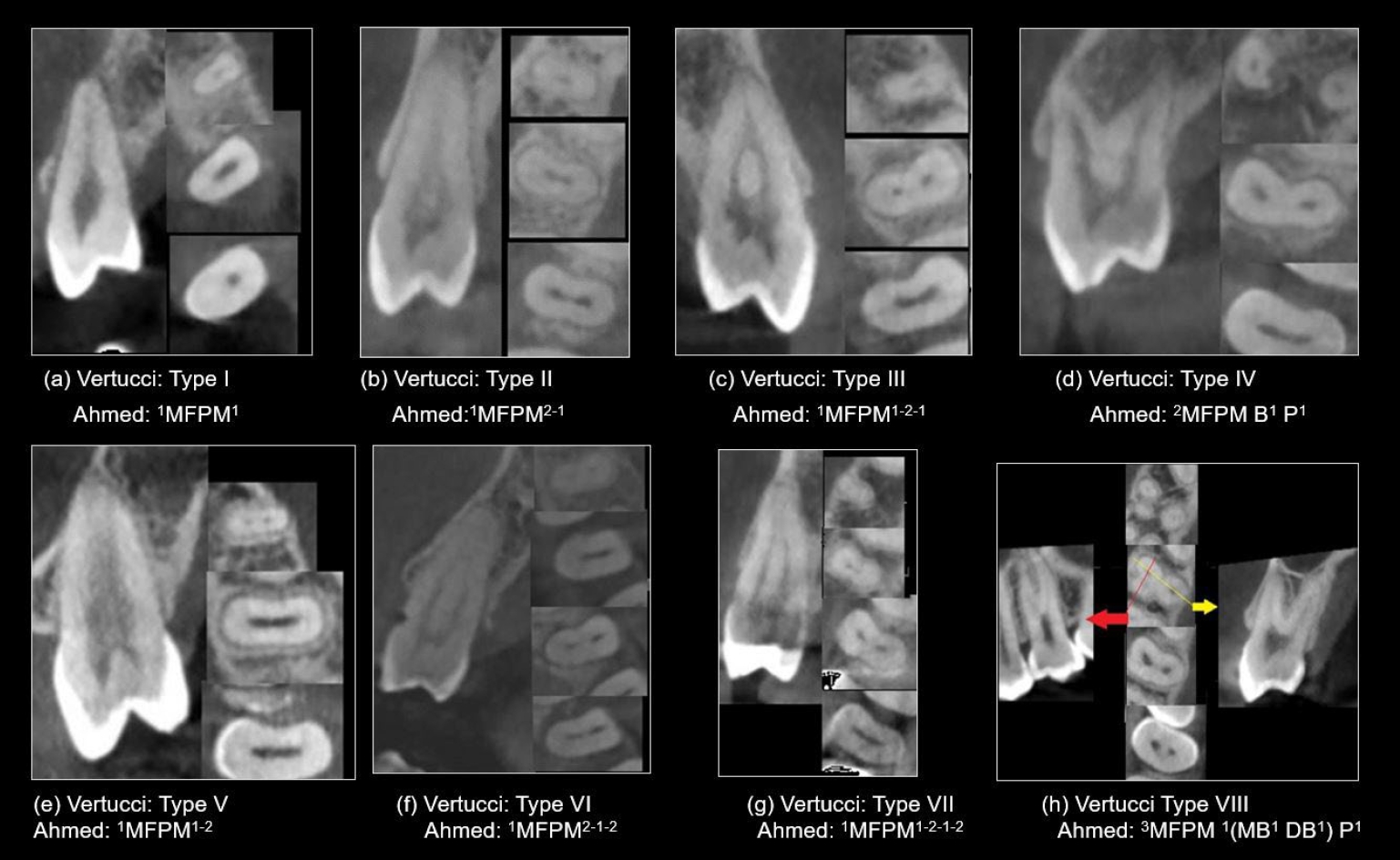
ePub - A comprehensive understanding of both the external and internal anatomy of teeth is fundamental for the effective diagnosis and management of pulp and periapical pathoses. Recent progress in noninvasive, high-resolution imaging modalities, including cone-beam computed tomography and micro-computed tomography, has significantly enhanced the ability to examine the complex morphology of dental structures. These technological advancements have facilitated a level of anatomical detail that was previously unattainable, particularly in the assessment of crown, root, and canal systems. In response to this wealth of new anatomical data, a novel classification framework has been developed, enabling the systematic coding of root and canal configurations across all tooth types. This system offers a more nuanced and comprehensive representation of root canal anatomy compared to earlier classification models. This narrative review explores the implementation of this contemporary classification scheme in education, with a particular focus on its utility in recognizing anatomical variations and accessory canals for the benefit of undergraduate and postgraduate dental students as well as general dental practitioners.
- 877 View
- 39 Download

Research Article
- Cone-beam computed tomography analysis of maxillary premolar canal anatomy: Ahmed’s versus Vertucci’s classifications in a Jordanian cohort
- Raidan Ba-Hattab, Muna M. Shaweesh, Nessrin A. Taha, Elham S. Abu Alhaija
- Restor Dent Endod 2026;51(1):e11. Published online February 26, 2026
- DOI: https://doi.org/10.5395/rde.2026.51.e11
-
Abstract
PDFPubReaderePub
- Objectives
This study analyzed the root and canal configurations of maxillary premolars in a Jordanian subpopulation using cone-beam computed tomography (CBCT) and classified them based on Vertucci’s and Ahmed’s systems.
Methods
Two hundred CBCT scans of 800 maxillary premolars were retrospectively assessed for root morphology, canal configurations, and root canal divergence and merging. Data was statistically analyzed.
Results
The study included 70 males and 130 females. Most right and left maxillary first premolars (RFPM, LFPM) had two roots (59.0% and 58.5%), with a significant association between sex and root number for RFPM and LFPM (p < 0.05). In contrast, the right and left maxillary second premolars (RSPM, LSPM) mostly had a single root (87.5% and 88.5%), with no association with sex. Vertucci’s classification showed type IV as the predominant configuration in first premolars (RFPM, 65.0% and LFPM, 67.0%) and type I in second premolars (RSPM, 44.0% and LSPM, 49.0%). A significant sex association was found only with RSPM. Ahmed’s classification revealed that maxillary premolar with two separated roots and two separated canals (2MP B1 P1) was mostly found in first premolars (RFPM, 58.0% and LFPM, 56.0%), and maxillary premolar with one root and one canal (1MP1) in second premolars (RSPM, 44.0% and LSPM, 49.0%), with a significant sex association for RSPM and LSPM (p < 0.05). Age had no impact, and symmetry was observed between the right and left sides. Three-rooted premolars were identified in four cases. Almost all of Vertucci’s types and numerous codes from Ahmed’s classification were documented.
Conclusions
CBCT revealed diverse anatomical variations in the Jordanian subpopulation, with Ahmed’s classification providing more detailed canal configurations than Vertucci’s, uncovering previously overlooked variations.
- 946 View
- 51 Download
 First
First Prev
Prev

