
Search
- Page Path
- HOME > Search
Research Articles
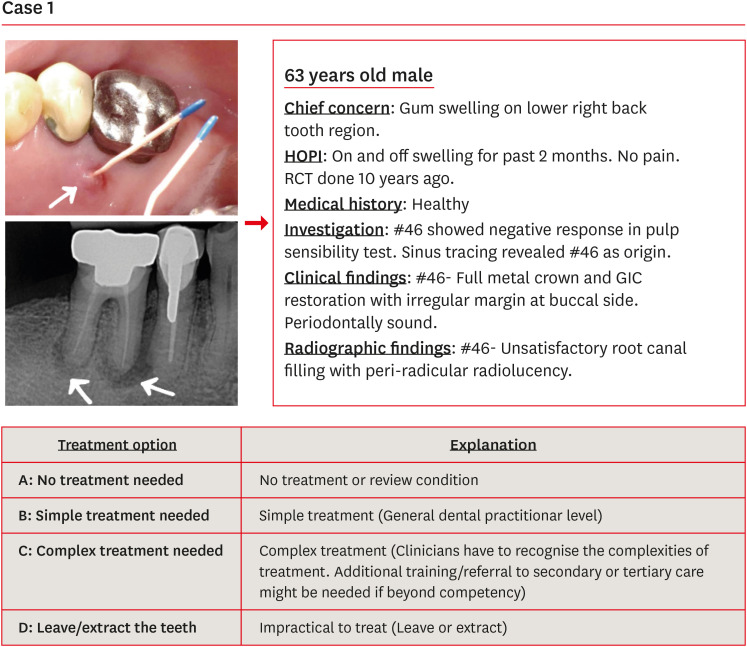
- Effect of Dental Practicality Index training using an online video on decision-making and confidence level in treatment planning by dental undergraduates
- Zhai Wei See, Ming Sern Lee, Abhishek Parolia, Shalini Kanagasingam, Shilpa Gunjal, Shanon Patel
- Restor Dent Endod 2024;49(1):e8. Published online January 24, 2024
- DOI: https://doi.org/10.5395/rde.2024.49.e8
-
 Abstract
 PDF
PDF PubReader
 ePub
ePub Objectives The purpose of this study was to evaluate the effect of Dental Practicality Index (DPI) training using an online video on the treatment planning decisions and confidence level of dental undergraduates (DUs).
Materials and Methods Ninety-four DUs were shown 15 clinical case scenarios and asked to decide on treatment plans based on 4 treatment options. The most appropriate treatment plan had been decided by a consensus panel of experienced dentists. DUs then underwent DPI training using an online video. In a post-DPI-training test, DUs were shown the same clinical case scenarios and asked to assign the best treatment option. After 6 weeks, DUs were retested to assess their knowledge retention. In all 3 tests, DUs completed the confidence level scale questionnaire. Data were analyzed using the related-samples Wilcoxon signed rank test and the independent-samples Mann-Whitney
U test with the level of significance set atp < 0.05.Results DPI training significantly improved the mean scores of the DUs from 7.53 in the pre-DPI-training test to 9.01 in the post-DPI-training test (
p < 0.001). After 6 weeks, the mean scores decreased marginally to 8.87 in the retention test (p = 0.563). DPI training increased their confidence level from 5.68 pre-DPI training to 7.09 post-DPI training.Conclusions Training DUs using DPI with an online video improved their decision-making and confidence level in treatment planning.
-
Citations
Citations to this article as recorded by
- STUDY OF THE EFFECTIVENESS OF THE USE OF DIRECT AND INDIRECT RESTORATION OVER TIME IN THE TREATMENT OF DEFECTS OF HARD DENTAL TISSUES AFTER ENDODONTIC INTERVENTION
V. V. Fedoriuk, М. М. Rozhko
Art of Medicine.2025; : 94. CrossRef
- STUDY OF THE EFFECTIVENESS OF THE USE OF DIRECT AND INDIRECT RESTORATION OVER TIME IN THE TREATMENT OF DEFECTS OF HARD DENTAL TISSUES AFTER ENDODONTIC INTERVENTION
- 3,466 View
- 74 Download
- 1 Crossref

- Radiographic patterns of periosteal bone reactions associated with endodontic lesions
- Poorya Jalali, Jessica Riccobono, Robert A. Augsburger, Mehrnaz Tahmasbi-Arashlow
- Restor Dent Endod 2023;48(3):e23. Published online June 8, 2023
- DOI: https://doi.org/10.5395/rde.2023.48.e23
-
Abstract
PDFPubReaderePub
Objectives The formation of new bone by periosteum due to an insult is called periosteal bone reaction (PBR). This study assessed the cone beam computed tomography (CBCT) patterns of periosteal bone reactions associated with periapical inflammatory lesion (apical periodontitis/periapical rarefying osteitis).
Materials and Methods Twenty-two small field of view CBCT images of patients with PBR were selected from a database of a private practice limited to endodontics. The volume of the periapical inflammatory lesion, the presence of cortical fenestration, the distance of the root apices to the affected cortex, and the location, pattern, and longest diameter of the periosteal reaction were recorded. Statistical analysis was performed using Wilcoxon Ranksum, Fischer’s exact, Spearman Correlation Coefficient, and paired
t -test.Results In all cases, periosteal bone reaction manifested as either parallel (90.9%) or irregular (9.1%). No correlation was found between periapical inflammatory lesion volume and the periosteal reaction's longest diameter (
p > 0.05). Cortical fenestration was noted in 72.7% of the cases. In addition, the findings showed that periosteal reactions were located mostly on the buccal and were present 53.8% and 100% of the time in the mandible and maxilla, respectively.Conclusions The periosteal reactions of endodontic origin had a nonaggressive form (
i.e ., parallel or irregular), and none of the lesions resulted in a periosteal reaction with an ominous Codman’s triangle or spicule pattern.-
Citations
Citations to this article as recorded by- ENDODONTIA E INTERCORRÊNCIAS: COMPREENDENDO OS ACIDENTES E OTIMIZANDO O PROGNÓSTICO
Ana Paula Oliveira Rocha, Flávia Cordeiro Antunes , Millena Alberto Luna , Raissa Danielle Muniz Da Silva , Gustavo Henrique Palma Durães , Juliano Magno de Valadares Bicalho , Lorena Miranda Lima , Barbara Quadros Tonelli
REMUNOM.2026; 2(03): 1. CrossRef - Endodontic Intervention in Chronic Osteomyelitis With Proliferative Periostitis: A Rare Case Report and Scoping Review
Gabriel Lima Braz, Ana Paula Neutzling Gomes, Lisandrea Rocha Schardosim, Nadia de Souza Ferreira, Jose Francisco Gomez-Sosa
Case Reports in Dentistry.2026;[Epub] CrossRef - The influence of endodontic treatment quality on periapical lesions' architecture in cone‐beam computed tomography
Ewa Mackiewicz, Tobias Bonsmann, Krzysztof Safranow, Patrycja Nowicka, Janusz Kołecki, Alicja Nowicka
Australian Endodontic Journal.2025; 51(1): 36. CrossRef - Novel radiographic pattern of maxillary periostitis induced by endodontic inflammation: A case report
Pai-Chun Huang, I-Hao Su, Meng-Ling Chiang, Jyh-Kwei Chen
Journal of Dental Sciences.2025; 20(3): 1982. CrossRef - Garre’s osteomyelitis of the mandible managed by nonsurgical re-endodontic treatment
Heegyun Kim, Jiyoung Kwon, Hyun-Jung Kim, Soram Oh, Duck-Su Kim, Ji-Hyun Jang
Restorative Dentistry & Endodontics.2024;[Epub] CrossRef
- ENDODONTIA E INTERCORRÊNCIAS: COMPREENDENDO OS ACIDENTES E OTIMIZANDO O PROGNÓSTICO
- 6,344 View
- 96 Download
- 4 Web of Science
- 5 Crossref
- Differential diagnosis of periapical cyst using collagen birefringence pattern of the cyst wall
- Hyo Jin Ji, Se-Hee Park, Kyung-Mo Cho, Suk Keun Lee, Jin Woo Kim
- Restor Dent Endod 2017;42(2):111-117. Published online February 9, 2017
- DOI: https://doi.org/10.5395/rde.2017.42.2.111
-
Abstract
PDFPubReaderePub
Objectives Periapical lesions, including periapical cyst (PC), periapical granuloma (PG), and periapical abscess (PA), are frequently affected by chemical/physical damage during root canal treatment or severe bacterial infection, and thus, the differential diagnosis of periapical lesions may be difficult due to the presence of severe inflammatory reaction. The aim of this study was to make differential diagnosis among PC, PG, and PA under polarizing microscope.
Materials and Methods The collagen birefringence patterns of 319 cases of PC (
n = 122), PG (n = 158), and PA (n = 39) obtained using a polarizing microscope were compared. In addition, 6 cases of periodontal fibroma (PF) were used as positive controls.Results Collagen birefringence was condensed with a thick, linear band-like pattern in PC, but was short and irregularly scattered in PG, and scarce or absent in PA. PF showed intense collagen birefringence with a short, palisading pattern but no continuous band-like pattern. The linear band-like birefringence in PC was ascribed to pre-existing expansile tensile stress of the cyst wall.
Conclusions In this study all PCs (
n = 122) were distinguishable from PGs and PAs by their characteristic birefringence, despite the absence of lining epithelium (n = 20). Therefore, the authors suggest that the presence of linear band-like collagen birefringence of the cyst wall aids the diagnostic differentiation of PC from PG and PA.-
Citations
Citations to this article as recorded by- Interplay of collagen and mast cells in periapical granulomas and periapical cysts: a comparative polarizing microscopic and immunohistochemical study
Deepty Bansal, Mala Kamboj, Anjali Narwal, Anju Devi, Nisha Marwah
Restorative Dentistry & Endodontics.2022;[Epub] CrossRef
- Interplay of collagen and mast cells in periapical granulomas and periapical cysts: a comparative polarizing microscopic and immunohistochemical study
- 2,521 View
- 7 Download
- 1 Crossref
Case Reports
- Diagnosis and treatment of teeth with primary endodontic lesions mimicking periodontal disease: three cases with long-term follow ups
- Jae-Hyung Lim, Ji-Hyun Lee, Su-Jung Shin
- Restor Dent Endod 2014;39(1):56-62. Published online January 20, 2014
- DOI: https://doi.org/10.5395/rde.2014.39.1.56
-
Abstract
PDFPubReaderePub
A tooth with primary endodontic disease that demonstrates a periodontal defect might be extracted because of misdiagnosis as severe periodontal disease or a vertical root fracture. The aim of this case report was to demonstrate the long-term survival of endodontically treated teeth, which had been initially considered unsavable. With meticulous evaluation including the patient's dental history, clinical and radiographic examinations, teeth with primary endodontic lesions could be differentiated and saved after proper root canal treatment. Pain history, vitality test, and radiographic examinations, as well as a general periodontal condition check with periodontal probing on an affected tooth, might be the key methods to differentiate endodontic pathosis from that of periodontal disease.
-
Citations
Citations to this article as recorded by- Variability in diagnostic and therapeutic decision-making for endodontic-periodontal lesions: evidence from a cross-sectional study
Yasir Dilshad Siddiqui, Malik Zain Ul Abideen, Muhammad Rizwan Memon, Farah Tahir, Ammar Ahmed Siddiqui, Muhammad Nadeem Baig, Mohammed Katib Alrowili, Azhar Iqbal, Raha Ahmed, Ahmad Salaar
Frontiers in Public Health.2026;[Epub] CrossRef - The morphological and functional relationship between dental pulp and periodontal tissue in the aspect of endo-perio lesions
D. A. Moiseev, S. I. Volkov, A. A. Konov, M. A. Kulyukina
Parodontologiya.2022; 26(4): 289. CrossRef - Evaluation of root morphology of maxillary and mandibular second molars lost due to periodontitis
Akiko Kato, Toshimitsu Hishikawa, Koji Inagaki, Genta Yamamoto, Akio Mitani, Masaki Honda
Journal of Periodontal Research.2020; 55(5): 753. CrossRef - Clinical and Radiographic Characteristics of Endoperiodontitis in Patients with Chronic Generalized Periodontitis
L.N. Dedova, Yu.L. Denisova, N.I. Rossenik
Stomatologist. Minsk.2017; (3(26)): 13. CrossRef - The importance of correct diagnosis and treatment in endo-periodontal lesions: a two cases comparison
Sara Bernardi, Christian Frascarelli, Giulia Fantozzi, Silvia Caruso, Robert Gatto, Gianna Maria Nardi, Maria Adelaide Continenza
Dental Update.2016; 43(8): 766. CrossRef - Surgical management with intentional replantation on a tooth with palato-radicular groove
Jorge Forero-López, Luis Gamboa-Martínez, Laura Pico-Porras, Javier Laureano Niño-Barrera
Restorative Dentistry & Endodontics.2015; 40(2): 166. CrossRef - Subgingival microbiome in smokers and non‐smokers in Korean chronic periodontitis patients
J.‐H. Moon, J.‐H. Lee, J.‐Y. Lee
Molecular Oral Microbiology.2015; 30(3): 227. CrossRef
- Variability in diagnostic and therapeutic decision-making for endodontic-periodontal lesions: evidence from a cross-sectional study
- 3,561 View
- 16 Download
- 7 Crossref
- Diagnostic challenges of nonodontogenic toothache
- Hyung-Ok Park, Jung-Hong Ha, Myoung-Uk Jin, Young-Kyung Kim, Sung-Kyo Kim
- Restor Dent Endod 2012;37(3):170-174. Published online August 29, 2012
- DOI: https://doi.org/10.5395/rde.2012.37.3.170
-
Abstract
PDFPubReaderePub
The objective of this article was to present two nonodontogenic conditions that may mimic odontogenic toothache: trigeminal neuralgia and burning mouth syndrome. Two cases are presented in which one is related to the upper left second premolar and the other is related to the upper left first molar. Both showed pain when chewing. These two cases highlight the complexities involved in diagnosing nonodontogenic toothache. This article demonstrates the importance of having a thorough knowledge of both odontogenic and nonodontogenic toothache, as well as the need for careful evaluation of the nature of the pain and history, clinical and radiographic examinations.
-
Citations
Citations to this article as recorded by- Analysis of Final Diagnosis of Patients with Suspected Nonodontogenic Toothache: A Retrospective Study
Jeong Yeop Chun, Young Joo Shim
Journal of Oral Medicine and Pain.2024; 49(3): 57. CrossRef - Interactions of Acetyl-11-Keto-Beta-Boswellic Acid on Catechol-O-Methyltransferase in the Management of Masticatory Myofascial Pain Syndrome
Ramya Suresh, Pradeep Kumar Yadalam, Ramya Ramadoss, Karthikeyan Ramalingam, Arvind Muthukrishnan
Cureus.2024;[Epub] CrossRef - Assessment of Concordance between Chairside Ultrasonography and Digital Palpation in Detecting Myofascial Trigger Points in Masticatory Myofascial Pain Syndrome
Mohamed Elbarbary, Michael Goldberg, Howard C. Tenenbaum, David K. Lam, Bruce V. Freeman, David J. Pustaka, David Mock, Joseph Beyene, Amir Azarpazhooh
Journal of Endodontics.2023; 49(2): 129. CrossRef - Masticatory Myofascial Pain Syndrome: Implications for Endodontists
Mohamed Elbarbary, Ariel Oren, Michael Goldberg, Bruce V. Freeman, David Mock, Howard C. Tenenbaum, Amir Azarpazhooh
Journal of Endodontics.2022; 48(1): 55. CrossRef - PRICE 2020 guidelines for reporting case reports in Endodontics: explanation and elaboration
V. Nagendrababu, B. S. Chong, P. McCabe, P. K. Shah, E. Priya, J. Jayaraman, S. J. Pulikkotil, P. M. H. Dummer
International Endodontic Journal.2020; 53(7): 922. CrossRef - Clinical Outline of Oral Diseases
Arvind Babu Rajendra Santosh, Doryck Boyd, Kumaraswamy Kikeri Laxminarayana
Dental Clinics of North America.2020; 64(1): 1. CrossRef - Nonodontogenic Sources of Dental Pain
Scott E. Schames, Michael Jordan, Hila Robbins, Lenard Katz, Kaitlyn Tarbert
Journal of the California Dental Association.2016; 44(8): 507. CrossRef - Nonodontogenic toothaches
Edward F. Wright
The Journal of the American Dental Association.2015; 146(6): 406. CrossRef - Síndrome de boca ardiente: claves diagnósticas y terapéuticas
Eduardo Chimenos-Küstner, Cristina Arcos-Guerra, Maria Sueli Marques-Soares
Medicina Clínica.2014; 142(8): 370. CrossRef
- Analysis of Final Diagnosis of Patients with Suspected Nonodontogenic Toothache: A Retrospective Study
- 2,892 View
- 32 Download
- 9 Crossref
Review Articles
- Early caries detection using optical coherence tomography: a review of the literature
- Young-Seok Park, Byeong-Hoon Cho, Seung-Pyo Lee, Won-Jun Shon
- J Korean Acad Conserv Dent 2011;36(5):367-376. Published online September 14, 2011
- DOI: https://doi.org/10.5395/JKACD.2011.36.5.367
-
Abstract
PDFPubReaderePub
Abstract Early detection of carious lesions increases the possibility of treatment without the need for surgical intervention. Optical coherence tomography (OCT) is an emerging three-dimensional imaging technique that has been successfully used in other medical fields, such as ophthalmology for optical biopsy, and is a prospective candidate for early caries detection. The technique is based on low coherence interferometry and is advantageous in that it is non-invasive, does not use ionizing radiation, and can render three-dimensional images. A brief history of the development of this technique and its principles are discussed in this paper. There have been numerous studies on caries detection, which were mostly
in vitro orex vivo experiments. Through these studies, the feasibility of OCT for caries detection was confirmed. However, further research should be performed, includingin vivo studies of OCT applications, in order to prove the clinical usefulness of this technique. In addition, some technological problems must be resolved in the near future to allow for the use of OCT in everyday practice.-
Citations
Citations to this article as recorded by- Differential diagnosis of periapical cyst using collagen birefringence pattern of the cyst wall
Hyo Jin Ji, Se-Hee Park, Kyung-Mo Cho, Suk Keun Lee, Jin Woo Kim
Restorative Dentistry & Endodontics.2017; 42(2): 111. CrossRef - How to designin situstudies: an evaluation of experimental protocols
Young-Hye Sung, Hae-Young Kim, Ho-Hyun Son, Juhea Chang
Restorative Dentistry & Endodontics.2014; 39(3): 164. CrossRef
- Differential diagnosis of periapical cyst using collagen birefringence pattern of the cyst wall
- 3,117 View
- 25 Download
- 2 Crossref
- Theory of X-ray microcomputed tomography in dental research: application for the caries research
- Young-Seok Park, Kwang-Hak Bae, Juhea Chang, Won-Jun Shon
- J Korean Acad Conserv Dent 2011;36(2):98-107. Published online March 31, 2011
- DOI: https://doi.org/10.5395/JKACD.2011.36.2.98
-
Abstract
PDFPubReaderePub
Caries remains prevalent throughout modern society and is the main disease in the field of dentistry. Although studies of this disease have used diverse methodology, recently, X-ray microtomography has gained popularity as a non-destructive, 3-dimensional (3D) analytical technique, and has several advantages over the conventional methods. According to X-ray source, it is classified as monochromatic or polychromatic with the latter being more widely used due to the high cost of the monochromatic source despite some advantages. The determination of mineral density profiles based on changes in X-ray attenuation is the principle of this method and calibration and image processing procedures are needed for the better image and reproducible measurements. Using this tool, 3D reconstruction is also possible and it enables to visualize the internal structures of dental caries. With the advances in the computer technology, more diverse applications are being studied, such automated caries assessment algorithms.
-
Citations
Citations to this article as recorded by- Synchrotron X-ray Studies of the Structural and Functional Hierarchies in Mineralised Human Dental Enamel: A State-of-the-Art Review
Cyril Besnard, Ali Marie, Sisini Sasidharan, Robert A. Harper, Richard M. Shelton, Gabriel Landini, Alexander M. Korsunsky
Dentistry Journal.2023; 11(4): 98. CrossRef - Revelation of microcracks as tooth structural element by X-ray tomography and machine learning
Irma Dumbryte, Donatas Narbutis, Arturas Vailionis, Saulius Juodkazis, Mangirdas Malinauskas
Scientific Reports.2022;[Epub] CrossRef - Three-dimensional non-destructive visualization of teeth enamel microcracks using X-ray micro-computed tomography
Irma Dumbryte, Arturas Vailionis, Edvinas Skliutas, Saulius Juodkazis, Mangirdas Malinauskas
Scientific Reports.2021;[Epub] CrossRef - Radiological Appraisal of Biodentine and Pulpotec Individually or in Combination with Photo-activated Disinfection as Pulp-capping Cements in Mature Teeth
Pratik Agrawal, Gaurav Patri, Surabhi Soumya, Prasanti K Pradhan, Vijeta Patri
The Journal of Contemporary Dental Practice.2021; 22(9): 1014. CrossRef - Ex vivoevaluation of new 2D and 3D dental radiographic technology for detecting caries
Laurence Gaalaas, Donald Tyndall, André Mol, Eric T Everett, Ananta Bangdiwala
Dentomaxillofacial Radiology.2016; 45(3): 20150281. CrossRef - Stationary intraoral digital tomosynthesis using a carbon nanotube X-ray source array
J Shan, A W Tucker, L R Gaalaas, G Wu, E Platin, A Mol, J Lu, O Zhou
Dentomaxillofacial Radiology.2015; 44(9): 20150098. CrossRef - Comparative efficacy of photo-activated disinfection and calcium hydroxide for disinfection of remaining carious dentin in deep cavities: a clinical study
Sidhartha Sharma, Ajay Logani, Naseem Shah
Restorative Dentistry & Endodontics.2014; 39(3): 195. CrossRef - Current status of dental caries diagnosis using cone beam computed tomography
Young-Seok Park, Jin-Soo Ahn, Ho-Beom Kwon, Seung-Pyo Lee
Imaging Science in Dentistry.2011; 41(2): 43. CrossRef
- Synchrotron X-ray Studies of the Structural and Functional Hierarchies in Mineralised Human Dental Enamel: A State-of-the-Art Review
- 3,275 View
- 13 Download
- 8 Crossref
Case Reports
- Partial pulp necrosis caused by excessive orthodontic force
- Min-Young Kim, Seung-Jong Lee, Il-Young Jung, Euiseong Kim
- J Korean Acad Conserv Dent 2011;36(2):149-153. Published online March 31, 2011
- DOI: https://doi.org/10.5395/JKACD.2011.36.2.149
-
Abstract
PDFPubReaderePub
As the dental pulp is encased with a rigid, noncompliant shell, changes in pulpal blood flow or vascular tissue pressure can have serious implication for the health of pulp. Numerous studies have demonstrated that orthodontic force application may influence both blood flow and cellular metabolism, leading degenerative and/or inflammatory responses in the dental pulp. The aim of this case report is to present a case about tooth with chronic periapical abscess which showed normal vital responses. Excessive orthodontic force is thought to be the prime cause of partial pulp necrosis. Owing to remaining vital tissue, wrong dianosis can be made, and tooth falsely diagnosed as vital may be left untreated, causing the necrotic tissue to destroy the supporting tissuses. Clinician should be able to utilize various diagnostic tools for the precise diagnosis, and be aware of the endodontic-orthodontic inter-relationship.
- 2,445 View
- 41 Download
- Diagnosis of periapical cemental dysplasia
- Soon-Young Lee, Chan-Young Lee, Byoung-Duck Roh
- J Korean Acad Conserv Dent 2005;30(1):66-71. Published online January 31, 2005
- DOI: https://doi.org/10.5395/JKACD.2005.30.1.066
-
Abstract
PDFPubReaderePub
Periapical cemental dysplasia(PCD) is a condition most commonly seen in the mandibular incisor region. Radiographically it passes through the three phases(osteolytic stage, intermediate stage, and mature stage). At osteolytic stage, the lesion is similar to features associated with granuloma or cyst that arise following pulpal necrosis. So, it is important to confirm the vitality of the pulp to diagnosis.
In this case, it is difficult to confirm the vitality of involved tooth because the tooth was covered with PFG bridge. And it is unusual that the PCD lesion at mandibular incisors has occurred at first and the lesion of mandibular canine and mandibular premolar were occurred afterward.
- 3,071 View
- 14 Download
 First
First Prev
Prev

