
Search
- Page Path
- HOME > Search
Research Articles
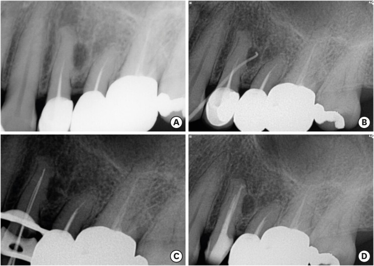
- Endodontic characteristics of mandibular premolar with dens evaginatus: a retrospective study
- Minjin Kim, Sujin Jeon, Min-Seock Seo
- Restor Dent Endod 2024;49(3):e28. Published online July 11, 2024
- DOI: https://doi.org/10.5395/rde.2024.49.e28
-
 Abstract
 PDF
PDF PubReader
 ePub
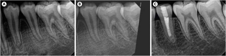
ePub Objectives This study aimed to investigate the endodontic characteristics of mandibular premolars with dens evaginatus (DE) that require endodontic treatment.
Materials and Methods Patients who underwent endodontic treatment were enrolled. The inclusion criteria were patients who underwent root canal treatment in the lower permanent teeth with DE and were followed up for at least 1 year. Preoperative clinical and radiographic variables were obtained. The frequency distribution of the preoperative variables was compared using the χ2 or Fisher’s exact tests. The significance of the change in periapical health index (PAI) and root development stages before and after treatment was examined using the Wilcoxon signed-rank test.
Results A total of 150 teeth of 134 patients with an average age of 15.3 years were included. The percentage distribution comparison of the preoperative variables and obturation techniques revealed significant differences in pulpal and periapical diagnosis, and percussion, and especially regarding age, root development stage, and PAI. Age was the only statistically significant preoperative variable associated with root growth (
p < 0.05).Conclusions Approximately, 60% of DEs requiring endodontic treatment had immature roots. Age being the most significant predisposing factor, early treatment provides the greatest opportunity for full root development.
-
Citations
Citations to this article as recorded by
- A tooth with multiple supernumerary cusps and taurodontism concurrently accompanied with other taurodont teeth: a rare case report
Zihui Tang, Hongchen Zhang, Rongrong Dang, Qiushi Zhang, Yan Huang, Yanwei Yang
Surgical and Radiologic Anatomy.2025;[Epub] CrossRef
- A tooth with multiple supernumerary cusps and taurodontism concurrently accompanied with other taurodont teeth: a rare case report
- 4,163 View
- 119 Download
- 1 Web of Science
- 1 Crossref

- Effect of an aluminum chloride hemostatic agent on the dentin shear bond strength of a universal adhesive
- Sujin Kim, Yoorina Choi, Sujung Park
- Restor Dent Endod 2023;48(2):e14. Published online March 22, 2023
- DOI: https://doi.org/10.5395/rde.2023.48.e14
-
Abstract
PDFPubReaderePub
Objectives This study investigated the effect of an aluminum chloride hemostatic agent on the shear bond strength (SBS) of a universal adhesive to dentin.
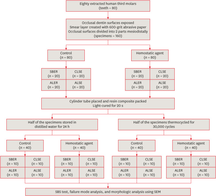
Materials and Methods Eighty extracted human molars were trimmed at the occlusal dentin surfaces and divided mesiodistally. According to hemostatic agent application, specimens were randomly allocated into control (C) and hemostatic agent (Traxodent; H) groups. Each group was divided into 4 subgroups according to the adhesive system (
n = 20): Scotchbond Multi-Purpose (SBER), Clearfil SE Bond (CLSE), All-Bond Universal etch-and-rinse mode (ALER), and All-Bond Universal self-etch mode (ALSE). SBS was measured for half of the specimens at 24 hours, and the other half were thermocycled in water baths (group T). Fracture surfaces were examined to determine the failure mode. The SBS was measured, and data were analyzed using 1-way analysis of variance, the Student’st -test, and the Tukey honestly significant difference test (p = 0.05).Results No significant differences in SBS were found between groups C and H for any adhesive system at 24 hours. After thermocycling, a statistically significant difference was observed between CT+ALSE and HT+ALSE (
p < 0.05). When All-Bond Universal was applied to hemostatic agent-contaminated dentin, the SBS of H+ALSE was significantly lower than that of H+ALER (p < 0.05). The SBER subgroups showed no significant differences in SBS regardless of treatment and thermocycling.Conclusions When exposed dentin was contaminated by an aluminum chloride hemostatic agent before dentin adhesive treatment, application of All-Bond Universal in etch-and-rinse mode was superior to self-etch mode.
-
Citations
Citations to this article as recorded by- Nature-driven blue-emissive N, S-CDs: Harnessing sequential "switch-off-on" fluorescence signals for detection of chrysin and Al³⁺ along with cellular imaging versatility
Maha Mohammad Abdel-Monem, Mohamed I. Walash, Asmaa Kamal El-Deen
Talanta Open.2025; : 100466. CrossRef - Comparative Evaluation of the Shear Bond Strength of Self-Adhesive and Glass Ionomer Cement to Dentin After Removal of Hemostatic Agents Using Different Cleansing Protocols: An In Vitro Study
Hemashree Namburajan, Mathew Chalakuzhiyil Abraham, Vidhyasankari N, Rajkumar K, Abhinayaa Suthagar, Vishnupriya Venkatasubramanian, Sindhuja Nagarajan
Cureus.2025;[Epub] CrossRef - Emalje- og dentinadhesiver: Avgjørende faser i klinisk behandling
Torgils Lægreid, Tom Paulseth, Arne Lund
Den norske tannlegeforenings Tidende.2024; 134(8): 604. CrossRef
- Nature-driven blue-emissive N, S-CDs: Harnessing sequential "switch-off-on" fluorescence signals for detection of chrysin and Al³⁺ along with cellular imaging versatility
- 3,840 View
- 80 Download
- 3 Web of Science
- 3 Crossref
- Buckling resistance, torque, and force generation during retreatment with D-RaCe, HyFlex Remover, and Mtwo retreatment files
- Yoojin Kim, Seok Woo Chang, Soram Oh
- Restor Dent Endod 2023;48(1):e10. Published online February 6, 2023
- DOI: https://doi.org/10.5395/rde.2023.48.e10
-
Abstract
PDFPubReaderePub
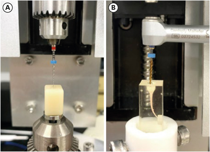
Objectives This study compared the buckling resistance of 3 nickel-titanium (NiTi) retreatment file systems and the torque/force generated during retreatment.
Materials and Methods The buckling resistance was compared among the D-RaCe (DR2), HyFlex Remover, and Mtwo R25/05 retreatment systems. J-shaped canals within resin blocks were prepared with ProTaper NEXT X3 and obturated by the single-cone technique with AH Plus. After 4 weeks, 4 mm of gutta-percha in the coronal aspect was removed with Gates-Glidden drills. Retreatment was then performed using DR1 (size 30, 10% taper) followed by DR2 (size 25, 4% taper), HyFlex Remover (size 30, 7% taper), or Mtrwo R25/05 (size 25, 5% taper) (15 specimens in each group). Further apical preparation was performed with WaveOne Gold Primary. The clockwise torque and upward force generated during retreatment were recorded. After retreatment, resin blocks were examined using stereomicroscopy, and the percentage of residual filling material in the canal area was calculated. Data were analyzed using 1-way analysis of variance with the Tukey test.
Results The HyFlex Remover files exhibited the greatest buckling resistance (
p < 0.05), followed by the Mtwo R25/05. The HyFlex Remover and Mtwo R25/05 files generated the highest maximum clockwise torque and upward force, respectively (p < 0.05). The DR1 and DR2 files generated the least upward force and torque (p < 0.05). The percentage of residual filling material after retreatment was not significantly different between file systems (p > 0.05).Conclusions NiTi retreatment instruments with higher buckling resistance generated greater clockwise torque and upward force.
-
Citations
Citations to this article as recorded by- Time Required to Retreat Carrier-Based Obturation: Comparison Between Two Techniques at Two Levels of Experience
Matteo Salvadori, Elisabetta Audino, Miriam Facchinetti, Vikas Kumar, Mario Alovisi, Luca Visconti, Stefano Salgarello
Dentistry Journal.2026; 14(3): 173. CrossRef - Comparative Evaluation of Rotary and Reciprocating Systems in Root Canal Retreatment with Subsequent Apical Enlargement
Burçin Arıcan, Ayşe Tuba Özalp Koca, Taha Özyürek, Fatma Macit Ermiş
European Journal of General Dentistry.2026;[Epub] CrossRef - Comparative evaluation of the efficacy of three different retreatment files in removing root canal filling material: An In vitro confocal microscopy study
Meghna Sarah Abraham, Aravind R. Kudva, Prathap M. S. Nair, Shravan Kini, Samreena Kalander, Faseeh Muhammed Bin Farookh
Endodontology.2025; 37(2): 136. CrossRef - Efficacy of Endodontic Files in Root Canal Retreatment: A Systematic Review of In Vitro Studies
Anna Soler-Doria, José Luis Sanz, Marcello Maddalone, Leopoldo Forner
Journal of Functional Biomaterials.2025; 16(8): 293. CrossRef - Postoperative Pain Following Single‐Visit Nonsurgical Retreatment Using Minimally Invasive Rotary vs. Reciprocating Nickel‐Titanium File Systems: A Two‐Arm Parallel Randomized Clinical Trial
Hüseyin Gürkan Güneç, Büşra Pehlivan, Celalettin Topbaş, Abdurrahman Kerim Kul, Dursun Ali Şirin, Sivakumar Nuvvula
Pain Research and Management.2025;[Epub] CrossRef - Comparative Evaluation of Canal Centering Ability of Single-file Retreatment System vs Multiple-file Retreatment System, with and without Gutta-Percha Solvent: An In Vitro Study
Sangkeetha Gnanasekaran, Arasappan Rajakumaran, Rajeswari Kalaiselvam, Mathan Rajan Rajendran, Seshan Rakkesh Ramesh, Manigandan Kuzhanchinathan
The Journal of Contemporary Dental Practice.2025; 26(9): 898. CrossRef - Effect of Different Downward Loads and Rotational Speeds on the Removal of Gutta-Percha and Root Canal Sealer Using a Nickel-Titanium Rotary Gutta-Percha Removal System: An Ex Vivo Study
Koki Toyoda, Shunsuke Kimura, Keiichiro Maki, Satoshi Omori, Keiko Hirano, Arata Ebihara, Takashi Okiji
Applied Sciences.2025; 16(1): 446. CrossRef - Cone-beam computed tomographic evaluation and fracture resistance of endodontically retreated teeth using hyflex remover, Mtwo, and ProTaper retreatment file systems: An in vitro study
Isha Singh, Dakshita Joy Sinha, Pallavi Sharma, Kunal Bedi, Priyanka Rani, Swapnil Vats
Saudi Endodontic Journal.2024; 14(1): 56. CrossRef - Comparison of torsional, bending, and buckling resistances of different nickel-titanium glide path files
Feyyaz Çeliker, İrem Çetinkaya
Matéria (Rio de Janeiro).2024;[Epub] CrossRef - Assessing the impact of obturation techniques, kinematics and irrigation protocols on apical debris extrusion and time required in endodontic retreatment
Eugenio Pedullà, Francesco Iacono, Martina Pitrolo, Giovanni Barbagallo, Giusy Rita Maria La Rosa, Chiara Pirani
Australian Endodontic Journal.2023; 49(3): 623. CrossRef
- Time Required to Retreat Carrier-Based Obturation: Comparison Between Two Techniques at Two Levels of Experience
- 3,358 View
- 62 Download
- 6 Web of Science
- 10 Crossref
- Push-out bond strength and intratubular biomineralization of a hydraulic root-end filling material premixed with dimethyl sulfoxide as a vehicle
- Ju-Ha Park, Hee-Jin Kim, Kwang-Won Lee, Mi-Kyung Yu, Kyung-San Min
- Restor Dent Endod 2023;48(1):e8. Published online January 20, 2023
- DOI: https://doi.org/10.5395/rde.2023.48.e8
-
Abstract
PDFPubReaderePub
Objectives This study was designed to evaluate the parameters of bonding performance to root dentin, including push-out bond strength and dentinal tubular biomineralization, of a hydraulic bioceramic root-end filling material premixed with dimethyl sulfoxide (Endocem MTA Premixed) in comparison to a conventional powder-liquid–type cement (ProRoot MTA).
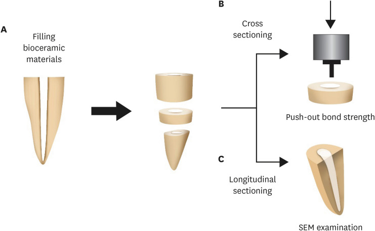
Materials and Methods The root canal of a single-rooted premolar was filled with either ProRoot MTA or Endocem MTA Premixed (
n = 15). A slice of dentin was obtained from each root. Using the sliced specimen, the push-out bond strength was measured, and the failure pattern was observed under a stereomicroscope. The apical segment was divided into halves; the split surface was observed under a scanning electron microscope, and intratubular biomineralization was examined by observing the precipitates formed in the dentinal tubule. Then, the chemical characteristics of the precipitates were evaluated with energy-dispersive X-ray spectroscopic (EDS) analysis. The data were analyzed using the Student’st -test followed by the Mann-WhitneyU test (p < 0.05).Results No significant difference was found between the 2 tested groups in push-out bond strength, and cohesive failure was the predominant failure type. In both groups, flake-shaped precipitates were observed along dentinal tubules. The EDS analysis indicated that the mass percentage of calcium and phosphorus in the precipitate was similar to that found in hydroxyapatite.
Conclusions Regarding bonding to root dentin, Endocem MTA Premixed may have potential for use as an acceptable root-end filling material.
-
Citations
Citations to this article as recorded by- Comparison of intratubular biomineralization between in vivo and in vitro conditions
Sieun Nam, Yeon-Jee Yoo, Mi-Kyung Yu, Kyung-San Min
Journal of Oral Science.2026; 68(1): 30. CrossRef - Effectiveness of Sectioning Method and Filling Materials on Roughness and Cell Attachments in Root Resection Procedure
Tarek Ashi, Naji Kharouf, Olivier Etienne, Bérangère Cournault, Pierre Klienkoff, Varvara Gribova, Youssef Haikel
European Journal of Dentistry.2025; 19(01): 240. CrossRef - Bond Strength and Adhesive Interface Quality of New Pre‐Mixed Bioceramic Root Canal Sealer
Gustavo Creazzo, Bruna Monteiro de Barros Ciribelli Alves, Helena Cristina de Assis, Karen Gisselle Garay Villamayor, Manoel Damião de Sousa‐Neto, Jardel Francisco Mazzi‐Chaves, Fabiane Carneiro Lopes‐Olhê
Microscopy Research and Technique.2025; 88(7): 1989. CrossRef - Evaluation of clinical and radiographic outcome of premixed injectable mineral trioxide aggregate and conventional mineral trioxide aggregate as pulpotomy medicaments in primary molars – A split-mouth randomized control trial
U. S. Aiswarya, Sharan S. Sargod, Sundeep K. Hegde, H. T. Ajay Rao, Nanditha Hegde
Journal of Indian Society of Pedodontics and Preventive Dentistry.2025; 43(4): 559. CrossRef - Evaluation of the root dentin bond strength and intratubular biomineralization of a premixed calcium aluminate-based hydraulic bioceramic endodontic sealer
Yu-Na Lee, Min-Kyeong Kim, Hee-Jin Kim, Mi-Kyung Yu, Kwang-Won Lee, Kyung-San Min
Journal of Oral Science.2024; 66(2): 96. CrossRef - Removal efficiency of a fast setting pozzalan-based bioactive cement: a micro CT study
Feyza Çetinkaya, Ahter Şanal Çıkman, Ali Keleş, Banu Arıcıoğlu
BMC Oral Health.2024;[Epub] CrossRef - Antibacterial Activity and Sustained Effectiveness of Calcium Silicate-Based Cement as a Root-End Filling Material against Enterococcus faecalis
Seong-Hee Moon, Seong-Jin Shin, Seunghan Oh, Ji-Myung Bae
Materials.2023; 16(18): 6124. CrossRef
- Comparison of intratubular biomineralization between in vivo and in vitro conditions
- 3,921 View
- 106 Download
- 7 Web of Science
- 7 Crossref
Case Report
- Surgical management of an accessory canal in a maxillary premolar: a case report
- Hee-Jin Kim, Mi-Kyung Yu, Kwang-Won Lee, Kyung-San Min
- Restor Dent Endod 2019;44(3):e30. Published online July 29, 2019
- DOI: https://doi.org/10.5395/rde.2019.44.e30
-
Abstract
PDFPubReaderePub
We report the surgical endodontic treatment of a maxillary first premolar with a lateral lesion that originated from an accessory canal. Although lesions originating from accessory canals frequently heal with simple conventional endodontic therapy, some lesions may need additional and different treatment. In the present case, conventional root canal retreatment led to incomplete healing with the need for further treatment (
i.e. , surgery). Surgical endodontic management with a fast-setting calcium silicate cement was performed on the accessory canal using a dental operating microscope. At the patient's 9-month recall visit, the lesion was resolved upon radiography.-
Citations
Citations to this article as recorded by- Predictive analysis of root canal morphology in relation to root canal treatment failures: a retrospective study
Mohmed Isaqali Karobari, Vishnu Priya Veeraraghavan, P. J. Nagarathna, Sudhir Rama Varma, Jayaraj Kodangattil Narayanan, Santosh R. Patil
Frontiers in Dental Medicine.2025;[Epub] CrossRef - Endodontic management of internal replacement resorption of two maxillary central incisors with the aid of cone-beam computed tomography as the diagnostic tool: a case report and review of literature
Fatemeh Eskandari, Safoora Sahebi, Negar Ghorbani Jahandizi, Hossein Mofidi
Journal of Medical Case Reports.2025;[Epub] CrossRef - The Impact of the Preferred Reporting Items for Case Reports in Endodontics (PRICE) 2020 Guidelines on the Reporting of Endodontic Case Reports
Sofian Youssef, Phillip Tomson, Amir Reza Akbari, Natalie Archer, Fayjel Shah, Jasmeet Heran, Sunmeet Kandhari, Sandeep Pai, Shivakar Mehrotra, Joanna M Batt
Cureus.2023;[Epub] CrossRef - Main and Accessory Canal Filling Quality of a Premixed Calcium Silicate Endodontic Sealer According to Different Obturation Techniques
Su-Yeon Ko, Hae Won Choi, E-Deun Jeong, Vinicius Rosa, Yun-Chan Hwang, Mi-Kyung Yu, Kyung-San Min
Materials.2020; 13(19): 4389. CrossRef
- Predictive analysis of root canal morphology in relation to root canal treatment failures: a retrospective study
- 2,201 View
- 20 Download
- 4 Crossref
Review Article
- Recognition and management of palatogingival groove for tooth survival: a literature review
- Hee-Jin Kim, Yoorina Choi, Mi-Kyung Yu, Kwang-Won Lee, Kyung-San Min
- Restor Dent Endod 2017;42(2):77-86. Published online April 12, 2017
- DOI: https://doi.org/10.5395/rde.2017.42.2.77
-
Abstract
PDFPubReaderePub
Palatogingival groove (PGG) is an anomaly in the maxillary anterior teeth, often accompanied by the area of bony destruction adjacent to the teeth with no carious or traumatic history. The hidden trap in the tooth can harbor plaque and bacteria, resulting in periodontal destruction with or without pulpal pathologic change. Related diseases can involve periodontal destruction, combined endodontic-periodontal lesions, or separate endodontic and periodontal lesions. Disease severity and prognosis related to PGG depend on several factors, including location, range, depth, and type of the groove. Several materials have been used and recommended for cases of extensive periodontal destruction from PGG to remove and block the inflammatory source and recover the health of surrounding periodontal tissues. Even in cases of severe periodontal destruction, several studies have reported favorable treatment outcomes with proper management. With new options in diagnosis and treatment, clinicians need a detailed understanding of the characteristics, treatment, and prognosis of PGG to successfully manage the condition.
-
Citations
Citations to this article as recorded by- Prevalence of Palatal Grooves on Maxillary Anterior Teeth Using Cone-beam Computed Tomography: A Systematic Review and Meta-Analysis
Oscar Lozano González, Marco Felipe Salas Orozco, Nuria Patiño Marín, Paul V. Abbott, Marc Garcia-Font, Francesc Abella Sans
Journal of Endodontics.2026; 52(1): 14. CrossRef - Endodontic bioceramics: current and futurity aspects
Roma M, Karthik Shetty, Laxmish Mallya, Krishna Prasad Shetty
Frontiers in Oral Health.2026;[Epub] CrossRef - A Unified Deep Learning Framework for Visual Diagnosis of Palatal Radicular Grooves in CBCT Scans: A Multicenter Validation Study
Qikui Zhu, Weitao Fu, Yeyu Lin, Jiaxing Li, Wenhui Tang, Ying Zhang, Rui Zhang, Guanfan Lu, Yao Lin, Jing Shen, Zhuan Bian, Liuyan Meng
Journal of Endodontics.2026;[Epub] CrossRef - Endodontic and Periodontal Treatment of a Two‐Rooted Maxillary Lateral Incisor With a Type III Palatoradicular Groove: A Case Report With 2‐Year Follow‐Up
Katsuhiro Takeda, Tomoya Naruse, Yohei Takahashi, Reina Kawai, Kimiaki Yuhi, Hideki Shiba, Barbara Lapinska
Case Reports in Dentistry.2026;[Epub] CrossRef - Morphological analysis of palatogingival grooves in an Iraqi population: a retrospective cone-beam computed tomography study
O. B. Taha, N. S. Irhayyim, H. Y. Mohammed, M. Z. AL-Rawas, M.A. A. Naw, J. Y. Abdullah, M. I. Karobari
Endodontics Today.2026;[Epub] CrossRef - Three-year follow-up case report: root canal treatment combined with intentional replantation for treating type III palatogingival groove in a maxillary lateral incisor
Jixu Jia, Miao Cheng, Sumeng Shi, Yanchun Qiao
Frontiers in Oral Health.2025;[Epub] CrossRef - Prevalence of palatogingival groove and its association with periapical lesions and periodontal bone loss: a cone beam computed tomography study
Dilan Pelin Yildirim, Selin Goker Kamali
BMC Oral Health.2025;[Epub] CrossRef - Evaluation of Morphology and Prevalence of Palatoradicular Grooves on Affected Maxillary Anterior Teeth Using Cone-Beam Computed Tomography: An Institutional Retrospective Study
Dilara Baştuğ, Leyla Benan Ayrancı
Applied Sciences.2025; 15(14): 8031. CrossRef - Sulco palato-gengival e suas consequências: Revisão de literatura
Marielli de Paula Gonçalves, Maria Júlia Ribeiro Chalita Vieira, Mikaelly Kawany Martins da Silva, Fabiana Tavares Lunardi Palhari, Maria Isabel Gonçalves Fialho
Research, Society and Development.2025; 14(8): e5014849388. CrossRef - Credibility of Intentional Reimplantation Techniques for Periodontally Compromised Teeth: A Report of Two Cases
Satarupa Suklabaidya, Ilakiya Mathi, Kennedy Babu, Gandhimadhi D, Manoj Margabandhu
Cureus.2025;[Epub] CrossRef - Prevalence of Palatal Radicular Groove in upper Lateral Incisors: A CBCT study at Isfahan Azad dental school
Amirreza Zefreh, Azadeh Torkzadeh, Hajar Shekarchizadeh, Maryam Zare Jahromi, Rojin Ardalani
Contemporary Orofacial Science.2025;[Epub] CrossRef - A classification of radicular grooves from the perspective of periodontology
Huxiao Li, Zhaowei Tai, Jiachen Dong, Zhongchen Song
BMC Oral Health.2025;[Epub] CrossRef - Advancements in Root Canal Therapy: Translational Innovations and the Role of Nanoparticles in Endodontic Treatment
Noha M. Badawi, Mohamed M. Kataia, Hadeel A. Mousa, Mozhgan Afshari
Journal of Nanotechnology.2025;[Epub] CrossRef - Cone-beam computed tomographic evaluation to estimate the prevalence of palatogingival groove in the maxillary anterior teeth and its radiographic characteristics: An institutional retrospective study
Mousumi Biswas, Dibyendu Mazumdar, Binayak Saha, Siddhi Agarwala, Kallol Kumar Saha, Kuntal Chowdhury
Journal of Conservative Dentistry and Endodontics.2024; 27(3): 233. CrossRef - A Three-Dimensional Assessment of a Type I Shallow Palatogingival Groove by Cone Beam Computed Tomography: A Case Report
Ramachandra Reddy Gowda Venkatesha, Karthik Rajaram Mohan, Saramma Mathew Fenn, Sabitha Gokulraj, Kumar Appusamy
Cureus.2024;[Epub] CrossRef - Diagnostic Approaches of Palatogingival Groove: A Systematic Review
Greta Venskutė
Journal of Dental Health and Oral Research.2024; : 1. CrossRef - Palatal groove associated with periodontal lesions: a systematic review illustrated by a decisional tree for management
Yvan Gaudex, Vianney Gandillot, Isabelle Fontanille, Philippe Bouchard, Stephane Kerner, Maria Clotilde Carra
BMC Oral Health.2024;[Epub] CrossRef - Palatogingival Groove: The Known–unknown Devourer
Sumedha Gupta, Sandeep Tandon, Ambika S Rathore, Rinku Mathur, Tripti S Rai, Kanchan Kumari Dhaker
International Journal of Clinical Pediatric Dentistry.2024; 17(S1): S95. CrossRef - Nomogram to predict radicular grooves in maxillary lateral incisors in preoperative orthodontic population
Xiuneng Zhou, Jie Deng, Nianke Liu, Chunhui Yang, Shiyu Li, Yaling Song
Clinical Oral Investigations.2024;[Epub] CrossRef - Management of Palatogingival Groove in Maxillary Lateral Incisor: A Report of a Rare Case With a Brief Review of Literature
Irfan Ansari, Sanjay Miglani, Vijay Yadav, Shamimul Hasan
Cureus.2023;[Epub] CrossRef - Prevalence of palatogingival groove affecting maxillary anterior teeth in Saudi subpopulation: A cone-beam computed tomographic study with literature review
Ali Ibrahim Aljuailan, Roqayah Aljuailan, Rahul N. Gaikwad, Shaul Hameed Kolarkodi, Nasser Rufaydan Alamri
The Saudi Dental Journal.2023; 35(8): 1039. CrossRef - Bioceramics in Endodontics: Updates and Future Perspectives
Xu Dong, Xin Xu
Bioengineering.2023; 10(3): 354. CrossRef - Interdisciplinary approach for diagnosis and management of the tooth with type III palatogingival groove
Harakh Chand Baranwal, Jyoti Yadav
Saudi Endodontic Journal.2023; 13(2): 211. CrossRef - Progress in Diagnosis and Treatment of Palatogingival Groove
倩 郑
Advances in Clinical Medicine.2022; 12(04): 2723. CrossRef - Palatogingival grooves associated with periodontal bone Loss of maxillary incisors in a Chinese population
Rui Zhang, Jie Xiong, Markus Haapasalo, Ya Shen, Liuyan Meng
Australian Endodontic Journal.2022; 48(2): 313. CrossRef - Surgical management of lateral lesions with intentional replantation in single-rooted mandibular first premolars with radicular groove
Ya-Hsin Yu, Minje Kim, Samuel Kratchman, Bekir Karabucak
The Journal of the American Dental Association.2022; 153(4): 371. CrossRef - Management of the palato-radicular groove with a periodontal regenerative procedure and prosthodontic treatment: A case report
Dan-Hua Ling, Wei-Ping Shi, Yan-Hong Wang, Dan-Ping Lai, Yan-Zhen Zhang
World Journal of Clinical Cases.2022; 10(17): 5732. CrossRef - Combined Periodontal and Endodontic Management of Palatal Radicular Groove with Platelet‐Rich Fibrin and Biodentine®
Arjun Hari Rijal, Bhageshwar Dhami, Pratistha Ghimire, Konstantinos Michalakis
Case Reports in Dentistry.2022;[Epub] CrossRef - Intentional replantation combined root resection therapy for the treatment of type III radicular groove with two roots: A case report
Dan Tan, Shi-Ting Li, Hao Feng, Zhong-Chao Wang, Cai Wen, Min-Hai Nie
World Journal of Clinical Cases.2022; 10(20): 6991. CrossRef - DENTAL DEFECTS WITH SUBGINGIVAL EXTENSION: A RESTORATIVE CONUNDRUM
Seema Yadav
INTERNATIONAL JOURNAL OF SCIENTIFIC RESEARCH.2021; : 20. CrossRef - Misdiagnosis or Missed Diagnosis? Cone-Beam Computed Tomography-Aided Multidisciplinary Management of Maxillary Central Incisor with Palatogingival Groove
R. Kurinji Amalavathy, K.M. Vidya, Sonali Nabil Sarooshi, Hrudi Sundar Sahoo
Indian Journal of Dental Sciences.2021; 13(1): 46. CrossRef - Root and Root Canal Morphology: Study Methods and Classifications
Duaa M Shihab , Anas F Mahdee
Journal of Baghdad College of Dentistry.2021; 33(4): 11. CrossRef - Prevalence and radiological characteristics of palatogingival groove: A retrospective cone-beam computed tomography study in an Indian cohort
MS Lekshmi, Sheetal Sharma, ShaliniR Gupta, Sidhartha Sharma, Vijay Kumar, Amrita Chawla, Ajay Logani
Journal of Conservative Dentistry.2021; 24(4): 359. CrossRef - Successful Multidisciplinary Management of an Endodontic‐Periodontal Lesion Associated With a Palato‐Radicular Groove: A Case Report
Diksha Katwal, Jennifer K. Fiorica, Jane Bleuel, Stephen J. Clark
Clinical Advances in Periodontics.2020; 10(2): 88. CrossRef - Anatomical, microbiological, and genetic considerations in treatment of Chinese periodontal patients
Edwin X. J. Goh, Marianne M. A. Ong
Journal of Investigative and Clinical Dentistry.2019;[Epub] CrossRef - A new system for classifying tooth, root and canal anomalies
H. M. A. Ahmed, P. M. H. Dummer
International Endodontic Journal.2018; 51(4): 389. CrossRef
- Prevalence of Palatal Grooves on Maxillary Anterior Teeth Using Cone-beam Computed Tomography: A Systematic Review and Meta-Analysis
- 10,497 View
- 221 Download
- 36 Crossref
Research Article
- Effects of proanthocyanidin, a crosslinking agent, on physical and biological properties of collagen hydrogel scaffold
- Yoorina Choi, Hee-Jin Kim, Kyung-San Min
- Restor Dent Endod 2016;41(4):296-303. Published online October 4, 2016
- DOI: https://doi.org/10.5395/rde.2016.41.4.296
-
Abstract
PDFPubReaderePub
Objectives The purpose of the present study was to evaluate the effects of proanthocyanidin (PAC), a crosslinking agent, on the physical properties of a collagen hydrogel and the behavior of human periodontal ligament cells (hPDLCs) cultured in the scaffold.
Materials and Methods Viability of hPDLCs treated with PAC was measured using the 3-(4,5-dimethylthiazol-2-yl)-2,5-diphenyltetrazolium bromide (MTT) assay. The physical properties of PAC treated collagen hydrogel scaffold were evaluated by the measurement of setting time, surface roughness, and differential scanning calorimetry (DSC). The behavior of the hPDLCs in the collagen scaffold was evaluated by cell morphology observation and cell numbers counting.
Results The setting time of the collagen scaffold was shortened in the presence of PAC (
p < 0.05). The surface roughness of the PAC-treated collagen was higher compared to the untreated control group (p < 0.05). The thermogram of the crosslinked collagen exhibited a higher endothermic peak compared to the uncrosslinked one. Cells in the PAC-treated collagen were observed to attach in closer proximity to one another with more cytoplasmic extensions compared to cells in the untreated control group. The number of cells cultured in the PAC-treated collagen scaffolds was significantly increased compared to the untreated control (p < 0.05).Conclusions Our results showed that PAC enhanced the physical properties of the collagen scaffold. Furthermore, the proliferation of hPDLCs cultured in the collagen scaffold crosslinked with PAC was facilitated. Conclusively, the application of PAC to the collagen scaffold may be beneficial for engineering-based periodontal ligament regeneration in delayed replantation.
-
Citations
Citations to this article as recorded by- Effect of collagen crosslinkers on sodium hypochlorite treated dentin bond strength: a systematic review and meta-analysis
Weiqing Zhou, Shuting Feng, Xiaojun Chu, Shuaimei Xu, Xiongqun Zeng
Frontiers in Bioengineering and Biotechnology.2025;[Epub] CrossRef - Proliferative Effect of Proanthocyanidins on HGF-1 and HPDLF Cells: An In Vitro Study
Evelina Alkimavičienė, Nomeda Basevičienė, Arvydas Strazdauskas, Rasa Banienė, Nijolė Savickienė
Medicina.2025; 61(12): 2098. CrossRef - A highly biocompatible CE-crosslinked collagen implant with exceptional anti-calcification and collagen regeneration capabilities for aging skin rejuvenation
Qi Wang, Huiyu Yan, Linyan Yao, Wenhua Li, Jianxi Xiao
Journal of Materials Chemistry B.2024; 12(18): 4467. CrossRef - Dexamethasone release from hyaluronic acid microparticle and proanthocyanidin-gelatin hydrogel in sciatic tissue regeneration
Kazem Javanmardi, Hamideh Shahbazi, Ava Soltani Hekmat, Mehdi Khanmohammadi, Arash Goodarzi
Journal of Materials Science: Materials in Medicine.2024;[Epub] CrossRef - New Materials Based on Collagen and Taxifolin Derivatives: Production and Properties
Yu. V. Shatalin, M. I. Kobyakova, V. S. Shubina
Биологические мембраны Журнал мембранной и клеточной биологии.2024; 41(1): 82. CrossRef - Modulation of Adhesion and Migration of NIH/3T3 Cells in Collagen Materials by Taxifolin Derivatives
Yu. V. Shatalin, M. I. Kobyakova, V. S. Shubina
Biochemistry (Moscow), Supplement Series A: Membrane and Cell Biology.2023; 17(S1): S85. CrossRef - Development and characterization of crosslinked k-carrageenan/sericin blend with covalent agents or thermal crosslink for indomethacin extended release
Wedja Timóteo Vieira, Meuris Gurgel Carlos da Silva, Laura de Oliveira Nascimento, Melissa Gurgel Adeodato Vieira
International Journal of Biological Macromolecules.2023; 246: 125558. CrossRef - New Challenges and Prospective Applications of Three-Dimensional Bioactive Polymeric Hydrogels in Oral and Craniofacial Tissue Engineering: A Narrative Review
Gamal Abdel Nasser Atia, Hany K. Shalaby, Naema Goda Ali, Shaimaa Mohammed Morsy, Mohamed Mohamady Ghobashy, Hager Abdel Nasser Attia, Paritosh Barai, Norhan Nady, Ahmad S. Kodous, Hasi Rani Barai
Pharmaceuticals.2023; 16(5): 702. CrossRef - Polyphenols: Bioavailability, Microbiome Interactions and Cellular Effects on Health in Humans and Animals
Michael B. Scott, Amy K. Styring, James S. O. McCullagh
Pathogens.2022; 11(7): 770. CrossRef - Advances of Hydrogel Therapy in Periodontal Regeneration—A Materials Perspective Review
Maoxue Li, Jiaxi Lv, Yi Yang, Guoping Cheng, Shujuan Guo, Chengcheng Liu, Yi Ding
Gels.2022; 8(10): 624. CrossRef - Collagen stabilization by natural cross-linkers: A qualitative and quantitative FTIR study on ultra-thin dentin collagen model
Rong WANG, Tyler STANLEY, Xiaomei YAO, Hang LIU, Yong WANG
Dental Materials Journal.2022; 41(3): 440. CrossRef - Cross-Linking Agents for Electrospinning-Based Bone Tissue Engineering
Dong-Jin Lim
International Journal of Molecular Sciences.2022; 23(10): 5444. CrossRef - Dense lamellar scaffold, biomimetically inspired, for reverse cardiac remodeling: Effect of proanthocyanidins and glutaraldehyde
Thais Alves, Juliana Ferreira Souza, Venancio Alves Amaral, Alessandra Candida Rios, Tais Costa, Kessi Crescencio, Fernando Batain, Denise Grotto, Renata Lima, Lindemberg Silveira Filho, Jose Oliveira Junior, Patricia Severino, Norberto Aranha, Marco Chau
Journal of Dispersion Science and Technology.2021; 42(2): 248. CrossRef - The effect of the cross-linker ratio used in gellan gum biomaterial synthesis on biomineralization
Serbülent TÜRK, Burak ÜNLÜ, Mahmut ÖZACAR
Bulletin of Biotechnology.2021; 2(2): 27. CrossRef - The recent advances in scaffolds for integrated periodontal regeneration
Hyun Nyun Woo, Young Joon Cho, Solaiman Tarafder, Chang H. Lee
Bioactive Materials.2021; 6(10): 3328. CrossRef - Plant based cross-linkers for tissue engineering applications
Abhishek Indurkar, Ashish Pandit, Ratnesh Jain, Prajakta Dandekar
Journal of Biomaterials Applications.2021; 36(1): 76. CrossRef - Plant-based biomaterials in tissue engineering
Abhishek Indurkar, Ashish Pandit, Ratnesh Jain, Prajakta Dandekar
Bioprinting.2021; 21: e00127. CrossRef - Traditional Chinese Medicine and orthopedic biomaterials: Host of opportunities from herbal extracts
Huijuan Tang, Andrell Hosein, Monica Mattioli-Belmonte
Materials Science and Engineering: C.2021; 120: 111760. CrossRef - Adsorption of Gold Ions onto Sericin and Alginate Particles Chemically Crosslinked by Proanthocyanidins: a Complete Fixed-Bed Column Study
Nilza Tatiane das Graças Santos, Richard Landers, Meuris Gurgel Carlos da Silva, Melissa Gurgel Adeodato Vieira
Industrial & Engineering Chemistry Research.2020; 59(1): 318. CrossRef - Proanthocyanidin as a crosslinking agent for fibrin, collagen hydrogels and their composites with decellularized Wharton’s-jelly-extract for tissue engineering applications
Elham Hasanzadeh, Narges Mahmoodi, Arefeh Basiri, Faezeh Esmaeili Ranjbar, Zahra Hassannejad, Somayeh Ebrahimi-Barough, Mahmoud Azami, Jafar Ai, Vafa Rahimi-Movaghar
Journal of Bioactive and Compatible Polymers.2020; 35(6): 554. CrossRef - Hydrogels for the Delivery of Plant-Derived (Poly)Phenols
Nicola Micale, Andrea Citarella, Maria Sofia Molonia, Antonio Speciale, Francesco Cimino, Antonella Saija, Mariateresa Cristani
Molecules.2020; 25(14): 3254. CrossRef - Natural biopolymer‐based hydrogels for use in food and agriculture
Miri Klein, Elena Poverenov
Journal of the Science of Food and Agriculture.2020; 100(6): 2337. CrossRef - Grape Seed-Inspired Smart Hydrogel Scaffolds for Melanoma Therapy and Wound Healing
Hongshi Ma, Quan Zhou, Jiang Chang, Chengtie Wu
ACS Nano.2019; 13(4): 4302. CrossRef - Improvement of the Physical Properties of Guided Bone Regeneration Membrane from Porcine Pericardium by Polyphenols-Rich Pomace Extract
Nazario Russo, Clara Cassinelli, Elisa Torre, Marco Morra, Giorgio Iviglia
Materials.2019; 12(16): 2564. CrossRef - Novel Biomedical Applications of Crosslinked Collagen
Lisha Gu, Tiantian Shan, Yu-xuan Ma, Franklin R. Tay, Lina Niu
Trends in Biotechnology.2019; 37(5): 464. CrossRef - The prospects of collagen as a basis for curable and activated osteoplastic materials
N. L. Fatkhudinova, A. V. Vasilyev, T. B. Bukharova, E. O. Osidak, N. V. Starikova, S. P. Domogatsky, D. V. Goldshtein, A. A. Kulakov
Stomatologiya.2018; 97(6): 78. CrossRef
- Effect of collagen crosslinkers on sodium hypochlorite treated dentin bond strength: a systematic review and meta-analysis
- 2,486 View
- 10 Download
- 26 Crossref
Case Reports
- Non-destructive management of white spot lesions by using tooth jewelry
- Hee-Jin Kim, Lorena Karanxha, Su-Jung Park
- Restor Dent Endod 2012;37(4):236-239. Published online November 21, 2012
- DOI: https://doi.org/10.5395/rde.2012.37.4.236
-
Abstract
PDFPubReaderePub
Although several methods including composite resin restoration and microabrasion have been used for management of white spot lesion, tooth jewelry can be considered as another noninvasive option. This case report describes the management of white spot lesions by using tooth jewelry. This report also highlights the patients' preference for tooth jewelry as an esthetic concern.
-
Citations
Citations to this article as recorded by- Putting the mouth back in the body – the neglected area of dental and oral travel health
Irmgard L Bauer
Tropical Diseases, Travel Medicine and Vaccines.2025;[Epub] CrossRef - Tooth adornments, gems, and grills
Harpuneet Kaur
International Journal of Oral Health Sciences.2022; 12(2): 50. CrossRef - Gold Enamel Choumps – A Case report
Sargam D. Kotecha, Y. Deepa Hedge, Kalpna Chaudhry, Ramakrishna Yeluri, Updesh Masih, Chanchal Singh
Egyptian Journal of Forensic Sciences.2016; 6(3): 303. CrossRef - Application of quantitative light-induced fluorescence to determine the depth of demineralization of dental fluorosis in enamel microabrasion: a case report
Tae-Young Park, Han-Sol Choi, Hee-Won Ku, Hyun-Su Kim, Yoo-Jin Lee, Jeong-Bum Min
Restorative Dentistry & Endodontics.2016; 41(3): 225. CrossRef
- Putting the mouth back in the body – the neglected area of dental and oral travel health
- 1,966 View
- 7 Download
- 4 Crossref
- Endodontic management of a C-shaped maxillary first molar with three independent buccal root canals by using cone-beam computed tomography
- Lorena Karanxha, Hee-Jin Kim, Sung-Ok Hong, Wan Lee, Pyung-Sik Kim, Kyung-San Min
- Restor Dent Endod 2012;37(3):175-179. Published online August 29, 2012
- DOI: https://doi.org/10.5395/rde.2012.37.3.175
-
Abstract
PDFPubReaderePub
The aim of this study was to present a method for endodontic management of a maxillary first molar with unusual C-shaped morphology of the buccal root verified by cone-beam computed tomography (CBCT) images. This rare anatomical variation was confirmed using CBCT, and nonsurgical endodontic treatment was performed by meticulous evaluation of the pulpal floor. Posttreatment image revealed 3 independent canals in the buccal root obturated efficiently to the accepted lengths in all 3 canals. Our study describes a unique C-shaped variation of the root canal system in a maxillary first molar, involving the 3 buccal canals. In addition, our study highlights the usefulness of CBCT imaging for accurate diagnosis and management of this unusual canal morphology.
-
Citations
Citations to this article as recorded by- Nonsurgical endodontic retreatment of C-shaped maxillary molars: case reports and review of literature
Ming Liu, Yanling Huang, Yixuan Wu, Yi Zhang, Zhisheng Zhang, Qianju Wu
BMC Oral Health.2024;[Epub] CrossRef - Analysis of Fused Rooted Maxillary First and Second Molars with Merged and C-shaped Canal Configurations: Prevalence, Characteristics, and Correlations in a Saudi Arabian Population
Mohammed Mashyakhy, Hemant Ramesh Chourasia, Ahmad Jabali, Abdulmajeed Almutairi, Gianluca Gambarini
Journal of Endodontics.2019; 45(10): 1209. CrossRef - C-shaped root canals of mandibular second molars in a Korean population: a CBCT analysis
Hee-Sun Kim, Daun Jung, Ho Lee, Yoon-Sic Han, Sohee Oh, Hye-Young Sim
Restorative Dentistry & Endodontics.2018;[Epub] CrossRef - Prevalence and Characteristics of the Maxillary C-shaped Molar
Jorge N.R. Martins, António Mata, Duarte Marques, Craig Anderson, João Caramês
Journal of Endodontics.2016; 42(3): 383. CrossRef - Use of cone-beam computed tomography and three-dimensional modeling for assessment of anomalous pulp canal configuration: a case report
Alper Sinanoglu, Dilek Helvacioglu-Yigit, Ibrahim Mutlu
Restorative Dentistry & Endodontics.2015; 40(2): 161. CrossRef - Endodontic management of a mandibular second molar with radix entomolaris: a case report
Rosaline Hannah, Deivanayagam Kandaswamy, Nachimuthu Jayaprakash
Restorative Dentistry & Endodontics.2014; 39(2): 132. CrossRef
- Nonsurgical endodontic retreatment of C-shaped maxillary molars: case reports and review of literature
- 1,884 View
- 4 Download
- 6 Crossref
Research Article
- Coronal microleakage of four temporary restorative materials in Class II-type endodontic access preparations
- Sang-Mi Yun, Lorena Karanxha, Hee-Jin Kim, Sung-Ho Jung, Su-Jung Park, Kyung-San Min
- Restor Dent Endod 2012;37(1):29-33. Published online March 2, 2012
- DOI: https://doi.org/10.5395/rde.2012.37.1.29
-
Abstract
PDFPubReaderePub
Objectives The purpose of this study was to evaluate the microleakage of 4 temporary materials in teeth with Class II-type endodontic access preparations by using a glucose penetration model.
Materials and Methods Glucose reaction test was performed to rule out the presence of any reaction between glucose and temporary material. Class II-type endodontic access preparations were made in extracted human premolars with a single root (
n = 10). Each experimental group was restored with Caviton (GC), Spacer (Vericom), IRM (Dentsply-Caulk), or Fuji II(GC). Microleakage of four materials used as temporary restorative materials was evaluated by using a glucose penetration model. Data were analyzed by the one-way analysis of variance followed by a multiple-comparison Tukey test. The interface between materials and tooth were examined under a scanning electron microscope (SEM).Results There was no significant reaction between glucose and temporary materials used in this study. Microleakage was significantly lower for Caviton and Spacer than for Fuji II and IRM. SEM observation showed more intimate adaptation of tooth-restoration interfaces in Caviton and Spacer than in IRM and Fuji II.
Conclusions Compared to IRM and Fuji II, Caviton and Spacer can be considered better temporary sealing materials in Class II-type endodontic access cavities.
-
Citations
Citations to this article as recorded by- Impact of spacers and thermocycling on porosity and gaps in class II endodontic temporary restorations evaluated by microcomputed tomography
Fahda N. Algahtani, Manal Alkadi, Hiba R. Talic, Sarah S. AlShalawi, Lujain M. Alqarni, Reem M. Barakat, Rasha Haridy, Sara M. ElKhateeb, Rahaf A. Almohareb
Scientific Reports.2025;[Epub] CrossRef - Comparative Evaluation of Sealing Ability, Water Absorption, and Solubility of Three Temporary Restorative Materials: An in vitro Study
AR Prabhakar, N Shantha Rani
International Journal of Clinical Pediatric Dentistry.2017; 10(2): 136. CrossRef - Sealing Ability of Three Different Materials Used as Retrograde Filling
Ji-Hoon Park, Seung-Bok Kang, Yong-Hoon Choi, Ji-Hyun Bae
Journal of Korean Dental Science.2012; 5(2): 60. CrossRef
- Impact of spacers and thermocycling on porosity and gaps in class II endodontic temporary restorations evaluated by microcomputed tomography
- 2,586 View
- 12 Download
- 3 Crossref
Basic Research
- The evaluation of periodontal ligament cells of rat teeth after low-temperature preservation under high pressure
- Jin-Ho Chung, Jin Kim, Seong-Ho Choi, Eui-Seong Kim, Jiyong Park, Seung-Jong Lee
- J Korean Acad Conserv Dent 2010;35(4):285-294. Published online July 31, 2010
- DOI: https://doi.org/10.5395/JKACD.2010.35.4.285
-
Abstract
PDFPubReaderePub
The purpose of this study was to evaluate the viability of periodontal ligament cells of rat teeth after low-temperature preservation under high pressure by means of MTT assay, WST-1 assay. 12 teeth of Sprague-Dawley white female rats of 4 week-old were used for each group.
Both side of the first and second maxillary molars were extracted as atraumatically as possible under tiletamine anesthesia. The experimental groups were group 1 (Immediate extraction), group 2 (Slow freezing under pressure of 3 MPa), group 3 (Slow freezing under pressure of 2 MPa), group 4 (Slow freezing under no additional pressure), group 5 (Rapid freezing in liquid nitrogen under pressure of 2 MPa), group 6 (Rapid freezing in liquid nitrogen under no additional pressure), group 7 (low-temperature preservation at 0℃ under pressure of 2 MPa), group 8 (low-temperature preservation at 0℃ under no additional pressure), group 9 (low-temperature preservation at -5℃ under pressure of 90 MPa). F-medium and 10% DMSO were used as preservation medium and cryo-protectant. For cryo-preservation groups, thawing was performed in 37℃ water bath, then MTT assay, WST-1 assay were processed. One way ANOVA and Tukey HSD method were performed at the 95% level of confidence. The values of optical density obtained by MTT assay and WST-1 were divided by the values of eosin staining for tissue volume standardization.
In both MTT and WST-1 assay, group 7 (0℃/2 MPa) showed higher viability of periodontal ligament cells than other group (2-6, 8) and this was statistically significant (p < 0.05), but showed lower viability than group 1, immediate extraction group (no statistical significance).
By the results of this study, low-temperature preservation at 0℃ under pressure of 2 MPa suggest the possibility for long term preservation of teeth.
-
Citations
Citations to this article as recorded by- Evaluation of the Viability of Rat Periodontal Ligament Cells after Storing at 0℃/2 MPa Condition up to One Week: In Vivo MTT Method
Sun Mi Jang, Sin-Yeon Cho, Eui-Seong Kim, Il-Young Jung, Seung Jong Lee
Journal of Korean Dental Science.2016; 9(1): 1. CrossRef
- Evaluation of the Viability of Rat Periodontal Ligament Cells after Storing at 0℃/2 MPa Condition up to One Week: In Vivo MTT Method
- 1,674 View
- 2 Download
- 1 Crossref
Original Articles
- THE EFFICACY OF PROGRAMMED CRYO-PRESERVATION UNDER PRESSURE IN RAT PERIODONTAL LIGAMENT CELLS
- Young-Eun Lee, Eui-Seong Kim, Jin Kim, Seung-Hoon Han, Seung-Jong Lee
- J Korean Acad Conserv Dent 2009;34(4):356-363. Published online January 14, 2009
- DOI: https://doi.org/10.5395/JKACD.2009.34.4.356
-
Abstract
PDFPubReaderePub
Abstract The purpose of this study was to evaluate the viability of periodontal ligament cells in rat teeth using slow cryo-preservation method under pressure by means of MTT assay and WST-1 assay. Eighteen teeth of Sprague-Dawley white female rats of 4 week-old were used for each group.
Both sides of the first and second maxillary molars were extracted as atraumatically as possible under Tiletamine anesthesia. The experimental groups were group 1 (Immediate control), group 2 (Cold preservation at 4°C for 1 week), group 3 (Slow freezing), group 4 (Slow freezing under pressure of 3 MPa). F-medium and 10% DMSO were used as preservation medium and cryo-protectant. For cryo-preservation groups, thawing was performed in 37°C water bath, then MTT assay and WST-1 assay were processed. One way ANOVA and Tukey method were performed at the 95% level of confidence. The values of optical density obtained by MTT assay and WST-1 were divided by the values of eosin staining for tissue volume standardization.
In both MTT and WST-1 assay, group 4 showed significantly higher viability of periodontal ligament cells than group 2 and 3 (p < 0.05), but showed lower viability than immediate control group.
By the results of this study, slow cryo-preservation method under pressure suggests the possibility for long term cryo-preservation of the teeth.
-
Citations
Citations to this article as recorded by- Effects of Slow Programmable Cryopreservation on Preserving Viability of the Cultured Periodontal Ligament Cells from Human Impacted Third Molar
Jin-Woo Kim, Tae-Yi Kim, Ye-mi Kim, Eun-Kyoung Pang, Sun-Jong Kim
Journal of Korean Dental Science.2015; 8(2): 57. CrossRef - The evaluation of periodontal ligament cells of rat teeth after low-temperature preservation under high pressure
Jin-Ho Chung, Jin Kim, Seong-Ho Choi, Eui-Seong Kim, Jiyong Park, Seung-Jong Lee
Journal of Korean Academy of Conservative Dentistry.2010; 35(4): 285. CrossRef - Comparison of viability of oral epithelial cells stored by different freezing methods
Do-Young Baek, Seung-Jong Lee, Han-Sung Jung, EuiSeong Kim
Journal of Korean Academy of Conservative Dentistry.2009; 34(6): 491. CrossRef
- Effects of Slow Programmable Cryopreservation on Preserving Viability of the Cultured Periodontal Ligament Cells from Human Impacted Third Molar
- 1,612 View
- 1 Download
- 3 Crossref
- Evaluation of the viability of periodontal ligament cell in rat teeth using slow cryopreservation method with magnetic field
- Hyun-Jung Ahn, Eui-Seong Kim, Jin Kim, Duck-Won Kim, Ki-Yeol Kim, Chan-Young Lee, Seung-Jong Lee
- J Korean Acad Conserv Dent 2008;33(4):332-340. Published online July 31, 2008
- DOI: https://doi.org/10.5395/JKACD.2008.33.4.332
-
Abstract
PDFPubReaderePub
The purpose of this study was to evaluate the viability of periodontal ligament cell in rat teeth using slow cryopreservation method with magnetic field through MTT assay and TUNEL test. For each group, 12 teeth of 4 weeks old white female Sprague-Dawley rat were used for MTT assay, and 6 teeth in TUNEL test. The Maxillary left and right, first and second molars were extracted as atraumatically as possible under tiletamine anesthesia. The experimental groups were group1 (immediately extraction), group 2 (cold preservation at 4℃ for 1 week), group 3 (rapid cryopreservation in liquid nitrogen), group 4 (slow cryopreservation with magnetic field of 1 G), and group 5 (slow cryopreservation). F medium was used as preservation medium and 10% DMSO as cryoprotectant. After preservation and thawing, the MTT assay and TUNEL test were processed. One way ANOVA and Scheffe method were performed at the 95% level of confidence. The value of optical density obtained after MTT analysis was divided by the value of eosin staining for tissue volume standardization. In both MTT assay and TUNEL test, it had showed no significant difference among group 3, 4, and 5. And group 3 had showed higher viability of periodontal ligament cell than group 2.
From this study, slow cryopreservation method with magnetic field can be used as one of cryopreservation methods.
-
Citations
Citations to this article as recorded by- The evaluation of periodontal ligament cells of rat teeth after low-temperature preservation under high pressure
Jin-Ho Chung, Jin Kim, Seong-Ho Choi, Eui-Seong Kim, Jiyong Park, Seung-Jong Lee
Journal of Korean Academy of Conservative Dentistry.2010; 35(4): 285. CrossRef - Comparison of viability of oral epithelial cells stored by different freezing methods
Do-Young Baek, Seung-Jong Lee, Han-Sung Jung, EuiSeong Kim
Journal of Korean Academy of Conservative Dentistry.2009; 34(6): 491. CrossRef - The efficacy of programmed cryo-preservation under pressure in rat periodontal ligament cells
Young-Eun Lee, Eui-Seong Kim, Jin Kim, Seung-Hoon Han, Seung-Jong Lee
Journal of Korean Academy of Conservative Dentistry.2009; 34(4): 356. CrossRef
- The evaluation of periodontal ligament cells of rat teeth after low-temperature preservation under high pressure
- 1,763 View
- 1 Download
- 3 Crossref
- Evaluation of periodontal ligament cell viability in rat teeth according to various extra-oral dry storage times using MTT assay
- In-Soo Jeon, Eui-Seong Kim, Jin Kim, Seung-Jong Lee
- J Korean Acad Conserv Dent 2006;31(5):398-408. Published online September 30, 2006
- DOI: https://doi.org/10.5395/JKACD.2006.31.5.398
-
Abstract
PDFPubReaderePub
The purpose of this study was to verify the usefulness of MTT analysis as a tool of measurement of the periodontal ligament cell viability from the extracted rat molar.
A total of 80 Sprague-Dawley white female rat of 4 week-old with a body weight of 100 grams were used. The maxillary left and right, first and second molars were extracted under Ketamine anesthesia. Twenty-four teeth of each group (divided as five groups depending upon the time-lapse after extraction such as immediate, 10, 20, 40 and 60 minutes) were immersed in 200 µl of MTT solution (0.5 mg/ml) and processed for optical density measurements . Another 10 teeth of each group were treated as same as above and sectioned at 10 µm for microscopic examination.
All measurements values were divided by the value of hematoxylin-eosin staining which represented the volume of each corresponding samples. Immediate and 10 minute groups showed highest MTT values followed by 20, 40, and 60 minutes consecutively. Statistical significance (p < 0.05) existed between all groups except in immediate versus 10 minute groups and 40 versus 60 minutes. Histological findings also showed similar findings with MTT results in crystal shape and crystal numbers between the experimental groups.
These data indicate that
in vivo MTT analysis may be of value for evaluation of the periodontal ligament cell viability without time- consuming cell culturing processes.-
Citations
Citations to this article as recorded by- Evaluation of the Viability of Rat Periodontal Ligament Cells after Storing at 0℃/2 MPa Condition up to One Week: In Vivo MTT Method
Sun Mi Jang, Sin-Yeon Cho, Eui-Seong Kim, Il-Young Jung, Seung Jong Lee
Journal of Korean Dental Science.2016; 9(1): 1. CrossRef
- Evaluation of the Viability of Rat Periodontal Ligament Cells after Storing at 0℃/2 MPa Condition up to One Week: In Vivo MTT Method
- 1,670 View
- 0 Download
- 1 Crossref
-
Evaluation of periodontal ligament cell viability in rat teeth after frozen preservation using
in-vivo MTT assay - Jae-Wook Kim, Eui-Sung Kim, Jin Kim, Seung-Jong Lee
- J Korean Acad Conserv Dent 2006;31(3):192-202. Published online May 31, 2006
- DOI: https://doi.org/10.5395/JKACD.2006.31.3.192
-
Abstract
PDFPubReaderePub
The purpose of this study was to examine the viability of PDL cells in rat molars by using
in vivo MTT assay, which was used to compare fast cryopreservation group by liquid nitrogen (-196℃) with 4℃ cold preservation group.A total of 74 Sprague-Dawley white female rats of 4 week-old with a body weight of 100 grams were used. The maxillary left and right, first and second molars were extracted as atraumatically as possible under ketamine anesthesia.
Ten teeth of each group were divided as six experimental groups depending upon the preservation. Cryopreservation groups were Group 1 (5% DMSO 6% HES in F medium), Group 2 (10% DMSO in F medium), Group 3 (5% DMSO 6% HES in Viaspan®), Group 4 (10% DMSO in Viaspan®) which were cryopreserved for 1 week and cold preservation groups were Group 5 (F medium), Group 6 (Viaspan®) at 4℃ for 1 week. Immediate extraction group was used as a control. After preservation and thawing, the
in vivo MTT assay was processed. Two way ANOVA and Duncan's Multiple Range Test was performed at the 95% level of confidence. Another 2 teeth of each group were treated as the same manner and frozen sections 10 µm thick for microscopic observation.The value of optical density obtained after
in vivo MTT analysis was divided by the value of eosin staining for tissue volume standardization. Group 1, 2 had significantly higher optical density than Group 3 and 4 which had the lowest OD value. Group 6 had higher OD value than in Group 5 (P < 0.05). Histological findings of periodontal ligament cell, after being stained with MTT solution were consistent with thein vivo MTT assay results.In this study, the groups which were frozen with DMSO as a cryoprotectant and the groups with F medium showed the best results.
-
Citations
Citations to this article as recorded by- Evaluation of the Viability of Rat Periodontal Ligament Cells after Storing at 0℃/2 MPa Condition up to One Week: In Vivo MTT Method
Sun Mi Jang, Sin-Yeon Cho, Eui-Seong Kim, Il-Young Jung, Seung Jong Lee
Journal of Korean Dental Science.2016; 9(1): 1. CrossRef - Comparative study on survival rate of human gingival fibroblasts stored in different storage media
Hee Su Lee, You Sun Lim
Journal of Korean society of Dental Hygiene.2012; 12(4): 733. CrossRef - The evaluation of periodontal ligament cells of rat teeth after low-temperature preservation under high pressure
Jin-Ho Chung, Jin Kim, Seong-Ho Choi, Eui-Seong Kim, Jiyong Park, Seung-Jong Lee
Journal of Korean Academy of Conservative Dentistry.2010; 35(4): 285. CrossRef - Comparison of viability of oral epithelial cells stored by different freezing methods
Do-Young Baek, Seung-Jong Lee, Han-Sung Jung, EuiSeong Kim
Journal of Korean Academy of Conservative Dentistry.2009; 34(6): 491. CrossRef - The efficacy of programmed cryo-preservation under pressure in rat periodontal ligament cells
Young-Eun Lee, Eui-Seong Kim, Jin Kim, Seung-Hoon Han, Seung-Jong Lee
Journal of Korean Academy of Conservative Dentistry.2009; 34(4): 356. CrossRef
- Evaluation of the Viability of Rat Periodontal Ligament Cells after Storing at 0℃/2 MPa Condition up to One Week: In Vivo MTT Method
- 1,685 View
- 0 Download
- 5 Crossref
- Microshear bond strength of adhesives according to the direction of enamel rods
- Young-Gon Cho, Jong-Jin Kim
- J Korean Acad Conserv Dent 2005;30(4):344-351. Published online July 30, 2005
- DOI: https://doi.org/10.5395/JKACD.2005.30.4.344
-
Abstract
PDFPubReaderePub
This study compared the microshear bond strength (µSBS) to end and side of enamel rod bonded by four adhesives including two total etch adhesives and two self-etch adhesives.
Crown segments of extracted human molars were cut mesiodistally. The outer buccal or lingual surface was used as specimens cutting the ends of enamel rods, and inner slabs used as specimens cutting the sides of enamel rods.
They were assigned to four groups by used adhesives: Group 1 (All-Bond 2), Group 2 (Single Bond), Group 3 (Tyrian SPE/One-Step Plus), Group 4 (Adper Prompt L-Pop). After each adhesive was applied to enamel surface, three composite cylinders were adhered to it of each specimen using Tygon tube. After storage in distilled water for 24 hours, the bonded specimens were subjected to µSBS testing with a crosshead speed of 1 mm/minute. The results of this study were as follows;
1. The µSBS of Group 2 (16.50 ± 2.31 MPa) and Group 4 (15.83 ± 2.33 MPa) to the end of enamel prism was significantly higher than that of Group 1 (11.93 ± 2.25 MPa) and Group 3 (11.97 ± 2.05 MPa) (p < 0.05).
2. The µSBS of Group 2 (13.43 ± 2.93 MPa) to the side of enamel prism was significantly higher than that of Group 1 (8.64 ± 1.53 MPa), Group 3 (9.69 ± 1.80 MPa), and Group 4 (10.56 ± 1.75 MPa) (p < 0.05).
3. The mean µSBS to the end of enamel rod was significantly higher than that to the side of enamel rod in all group (p < 0.05).
-
Citations
Citations to this article as recorded by- Enamel adhesion of light- and chemical-cured composites coupled by two step self-etch adhesives
Sae-Hee Han, Eun-Soung Kim, Young-Gon Cho
Journal of Korean Academy of Conservative Dentistry.2007; 32(3): 169. CrossRef
- Enamel adhesion of light- and chemical-cured composites coupled by two step self-etch adhesives
- 1,574 View
- 6 Download
- 1 Crossref
- The Effect of Estrogen Deficiency on Rat Pulpodentinal Complex
- Miri Kim, Won-Kyung Yang, Jin Baek, Jong-Jin Kim, Won-Kyung Kim, Young-Kyoo Lee
- J Korean Acad Conserv Dent 2005;30(5):402-408. Published online January 14, 2005
- DOI: https://doi.org/10.5395/JKACD.2005.30.5.402
-
Abstract
PDFPubReaderePub
Abstract The purpose of this study was to investigate the effects of estrogen deficiency on pulpodentinal complex of tooth in ovariectomized rats. Thirty female Sprague-Dawley rats, 10 weeks old, were used. Rats were grouped into two groups. One group (n = 15) was subjected to sham surgery (SHAM) and the other group (n = 15) was ovariectomized bilaterally (OVX). Animals were sacrificed 12 weeks later, and their mandibular molars and associated periodontal supporting tissues were dissected out, and fixed in 10% buffered formalin. For comparison of groups, immunostained for osteonectin. Histomorphometrical measurement of change of teeth was performed using an image analysis system and paired t-test was used and the level of significance for overall differences was set at p < 0.05.
In immunostaining of osteonectin, they were significantly different from each other. The predentin thickness in OVX rats was wider than in SHAM rats. And in SHAM rats, osteonectin was more specifically stained in predentin areas than in OVX rats. These results indicate that estrogen deficiency increased the unmineralized predentin areas and decreased osteonectin content in pulpal tissues in rats. If our result is applicable to human studies, odotoblast is affected by estrogen deficiency.
-
Citations
Citations to this article as recorded by- Characterization of craniofacial tissue aging in genetically diverse HET3 male mice with longevity treatment of 17-alpha estradiol
Rami Alsabbagh, Leah LaVerde, Emma Chufar, Jake W. Willows, Kristy L. Townsend, Sarah B. Peters
Archives of Oral Biology.2025; 171: 106170. CrossRef - Association between Metabolic Syndrome and Tooth Loss among Korean Adults: Analysis of the 2007~2018 Korea National Health and Nutrition Examination Survey
Jung-Hui Son, Soo-Myoung Bae
Journal of Dental Hygiene Science.2024; 24(4): 335. CrossRef - Investigation of Changes in Dental Pulp Tissue in Rats with Bilateral Ovariectomy by Histopathological Methods
Elif Pinar Bakir, Şeyhmus Bakir, Büşra Deveci, Firat Şahin, Firat Aşir, Engin Deveci
The Journal of Dentists.2021; 9: 16. CrossRef - Short-term and long-term effects of osteoporosis on incisor teeth and femoral bones evaluated by Raman spectroscopy and energy dispersive X-ray analysis in ovariectomized rats
Fernanda Rossi Paolillo, Renan Arnon Romano, Luciana de Matos, Airton Abrahão Martin, Francisco Eduardo Gontijo Guimarães, Jarbas Caiado de Castro Neto, Vanderlei Salvador Bagnato
Journal of Bone and Mineral Metabolism.2019; 37(1): 18. CrossRef
- Characterization of craniofacial tissue aging in genetically diverse HET3 male mice with longevity treatment of 17-alpha estradiol
- 1,662 View
- 0 Download
- 4 Crossref
- The effect of adhesive property on microtensile bond strength to human dentin
- Hyoun-Jin Kim, Bock Hur, Hyun-Cheol Kim
- J Korean Acad Conserv Dent 2004;29(3):281-287. Published online May 31, 2004
- DOI: https://doi.org/10.5395/JKACD.2004.29.3.281
-
Abstract
PDFPubReaderePub
The purposes of this study were to evaluate the effect of adhesive property on microtensile bond strength and to determine the failure mode.
Flat occlusal dentin surfaces were prepared using low-speed diamond saw. The dentin was etched with 37% phosphoric acid. The following adhesives were applied to the etched dentin to manufacturer's directions; Scotchbond Multi-Purpose in group SM, Prime&Bond NT in group NT, Scotchbond Multi-Purpose followed by Tetric-flow in group TR. After adhesive application, a cylinder of resin-based composite was built up on the occlusal surface. Each tooth was sectioned vertically to obtain the 1 × 1mm2 "sticks". Microtensile bond strength were determined. Each specimen was observed under stereomicroscope and scanning electron microscope (SEM) to examine the failure mode. Data were analyzed using one way ANOVA.
The results of this study were as follows;
1. The microtensile bond strength value were; group SM (18.98 ± 3.01MPa), group NT (16.01 ± 4.82MPa) and group TR (17.56 ± 3.22MPa). No significant statistical differences were observed among the groups (P>0.05).
2. Most of specimens showed mixed failure. In group TR, there was a higher number of specimens showing areas of cohesive failure in resin.
- 1,260 View
- 5 Download
- The effect of delayed composite resin filling on microtensile bond strength
- Hyun-Sik Park, Young-Gon Cho, Byung-Cheul Park, Jong-Uk Kim, Hee-Young Choi, Jong-Jin Kim, Cheul-Hee Jin, Sang-Hoon Yoo, Young-Jae Ki
- J Korean Acad Conserv Dent 2004;29(3):233-238. Published online May 31, 2004
- DOI: https://doi.org/10.5395/JKACD.2004.29.3.233
-
Abstract
PDFPubReaderePub
The purpose of this study was to evaluate the effect of immediate or delayed composite resin filling on dentinal microtensile bond strength (µTBS) after applied the adhesive.
The coronal dentin of human third molars was exposed. Single-Bond or One-Step was applied on the dentin surfaces, and composite resin were constructed immediately (group 1) or 5 min., 10 min., 15 min., 20 min. and 30 min. (groups 2-6) after an adhesive was applied. The specimens were sectioned and made bar-shaped. Each surface area of them was about 1mm2. The µTBS test was performed by EZ test. The results were analysed by One-way ANOVA and Tukey's test at 95% significance level.
The results suggested that the µTBS of Single-Bond to dentin was decreased when the composite resin was constructed 20 min. and 30 min. after Single-Bond was applied. But the µTBS of One-Step was not affected by delayed composite resin filling.
- 1,186 View
- 1 Download
- Effect of wetness on the enamel bonding
- Keun-Ho Ko, Young-Gon Cho, Cheul-Hee Jin, Sang-Hoon Yoo, Jong-Uk Kim, Byung-Cheul Park, Young-Jae Ki, Hee-Young Choi, Jong-Jin Kim
- J Korean Acad Conserv Dent 2004;29(3):205-211. Published online May 31, 2004
- DOI: https://doi.org/10.5395/JKACD.2004.29.3.205
-
Abstract
PDFPubReaderePub
This study evaluated the microleakage and interfacial gap between enamel and composite resin under the dry and wet condition of the enamel surface. V shaped class 5 cavities were prepared on the occlusal portion of extracted human molars. Samples were divided into three groups: D group (air dry for 10-15 s), BD group (blot dry with moist cotton pellet), and DR group (air dry for 10-15 s and rewet with Aqua-Prep F for 20 s).
Cavities were filled using Aelitefil composite resin after applied One-Step. Microleakage was tested by 2% methylene blue dye solution and the data were statistically analysed by Kruskal-Wallis test and Mann-Whitney test. Also Enamel-resin interface was observed under SEM. Group BD showed statistically lower microleakage than group D (p < 0.05), but there was no statistically significant difference between group BD and DR (p > 0.05). At the enamel-resin interface, group D showed the gap of 2 µm thickness, but group BD and DR showed close adaptation.
In conclusion, the use of blot dry and rewetting agent (Aqua-Prep F) resulted in decreased microleakage and improved adhesion between enamel and resin when using One-Step.
-
Citations
Citations to this article as recorded by- Effect of an intermediate bonding resin and flowable resin on the compatibility of two-step total etching adhesives with a self-curing composite resin
Sook-Kyung Choi, Ji-Wan Yum, Hyeon-Cheol Kim, Bock Hur, Jeong-Kil Park
Journal of Korean Academy of Conservative Dentistry.2009; 34(5): 397. CrossRef
- Effect of an intermediate bonding resin and flowable resin on the compatibility of two-step total etching adhesives with a self-curing composite resin
- 1,475 View
- 2 Download
- 1 Crossref
- INFLUENCE OF APPICATION TIME OF SELF-ETCHING PRIMERS ON DENTINAL MICROTENSILE BOND STRENGTH
- Young-Gon Cho, Young-Gon Lee, Jong-Uk Kim, Byung-Cheul Park, Jong-Jin Kim, Hee-Young Choi, Cheul-Hee Jin, Sang-Hoon Yoo
- J Korean Acad Conserv Dent 2004;29(5):430-438. Published online January 14, 2004
- DOI: https://doi.org/10.5395/JKACD.2004.29.5.430
-
Abstract
PDFPubReaderePub
ABSTRACT This study evaluated the influence of application time of self-etching primers on microtensile bond strength (μTBS) to dentin using three self-etching primer adhesive systems.
Dentin surfaces were exposed from forty-eight human molars. They were conditioned with three self-etching primers (Clearfil SE Bond [SE], Unifil Bond [UF], Tyrian SPE + One Step Plus [TY]) and different primining times (10s, 20s, 30s and 40s). Composite resins were bonded to dentin surfaces and specimens were made. μTBS was tested and statistically compared using by one-way ANOVA and Tukey’s Test.
The results of this study presented that priming time for 10s in SE and UF groups and for 30s and 40s in TY group was highly decreased μTBS to dentin.
-
Citations
Citations to this article as recorded by- The utility of dental health capacity of the first permanent molars as an oral health indicator among children and adolescents
Sun-Ho Lee, Heung-Soo Lee, Hyo-Won Oh, Hyun-Jeong Ju, So-Young Park
Journal of Korean Academy of Oral Health.2015; 39(3): 174. CrossRef - Logistic regression analysis of factors affecting the survival of first molars in Korean adults
Sung-Suk Chung, Jong-Bae Kim, Young-Sam Kim, Young-Ran Jung, Jin-Sil Hong, Ju-Yeon Kim, Kee-Wan Chang
Journal of Korean Academy of Oral Health.2015; 39(2): 88. CrossRef - Dental health capacity of the first permanent molars among children and adolescents in Korea for the year 2010
Sun-Ho Lee, Hyun-Jeong Ju, So-Young Park, Hyo-Won Oh, Heung-Soo Lee
Journal of Korean Academy of Oral Health.2013; 37(2): 103. CrossRef
- The utility of dental health capacity of the first permanent molars as an oral health indicator among children and adolescents
- 1,519 View
- 0 Download
- 3 Crossref
- The verification of the MTT assay on the viability of periodontal ligamental cells in rat molars through the histologic examination
- Hyun-Ki Kim, Eui-Seoung Kim, In-Bok Choi, Jin Kim, Seung-Jong Lee
- J Korean Acad Conserv Dent 2003;28(5):385-391. Published online September 30, 2003
- DOI: https://doi.org/10.5395/JKACD.2003.28.5.385
-
Abstract
PDFPubReaderePub
The purpose of this study is to examine the viability of PDL cells in rat molars by using MTT assay and to verify the MTT assay through the histologic observation. Thirty of Sprague-Dawley white female rats of 4-weeks old with a body weight of about 100 grams were used. Groupings are as follows:
Immediate Group : Positive control group(n=10)-after extraction immediately.
Dried Group : Negative control group(n=10)-after drying for an hour under warm dry.
ViaSpan® Group : 1hour ViaSpan® group(n=10)-after storing in ViaSpan® at 4℃ for 1hour.
Ten teeth of each group were treated as same as above and replanted to the original socket of experimental animals. After two weeks of replantation, all the experimental animals were sacrificed. And after fixation, extracted maxillary jaw was dimineralized. After it was embedded in paraffin, serial section by 5µm was carried out and for construction of specimen, hematoxylin-eosin dye was used.
The mean MTT measurement of immediate group(positive control) is 2.81 and the mean measurement of dried group(negative control) is 0.98 which is significant differnt(P<0.05). The mean measurement of ViaSpan® group is 2.65 and there is significant difference between dried group and ViaSpan® group(P<0.05). However, there is no difference between immediate group and ViaSpan® group. The average resorption points of immediate group is 3.03 points. In the dried group, average 6.44 points resorption and 2.68 points showed resorption in the ViaSpan® group. Unlike with MTT assay, there was no significant difference between the immediate group and ViaSpan® group.
The usage of MTT assay as a viable cell marker may give us a better indication of the maintenance of periodontal ligament cell vitality.
-
Citations
Citations to this article as recorded by- Concentration of propolis as a storage medium for avulsed teeth: a systematic review
Chaya Chhabra, Kumar Gaurav Chhabra, Seemadevi Thangeswaran, Shraddha Shere
Frontiers in Medicine.2025;[Epub] CrossRef - Effects of Trichosanthis Radix Extract on Collagen Production
Mi-Young Yun, Hye-Won Kim
Journal of the Korean Society of Cosmetology.2024; 30(2): 408. CrossRef - Evaluation of periodontal ligament cell viability in rat teeth according to various extra-oral dry storage times using MTT assay
In-Soo Jeon, Eui-Seong Kim, Jin Kim, Seung-Jong Lee
Journal of Korean Academy of Conservative Dentistry.2006; 31(5): 398. CrossRef
- Concentration of propolis as a storage medium for avulsed teeth: a systematic review
- 1,440 View
- 2 Download
- 3 Crossref
-
In Vitro assessment on viability of human periodontal ligament cells after storage in chlorophyllin-added medium - Won-Gyun Chung, Jin Kim, Eun-Ju Lee, Seung-Jong Lee
- J Korean Acad Conserv Dent 2002;27(6):600-611. Published online November 30, 2002
- DOI: https://doi.org/10.5395/JKACD.2002.27.6.600
-
PDFPubReaderePub
-
Citations
Citations to this article as recorded by- Comparative Evaluation of the Efficacy of Human Periostin and Enamel Matrix Derivative (Emdogain®) on Proliferation and Migration of Periodontal Ligament Fibroblasts
Swastik Mishra, Jothi M. Varghese, Shama Prasada Kabekkodu
Pesquisa Brasileira em Odontopediatria e Clínica Integrada.2026;[Epub] CrossRef - Comparative Evaluation of Corneal Storage Medias Used as Tooth Avulsion Medias in Maintaining the Viability of Periodontal Ligament Cells Using the Cell Counting Kit-8 Assay
Nidhi James, Sandya Kini, Swathi Pai, Neetha Shenoy, Shama Prasada Kabekkodu
Clinical, Cosmetic and Investigational Dentistry.2022; Volume 14: 87. CrossRef - The verification of the MTT assay on the viability of periodontal ligamental cells in rat molars through the histologic examination
Hyun-Ki Kim, Eui-Seoung Kim, In-Bok Choi, Jin Kim, Seung-Jong Lee
Journal of Korean Academy of Conservative Dentistry.2003; 28(5): 385. CrossRef
- Comparative Evaluation of the Efficacy of Human Periostin and Enamel Matrix Derivative (Emdogain®) on Proliferation and Migration of Periodontal Ligament Fibroblasts
- 3,051 View
- 6 Download
- 3 Crossref
 First
First Prev
Prev

