
Search
- Page Path
- HOME > Search
Research Articles
- Interplay of hypoxia, angiogenesis, and macrophages in pulp and periapical lesions: an immunohistochemical cross-sectional study
- Puja Chatterjee, Mala Kamboj, Shweta Mittal, Anjali Narwal, Anju Devi
- Restor Dent Endod 2026;51(2):e22. Published online April 23, 2026
- DOI: https://doi.org/10.5395/rde.2026.51.e22
-
 Abstract
 PDF
PDF PubReader
 ePub
ePub - Objectives
This study evaluated and correlated the immune expression of hypoxia and angiogenesis with macrophages in periapical granuloma (PG), radicular cyst (RC), and healthy pulp (HP).
Methods
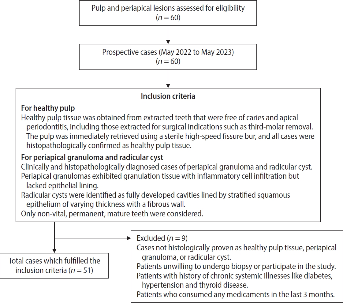
An observational study was performed on 51 tissue blocks equally divided among the groups, stained immunohistochemically for hypoxia-inducible factor (HIF)-1α, vascular endothelial growth factor (VEGF), and CD68, and the mean expression was calculated. Data were analyzed using Kruskal-Wallis, Mann-Whitney, Spearman correlation tests (p < 0.001), and multiple linear regression analysis (p ≤ 0.05).
Results
HIF-1α expression was highest in PG than RC and HP (p < 0.001). Significant differences were found between HP, PG, and RC (both p < 0.001). VEGF expression was highest in RC than in PG and HP (p < 0.001), with significant differences between HP and both PG and RC (p < 0.001); pairwise comparisons were significant between all groups (p < 0.001, p < 0.001, p = 0.018). Correlation analysis showed significant correlations between VEGF and CD68 in HP and PG (p = 0.007 and p = 0.028, respectively). Linear regression showed that study groups were significantly associated with mean scores of HIF-1α, VEGF, and CD68 (p = 0.002, p = 0.001, p < 0.001).
Conclusions
HIF-1α, VEGF, and CD68 showed increased expression in PGs and RCs, suggesting an association between hypoxic conditions, enhanced angiogenic activity, and macrophage presence within the periapical inflammatory microenvironment. Future studies exploring HIF-1α and VEGF inhibitors as potential treatment modalities for periapical lesions are warranted.
- 876 View
- 48 Download

- Interplay of collagen and mast cells in periapical granulomas and periapical cysts: a comparative polarizing microscopic and immunohistochemical study
- Deepty Bansal, Mala Kamboj, Anjali Narwal, Anju Devi, Nisha Marwah
- Restor Dent Endod 2022;47(1):e12. Published online February 14, 2022
- DOI: https://doi.org/10.5395/rde.2022.47.e12
-
Abstract
PDFPubReaderePub
Objectives This pilot study aimed to establish the interrelationship between collagen and mast cells in periapical granulomas and periapical cysts.
Materials and Methods An observational cross-sectional study was conducted on the paraffin-embedded tissue sections of 68 specimens (34 periapical granulomas and 34 periapical cysts). The specimens were stained with picrosirius to observe collagen fiber birefringence and anti-tryptase antibody to evaluate the mast cell count immunohistochemically. The mean number and birefringence of collagen fibers, as well as the mean number of mast cells (total, granulated, and degranulated), and the mean inflammatory cell density were calculated. The data obtained were analyzed using the Kruskal Wallis test, Mann Whitney
U test, and Spearman correlation test (p < 0.05).Results The mean number of thick collagen fibers was higher in periapical cysts, while that of thin fibers was higher in granulomas (
p = 0.00). Cysts emitted orange-yellow to red birefringence, whereas periapical granulomas had predominantly green fibers (p = 0.00). The mean inflammatory cell density was comparable in all groups (p = 0.129). The number of total, degranulated, and granulated mast cells exhibited significant results (p = 0.00) in both groups. Thick cyst fibers showed significant inverse correlations with inflammation and degranulated mast cells (p = 0.041, 0.04 respectively).Conclusions Mast cells and inflammatory cells influenced the nature of collagen fiber formation and its birefringence. This finding may assist in the prediction of the nature, pathogenesis, and biological behavior of periapical lesions.
-
Citations
Citations to this article as recorded by
- Chronic periapical lesions: clinical and morphological characterization of 508 cases
Talytha Barbosa da Rocha, Raelly Katharinne Lima de Meneses, Nathália Yvia Assis Henriques, Jonh Lennon Silva Cunha, Manuel Antonio Gordón-Núñez, Cassiano Francisco Weege Nonaka, Pollianna Muniz Alves
JORDI - Journal of Oral Diagnosis.2026;[Epub] CrossRef - The Mystifying Role of Mast Cells in the Pathogenesis of Periapical Pathologies - A Systematic Review
Mala Kamboj, Shashibala Malik, R. Keerthika, Anjali Narwal, Anju Devi, Gopikrishnan Vijayakumar, Adarsh Kumar
Journal of Endodontics.2025; 51(7): 845. CrossRef - Immunohistochemical Analysis of CD117 in the Mast Cells of Odontogenic Keratocysts
Sujatha Varma, Shameena PM, Plakkil Viswanathan Deepthi, Indu G
Cureus.2024;[Epub] CrossRef - Immunohistochemical evaluation of cyclooxygenase‐2 and mast cell density in periapical lesions
Shashibala Malik, Mala Kamboj, Anjali Narwal, Anju Devi
International Endodontic Journal.2023; 56(8): 980. CrossRef
- Chronic periapical lesions: clinical and morphological characterization of 508 cases
- 3,267 View
- 31 Download
- 2 Web of Science
- 4 Crossref
 First
First Prev
Prev

