
Search
- Page Path
- HOME > Search
Research Articles
-
Comparative evaluation of
Emblica officinalis as an etchant and an MMP inhibitor with orthophosphoric acid and chlorhexidine on the microshear bond strength of composite resin: anex vivo study - Divya Sangeetha Rajkumar, Annapoorna Ballagere Mariswamy
- Restor Dent Endod 2021;46(3):e36. Published online June 8, 2021
- DOI: https://doi.org/10.5395/rde.2021.46.e36
-
 Abstract
 PDF
PDF PubReader
 ePub
ePub Objectives This study aimed to evaluate
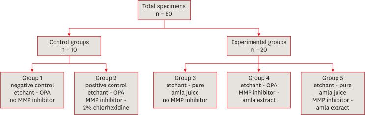
Emblica officinalis (Indian gooseberry or amla) as an acid etchant and matrix metalloproteinase (MMP) inhibitor, and to compare its effect on the microshear bond strength of composite resin with orthophosphoric acid (OPA) and 2% chlorhexidine (CHX) as an acid etchant and MMP inhibitor, respectively.Materials and Methods The etching effect and MMP-inhibiting action of amla on dentin samples were confirmed by scanning electron microscopy (SEM) and gelatin zymography, respectively. Dentinal slabs (3 mm thick) from 80 extracted human molars were divided into 10 and 20 samples to form 2 control groups and 3 experimental groups. Groups 1, 2, and 4 were etched with OPA and groups 3 and 5 with amla juice. An MMP inhibitor was then applied: CHX for group 2 and amla extract for groups 4 and 5. Groups 1 and 3 received no MMP inhibitor. All specimens received a standardized bonding protocol and composite resin build-up, and were subjected to microshear bond strength testing. The force at which the fracture occurred was recorded and statistically analyzed.
Results Amla juice had a similar etching effect as a self-etch adhesive in SEM and 100% amla extract was found to inhibit MMP-9 by gelatin zymography. The microshear bond strength values of amla were lower than those obtained for OPA and CHX, but the difference was not statistically significant.
Conclusions Amla has a promising role as an acid etchant and MMP inhibitor, but further studies are necessary to substantiate its efficacy.
-
Citations
Citations to this article as recorded by
- In vitro assessment of anti-glioblastoma potential of Emblica officinalis methanolic fruit extract and green nanoparticles in U87-MG cells
Kokkonda Jackson Sugunakara Chary, Anuradha Sharma, Amrita Singh
Medical Oncology.2025;[Epub] CrossRef - Eco-conscious synthesis of novel 1,2,4-triazolo[1,5-a]pyrimidine derivatives as potent Anti-microbial agent and comparative study of cell viability and cytotoxicity in HEK-293 cell line utilizing Indian gooseberry (Phyllanthus emblica) fruit extract
Bhaktiben R. Bhatt, Kamalkishor Pandey, Tarosh Patel, Anupama Modi, Chandani Halpani, Vaibhav D. Bhatt, Bharat C. Dixit
Bioorganic Chemistry.2024; 153: 107936. CrossRef - Cell mediated ECM-degradation as an emerging tool for anti-fibrotic strategy
Peng Zhao, Tian Sun, Cheng Lyu, Kaini Liang, Yanan Du
Cell Regeneration.2023;[Epub] CrossRef - Insight into the development of versatile dentin bonding agents to increase the durability of the bonding interface
Isabel Cristina Celerino de Moraes Porto, Teresa de Lisieux Guedes Ferreira Lôbo, Raphaela Farias Rodrigues, Rodrigo Barros Esteves Lins, Marcos Aurélio Bomfim da Silva
Frontiers in Dental Medicine.2023;[Epub] CrossRef
- In vitro assessment of anti-glioblastoma potential of Emblica officinalis methanolic fruit extract and green nanoparticles in U87-MG cells
- 2,419 View
- 21 Download
- 4 Web of Science
- 4 Crossref

- Influence of silver nanoparticles on resin-dentin bond strength durability in a self-etch and an etch-and-rinse adhesive system
- Zahra Jowkar, Fereshteh Shafiei, Elham Asadmanesh, Fatemeh Koohpeima
- Restor Dent Endod 2019;44(2):e13. Published online March 29, 2019
- DOI: https://doi.org/10.5395/rde.2019.44.e13
-
Abstract
PDFPubReaderePub
Objectives This study evaluated the effect of dentin pretreatment with silver nanoparticles (SNPs) and chlorhexidine (CHX) on the microshear bond strength (µSBS) durability of different adhesives to dentin.
Materials and Methods Occlusal surfaces of 120 human molars were ground to expose flat dentin surfaces. The specimens were randomly assigned to six groups (
n = 20). Three groups (A, B, and C) were bonded with Adper Single Bond 2 (SB) and the other groups (D, E, and F) were bonded with Clearfil SE Bond (SEB). Dentin was pretreated with CHX in groups B and E, and with SNPs in groups C and F. The specimens were restored with Z250 composite. Half of the bonded surfaces in each group underwent µSBS testing after 24 hours and the other half was tested after 6 months of water storage.Results SNP application was associated with a higher µSBS than was observed in the CHX and control groups for SEB after 24 hours (
p < 0.05). A significantly lower µSBS was observed when no dentin pretreatment was applied compared to dentin pretreatment with CHX and SNPs for SB after 24 hours (p < 0.05). The µSBS values of the 6-month specimens were significantly lower than those obtained from the 24-hour specimens for all groups (p < 0.05). This decrease was much more pronounced when both adhesives were used without any dentin pretreatment (p < 0.05).Conclusions SNPs and CHX reduced the degradation of resin-dentin bonds over a 6-month period for both adhesive systems.
-
Citations
Citations to this article as recorded by- Effect of nano silver fluoride pre-treatment on the microtensile bond strength of a universal adhesive to dentin: an in vitro study
Larissa Alexsandra dos Santos Silva, Anna Caroline Lima Dos Santos, Luis Felipe Espíndola-Castro, Ana Luísa Cassiano Alves, Márcia de Almeida Durão, Maria Clara Müller De Andrade, André Galembeck, Gabriela Queiroz de Melo Monteiro
Biomaterial Investigations in Dentistry.2026; 13: 198. CrossRef - An in vitro comparative evaluation of silver and chitosan nanoparticles on shear bond strength of nanohybrid composite using different adhesion protocols
Roopadevi Garlapati, Nagesh Bolla, Mayana Aameena Banu, Anila Bandlapally Sreenivasa Guptha, Niharika Halder, Ram Chowdary Basam
Journal of Conservative Dentistry and Endodontics.2025; 28(6): 522. CrossRef - Nanoparticle-enhanced dental adhesives: improving dentin bond strength through multifunctional nanotechnology
Suleiman Ibrahim Mohammad, Asokan Vasudevan, Lashin Saad Ali, Wenchang Chen
The Journal of Adhesion.2025; : 1. CrossRef - The Effect of Silver Nanoparticles on Bond Strength of Calcium Silicate-Based Sealer: An In Vitro Study
Sundus Bukhary, Sarah Alkahtany, Dalal AlDabeeb
Applied Sciences.2024; 14(21): 9817. CrossRef - Performance of self-etching adhesives on caries-affected primary dentin treated with glutaraldehyde or silver diamine fluoride
Marcelly Tupan Christoffoli Wolowski, Andressa Mioto Stabile Grenier, Victória Alícia de Oliveira, Caroline Anselmi, Mariana Sversut Gibin, Lidiane Vizioli de Castro-Hoshino, Francielle Sato, Cristina Perez, Régis Henke Scheffel, Josimeri Hebling, Mauro L
Journal of the Mechanical Behavior of Biomedical Materials.2024; 150: 106293. CrossRef - The Impact of Silver Nanoparticles on Dentinal Tubule Penetration of Endodontic Bioceramic Sealer
Sundus Bukhary, Sarah Alkahtany, Amal Almohaimede, Nourah Alkhayatt, Shahad Alsulaiman, Salma Alohali
Applied Sciences.2024; 14(24): 11639. CrossRef - Effect of silver diamine fluoride on the longevity of the bonding properties to caries-affected dentine
LP Muniz, M Wendlinger, GD Cochinski, PHA Moreira, AFM Cardenas, TS Carvalho, AD Loguercio, A Reis, FSF Siqueira
Journal of Dentistry.2024; 143: 104897. CrossRef - Evaluation of Chitosan-Oleuropein Nanoparticles on the Durability of Dentin Bonding
Shuya Zhao, Yunyang Zhang, Yun Chen, Xianghui Xing, Yu Wang, Guofeng Wu
Drug Design, Development and Therapy.2023; Volume 17: 167. CrossRef - Influence of silver nanoparticles on the resin-dentin bond strength and antibacterial activity of a self-etch adhesive system
Jia Wang, Wei Jiang, Jingping Liang, Shujun Ran
The Journal of Prosthetic Dentistry.2022; 128(6): 1363.e1. CrossRef - Marginal Integrity of Composite Restoration with and without Surface Pretreatment by Gold and Silver Nanoparticles vs Chlorhexidine: A Randomized Controlled Trial
Aya AEM Nemt-Allah, Shereen H Ibrahim, Amira F El-Zoghby
The Journal of Contemporary Dental Practice.2022; 22(10): 1087. CrossRef - Effect of Cavity Disinfectants on Dentin Bond Strength and Clinical Success of Composite Restorations—A Systematic Review of In Vitro, In Situ and Clinical Studies
Ana Coelho, Inês Amaro, Beatriz Rascão, Inês Marcelino, Anabela Paula, José Saraiva, Gianrico Spagnuolo, Manuel Marques Ferreira, Carlos Miguel Marto, Eunice Carrilho
International Journal of Molecular Sciences.2020; 22(1): 353. CrossRef
- Effect of nano silver fluoride pre-treatment on the microtensile bond strength of a universal adhesive to dentin: an in vitro study
- 2,072 View
- 18 Download
- 11 Crossref
- Bonding of the silane containing multi-mode universal adhesive for lithium disilicate ceramics
- Hyun-Young Lee, Geum-Jun Han, Juhea Chang, Ho-Hyun Son
- Restor Dent Endod 2017;42(2):95-104. Published online January 25, 2017
- DOI: https://doi.org/10.5395/rde.2017.42.2.95
-
Abstract
PDFPubReaderePub
Objectives This study evaluated the influence of a multi-mode universal adhesive (MUA) containing silane (Single Bond Universal, 3M EPSE) on the bonding of resin cement to lithium disilicate.
Materials and Methods Thirty IPS e.max CAD specimens (Ivoclar Vivadent) were fabricated. The surfaces were treated as follows: Group A, adhesive that did not contain silane (ANS, Porcelain Bonding Resin, Bisco); Group B, silane (S) and ANS; Group C, hydrofluoric acid (HF), S, and ANS; Group D, MUA; Group E, HF and MUA. Dual-cure resin cement (NX3, Kerr) was applied and composite resin cylinders of 0.8 mm in diameter were placed on it before light polymerization. Bonded specimens were stored in water for 24 hours or underwent a 10,000 thermocycling process prior to microshear bond strength testing. The data were analyzed using multivariate analysis of variance (
p < 0.05).Results Bond strength varied significantly among the groups (
p < 0.05), except for Groups A and D. Group C showed the highest initial bond strength (27.1 ± 6.9 MPa), followed by Group E, Group B, Group D, and Group A. Thermocycling significantly reduced bond strength in Groups B, C, and E (p < 0.05). Bond strength in Group C was the highest regardless of the storage conditions (p < 0.05).Conclusions Surface treatment of lithium disilicate using HF and silane increased the bond strength of resin cement. However, after thermocycling, the silane in MUA did not help achieve durable bond strength between lithium disilicate and resin cement, even when HF was applied.
-
Citations
Citations to this article as recorded by- Clinical Roles of Nanoparticles in Orthodontic Bonding Materials
Maria Arampatzi, Ellas Spyratou, Iosif Sifakakis, Efstathios P. Efstathopoulos
Applied Sciences.2026; 16(4): 1996. CrossRef - The influence of different factors on the bond strength of lithium disilicate-reinforced glass–ceramics to Resin: a machine learning analysis
Jiawen Liu, Suqing Tu, Mingjuan Wang, Du Chen, Chen Chen, Haifeng Xie
BMC Oral Health.2025;[Epub] CrossRef - Influence of different primers and adhesive system combinations on the durability of resin bonding to lithium disilicate
Christine Yazigi, Shila Alawi, Sebastian Wille, Matthias Kern
The Journal of Prosthetic Dentistry.2025; 134(3): 749. CrossRef - Shear Bond Strength and Finite Element Stress Analysis of Composite Repair Using Various Adhesive Strategies With and Without Silane Application
Elif Ercan Devrimci, Hande Kemaloglu, Cem Peskersoy, Tijen Pamir, Murat Turkun
Applied Sciences.2025; 15(15): 8159. CrossRef - Effect of multiple firings on mechanical and optical properties of CAD/CAM lithium disilicate-based glass ceramics
Chawal Padunglappisit, Pitsucha Charoensakthanakul, Sintwo Wongthongdee, Kan Wongkamhaeng
BMC Oral Health.2025;[Epub] CrossRef - Effect of universal adhesives and self-etch ceramic primers on bond strength to glass-ceramics: A systematic review and meta-analysis of in vitro studies
Renally Bezerra Wanderley Lima, Isis de Araújo Ferreira Muniz, Débora e Silva Campos, Fabián Murillo-Gómez, Ana Karina Maciel de Andrade, Rosângela Marques Duarte, Grace Mendonça de Souza
The Journal of Prosthetic Dentistry.2024; 131(3): 392. CrossRef - Effect of the difference water amounts and hydrolysis times of silane coupling agent on the shear bond strength between lithium disilicate glass ceramic and composite resin
Pimchanok OSOTPRASIT, Sasipin LAUVAHUTANON, Yosnarong SIRIMETHAWONG, Patcharanun CHAIAMORNSUP, Pornpot JIANGKONGKHO
Dental Materials Journal.2024; 43(3): 375. CrossRef - Is additional silane application necessary for a new silane‐containing universal adhesive to bond to glass ceramics?
Priscila Luciane da Silva, Hélio Radke Bittencourt, Luiz Henrique Burnett, Ana Maria Spohr
Journal of Esthetic and Restorative Dentistry.2024; 36(10): 1452. CrossRef - The Effect of Various Lasers on the Bond Strength Between Orthodontic Brackets and Dental Ceramics: A Systematic Review and Meta-Analysis
Seyed Ali Mosaddad, Jaafar Abduo, Mehrnaz Zakizade, Hamid Tebyaniyan, Ahmed Hussain
Photobiomodulation, Photomedicine, and Laser Surgery.2024; 42(1): 20. CrossRef - Long-Term Bonding Performance of One-Bottle vs. Two-Bottle Bonding Agents to Lithium Disilicate Ceramics
Masao Irie, Masahiro Okada, Yukinori Maruo, Goro Nishigawa, Takuya Matsumoto
Polymers.2024; 16(16): 2266. CrossRef - Bond strength to different CAD/CAM lithium disilicate reinforced ceramics
Mona Alhomuod, Jin‐Ho Phark, Sillas Duarte
Journal of Esthetic and Restorative Dentistry.2023; 35(1): 129. CrossRef - Surface Treatment Effect on Shear Bond Strength between Lithium Disilicate Glass-Ceramic and Resin Cement
Siripan Simasetha, Awiruth Klaisiri, Tool Sriamporn, Kraisorn Sappayatosok, Niyom Thamrongananskul
European Journal of Dentistry.2022; 16(02): 373. CrossRef - Bonding of Clear Aligner Composite Attachments to Ceramic Materials: An In Vitro Study
Bashair A. Alsaud, Maher S. Hajjaj, Ahmad I. Masoud, Ensanya A. Abou Neel, Dalia A. Abuelenain, Amal I. Linjawi
Materials.2022; 15(12): 4145. CrossRef - Bonding of different resin luting materials to composite, polymer-infiltrated and feldspathic ceramic CAD/CAM blocks
Burcu Dikici, Esra Can Say
Journal of Adhesion Science and Technology.2022; 36(14): 1572. CrossRef - Influence of mechanical and chemical pre-treatments on the repair of a hybrid ceramic
Sascha Niklas Jung, Stefan Rüttermann
Dental Materials.2022; 38(7): 1140. CrossRef - Effect of Silane-Containing Universal Adhesives on the Bonding Strength of Lithium Disilicate
Yu-Ri Kim, Jae-Hoon Kim, Sung-Ae Son, Jeong-Kil Park
Materials.2021; 14(14): 3976. CrossRef - Ceramics in dentistry: which material is appropriate for the anterior or posterior Dentition? Part 1: materials science
Loo Chien Win, Peter Sands, Stephen J Bonsor, FJ Trevor Burke
Dental Update.2021; 48(8): 680. CrossRef - The effect of different ceramic surface treatments on the repair bond strength of resin composite to lithium disilicate ceramic
Nanako UEDA, Tomohiro TAKAGAKI, Toru NIKAIDO, Rena TAKAHASHI, Masaomi IKEDA, Junji TAGAMI
Dental Materials Journal.2021; 40(5): 1073. CrossRef - Bonding Strength of Universal Adhesives to Indirect Substrates: A Meta‐Analysis of in Vitro Studies
Carlos Enrique Cuevas‐Suárez, Wellington Luiz de Oliveira da Rosa, Rafael Pino Vitti, Adriana Fernandes da Silva, Evandro Piva
Journal of Prosthodontics.2020; 29(4): 298. CrossRef - Effect of different surface treatments and multimode adhesive application on the Weibull characteristics, wettability, surface topography and adhesion to CAD/CAM lithium disilicate ceramic
Karina Barbosa Souza, Dayanne Monielle Duarte Moura, Sarah Emille Gomes da Silva, Gabriela Monteiro de Araújo, Rafael de Almeida Spinelli Pinto, Fabíola Pessôa Pereira Leite, Mutlu Özcan, Rodrigo Othávio de Assunção e Souza
Journal of Applied Oral Science.2020;[Epub] CrossRef - Effects of the ratio of silane to 10-methacryloyloxydecyl dihydrogenphosphate (MDP) in primer on bonding performance of silica-based and zirconia ceramics
Minkhant Koko, Tomohiro Takagaki, Ahmed Abdou, Masanao Inokoshi, Masaomi Ikeda, Takahiro Wada, Motohiro Uo, Toru Nikaido, Junji Tagami
Journal of the Mechanical Behavior of Biomedical Materials.2020; 112: 104026. CrossRef - Influence of surface treatments and repair materials on the shear bond strength of CAD/CAM provisional restorations
Ki-Won Jeong, Sung-Hun Kim
The Journal of Advanced Prosthodontics.2019; 11(2): 95. CrossRef - Microtensile bond strengths of adhesively bonded polymer-based CAD/CAM materials to dentin
Nuray CAPA, Esra CAN SAY, Cansin CELEBI, Ayca CASUR
Dental Materials Journal.2019; 38(1): 75. CrossRef - Simplified Surface Treatments for Ceramic Cementation: Use of Universal Adhesive and Self-Etching Ceramic Primer
Heloísa A. B. Guimarães, Paula C. Cardoso, Rafael A. Decurcio, Lúcio J. E. Monteiro, Letícia N. de Almeida, Wellington F. Martins, Ana Paula R. Magalhães
International Journal of Biomaterials.2018; 2018: 1. CrossRef - Effects of surface treatments on repair bond strength of a new CAD/CAM ZLS glass ceramic and two different types of CAD/CAM ceramics
Ayse Seda Ataol, Gulfem Ergun
Journal of Oral Science.2018; 60(2): 201. CrossRef - An in vitro evaluation of fracture load of implant‐supported zirconia‐based prostheses fabricated with different veneer materials
Hiroki Takata, Futoshi Komine, Junichi Honda, Markus B. Blatz, Hideo Matsumura
Clinical Oral Implants Research.2018; 29(4): 396. CrossRef - Effects of multiple firings on mechanical properties and resin bonding of lithium disilicate glass-ceramic
Hongliang Meng, Haifeng Xie, Lu Yang, Bingzhuo Chen, Ying Chen, Huaiqin Zhang, Chen Chen
Journal of the Mechanical Behavior of Biomedical Materials.2018; 88: 362. CrossRef
- Clinical Roles of Nanoparticles in Orthodontic Bonding Materials
- 4,789 View
- 28 Download
- 27 Crossref
Basic Researchs
- Effect of Er:YAG lasing on the dentin bonding strength of two-step adhesives
- Byeong-Choon Song, Young-Gon Cho, Myung-Seon Lee
- J Korean Acad Conserv Dent 2011;36(5):409-418. Published online September 30, 2011
- DOI: https://doi.org/10.5395/JKACD.2011.36.5.409
-
Abstract
PDFPubReaderePub
Objectives The purpose of this study was to compare the microshear bond strength (µSBS) and bonding interfaces of two-step total-etching and self-etching adhesive systems to three etch types of dentin either the acid etched, laser etched or laser and acid etched.
Materials and Methods The occlusal dentinal surfaces of thirty human molars were used. They were divided into six groups: group 1, 37% H3PO4 + Single Bond 2 (3M ESPE); group 2, Er:YAG laser (KEY Laser 3, KaVo) + Single Bond 2; group 3, Er:YAG laser + 37% H3PO4 + Single Bond 2; group 4, Clearfil SE Primer + Bond (Kuraray); group 5, Er:YAG laser + Clearfil SE Bond; group 6, Er:YAG laser + Clearfil SE Primer + Bond. The samples were subjected to µSBS testing 24 hr after bonding. Also scanning microscopic evaluations were made on the resin-dentin interfaces of six specimens.
Results The µSBS of group 2 was significantly lower than that of groups 1 and 3 in Single Bond 2 (
p < 0.05). There were significant differences among the uSBS of groups 4, 5, and 6 in Clearfil SE Bond (p < 0.05). Very short and slender resin tags were observed in groups 2 and 5. Long and slender resin tags and lateral branches of tags were observed in groups 3 and 6.Conclusions Treatment of dentin surface using phosphoric acid or self-etching primer improved the adhesion of Er:YAG lased dentin.
-
Citations
Citations to this article as recorded by- Effect of Acid or Laser Treatment on Degradation of Dentin Matrix
Aslihan Usumez, Tugrul Sari, Roda Seseogullari Dirihan, Mehmet Esad Guven, Serra Oguz Ahmet, Norbert Gutknecht, Arzu Tezvergil Mutluay
Lasers in Dental Science.2022; 6(2): 99. CrossRef - Ablation of carious dental tissue using an ultrashort pulsed laser (USPL) system
Christoph Engelbach, Claudia Dehn, Christoph Bourauel, Jörg Meister, Matthias Frentzen
Lasers in Medical Science.2015; 30(5): 1427. CrossRef
- Effect of Acid or Laser Treatment on Degradation of Dentin Matrix
- 1,408 View
- 1 Download
- 2 Crossref
- Microshear bond strength of a flowable resin to enamel according to the different adhesive systems
- Jeong-Ho Kim, Young-Gon Cho
- J Korean Acad Conserv Dent 2011;36(1):50-58. Published online January 31, 2011
- DOI: https://doi.org/10.5395/JKACD.2011.36.1.50
-
Abstract
PDFPubReaderePub
Objectives The purpose of this study was to compare the microshear bond strength (uSBS) of two total-etch and four self-etch adhesive systems and a flowable resin to enamel.
Materials and Methods Enamels of sixty human molars were used. They were divided into one of six equal groups (
n = 10) by adhesives used; OS group (One-Step Plus), SB group (Single Bond), CE group (Clearfil SE Bond), TY group (Tyrian SPE/One-Step Plus), AP group (Adper Prompt L-Pop) and GB group (G-Bond).After enamel surfaces were treated with six adhesive systems, a flowable composite resin (Filek Z 350) was bonded to enamel surface using Tygon tubes. the bonded specimens were subjected to uSBS testing and the failure modes of each group were observed under FE-SEM.
Results 1. The
u SBS of SB group was statistically higher than that of all other groups, and theu SBS of OS, SE and AP group was statistically higher than that of TY and GB group (p < 0.05).2. The
u SBS for TY group was statistically higher than that for GB group (p < 0.05).3. Adhesive failures in TY and GB group and mixed failures in SB group and SE group were often analysed. One cohesive failure was observed in OS, SB, SE and AP group, respectively.
Conclusions Although adhesives using the same step were applied the enamel surface, the uSBS of a flowable resin to enamel was different.
-
Citations
Citations to this article as recorded by- Enamel pretreatment with Er:YAG laser: effects on the microleakage of fissure sealant in fluorosed teeth
Mahtab Memarpour, Nasrin Kianimanesh, Bahareh Shayeghi
Restorative Dentistry & Endodontics.2014; 39(3): 180. CrossRef
- Enamel pretreatment with Er:YAG laser: effects on the microleakage of fissure sealant in fluorosed teeth
- 1,570 View
- 1 Download
- 1 Crossref
- Effect of cutting instruments on the dentin bond strength of a self-etch adhesive
- Young-Gon Lee, So-Ra Moon, Young-Gon Cho
- J Korean Acad Conserv Dent 2010;35(1):13-19. Published online January 31, 2010
- DOI: https://doi.org/10.5395/JKACD.2010.35.1.013
-
Abstract
PDFPubReaderePub
The purpose of this study was to compare the microshear bond strength of a self-etching primer adhesive to dentin prepared with different diamond points, carbide burs and SiC papers, and also to determine which SiC paper yield similar strength to that of dentinal surface prepared with points or burs.
Fifty-six human molar were sectioned to expose the occlusal dentinal surfaces of crowns and slabs of 1.2 mm thick were made. Dentinal surfaces were removed with three diamond points, two carbide burs, and three SiC papers. They were divided into one of eight equal groups (n = 7); Group 1: standard diamond point(TF-12), Group 2: fine diamond point (TF-12F), Group 3: extrafine diamond point (TF-12EF), Group 4: plain-cut carbide bur (no. 245), Group 5: cross-cut carbide bur (no. 557), Group 6 : P 120-grade SiC paper, Group 7: P 220-grade SiC paper, Group 8: P 800-grade SiC paper.
Clearfil SE Bond was applied on dentinal surface and Clearfil AP-X was placed on dentinal surface using Tygon tubes. After the bonded specimens were subjected to uSBS testing, the mean uSBS (n = 20 for each group) was statistically compared using one-way ANOVA and Tukey HSD test.
In conclusion, the use of extrafine diamond point is recommended for improved bonding of Clearfil SE Bond to dentin. Also the use of P 220-grade SiC paper in vitro will be yield the results closer to dentinal surface prepared with fine diamond point or carbide burs
in vivo .-
Citations
Citations to this article as recorded by- Evaluation of the flexural and repair bond strengths of 3D-printed temporary restorations
Nazmi Dinçer, Şafak Külünk, Seniha Kısakürek, Ibrahim Duran
BMC Oral Health.2025;[Epub] CrossRef - Comparison of shear bond strength between various temporary prostheses resin blocks fabricated by subtractive and additive manufacturing methods bonded to self-curing reline resin
Hyo-Min Ryu, Jin-Han Lee
The Journal of Korean Academy of Prosthodontics.2023; 61(3): 189. CrossRef - The Effect of Aging and Different Surface Treatments on Temporary Cement Bonding of Temporaray Crown Materials
Sebahat FINDIK AYDINER, Nuran YANIKOĞLU, Zeynep YEŞİL DUYMUŞ
Cumhuriyet Dental Journal.2023; 26(2): 144. CrossRef - Influence of surface treatments and repair materials on the shear bond strength of CAD/CAM provisional restorations
Ki-Won Jeong, Sung-Hun Kim
The Journal of Advanced Prosthodontics.2019; 11(2): 95. CrossRef - Shear bond strength of dental CAD-CAM hybrid restorative materials repaired with composite resin
Yun-Hee Moon, Jonghyuk Lee, Myung-Gu Lee
The Journal of Korean Academy of Prosthodontics.2016; 54(3): 193. CrossRef - Microshear bond strength of a self-etching primer adhesive to enamel according to the type of bur
Jin-Ho Jeong, Young-Gon Cho, Myung-Seon Lee
Journal of Korean Academy of Conservative Dentistry.2011; 36(6): 477. CrossRef
- Evaluation of the flexural and repair bond strengths of 3D-printed temporary restorations
- 1,671 View
- 15 Download
- 6 Crossref
Original Articles
- Effect of the application time of self-etching primers on the bonding of enamel
- Cheol-Hee Jin, Young-Gon Cho, Soo-Mee Kim, Myeong-Seon Lee
- J Korean Acad Conserv Dent 2008;33(3):224-234. Published online May 31, 2008
- DOI: https://doi.org/10.5395/JKACD.2008.33.3.224
-
Abstract
PDFPubReaderePub
The purpose of this study was to compare the normal and two times of application time of six self-etching primers applied to enamel using microshear bond strength (uSBS) test and the finding of scanning electronic microscope (SEM).
Crown of sixty human molars were bisected mesiodistally and buccal and lingual enamel of crowns were partially exposed and polished with 600 grit SiC papers. They were divided into one of two equal groups subdivided into one of six equal groups (n = 10) by self-etching primer adhesives.
After the same manufacture's adhesive resin and composites were bonded on the enamel surface of each group, the bonded specimens were subjected to uSBS testing and also observed under SEM.
In conclusion, generally two times of primer application time increased the enamel uSBS, especially with the statistical increase of bond strength in adhesives involving high-pH primers.
-
Citations
Citations to this article as recorded by- Pre-cure contact time and dentin surface roughness in modulating bonding and impregnation of self-adhesive composites to dentin
Inês Miranda, Teresa Andrade e Sousa, Pedro Pereira, António HS Delgado
BMC Oral Health.2026;[Epub] CrossRef
- Pre-cure contact time and dentin surface roughness in modulating bonding and impregnation of self-adhesive composites to dentin
- 1,203 View
- 4 Download
- 1 Crossref
- Enamel adhesion of light- and chemical-cured composites coupled by two step self-etch adhesives
- Sae-Hee Han, Eun-Soung Kim, Young-Gon Cho
- J Korean Acad Conserv Dent 2007;32(3):169-179. Published online May 31, 2007
- DOI: https://doi.org/10.5395/JKACD.2007.32.3.169
-
Abstract
PDFPubReaderePub
This study was to compare the microshear bond strength (µSBS) of light- and chemically cured composites to enamel coupled with four 2-step self-etch adhesives and also to evaluate the incompatibility between 2-step self-etch adhesives and chemically cured composite resin.
Crown segments of extracted human molars were cut mesiodistally, and a 1 mm thickness of specimen was made. They were assigned to four groups by adhesives used: SE group (Clearfil SE Bond), AdheSE group (AdheSE), Tyrian group (Tyrian SPE/One-Step Plus), and Contax group (Contax). Each adhesive was applied to a cut enamel surface as per the manufacturer's instruction. Light-cured (Filtek Z250) or chemically cured composite (Luxacore Smartmix Dual) was bonded to the enamel of each specimen using a Tygon tube. After storage in distilled water for 24 hours, the bonded specimens were subjected to µSBS testing with a crosshead speed of 1 mm/minute. The mean µSBS (n=20 for each group) was statistically compared using two-way ANOVA, Tukey HSD, and t test at 95% level. Also the interface of enamel and composite was evaluated under FE-SEM.
The results of this study were as follows;
1. The µSBS of the SE Bond group to the enamel was significantly higher than that of the AdheSE group, the Tyrian group, and the Contax group in both the light-cured and the chemically cured composite resin (p < 0.05).
2. There was not a significant difference among the AdheSE group, the Tyrian group, and the Contax group in both the light-cured and the chemically cured composite resin.
3. The µSBS of the light-cured composite resin was significantly higher than that of the chemically cured composite resin when same adhesive was applied to the enamel (p < 0.05).
4. The interface of enamel and all 2-step self-etch adhesives showed close adaptation, and so the incompatibility of the chemically cured composite resin did not show.
-
Citations
Citations to this article as recorded by- Effect of pre-heating on some physical properties of composite resin
Myoung Uk Jin, Sung Kyo Kim
Journal of Korean Academy of Conservative Dentistry.2009; 34(1): 30. CrossRef
- Effect of pre-heating on some physical properties of composite resin
- 1,562 View
- 3 Download
- 1 Crossref
- Comparative enamel bond strength between light- and dual-cured composites bonded by self-etching adhesives
- Young-Gon Cho, Sang-Hoon Yoo
- J Korean Acad Conserv Dent 2007;32(1):1-8. Published online January 31, 2007
- DOI: https://doi.org/10.5395/JKACD.2007.32.1.001
-
Abstract
PDFPubReaderePub
This study compared the microshear bond strength (µSBS) of light-cured and dual-cured composites to enamel bonded with three self-etching adhesives. Crown segments of extracted human molars were cut mesiodistally, and 1 mm thickness of specimen was made. They were assigned to three groups by used adhesives: Xeno group (Xeno III), Adper group (Adper Prompt L-Pop), and AQ group (AQ Bond). Each adhesive was applied to cut enamel surface as per manufacturer's instruction. Light-cured (Filtek Z 250) or dual-cured composite (Luxacore) was bonded to enamel of each specimen using Tygon tube.
After storage in distilled water for 24 hours, the bonded specimens were subjected to µSBS testing with a crosshead speed of 1 mm/minute. The mean µSBS (n = 20 for each group) was statistically compared using two-way ANOVA, Tukey HSD, and t test at the 0.05 probability level. The results of this study were as follows;
1. The µSBS of light-cured composite was significantly higher than that of dual-cured composite when same adhesive was applied to enamel.
2. For Z 250, the µSBS of AQ group (9.95 ± 2.51 MPa) to enamel was significantly higher than that of Adper goup (6.74 ± 1.80 MPa), but not significantly different with Xeno group (7.73 ± 2.01 MPa).
3. For Luxacore, the µSBS of Xeno group (5.19 ± 1.32 MPa) to enamel was significantly higher than that of Adper goup (3.41 ± 1.19 MPa), but not significantly different with AQ group (4.50 ± 0.96 MPa).
-
Citations
Citations to this article as recorded by- Comparative Evaluation of Bond Strengths Between Dual Cure Resin Cement and Light Cure Resin Cement in Root Surface Indirect Restorations: An In Vitro Analysis Study
Karishma Desai, Karthickraj S M
Cureus.2024;[Epub] CrossRef - Difference in bond strength according to filling techniques and cavity walls in box-type occlusal composite resin restoration
Eun-Joo Ko, Dong-Hoon Shin
Journal of Korean Academy of Conservative Dentistry.2009; 34(4): 350. CrossRef - Effect of an intermediate bonding resin and flowable resin on the compatibility of two-step total etching adhesives with a self-curing composite resin
Sook-Kyung Choi, Ji-Wan Yum, Hyeon-Cheol Kim, Bock Hur, Jeong-Kil Park
Journal of Korean Academy of Conservative Dentistry.2009; 34(5): 397. CrossRef
- Comparative Evaluation of Bond Strengths Between Dual Cure Resin Cement and Light Cure Resin Cement in Root Surface Indirect Restorations: An In Vitro Analysis Study
- 1,543 View
- 1 Download
- 3 Crossref
- Microshear bond strength of adhesives according to the direction of enamel rods
- Young-Gon Cho, Jong-Jin Kim
- J Korean Acad Conserv Dent 2005;30(4):344-351. Published online July 30, 2005
- DOI: https://doi.org/10.5395/JKACD.2005.30.4.344
-
Abstract
PDFPubReaderePub
This study compared the microshear bond strength (µSBS) to end and side of enamel rod bonded by four adhesives including two total etch adhesives and two self-etch adhesives.
Crown segments of extracted human molars were cut mesiodistally. The outer buccal or lingual surface was used as specimens cutting the ends of enamel rods, and inner slabs used as specimens cutting the sides of enamel rods.
They were assigned to four groups by used adhesives: Group 1 (All-Bond 2), Group 2 (Single Bond), Group 3 (Tyrian SPE/One-Step Plus), Group 4 (Adper Prompt L-Pop). After each adhesive was applied to enamel surface, three composite cylinders were adhered to it of each specimen using Tygon tube. After storage in distilled water for 24 hours, the bonded specimens were subjected to µSBS testing with a crosshead speed of 1 mm/minute. The results of this study were as follows;
1. The µSBS of Group 2 (16.50 ± 2.31 MPa) and Group 4 (15.83 ± 2.33 MPa) to the end of enamel prism was significantly higher than that of Group 1 (11.93 ± 2.25 MPa) and Group 3 (11.97 ± 2.05 MPa) (p < 0.05).
2. The µSBS of Group 2 (13.43 ± 2.93 MPa) to the side of enamel prism was significantly higher than that of Group 1 (8.64 ± 1.53 MPa), Group 3 (9.69 ± 1.80 MPa), and Group 4 (10.56 ± 1.75 MPa) (p < 0.05).
3. The mean µSBS to the end of enamel rod was significantly higher than that to the side of enamel rod in all group (p < 0.05).
-
Citations
Citations to this article as recorded by- Enamel adhesion of light- and chemical-cured composites coupled by two step self-etch adhesives
Sae-Hee Han, Eun-Soung Kim, Young-Gon Cho
Journal of Korean Academy of Conservative Dentistry.2007; 32(3): 169. CrossRef
- Enamel adhesion of light- and chemical-cured composites coupled by two step self-etch adhesives
- 1,566 View
- 6 Download
- 1 Crossref
- The effect of various commercially available bleaching agents on the microshear bond strength of composite resin to enamel
- Hoon-Sang Chang, Kyung-Mo Cho, Jin-Woo Kim
- J Korean Acad Conserv Dent 2004;29(3):219-225. Published online May 31, 2004
- DOI: https://doi.org/10.5395/JKACD.2004.29.3.219
-
Abstract
PDFPubReaderePub
This study evaluated the microshear bond strength of composte resin to teeth bleached with commercial whitening strips and compared with those bleached with home bleaching gel. Twelve extracted human central incisors were cut into pieces and central four segments were chosen from each tooth and embedded in acrylic resin. Four blocks with 12 tooth segments embedded in acrylic resin were acquired and numbered from group one to group four. Group 1 was bleached with Crest Whitestrips, group 2 with Claren, group 3 with Opalescence tooth whitening gel (10% carbamide peroxide). Group 4 was used as control. The bleaching procedure was conducted for 14 days according to the manufacturer's instructions; the bleaching strips twice a day for 30 min and the bleaching gel once a day for 2 hr. After bleaching, composite resin (Filtek Supreme) was bonded to the enamel surfaces with a self-etching adhesive (Adper Prompt L-Pop) using Tygon tube. Microshear bond strength was tested with a universal testing machine (EZ-test). The data were statistically analysed by one-way ANOVA. The study resulted in no statistical differences in microshear bond strength between the tooth segments bleached with 2 different whitening strips and bleaching gel. It can be concluded that the effect of bleaching with either commercial whitening strips or bleaching gel on enamel is minimal in bonding with self-etching adhesive to composite resin.
- 1,531 View
- 2 Download
 First
First Prev
Prev

