
Search
- Page Path
- HOME > Search
Research Articles
- Effects of eye dominance on shade matching and color perception among the dentist population
- Pattnaik Kalyani, Kannan Subiksha, Amit Jena, Govind Shashirekha, Saumyakanta Mohanty, Gaurav Sharma
- Restor Dent Endod 2023;48(4):e40. Published online November 9, 2023
- DOI: https://doi.org/10.5395/rde.2023.48.e40
-
 Abstract
 PDF
PDF PubReader
 ePub
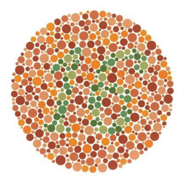
ePub Objectives The purpose of this study was to evaluate the influence of eye dominance on color perception, and shade matching.
Materials and Methods A total of 104 participants were selected for the study. There were 3 groups: Group I: 3rd and 4th year dental students and interns (
n = 40); Group II: postgraduates (n = 34); Group III: senior residents and faculty members (≥ 6 years of clinical experience) (n = 30). All participants were evaluated for congenital color blindness with Ishihara plates, their dominant eye with Mile's test, and their color perception with the Farnsworth-Munsell 100 hue test. The shade guide test was used for shade matching with a second corresponding set of Vitapan classical shade guides.Results The results of Mile’s test revealed that 60.6% were right-eye dominant and 39.4% were left-eye dominant. There was a statistically significant difference among all participants between the dominant eye and the non-dominant eye in shade matching.
Conclusions The dominant eye has a positive effect on shade matching and the ability to match shades becomes better with an increase in clinical experience.
-
Citations
Citations to this article as recorded by
- Influence of binocular vision, ocular dominance, and sex on color discrimination
Heba F. Elghorab, Ihab A. Hammad, Amir S. Azer, Mohamed A. Nassif, Mahinour A. Yousry
The Journal of Prosthetic Dentistry.2026; 136(1): e362. CrossRef - A Multidimensional Analysis of Shade Selection Difficulty for Indirect Restorations Among Dental Students and Professionals
Roxana-Ionela Vasluianu, Andreas Katsonis, Monica Silvia Tatarciuc, Anca Mihaela Vitalariu, Adina Oana Armencia, Andrea-Simoni Katsoni, Panagiotis Perperidis, Catalina Cioloca Holban, Irina Gradinaru, Ovidiu Stamatin, Magda Ecaterina Antohe
Dentistry Journal.2026; 14(4): 234. CrossRef - Analysis of Aesthetic Preferences Regarding Gingival–Dental Color Combinations
Cristina Gómez Polo, Ana María Martín Casado
Journal of Esthetic and Restorative Dentistry.2025; 37(9): 2060. CrossRef - Ocular dominance and visual color perception: A study on the overlooked factor in dental esthetics
Navjot Singh Mann, Ashu Jhamb, Divya Batra, Navneet Kaur Mann, Tanya Sharma, Rajat Kapur
Journal of Conservative Dentistry and Endodontics.2025; 28(12): 1258. CrossRef
- Influence of binocular vision, ocular dominance, and sex on color discrimination
- 2,590 View
- 59 Download
- 3 Web of Science
- 4 Crossref

- Evaluation of at-home bleaching protocol with application on different surfaces: bleaching efficacy and hydrogen peroxide permeability
- Heloisa Forville, Michael Willian Favoreto, Michel Wendlinger, Roberta Micheten Dias, Christiane Philippini Ferreira Borges, Alessandra Reis, Alessandro D. Loguercio
- Restor Dent Endod 2023;48(4):e33. Published online October 6, 2023
- DOI: https://doi.org/10.5395/rde.2023.48.e33
-
Abstract
PDFPubReaderePub
Objectives This study aimed to evaluate the bleaching efficacy and hydrogen peroxide permeability in the pulp chamber by the at-home bleaching gel in protocols applied on different dental surfaces.
Materials and Methods Forty premolars were randomly into 4 groups: control group no bleaching, only application on the buccal surface (OB), only application on the lingual surface (OL) and application in buccal and lingual surfaces, simultaneously (BL). At-home bleaching gel (White Class 7.5%) was used for the procedure. The bleaching efficacy was evaluated with a digital spectrophotometer (color change in CIELAB [Δ
E ab] and CIEDE 2000 [ΔE 00] systems and Whitening Index for Dentistry [ΔWID]). The hydrogen peroxide permeability in the pulp chamber (µg/mL) was assessed using UV-Vis spectrophotometry and data were analyzed for a 1-way analysis of variance and Tukey’s test (α = 0.05).Results All groups submitted to bleaching procedure showed bleaching efficacy when measured with Δ
E ab and ΔE 00 (p > 0.05). Therefore, when analyzed by ΔWID, a higher bleaching efficacy were observed for the application on the groups OB and BL (p = 0.00003). Similar hydrogen peroxide permeability was found in the pulp chambers of the teeth undergoing different protocols (p > 0.05).Conclusions The application of bleaching gel exclusively on the OB is sufficient to achieve bleaching efficacy, when compared to BL. Although the OL protocol demonstrated lower bleaching efficacy based on the ΔWID values, it may still be of interest and relevant in certain clinical scenarios based on individual needs, requiring clinical trials to better understand its specificities.
-
Citations
Citations to this article as recorded by- Effect of whitening pens on hydrogen peroxide permeability in the pulp chamber, color change and surface morphology
Laryssa Mylenna Madruga Barbosa, Gabrielle Gomes Centenaro, Deisy Cristina Ferreira Cordeiro, Maria Alice de Matos Rodrigues, Letícia Condolo, Michael Willian Favoreto, Alessandra Reis, Alessandro D. Loguercio
Journal of Dentistry.2025; 154: 105595. CrossRef - Evaluation of bleaching efficiency of carbamide peroxide applied on different dental surfaces: An in vitro study
R. Gokulnath, R. S. Mohan Kumar, A. Jayasenthil, R. Anjana, G. Sree Vidya
Journal of Conservative Dentistry and Endodontics.2025; 28(4): 366. CrossRef - Characterization and effects on enamel of low-concentration bleaching gels containing hyaluronic acid, NF_TiO2 nanoparticles and irradiated with violet LED light
Marcos Roberto Lima Benati, Matheus Kury, Priscila Borges Gobbo de Melo, Iago César Ribeiro Teles Matos, Roberta Tarkany Basting, Rosanna Tarkany Basting, Fernando Luis Esteban Florez, Vanessa Cavalli
Clinical Oral Investigations.2025;[Epub] CrossRef - Impact of bleaching on white spot lesions: hydrogen peroxide permeability and color alteration
Laryssa Mylenna Madruga Barbosa, Bruno Baracco, Taynara S. Carneiro, Michael Willian Favoreto, Michel Wendlinger, Daniel Jiménez-Díez, Laura Ceballos, Alessandro D. Loguercio
Clinical Oral Investigations.2025;[Epub] CrossRef - Efficacy of a buccal and lingual at‐home bleaching protocol—A randomized, split‐mouth, single‐blind controlled trial
Heloisa Forville, Laís Giacomini Bernardi, Michael Willian Favoreto, Felipe Coppla, Taynara de Souza Carneiro, Fabiana Madalozzo Coppla, Alessandro D. Loguercio, Alessandra Reis
Journal of Esthetic and Restorative Dentistry.2024; 36(9): 1301. CrossRef - REANATOMIZAÇÃO DE DENTE CONOIDE ASSOCIADA A ESTÉTICA VERMELHA: RELATO DE CASO
Ana Karolayne Sousa de Morais, Daniele Fernanda Sousa Barros, Daniel Messias Limeira, Rhana Leticia de Oliveira Faria, Roberta Furtado Carvalho, Sandna Nolêto de Araújo, Laura Barbosa Santos Di Milhomem
Revista Contemporânea.2024; 4(10): e6299. CrossRef - Effect of the reduction in the exposure time to at-home bleaching gel on color change and tooth sensitivity: A systematic review and meta-analysis
Priscila Borges Gobbo de Melo, Letícia Vasconcelos Silva Souza, Lucianne Cople Maia, Guido Artemio Marañón-Vásquez, Matheus Kury, Vanessa Cavalli
Clinical Oral Investigations.2024;[Epub] CrossRef
- Effect of whitening pens on hydrogen peroxide permeability in the pulp chamber, color change and surface morphology
- 5,345 View
- 91 Download
- 5 Web of Science
- 7 Crossref
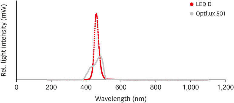
- Effects of 3 different light-curing units on the physico-mechanical properties of bleach-shade resin composites
- Azin Farzad, Shahin Kasraei, Sahebeh Haghi, Mahboubeh Masoumbeigi, Hassan Torabzadeh, Narges Panahandeh
- Restor Dent Endod 2022;47(1):e9. Published online February 7, 2022
- DOI: https://doi.org/10.5395/rde.2022.47.e9
-
Abstract
PDFPubReaderePub
Objectives This study investigated the microhardness, flexural strength, and color stability of bleach-shade resin composites cured with 3 different light-curing units.
Materials and Methods In this
in vitro experimental study, 270 samples were fabricated of bleach and A2 shades of 3 commercial resin composites (Point 4, G-aenial Anterior, and Estelite Sigma Quick). Samples (n = 5 for each trial) were cured with Bluephase N, Woodpecker LED.D, and Optilux 501 units and underwent Vickers microhardness and flexural strength tests. The samples were tested after 24 hours of storage in distilled water. Color was assessed using a spectrophotometer immediately after preparation and 24 hours after curing. Data were analyzed using 3-way analysis of variance and the Tukey test (p ≤ 0.001).Results Samples cured with Optilux exhibited the highest and those cured with LED.D exhibited the lowest microhardness (
p = 0.023). The bleach shade of Point 4 composite cured with Optilux displayed the highest flexural strength, while the same composite and shade cured with Sigma Quick exhibited the lowest (p ≤ 0.001). The color change after 24 hours was greatest for the bleach shade of G-aenial cured with Bluephase N and least for the A2 shade of Sigma Quick cured with Optilux (p ≤ 0.001).Conclusions Light curing with polywave light-emitting diode (LED) yielded results between or statistically similar to those of quartz-tungsten-halogen and monowave LED in the microhardness and flexural strength of both A2 and bleach shades of resin composites. However, the brands of light-curing devices showed significant differences in color stability.
-
Citations
Citations to this article as recorded by- Mechanical Behaviour of Novel Nanohybrid Resin Composite Using Two Light Cure Systems
Ghada H. Naguib, Jumana Mazhar, Abeer Alnowaiser, Abdulghani Mira, Hisham Mously, Rabab Aljawi, Samar H. Abuzinadah, Mohamed T. Hamed
International Dental Journal.2025; 75(2): 1136. CrossRef - Repair Bond Strength of Aged Composite: Effect of Thermocycling and Surface Treatment
Sina Yarmoradian, Ladan Ranjbar Omrani, Elham Ahmadi, Niyousha Rafeie, Mahdi Abbasi, Nastaran Dabiri Shahabi
Journal of Research in Dental and Maxillofacial Sciences.2025; 10(3): 228. CrossRef - Evaluation of the Depth of Cure by Microhardness of Bulk-Fill Composites with Monowave and Polywave LED Light-Curing Units
Socratis Thomaidis, Dimitris Kampouropoulos, Maria Antoniadou, Afrodite Kakaboura
Applied Sciences.2024; 14(24): 11532. CrossRef - Effect of hard segment chemistry and structure on the self‐healing properties of UV‐curable coatings based on the urethane acrylates with built‐in Diels–Alder adduct
Paulina Bednarczyk, Karolina Mozelewska, Małgorzata Nowak, Joanna Klebeko, Joanna Rokicka, Paula Ossowicz‐Rupniewska
Journal of Applied Polymer Science.2023;[Epub] CrossRef - Effects of Dental Bleaching Agents on the Surface Roughness of Dental Restoration Materials
Alexandru Dan Popescu, Mihaela Jana Tuculina, Oana Andreea Diaconu, Lelia Mihaela Gheorghiță, Claudiu Nicolicescu, Cristian Niky Cumpătă, Cristiana Petcu, Jaqueline Abdul-Razzak, Ana Maria Rîcă, Ruxandra Voinea-Georgescu
Medicina.2023; 59(6): 1067. CrossRef - Effect of Polywave and Monowave Light Curing Units on Color Change of Composites Containing Trime-thylbenzoyl-Diphenyl-Phosphine Before and After Aging
Negar Madihi, Maryam Hoorizad ganjkar, Negin Nasoohi, Ali Kaboudanian Ardestani
Journal of Research in Dental and Maxillofacial Sciences.2023; 8(4): 249. CrossRef
- Mechanical Behaviour of Novel Nanohybrid Resin Composite Using Two Light Cure Systems
- 3,035 View
- 47 Download
- 4 Web of Science
- 6 Crossref
- Shade reproduction and the ability of lithium disilicate ceramics to mask dark substrates
- Maryam Iravani, Sayna Shamszadeh, Narges Panahandeh, Seyedeh Mahsa Sheikh-Al-Eslamian, Hassan Torabzadeh
- Restor Dent Endod 2020;45(3):e41. Published online July 16, 2020
- DOI: https://doi.org/10.5395/rde.2020.45.e41
-
Abstract
PDFPubReaderePub
Objectives This study aimed to evaluate the ability of lithium disilicate ceramics to reproduce the A2 shade and to mask A4 substrates.
Materials and Methods Twenty-four discs (8 mm in diameter, shade A2) of high translucency (groups 1–3) and low translucency (groups 4–6) of IPS e.max ceramic with different thicknesses (0.5, 0.75, and 1 mm) were fabricated as monolithic structures. In addition, discs of medium opacity (group 7–8) with different core/veneer combinations (0.3 mm/0.7 mm and 0.5 mm/0.5 mm) were fabricated as bilayer structures. Specimens were superimposed on an A4 substrate (complex). The color changes of the complex were measured using a spectrophotometer on a black background, and the ΔE values of the complex were compared with either the A4 substrate or the A2 shade tab. One-way analysis of variance, the Tukey honest significant difference test, and the Fisher test were used to analyze the data (
p < 0.05).Results Significant between-group differences were found for comparisons to both the A4 substrate and the A2 shade (
p < 0.05). When compared with the A4 substrate, the ΔE values in all groups were in the non-acceptable range. When compared with the A2 shade, the ΔE values in all groups, except groups 2 and 3, were in the clinically acceptable range.Conclusions All translucencies and thicknesses masked the underlying dark substrate. However, the low-translucency IPS e.max Press better reproduced the A2 shade.
-
Citations
Citations to this article as recorded by- The effects of material thicknesses and substrates on the translucency and color masking ability of additively manufactured definitive crown materials
Ting Wang, Yun‐Ju Wang, Chao‐Chieh Yang, John A. Levon, Tien‐Min G. Chu, Wei‐Shao Lin
Journal of Prosthodontics.2026;[Epub] CrossRef - The effect of thickness, cement type, and substrate color on the optical properties of three-dimensional-printed composite resin
Elif Önal, Zeynep Şahin, Lale Karaağaçlioğlu
The Journal of Indian Prosthodontic Society.2026; 26(2): 150. CrossRef - The Impact of Surface Treatments and 3D Printing Machines on the Biaxial Flexural Strength of 3D-Printed Composite Resins
Mohammed K. Fahmi
The Open Dentistry Journal.2025;[Epub] CrossRef - Masking Ability of the Combined Application of Opaque Resin Composite and High‐Translucency Zirconia on Discolored Substrates
Shuping Chen, Lei Jiang, Run Chen
Journal of Esthetic and Restorative Dentistry.2025; 37(10): 2298. CrossRef - Comparative Evaluation of the Translucency of Polyetheretherketone (PEEK) Veneered With Two Different Materials: An In Vitro Study
M P Chinmayi, Gautam Shetty, S M Kedar, Lokesh B Kanchan, Rohit S Kundu, Krishna Kumar U, Maria Jenifer
Cureus.2025;[Epub] CrossRef - Masking capacity of minimally invasive lithium disilicate restorations on discolored teeth—The impact of ceramic thickness, the material's translucency, and the cement color
Kevser Pala, Eva Maria Reinshagen, Thomas Attin, Jürg Hüsler, Ronald E. Jung, Alexis Ioannidis
Journal of Esthetic and Restorative Dentistry.2024; 36(1): 107. CrossRef - Comparing the color match of monolithic CAD-CAM dental ceramics with the VITA Classical shade guide
Mohammadjavad Shirani, Maryam Emami, Ramin Mosharraf, Omid Savabi, Mehrdad Akhavankhaleghi, Kamran Azadbakht
The Journal of Prosthetic Dentistry.2024; 132(3): 605. CrossRef - Quantitative examination of factors influencing the colour reproduction ability of lithium disilicate glass-ceramics
József Saláta, Ferenc Szabó, Péter Csuti, Melinda Antal, Péter Márton, Péter Hermann, Judit Borbély, Emese Ábrám
BMC Oral Health.2024;[Epub] CrossRef - Aesthetic restoration using liner-treated lithium disilicate laminate veneers in discolored teeth after endodontic treatment : A case report
Ji-Hyun Kim, Min-Soo Bae, Yeon-Hee Park, Jung-Jin Lee, Tae-Sung Bae3, Jae-Min Seo
Korean Journal of Dental Materials.2023; 50(2): 91. CrossRef - Can we use the translucency parameter to predict the CAD/CAM ceramic restoration aesthetic?
Jie Wang, Jiawei Yang, Kaige Lv, Hongming Zhang, Hui Huang, Xinquan Jiang
Dental Materials.2023; 39(3): e1. CrossRef - Final Color of CAD-CAM Produced Thin Lithium Disilicate Ceramics Cemented with Different Colored Resin Cements on Darker Backgrounds
Merve BANKOĞLU GÜNGÖR
ADO Klinik Bilimler Dergisi.2023; 12(2): 234. CrossRef - Masking Ability of Monolithic and Layered Zirconia Crowns on Discolored Substrates
Cristina Gasparik, Manuela Maria Manziuc, Alexandru Victor Burde, Javier Ruiz-López, Smaranda Buduru, Diana Dudea
Materials.2022; 15(6): 2233. CrossRef - Effects of background color and thickness on the optical properties of CAD-CAM resin-matrix ceramics
Afnan F. Alfouzan, Sarah M. Alnafaiy, Lama S. Alsaleh, Noor H. Bawazir, Hanan N. Al-Otaibi, Sara M. Al Taweel, Huda A. Alshehri, Nawaf Labban
The Journal of Prosthetic Dentistry.2022; 128(3): 497.e1. CrossRef - Effect of CAD/CAM Ceramic Thickness on Shade Masking Ability of Discolored Teeth: In Vitro Study
Passent Ellakany, Marwa Madi, Nourhan M. Aly, Zainb S. Al-Aql, Maher AlGhamdi, Abdulrahman AlJeraisy, Adel S. Alagl
International Journal of Environmental Research and Public Health.2021; 18(24): 13359. CrossRef
- The effects of material thicknesses and substrates on the translucency and color masking ability of additively manufactured definitive crown materials
- 2,765 View
- 25 Download
- 14 Crossref
- Effect of immersion into solutions at various pH on the color stability of composite resins with different shades
- Ji-Deok Moon, Eun-Mi Seon, Sung-Ae Son, Kyoung-Hwa Jung, Yong-Hoon Kwon, Jeong-Kil Park
- Restor Dent Endod 2015;40(4):270-276. Published online August 28, 2015
- DOI: https://doi.org/10.5395/rde.2015.40.4.270
-
Abstract
PDFPubReaderePub
Objectives This study examined the color changes of a resin composite with different shades upon exposure to water with different pH.
Materials and Methods Nanohybrid resin composites (Filtek Z350XT, 3M ESPE) with four different shades (A2, A3, B1, and B2) were immersed in water with three different pH (pH 3, 6, and 9) for 14 day. The CIE
L*a*b* color coordinates of the specimens were evaluated before and after immersion in the solutions. The color difference (ΔE* ) and the translucency parameter (TP ) were calculated using the color coordinates.Results ΔE* ranged from 0.33 to 1.58, and the values were affected significantly by the pH. The specimens immersed in a pH 6 solution showed the highestΔE* values (0.87 - 1.58). The specimens with a B1 shade showed the lowestΔE* change compared to the other shades.TP ranged from 7.01 to 9.46 depending on the pH and resin shade. TheTP difference between before and after immersion in the pH solutions was less than 1.0.Conclusions The resulting change of color of the tested specimens did not appear to be clinically problematic because the color difference was < 1.6 in the acidic, neutral, and alkaline solutions regardless of the resin shade, i.e., the color change was imperceptible.
-
Citations
Citations to this article as recorded by- Effect of Acidic Environment on the Mechanical Strength and Surface Properties of Three-Dimensional-Printed Denture Base Resin
Eun-Ju Kim, Se-Eun Kim, Ye-Jin Kim, Ji-Eun Kim, Yun-Yeong Hwang, Hye-Bin Go, Song-Yi Yang
International Dental Journal.2026; 76(5): 109757. CrossRef - Effect of mouth rinses on roughness and optical properties of restorative materials for oral rehabilitation
Laura Firmo de Carvalho, Edmara T. P. Bergamo, Ernesto B. Benalcázar-Jalkh, Tiago M. B. Campos, Abbas Zahoui, Elisa De Souza Fermino, Ana Clara Mota de Oliveira, Ana Carolina Magalhães, Estevam A. Bonfante, Fábio José B. Bezerra, Larissa M.M. Alves
Biomaterial Investigations in Dentistry.2025; 12: 25. CrossRef - Effect of Printing Layer Thickness on the Color Stability and Surface Roughness of Three-dimensional Printed Resin Material Immersed in Different Aging Media: An In Vitro Study
Vilas Rajguru, Smita Khalikar, Sonali Mahajan, Gopika Gopan, Siddhi D Bhatawadekar, Kishor Mahale
International Journal of Prosthodontics and Restorative Dentistry.2025; 15(1): 36. CrossRef - Colour Retaining Ability of Three Restorative Materials Used in Pediatric Dentistry with the Use of Health Drinks and Beverages - An In Vitro Study
Arnab Mondal, Swati Singh, Seema Qamar, Barun Dasgupta, Shovan Roy
Indian Journal of Dental Research.2025; 36(1): 89. CrossRef - Effects of different antiviral mouthwashes on the surface roughness, hardness, and color stability of composite CAD/CAM materials
Ahmet Hazar, Ecehan Hazar
Journal of Applied Biomaterials & Functional Materials.2024;[Epub] CrossRef - Flexural Properties of Contemporary Bioactive Restorative Materials: Effect of Environmental pH
JEX Ong, AU Yap, A Abdul Aziz, NA Yahya
Operative Dentistry.2023; 48(1): 90. CrossRef - Effect of Mouthwashes for COVID-19 Prevention on Surface Changes of Resin Composites
Saijai Tanthanuch, Boonlert Kukiattrakoon, Chailuck Naiyanart, Tanyanat Promtong, Panuwit Yothinwatthanabamrung, Suttida Pumpua
International Dental Journal.2023; 73(4): 511. CrossRef - Can Modification with Urethane Derivatives or the Addition of an Anti-Hydrolysis Agent Influence the Hydrolytic Stability of Resin Dental Composite?
Agata Szczesio-Wlodarczyk, Izabela M. Barszczewska-Rybarek, Marta W. Chrószcz-Porębska, Karolina Kopacz, Jerzy Sokolowski, Kinga Bociong
International Journal of Molecular Sciences.2023; 24(5): 4336. CrossRef - Understanding the Mechanical, Surface, and Color Behavior of Oral Bioactive Prosthetic Polymers under Biodegradation Processes
Cristina B. Neves, Joana Costa, Jaime Portugal, Ana F. Bettencourt
Polymers.2023; 15(11): 2549. CrossRef - An Evaluation of the Hydrolytic Stability of Selected Experimental Dental Matrices and Composites
Agata Szczesio-Wlodarczyk, Karolina Kopacz, Malgorzata Iwona Szynkowska-Jozwik, Jerzy Sokolowski, Kinga Bociong
Materials.2022; 15(14): 5055. CrossRef - Evaluation of the Color Stability, Water Sorption, and Solubility of Current Resin Composites
Wenkai Huang, Ling Ren, Yuyao Cheng, Minghua Xu, Wenji Luo, Desong Zhan, Hidehiko Sano, Jiale Fu
Materials.2022; 15(19): 6710. CrossRef - Comparative Evaluation of Surface Roughness of different Composites and its effect on Colour Stability of the Restoration
Nalini B, Srinivas Kumar Ch, Narsimha Rao VV
Research Journal of Pharmacy and Technology.2022; : 3854. CrossRef - Comparison of instrumental methods for color change assessment of Giomer resins
Luiza de Almeida Queiroz Ferreira, Rogéli Tibúrcio Ribeiro da Cunha Peixoto, Cláudia Silami de Magalhães, Tassiana Melo Sá, Monica Yamauti, Francisca Daniele Moreira Jardilino
Restorative Dentistry & Endodontics.2022;[Epub] CrossRef - Effect of hydrothermal aging on the microhardness of high- and low-viscosity conventional and additively manufactured polymers
Nadin Al-Haj Husain, Albert J. Feilzer, Cornelis J. Kleverlaan, Samir Abou-Ayash, Mutlu Özcan
The Journal of Prosthetic Dentistry.2022; 128(4): 822.e1. CrossRef - KARANFİL VE YEŞİL ÇAYIN RESTORATİF MATERYALLERİN YÜZEY VE OPTİK ÖZELLİKLERİ ÜZERİNE ETKİSİ: İN VİTRO ÇALIŞMA
Zeynep BUKET KAYNAR, Tolgahan DOĞAN, Nazmiye DÖNMEZ, Mağrur KAZAK
Atatürk Üniversitesi Diş Hekimliği Fakültesi Dergisi.2022; : 1. CrossRef - Effects of Red Dragon Fruit on Color Stability of Self-Adhesive Flowable Composite
Rosalina Tjandrawinata, Brigitta Heidy, Octarina
Applied Mechanics and Materials.2022; 910: 1. CrossRef - Effect of different cement systems and aging on the bond strength of chairside CAD-CAM ceramics
Seda Ustun, Elif Aydogan Ayaz
The Journal of Prosthetic Dentistry.2021; 125(2): 334. CrossRef - Influence of Selected Restorative Materials on the Environmental pH: In Vitro Comparative Study
Anna Lehmann, Kacper Nijakowski, Michalina Nowakowska, Patryk Woś, Maria Misiaszek, Anna Surdacka
Applied Sciences.2021; 11(24): 11975. CrossRef - Color stability of an artificially aged nanofilled composite resin post-cured with different techniques
Lais Sampaio Souza, Tais Rocha Donato, Gabriela Alves Cerqueira, Andrea Nobrega Cavalcanti, Paula Mathias
Journal of Dental Research, Dental Clinics, Dental Prospects.2021; 15(1): 53. CrossRef - Ageing of Dental Composites Based on Methacrylate Resins—A Critical Review of the Causes and Method of Assessment
Agata Szczesio-Wlodarczyk, Jerzy Sokolowski, Joanna Kleczewska, Kinga Bociong
Polymers.2020; 12(4): 882. CrossRef - Use of Resin Solvent as a Facilitator for Removal of Resin Composite Restoratives by Influencing their Mechanical Properties: Is this Possible?
Inas A Elghandour
Journal of Operative Dentistry & Endodontics.2019; 4(1): 1. CrossRef - Effect of powder-liquid ratios and powder colors on color stability of 4-META / MMA-TBB resin after immersion in coffee.
Yoorina Choi, Seo-Jin Jang, Su-Jung Park
Korean Journal of Dental Materials.2018; 45(3): 187. CrossRef - O uso do gel de glicerina melhora a estabilidade de cor de resinas compostas?
Marcus Vinicius Loureiro BERTOLO, Mário Alexandre Coelho SINHORETI, Julia Puppin RONTANI, Pedro Paulo Albuquerque Cavalcanti de ALBUQUERQUE, Luis Felipe Jochims SCHNEIDER
Revista de Odontologia da UNESP.2018; 47(4): 256. CrossRef - In situ evaluation of color stability and hardness' decrease of resin‐based composites
Juliana Silva Ribeiro, Sonia Luque Peralta, Vinícius Esteves Salgado, Rafael Guerra Lund
Journal of Esthetic and Restorative Dentistry.2017; 29(5): 356. CrossRef - Discoloration Effects of Traditional Turkish Beverages on different Composite Restoratives
Serdar Baglar, Erol Keskin, Tahir Orun, Abdulhamit Es
The Journal of Contemporary Dental Practice.2017; 18(2): 83. CrossRef - The effect of red and white wine on color changes of nanofilled and nanohybrid resin composites
Saijai Tanthanuch, Boonlert Kukiattrakoon, Thanwalee Peerasukprasert, Nilobon Chanmanee, Parnchanok Chaisomboonphun, Apisara Rodklai
Restorative Dentistry & Endodontics.2016; 41(2): 130. CrossRef
- Effect of Acidic Environment on the Mechanical Strength and Surface Properties of Three-Dimensional-Printed Denture Base Resin
- 2,528 View
- 16 Download
- 26 Crossref
- Colorimetric comparison of single layered dental composite with double layered dental composite
- Young-Sang Song, Ja-Hyun Kim, Bin-Na Lee, Ji-Hyun Jang, Hoon-Sang Chang, Yun-Chan Hwang, Won-Mann Oh, In-Nam Hwang
- Restor Dent Endod 2012;37(2):84-89. Published online May 18, 2012
- DOI: https://doi.org/10.5395/rde.2012.37.2.84
-
Abstract
PDFPubReaderePub
Objectives This study analyzed the difference in color caused by different thickness in enamel layer of composite resins when applied with single and layering placement technique, and evaluated if the results agreed with the shade guide from the manufacturers to verify reliability of the color matching process of the manufacturers.
Materials and Methods For single composite resin samples, 6 mm diameter and 4 mm thickness cylindrical samples were fabricated using Ceram-X mono (DENTSPLY DeTrey) and CIE L*a*b* values were measured with spectrophotometer. Same process was done for layering composite resin samples, making 3 dentinal shade samples, 4 mm thickness, for each shade using Ceram-X duo (DENTSPLY DeTrey) and enamel shade resins were layered in 2 mm thickness and CIE L*a*b* values were measured. These samples were ground to 0.2 mm thickness each time, and CIE L*a*b* values were measured to 1 mm thickness of enamel shade resin.
Results Color difference (ΔE*) between single and layering composite resin was 1.37 minimum and 10.53 maximum when layering thicknesses were between 1 mm and 2 mm and 6 out of 10 same shade groups suggested by manufacturer showed remarkable color difference at any thickness (ΔE* > 3.3).
Conclusion When using Ceram-X mono and duo for composite resin restoration, following the manufacturer's instructions for choosing the shade is not appropriate, and more accurate information for Ceram-X duo is needed on the variation and expression of the shades depending on the thickness of the enamel.
-
Citations
Citations to this article as recorded by- Improvement of mechanical strength and water repellency of Hanji (traditional Korean paper) through acetylation in supercritical CO2
Seungmok Shin, Hwi-Sung Lee, Hee Suk Woo, Mulugeta G. Aregay, Tae Jun Yoon, Youn-Woo Lee
The Journal of Supercritical Fluids.2022; 190: 105735. CrossRef - Color Change in Tooth Induced by Various Calcium Silicate-Based Pulp-Capping Materials
Jiyoon Jeon, Namki Choi, Seonmi Kim
THE JOURNAL OF THE KOREAN ACADEMY OF PEDTATRIC DENTISTRY.2021; 48(3): 280. CrossRef
- Improvement of mechanical strength and water repellency of Hanji (traditional Korean paper) through acetylation in supercritical CO2
- 1,683 View
- 5 Download
- 2 Crossref
Basic Researchs
- Effects of the color components of light-cured composite resin before and after polymerization on degree of conversion and flexural strength
- Ji-A Yoo, Byeong-Hoon Cho
- J Korean Acad Conserv Dent 2011;36(4):324-335. Published online July 31, 2011
- DOI: https://doi.org/10.5395/JKACD.2011.36.4.324
-
Abstract
PDFPubReaderePub
Objectives This study investigated the effects of the color components of light-cured composite resin before and after polymerization on degree of conversion (DC) and biaxial flexural strength (FS).
Materials and Methods Four enamel shades (A1, A2, A3, A4) and two dentin shades (A2O, A3O) of Premisa (Kerr Co.) and Denfil (Vericom Co.) were evaluated on their CIE L*, a*, b* color components using the spectrophotometer before curing, after curing and at 7 day. The DC of same specimens were measured with Near-infrared spectrometer (Nexus, Thermo Nicolet Co.) at 2 hr after cure and at 7 day. Finally, the FS was obtained after all the other measurements were completed at 7 day. The correlations between each color component and DC and FS were evaluated.
Results The light-curing of composite resin resulted in color changes of Premisa in red-blue direction and Denfil in green-blue direction. The DC and FS were affected by product, time and shade (3-way ANOVA,
p < 0.05) and product and shade (2-way ANOVA,p < 0.05), respectively. Premisa only showed a significant correlation between the DC and CIE a* component - before and after polymerization (Pearson product moment correlation,p < 0.05). The FS of Premisa showed significant negative correlations with CIE a* and CIE b* components.Conclusions The DC and FS of the light-curing composite resin were affected by the color components of the material before and after polymerization.
-
Citations
Citations to this article as recorded by- Evaluation of Color Stability according to Shade of Temporary Crown Resin Using Digital Spectrophotometer: In Vitro Study
Hye-min Ku, Mi-Kyoung Jun
Journal of Dental Hygiene Science.2022; 22(3): 139. CrossRef - The properties of UDMA dental composite resin with novel photosensitizers
Gum Ju Sun
Journal of Korean Acedemy of Dental Technology.2013; 35(3): 209. CrossRef
- Evaluation of Color Stability according to Shade of Temporary Crown Resin Using Digital Spectrophotometer: In Vitro Study
- 2,021 View
- 4 Download
- 2 Crossref
- Clinical assessment of whitening efficacy and safety of in-office tooth whitening system containing 15% hydrogen peroxide with or without light activation
- Young-Suk Noh, Young-Jee Rho, Yeon-Jee Yoo, Hyang-Ok Lee, Sang-Min Lim, Hyun-Jeong Kweon, Yeun Kim, Seong-Yeon Park, Hee-Young Yoon, Jung-Hyun Lee, Chan-Hee Lee, So-Ram Oh, Kee-Yeon Kum
- J Korean Acad Conserv Dent 2011;36(4):306-312. Published online July 31, 2011
- DOI: https://doi.org/10.5395/JKACD.2011.36.4.306
-
Abstract
PDFPubReaderePub
Objectives This clinical study evaluated the effect of light activation on the whitening efficacy and safety of in-office bleaching system containing 15% hydrogen peroxide gel.
Materials and Methods Thirty-three volunteers were randomly treated with (n = 17, experimental group) or without light activation (n = 16, control group), using Zoom2 white gel (15% H2O2, Discus Dental) for a total treatment time of 45 min. Visual and instrumental color measurements were obtained using Vitapan Classical shade guide and Shadepilot (DeguDent) at screening test, after bleaching, and 1 month and 3 month after bleaching. Data were analyzed using
t -test, repeated measure ANOVA, and chi-squared test.Results Zoom2 white gel produced significant shade changes in both experimental and control group when pre-treatment shade was compared with that after bleaching. However, shade difference between two groups was not statistically significant (
p > 0.05). Tooth shade relapse was not detected at 3 months after bleaching. The incidence of transient tooth sensitivity was 39.4%, with being no differences between two groups.Conclusions The application of light activation with Zoom2 white gel system neither achieved additional whitening effects nor showed more detrimental influences.
-
Citations
Citations to this article as recorded by- Comparative Effects of Hydrogen Peroxide–Containing Whitening Toothpaste and Gel on Tooth Lightness: An In Vitro Study
Ye-Bin Kim, Hyon-Mo Ku, Ji-Hyeon Park
International Journal of Clinical Preventive Dentistry.2025; 21(4): 148. CrossRef - Effect of preference drinks on tooth color reduction after tooth bleaching: A 12-momth follow-up study
Hyo Jin Goo, Min Jeong Cho, Yun Sook Jung, Ji Hye Kim, Fan Dong, Keun Bae Song
Journal of Korean Academy of Oral Health.2016; 40(1): 55. CrossRef - Development of a Piezoelectric Ultrasonic Tooth-whitening Apparatus
Young-Jin Lee, Jong-Hoo Paik, Jeong-Bae Lee, Seung-Jae Choi
Transactions on Electrical and Electronic Materials.2013; 14(5): 268. CrossRef
- Comparative Effects of Hydrogen Peroxide–Containing Whitening Toothpaste and Gel on Tooth Lightness: An In Vitro Study
- 2,875 View
- 8 Download
- 3 Crossref
- The evaluation of color and color difference according to the layering placement of Incisal shade composites on the body composites of the indirect resin restoration
- Su-Jung Park, Han-Young Lee, Myong-Yun Nah, Hoon-Sang Chang, Yun-Chan Hwang, Won-Mann Oh, In-Nam Hwang
- J Korean Acad Conserv Dent 2011;36(1):37-49. Published online January 14, 2011
- DOI: https://doi.org/10.5395/JKACD.2011.36.1.37
-
Abstract
PDFPubReaderePub
Abstract Objectives: The aim of this study was to evaluate the surface color of indirect resin restoration according to the layering placement of different shade of incisal composite.
Materials and Methods: In this study, CIE L*a*b* value of 16 Body composite of Tescera ATL (Bisco, Schaumburg IL, USA) was measured by spectrophotometer (NF999, Nippon Denshuku, Japan), and compared to CIE L*a*b* value of Vitapan shade guide. Nine shade Incisal composite of Tescera ATL were buildup to 1 mm thickness on Body composites inlay block, and CIE L*a*b* value was measured. Incisal composite was ground to 0.5 mm thickness and CIE L*a*b* value was re-measured. Color difference between Body composite and Incisal composites layered on Body composite was calculated as a function of thickness.
Results: Color difference between corresponding shade of Tescera Body composite and Vitapan shade guide was from 6.88 to 12.80.
L* and b*value was decreased as layering thickness of Incisal composite on Body composite was increased. But, a* value did not show specific change tendency.
Conclusions: Surface color difference between Body composites and Incisal composites layered on Body composite was increased as the layering thickness of Incisal composite increased (
p < 0.05).-
Citations
Citations to this article as recorded by- Evaluation of the color stability of materials used in the resin infiltration technique for artificial white spot lesions
Kkot-Byeol Bae, Tae-geon Kim, Su-Jung Park, Yun-Chan Hwang, In-Nam Hwang
Journal of Dental Rehabilitation and Applied Science.2026; 42(2): 90. CrossRef - Color stability of esthetic restorative materials after application of fluoride varnishes
Chul-Hoon Jang, Dong-Gil Jang, Bin-Na Lee, Hoon-Sang Chang, Yun-Chan Hwang, Won-Mann Oh, In-Nam Hwang
Korean Journal of Dental Materials.2021; 48(3): 147. CrossRef - Color Change in Tooth Induced by Various Calcium Silicate-Based Pulp-Capping Materials
Jiyoon Jeon, Namki Choi, Seonmi Kim
THE JOURNAL OF THE KOREAN ACADEMY OF PEDTATRIC DENTISTRY.2021; 48(3): 280. CrossRef - Discrimination between FRC-post and core according to the color difference
Jou-Hwe Kim, Jin-Woo Kim, Kyung-Mo Cho, Se-Hee Park
Journal of Dental Rehabilitation and Applied Science.2015; 31(2): 75. CrossRef - Optical characteristics of resin composite before and after polymerization
Ah-Hyang Eom, Duck-Su Kim, Soo-Hee Lee, Chang-Won Byun, Noh-Hoon Park, Kyoung-Kyu Choi
Journal of Korean Academy of Conservative Dentistry.2011; 36(3): 219. CrossRef
- Evaluation of the color stability of materials used in the resin infiltration technique for artificial white spot lesions
- 1,779 View
- 3 Download
- 5 Crossref
Clinical Research
- The evaluation of clinical efficacy and longevity of home bleaching without combined application of In-office bleaching
- Byunk-Gyu Shin, Sung-Eun Yang
- J Korean Acad Conserv Dent 2010;35(5):387-394. Published online September 30, 2010
- DOI: https://doi.org/10.5395/JKACD.2010.35.5.387
-
Abstract
PDFPubReaderePub
Objectives The purpose of this study was to evaluate the whitening efficacy and longevity of home bleaching.
Materials and Methods A total of 28 patients were divided into either experimental group (Opalescence F; 15% carbamide peroxide) or control group randomly. The patients in experimental group were instructed to wear individual trays applied with bleaching gel for 2 hours a day for 4 weeks. Any treatments weren't applied to the patients in control group. The color measurements of central incisors, lateral incisors & canines of upper and lower arch were recorded at base line, immediately after the finishment of treatmemt (4 weeks), 8 weeks and 12 weeks using Colorimeter (Chroma Meter, 2600d Konica Minolta co.) and Vitapan classical shade guide (Vita Zahnfabrik).
Results A significantly stronger color change was observed for overall teeth samples in experimental group immediately after treatment (at 4 weeks) compared to ones in control group (
p < 0.05). There was also a significant difference between baseline and 8 weeks or 12 weeks separately though color rebouncing phenomenon occurred as time went by (p < 0.05).Conclusions The clinical effecacy and longevity of home bleaching without combined application of in-office bleaching was observed through this experiment.
-
Citations
Citations to this article as recorded by- Effect of at-home bleaching agents and concentrations on tooth sensitivity: A systematic review and network meta-analysis
Renata Maria Oleniki Terra, Michael Willian Favoreto, Tom Morris, Alessandro D. Loguercio, Alessandra Reis
Journal of Dentistry.2025; 160: 105891. CrossRef - Effect of at-home agents and concentrations on bleaching efficacy: A systematic review and network meta-analysis
Renata Maria Oleniki Terra, Michael Willian Favoreto, Tom Morris, Alessandro D. Loguercio, Alessandra Reis
Journal of Dentistry.2025; 160: 105857. CrossRef - Effects of Citrus limon Extract on Oxidative Stress-Induced Nitric Oxide Generation and Bovine Teeth Bleaching
Soon-Jeong Jeong
Journal of Dental Hygiene Science.2021; 21(2): 96. CrossRef - Efficacy of a self - applied paint - on whitening gel combined with wrap
Soo-Yeon Kim, Jae-Hyun Ahn, Ji-Young Kim, Jin-Woo Kim, Se-Hee Park, Kyung-Mo Cho
Journal of Dental Rehabilitation and Applied Science.2018; 34(3): 175. CrossRef - Home-based chemically-induced whitening (bleaching) of teeth in adults
Prashanti Eachempati, Sumanth Kumbargere Nagraj, Salian Kiran Kumar Krishanappa, Puneet Gupta, Ibrahim Ethem Yaylali
Cochrane Database of Systematic Reviews.2018;[Epub] CrossRef - Effect of preference drinks on tooth color reduction after tooth bleaching: A 12-momth follow-up study
Hyo Jin Goo, Min Jeong Cho, Yun Sook Jung, Ji Hye Kim, Fan Dong, Keun Bae Song
Journal of Korean Academy of Oral Health.2016; 40(1): 55. CrossRef - Tooth color reduction and consequent patient satisfaction after office and home bleaching: a 6-month follow-up study
Hyo-Jin Goo, Hyeon-Sook Kwun, Jeong-Hee Park, Min-Jeong Cho, Eun-Kyong Kim, Youn-Hee Choi, Keun-Bae Song
Journal of Korean Academy of Oral Health.2015; 39(1): 3. CrossRef
- Effect of at-home bleaching agents and concentrations on tooth sensitivity: A systematic review and network meta-analysis
- 1,787 View
- 2 Download
- 7 Crossref
Original Articles
- Clinical study of shade improvement and safety of polymer-based pen type BlancTic Forte whitening agent containing 8.3% Carbamide peroxide
- Jin-Kyung Lee, Sun-Hong Min, Sung-Tae Hong, So-Ram Oh, Shin-Hye Chung, Young-Hye Hwang, Sung-Yeop You, Kwang-Shik Bae, Seung-Ho Baek, Woo-Cheol Lee, Won-Jun Son, Kee-Yeon Kum
- J Korean Acad Conserv Dent 2009;34(2):154-161. Published online March 31, 2009
- DOI: https://doi.org/10.5395/JKACD.2009.34.2.154
-
Abstract
PDFPubReaderePub
This clinical study evaluated the whitening effect and safety of polymer based-pen type BlancTis Forte (NIBEC) containing 8.3% carbamide peroxide. Twenty volunteers used the BlancTis Forte whitening agent for 2 hours twice a day for 4 weeks. As a control, Whitening Effect Pen (LG) containing 3% hydrogen peroxide was used by 20 volunteers using the same protocol. The change in shade (ΔE*, color difference) was measured using Shadepilot™ (DeguDent) before, during, and after bleaching (2 weeks, 4 weeks, and post-bleaching 4 weeks). A clinical examination for any side effects (tooth hypersensitivity or soft tissue complications) was also performed at each check-up. The following results were obtained.
1. Both the experimental and control groups displayed a noticeable change in shade (ΔE) of over 2. No significant differences were found between the two groups (p > 0.05), implying that the two agents have a similar whitening effect.
2. The whitening effect was mainly due to changes in a and b values rather than in L value (brightness). The experimental group showed a significantly higher change in b value, thus yellow shade, than the control (p < 0.05).
3. None of the participants complained of tooth hypersensitivity or soft tissue complications, confirming the safety of both whitening agents.
-
Citations
Citations to this article as recorded by- Surface Damage and Bleaching Effect according to the Application Type of Home Tooth Bleaching Applicants
Na-Yeoun Tak, Do-Seon Lim, Hee-Jung Lim, Im-Hee Jung
Journal of Dental Hygiene Science.2020; 20(4): 252. CrossRef - Efficacy of a self - applied paint - on whitening gel combined with wrap
Soo-Yeon Kim, Jae-Hyun Ahn, Ji-Young Kim, Jin-Woo Kim, Se-Hee Park, Kyung-Mo Cho
Journal of Dental Rehabilitation and Applied Science.2018; 34(3): 175. CrossRef
- Surface Damage and Bleaching Effect according to the Application Type of Home Tooth Bleaching Applicants
- 2,125 View
- 2 Download
- 2 Crossref
- The effectiveness of sealing technique on in-office bleaching
- Yoon Lee, So-Ran Kwon, Jeong-Won Park
- J Korean Acad Conserv Dent 2008;33(5):463-471. Published online September 30, 2008
- DOI: https://doi.org/10.5395/JKACD.2008.33.5.463
-
Abstract
PDFPubReaderePub
This study investigated the clinical effectiveness and safety of sealed bleaching compared to conventional in-office bleaching using a randomized clinical trial of split arch design. Ten participants received a chairside bleaching treatment on the upper anterior teeth, and each side was randomly designated as sealed or control side. A mixture of Brite powder (PacDent, Walnut, USA), 3% hydrogen peroxide and carbamide peroxide (KoolWhite, PacDent, Walnut, USA) were used as bleaching agent. The control side was unwrapped and the experimental side was covered with a linear low density polyethylene (LLDPE) wrap for sealed bleaching. The bleaching gel was light activated for 1 hour. The tooth shades were evaluated before treatment, after treatment, and at one week check up by means of a visual shade (VS) assessment using a value oriented shade guide and a computer assisted shade assessment using a spectrophotometer (SP). The data were analyzed by paired t-test.
In the control and sealed groups, the visual shade scores after bleaching treatment and at check up showed statistically significant difference from the preoperative shade scores (p < .05). The shade scores of the sealed group were significantly lighter than the control immediately after bleaching and at the check-up appointment (p < 0.05). Compared to prebleaching status, the ΔE values at post-bleaching condition were 4.35 ± 1.38 and 5.08 ± 1.34 for the control and sealed groups, respectively. The ΔE values at check up were 3.73 ± 1.95 and 4.38 ± 2.08 for the control and sealed groups. ΔE values were greater for the sealed group both after bleaching (p < .05) and at check up (p < .05).
In conclusion, both ΔE and shade score changes were greater for the sealed bleaching group than the conventional bleaching group, effectively demonstrating the improvement of effectiveness through sealing.
- 2,912 View
- 4 Download
- Evaluating the reliability and repeatability of the digital color analysis system for dentistry
- Joong-Jae Jeong, Su-Jung Park, Hyun-Gu Cho, Yun-Chan Hwang, Won-Mann Oh, In-Nam Hwang
- J Korean Acad Conserv Dent 2008;33(4):352-368. Published online July 31, 2008
- DOI: https://doi.org/10.5395/JKACD.2008.33.4.352
-
Abstract
PDFPubReaderePub
This study was done to evaluate the reliability of the digital color analysis system (ShadeScan, CYNOVAD, Montreal, Canada) for dentistry.
Sixteen tooth models were made by injecting the A2 shade chemical cured resin for temporary crown into the impression acquired from 16 adults. Surfaces of the model teeth were polished with resin polishing cloth. The window of the ShadeScan handpiece was placed on the labial surface of tooth and tooth images were captured, and each tooth shade was analyzed with the ShadeScan software. Captured images were selected in groups, and compared one another.
Two models were selected to evaluate repeatability of ShadeScan, and shade analysis was performed 10 times for each tooth.
And, to ascertain the color difference of same shade code analyzed by ShadeScan, CIE L*a*b*values of shade guide of Gradia Direct (GC, Tokyo, Japan) were measured on the white and black background using the Spectrolino (GretagMacbeth, USA), and Shade map of each shade guide was captured using the ShadeScan.
There were no teeth that were analyzed as A2 shade and unique shade. And shade mapping analyses of the same tooth revealed similar shade and distribution except incisal third.
Color difference (ΔE*) among the Shade map which analyzed as same shade by ShadeScan were above 3.
Within the limits of this study, digital color analysis instrument for dentistry has relatively high repeatability, but has controversial in accuracy.
-
Citations
Citations to this article as recorded by- Development of polarization dental imaging modality and evaluation of its clinical feasibility
Eunji Kim, Taeyoon Son, Yoon Lee, Byungjo Jung
Journal of Dentistry.2012; 40: e18. CrossRef
- Development of polarization dental imaging modality and evaluation of its clinical feasibility
- 1,940 View
- 2 Download
- 1 Crossref
- Opacity and masking effect of the opaque shade composite resins
- Su Jung Park, Yun-Chan Hwang, Wonmann Oh, In-Nam Hwang
- J Korean Acad Conserv Dent 2007;32(4):356-364. Published online July 31, 2007
- DOI: https://doi.org/10.5395/JKACD.2007.32.4.356
-
Abstract
PDFPubReaderePub
The purpose of this study was to assess the background color-interceptive ability and opacity of opaque shade composites (Universal composite, Filtek Z350, Charisma, Clearfil ST, Palpaque Estelite, Esthet-X, and Metafil Flo).
Twenty four background specimens (diameter 5.5 mm, thickness 3.0 mm) with Root dentin Mustard (Bisco, Schaumburg, IL, USA) were made. The CIE L*a*b* value of background specimens was measured by a spectrophotometer (Spectrolino, GretagMacbeth, Regensdorf, Switzerland). Three specimens in every group were filled on the background specimens. The surface color of samples was measured by a spectrophotometer in 3.0 mm and every thickness to 0.5 mm while grinding. The color difference in the background color along with 3.0 mm specimen gauged the masking effect in each thickness while grinding and polishing. The opacity was calculated in 1 mm thick specimens.
The opacity was in the decreasing order of Clearfil ST, Metafil Flo, Filtek Z350, Palpaque Estelite, Universal composite, Charisma, and Esthet-X (p < 0.05). As the thickness get reduced, L* value showed decreasing, a* increasing tendency. The surface color difference between pair of the 3.0 mm thick specimen and after grinding in same opaque resin was above 3.3 except Clearfil ST and Metafil Flo. The color difference (ΔE*) between pair of background specimen and opaque resin builtup specimen showed more than 10.0 regardless kinds and thickness.
The variance in opacity characteristics and color of the opaque composites is dependent upon manufacturer. When using the opaque resin, the optical properties of each material must be considered as well as cavity.
-
Citations
Citations to this article as recorded by- Fabrication of eco-friendly nanocellulose-chitosan-calcium phosphate ternary nanocomposite for wastewater remediation
Sherin Peter, Nathalie Lyczko, Sabu Thomas, Denis Leruth, Alain Germeau, Dorina Fati, Ange Nzihou
Chemosphere.2024; 363: 142779. CrossRef - Optical characteristics of resin composite before and after polymerization
Ah-Hyang Eom, Duck-Su Kim, Soo-Hee Lee, Chang-Won Byun, Noh-Hoon Park, Kyoung-Kyu Choi
Journal of Korean Academy of Conservative Dentistry.2011; 36(3): 219. CrossRef
- Fabrication of eco-friendly nanocellulose-chitosan-calcium phosphate ternary nanocomposite for wastewater remediation
- 2,080 View
- 12 Download
- 2 Crossref
- Influence of the shade guide design on color matching
- Geol Park, Dong-Jun Kim, Shee-Eun Lee, Yun-Chan Hwang, Won-Mann Oh, In-Nam Hwang
- J Korean Acad Conserv Dent 2005;30(3):170-177. Published online May 31, 2005
- DOI: https://doi.org/10.5395/JKACD.2005.30.3.170
-
Abstract
PDFPubReaderePub
This study was conducted in order to assess whether the form of the shade guide affects in deciding the color of the teeth using the shade guide.
Eight shade light cured composite resins (Esthet-X, Dentsply, Milford, USA) were used in this study. Shade guides including the model of maxillary central incisors, teeth-form shade guide, doughnut form shade guide, and shade guide with perforated gray shield were prepared with eight shade composite resins and provided the codes randomly.
After arranging the models of teeth, 19 dentists working at the clinic of the Dentistry of Chonnam University Hospital and 65 students of college of dentistry, Chonnnam University selected the shade guides corresponding to the color of each tooth on the gray board under the D65 standard illuminant.
B1 shade showed highest accuracy of about 95% among all shade guides of 3 forms applied to the test and regardless of observer, tooth form shade guide showed the highest accuracy (p < 0.05), and the doughnut form showed the lowest accuracy (p < 0.05).
At the time of deciding on the color of the teeth using the shade guides as a result of above, the forms of the shade guides can affect the accuracy, and it suggests that the development of the diversified forms of shade guides, which may obtain more accurate results, is required.
- 1,319 View
- 4 Download
- Metamerism in composite resins under five standard illuminants - D65, A, C, FCW and TL84
- Ki-Jeong Park, Yun-Chan Hwang, Sun-Ho Kim, Won-Mann Oh, In-Nam Hwang
- J Korean Acad Conserv Dent 2003;28(5):402-408. Published online September 30, 2003
- DOI: https://doi.org/10.5395/JKACD.2003.28.5.402
-
Abstract
PDFPubReaderePub
This study was done to present a criterion in selection of the most proper light sources and materials by measuring metamerism index(MI) of the light curing composite resins with spectrocolorimeter. Metamerism is defined when two objects appear to be the same color in one illuminant but different in another. This is due to the fact that they have different spectral curves that fail to match under the second illuminant.
In this study, A1 & A3 shade of five light curing composite resins (Esthet-X, Filteck Z250, Filteck A110, Charisma, Vitalescence) were chosen based on Vita shade. Five samples were made for shade of each product with Teflon mold (diameter: 15mm, thickness: 2mm).
Metamerism index of each samples on a Barium sulfate plate (L*=96.54, a*=0.19, b*=0.01) prepared for sample fixation were measured with spectrocolorimeter(Miniscan XE plus, Model 4000s, Hunter Lab, USA) by applying standard light source D65, C, Fcw, TL84 and A. Standardization was done with reference standard (X=80.8, Y=85.7, Z=90.8) and light trap. The results were as follows.
Different resins with same Vita shade showed recognizable color difference(ΔE*>2).
All composites had MI below accepted value 0.5 between standard illuminant(D65, C, & A) and below 1.5 under fluorescent condition (Fcw & TL84).
MI value between D65 and A showed higher value than MI value between other source of light(p<0.01).
All resins except Z250 showed MI value that A3 is higher than A1 between D65 and A(p<0.05).
-
Citations
Citations to this article as recorded by- Color matching and metameric behavior of composite resins applied on anodized titanium surfaces: an in vitro spectrophotometric study
Umur Davut, Ozge Doganay-Ozyilmaz, Ozgun Yusuf Ozyilmaz
BMC Oral Health.2026;[Epub] CrossRef - Illuminant metameric effects on interbrand and intrabrand color differences of direct composite resins
Ioannis Brokos, Nick Polychronakis, Gregory Polyzois, Panagiotis Lagouvardos, Ivo Krejci
The Journal of Prosthetic Dentistry.2022; 128(6): 1342. CrossRef - Fluorescence intensities of composite resins on photo images
Ioannis Brokos, Minos Stavridakis, Panos Lagouvardos, Ivo Krejci
Odontology.2021; 109(3): 615. CrossRef - Color Perception under Standard Illuminants D65, TL84, and A with Farnsworth-Munsell 100 Hue Test
Gyeong Sun Lee, Chang Jin Kim, Yong Gwon Kim, Eun Jung Choi
Journal of Korean Ophthalmic Optics Society.2017; 22(1): 51. CrossRef
- Color matching and metameric behavior of composite resins applied on anodized titanium surfaces: an in vitro spectrophotometric study
- 2,793 View
- 24 Download
- 4 Crossref
 First
First Prev
Prev

