
 KACD
KACD
Search
- Page Path
- HOME > Search
Research Articles
- Effect of light-curing modes on degree of conversion and color stability of flowable bulk-fill composites: an in vitro experimental study
- Eda Karaaslan, Sultan Aktuğ Karademir
- Restor Dent Endod 2026;51(3):e35. Published online August 4, 2026
- DOI: https://doi.org/10.5395/rde.2026.51.e35
-
 Abstract
 PDF
PDF - Objectives
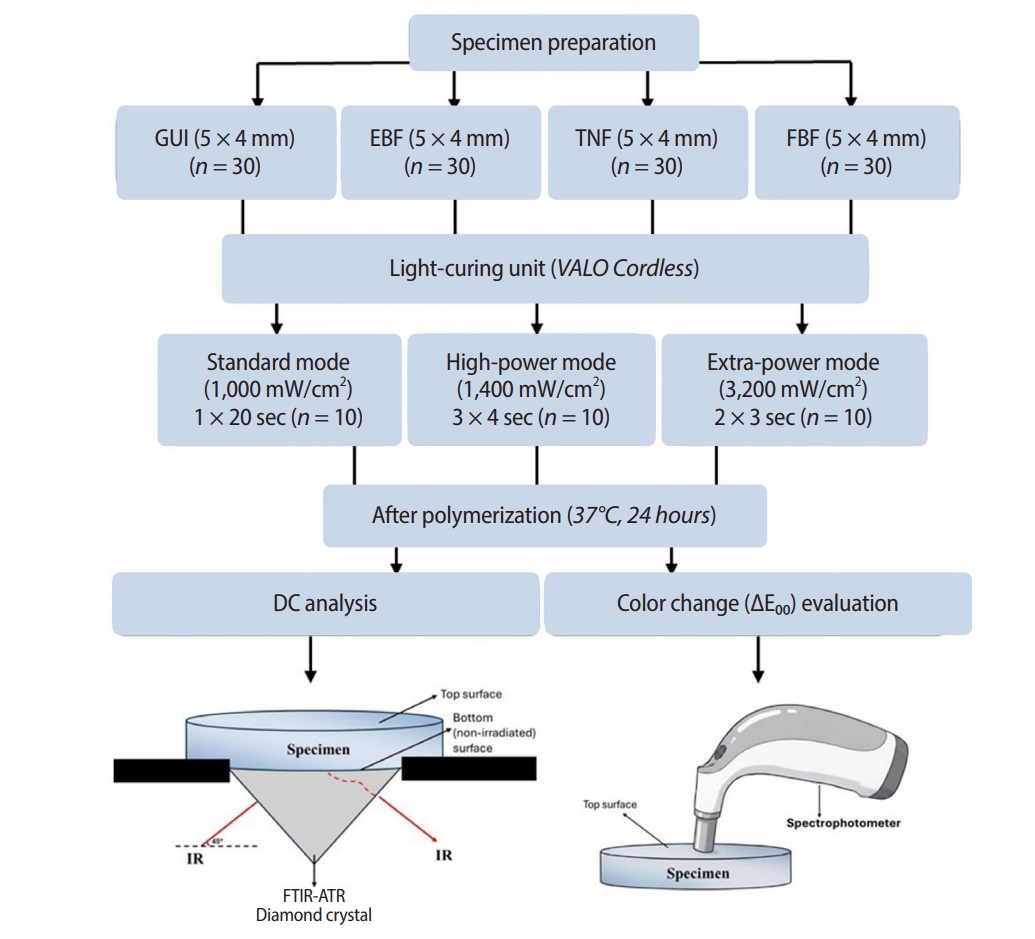
This study aims to evaluate the impact of light-curing modes on the degree of conversion (DC) and color change (CIEDE2000 color difference, ΔE00) of different flowable composite resins. Methods: A total of 120 specimens (n = 10 per group) were prepared from four materials: G-aenial Universal Injectable (GUI; GC), Estelite Bulk Fill Flow (EBF; Tokuyama Dental), Tetric N-Flow Bulk Fill (TNF; Ivoclar Vivadent), and Filtek Bulk Fill Flowable (FBF; 3M ESPE). The specimens were polymerized using an LED light-curing unit under three light-curing modes: standard (1,000 mW/cm², 20 seconds), high-power (1,400 mW/cm², 3 × 4 seconds), and extra-power (3,200 mW/cm², 2 × 3 seconds). The DC was quantified using Fourier transform infrared spectroscopy. Color measurements were performed at baseline, and on the 7th and 14th days after immersion in coffee, using a spectrophotometer. The ΔE₀₀ was calculated using the CIEDE2000 formula. The data were analyzed using IBM SPSS software (α = 0.05). Results: Material type and light-curing mode significantly affected both the DC and ΔE00 (p < 0.05). All composites exhibited lower DC in the extra-power mode compared to the high-power mode. On day 14, the extra-power mode resulted in significantly greater discoloration in TNF, FBF, and EBF compared to the high-power mode (p < 0.05). Conclusions: Although curing with the extra-power mode saves time, it may result in a lower DC and an increase in ΔE00 in flowable composite resin.
- 911 View
- 65 Download

- Discoloration of teeth due to different intracanal medicaments
- Farzaneh Afkhami, Sadaf Elahy, Alireza Mahmoudi Nahavandi, Mohamad Javad Kharazifard, Aidin Sooratgar
- Restor Dent Endod 2019;44(1):e10. Published online February 12, 2019
- DOI: https://doi.org/10.5395/rde.2019.44.e10
- Retraction in: Restor Dent Endod 2026;51(3):e38
- 5,410 View
- 72 Download
- 14 Crossref
- Colorimetric comparison of single layered dental composite with double layered dental composite
- Young-Sang Song, Ja-Hyun Kim, Bin-Na Lee, Ji-Hyun Jang, Hoon-Sang Chang, Yun-Chan Hwang, Won-Mann Oh, In-Nam Hwang
- Restor Dent Endod 2012;37(2):84-89. Published online May 18, 2012
- DOI: https://doi.org/10.5395/rde.2012.37.2.84
-
Abstract
PDF
Objectives This study analyzed the difference in color caused by different thickness in enamel layer of composite resins when applied with single and layering placement technique, and evaluated if the results agreed with the shade guide from the manufacturers to verify reliability of the color matching process of the manufacturers.
Materials and Methods For single composite resin samples, 6 mm diameter and 4 mm thickness cylindrical samples were fabricated using Ceram-X mono (DENTSPLY DeTrey) and CIE L*a*b* values were measured with spectrophotometer. Same process was done for layering composite resin samples, making 3 dentinal shade samples, 4 mm thickness, for each shade using Ceram-X duo (DENTSPLY DeTrey) and enamel shade resins were layered in 2 mm thickness and CIE L*a*b* values were measured. These samples were ground to 0.2 mm thickness each time, and CIE L*a*b* values were measured to 1 mm thickness of enamel shade resin.
Results Color difference (ΔE*) between single and layering composite resin was 1.37 minimum and 10.53 maximum when layering thicknesses were between 1 mm and 2 mm and 6 out of 10 same shade groups suggested by manufacturer showed remarkable color difference at any thickness (ΔE* > 3.3).
Conclusion When using Ceram-X mono and duo for composite resin restoration, following the manufacturer's instructions for choosing the shade is not appropriate, and more accurate information for Ceram-X duo is needed on the variation and expression of the shades depending on the thickness of the enamel.
-
Citations
Citations to this article as recorded by
- Improvement of mechanical strength and water repellency of Hanji (traditional Korean paper) through acetylation in supercritical CO2
Seungmok Shin, Hwi-Sung Lee, Hee Suk Woo, Mulugeta G. Aregay, Tae Jun Yoon, Youn-Woo Lee
The Journal of Supercritical Fluids.2022; 190: 105735. CrossRef - Color Change in Tooth Induced by Various Calcium Silicate-Based Pulp-Capping Materials
Jiyoon Jeon, Namki Choi, Seonmi Kim
THE JOURNAL OF THE KOREAN ACADEMY OF PEDTATRIC DENTISTRY.2021; 48(3): 280. CrossRef
- Improvement of mechanical strength and water repellency of Hanji (traditional Korean paper) through acetylation in supercritical CO2
- 2,169 View
- 5 Download
- 2 Crossref
Review Article
- Understanding of the color in composite resin
- Jeong-Won Park
- J Korean Acad Conserv Dent 2011;36(4):271-279. Published online July 31, 2011
- DOI: https://doi.org/10.5395/JKACD.2011.36.4.271
-
Abstract
PDF
In clinic, esthetic restoration of a defective natural tooth with composite resin is challenging procedure and needs complete understanding of the color of tooth itself and materials used. The optical characteristics of the composites are different because the chemical compositions and microstructures are not same.
This review provided basic knowledge of the color and the color measurement devices, and analyze the color of the natural tooth. Further, the accuracy of the shade tab, color of the composite resins before and after curing, effect of the water, food and bleaching agent, and translucency, opalescence, and fluorescence effects were evaluated.
-
Citations
Citations to this article as recorded by- Translucency and Masking Ability of Single-Shade Opaque Composite Resin Compared with Conventional Opaque Composite Resins According to Thickness
Junmo Jeong, Jongsoo Kim, Joonhaeng Lee, Jongbin Kim, Jisun Shin, Miran Han
THE JOURNAL OF THE KOREAN ACADEMY OF PEDTATRIC DENTISTRY.2025; 52(3): 312. CrossRef - Evaluation of Color Stability according to Shade of Temporary Crown Resin Using Digital Spectrophotometer: In Vitro Study
Hye-min Ku, Mi-Kyoung Jun
Journal of Dental Hygiene Science.2022; 22(3): 139. CrossRef - Effects of Children's Drinks on the Color Stability of Strip and Zirconia crown
Ilyong Jeong, Seoksoon Yi, Haney Lee, Daewoo Lee, Yeonmi Yang, Jaegon Kim
THE JOURNAL OF THE KOREAN ACADEMY OF PEDTATRIC DENTISTRY.2017; 44(3): 306. CrossRef - Color and Translucency of Multi-Shade Layered Composites
Chang-Ha Lee, Bum-Soon Lim, In-Bog Lee
Korean Journal of Dental Materials.2016; 43(4): 369. CrossRef - Color evaluation of composite resin using dental colorimeter according to the specimen size
Ji-Hye Jung, 심규리, 장훈상
Oral Biology Research.2016; 40(4): 198. CrossRef
- Translucency and Masking Ability of Single-Shade Opaque Composite Resin Compared with Conventional Opaque Composite Resins According to Thickness
- 3,802 View
- 75 Download
- 5 Crossref
Original Articles
- Color difference of the dental composites measured by different color measuring instruments
- Su-Jung Park, Eun-Young Noh, Hyun-Gu Cho, Yun-Chan Hwang, Won-Mann Oh, In-Nam Hwang
- J Korean Acad Conserv Dent 2009;34(3):199-207. Published online May 31, 2009
- DOI: https://doi.org/10.5395/JKACD.2009.34.3.199
-
Abstract
PDF
The objective of this study was to evaluate the effect of color measuring instrument by measuring the color of dental composite resins.
Nine shade light cured composite resin disks were prepared (diameter : 15 mm, thickness : 4 mm). CIE L*a*b* color scale of each disk was measured with 3 different types of spectrophotometer [MiniScan XE plus (Model 4000S, Hunter Lab, USA), CM-3500d (Minolta, Japan) and Specbos 2100 Miniature VIS Reflection spectrometer (Serial No: 319416, JETI Technishe VIS Instrumentic GmbH, Germany)]. Miniscan XE Plus and CM-3500d using identical measuring geometry with different size of viewing aperture. But Specbos 2100 using different measuring geometry.
Within the limitation of this study, there were color difference (ΔE*) from 2.4 to 7.8 between Miniscan XE Plus and CM-3500d, but L*, a*, b* values showed the high correlation. However, there were great color difference (ΔE*) in the extent of about 20 between instruments with the different measuring geometry.
Therefore, color scale measured by color measuring instrument should be used as a relative value rather than an absolute value in the field of dentistry.
-
Citations
Citations to this article as recorded by- Color Change in Tooth Induced by Various Calcium Silicate-Based Pulp-Capping Materials
Jiyoon Jeon, Namki Choi, Seonmi Kim
THE JOURNAL OF THE KOREAN ACADEMY OF PEDTATRIC DENTISTRY.2021; 48(3): 280. CrossRef - Effects of the color components of light-cured composite resin before and after polymerization on degree of conversion and flexural strength
Ji-A Yoo, Byeong-Hoon Cho
Journal of Korean Academy of Conservative Dentistry.2011; 36(4): 324. CrossRef
- Color Change in Tooth Induced by Various Calcium Silicate-Based Pulp-Capping Materials
- 2,062 View
- 5 Download
- 2 Crossref
- Effect of the bleaching light on whitening efficacy
- Jong-Hyun Park, Hye-Jin Shin, Deok-Young Park, Se-Hee Park, Jin-Woo Kim, Kyung-Mo Cho
- J Korean Acad Conserv Dent 2009;34(2):95-102. Published online March 31, 2009
- DOI: https://doi.org/10.5395/JKACD.2009.34.2.095
-
Abstract
PDF
The aim of this study was to evaluate the influence of light energy on the tooth whitening effect of bleaching agent in vitro. Extracted human mandibular molars were sectioned to 2 fragments(mesial, distal) and lingual portions of crown were used in this study. All specimens were stained using a red wine for 24 hours and immersed in artificial saliva. Specimens divided into four groups, group 1 and 2 light-activated by LumaCool (LED, LumaLite, Inc., Spring Valley, USA), group 3 and 4 light-activated by FlipoWhite2 (Plasma acr lamp, Lokki, Australia). Group 1 and 3 bleached with LumaWhite(LumaLite, Inc., Spring Valley, USA), group 2 and 4 bleached with Polaoffice(SDI, Victoria, Australia). Bleaching treatment performed during 10 minutes every 24 hours and repeated 6 times. During bleaching treatment , distal fragments was light-activated(L) but mesial fragments was not(NL). Shade assessment employed before and after bleaching treatment using spectrophotometer. The results of the change in shade was compared and analysed between NL and L by using paired-sample T test with 95% level of confidence.
There were no significant differences between NL and L with a few exceptions. In group 2, a* value more change in L, in group 3, b* value more change in L, in group 4, a* value less change in L. After bleaching, L* value and ΔE increased in all groups and the value of a* and b* decreased in all groups.
Within the limitation of this test conditions, the results of this study indicate that the light energy has no obvious improving impact on the tooth whitening effect of a bleaching agent.
-
Citations
Citations to this article as recorded by- Development of a Piezoelectric Ultrasonic Tooth-whitening Apparatus
Young-Jin Lee, Jong-Hoo Paik, Jeong-Bae Lee, Seung-Jae Choi
Transactions on Electrical and Electronic Materials.2013; 14(5): 268. CrossRef - Clinical assessment of whitening efficacy and safety of in-office tooth whitening system containing 15% hydrogen peroxide with or without light activation
Young-Suk Noh, Young-Jee Rho, Yeon-Jee Yoo, Hyang-Ok Lee, Sang-Min Lim, Hyun-Jeong Kweon, Yeun Kim, Seong-Yeon Park, Hee-Young Yoon, Jung-Hyun Lee, Chan-Hee Lee, So-Ram Oh, Kee-Yeon Kum
Journal of Korean Academy of Conservative Dentistry.2011; 36(4): 306. CrossRef
- Development of a Piezoelectric Ultrasonic Tooth-whitening Apparatus
- 2,395 View
- 6 Download
- 2 Crossref
- Color changes in composite resins exposed to xenon lamp
- Young-Gon Cho, Jeong-Il Seo, Soo-Mee Kim, Jin-Ho Jeong, Young-Gon Lee
- J Korean Acad Conserv Dent 2003;28(3):195-202. Published online May 31, 2003
- DOI: https://doi.org/10.5395/JKACD.2003.28.3.195
-
Abstract
PDF
The purpose of this study was to evaluate the color changes of the composite resin resulting from xenon lamp exposure in different environments. Composite resin (Z 250 ; shade A1, A2, A3, A3.5, and A4) were applied in a cylindrical metal mold. Seventy five specimens according to environments of exposure were made as follows;
Group I: aluminum foiling of the specimens in the air at 37℃ for 1 day and 7 days.
Group II: exposure of xenon lamp to the specimens in the air at 37℃ for 1 day and 7 days.
Group III: exposure of xenon lamp to the specimens in distilled water at 37℃ for 1 day and 7 days.
The color characteristics (L*,a*,b*) of the specimens before and after exposure of xenon lamp were measured by spectrophotometer and the total color differences (ΔE*) were computed.
The results obtained were as follows:
In all groups except A1 shade of group III, the ΔE* values presented below 2.0, and group III showed the highest ΔE* values followed by group II and group I in a decreasing order(p<0.05).
In all shades and groups, the more the exposure time of xenon lamp and the lighter the shade were, the higher the tendency for discoloration (p<0.05).
The composite resins which was exposed to xenon lamp in the distilled water was more discolored than those in the air (p<0.05).
The major changes of composite resins which were exposed to xenon lamp in the air were an increase in yellowness through a positive shift of the b* value, and those in the distilled water were an increase in darkness and yellowness through a negative shift of the L* value and a positive shift of the b* value.
-
Citations
Citations to this article as recorded by- Effects of the color components of light-cured composite resin before and after polymerization on degree of conversion and flexural strength
Ji-A Yoo, Byeong-Hoon Cho
Journal of Korean Academy of Conservative Dentistry.2011; 36(4): 324. CrossRef
- Effects of the color components of light-cured composite resin before and after polymerization on degree of conversion and flexural strength
- 2,114 View
- 1 Download
- 1 Crossref
 First
First Prev
Prev


