
Articles
- Page Path
- HOME > Restor Dent Endod > Volume 36(1); 2011 > Article
- Basic Research Comparison of apical transportation and change of working length in K3, NRT AND PROFILE rotary instruments using transparent resin block
- Min-Jung Yoon, DDS, MSD1, Min-Ju Song, DDS, MSD2, Su-Jung Shin, DDS, MSD2, Euiseong Kim, DDS, MSD, PhD1
-
2011;36(1):-65.
DOI: https://doi.org/10.5395/JKACD.2011.36.1.59
Published online: January 31, 2011
1Department of Conservative Dentistry, Yonsei University College of Dentistry, Seoul, Korea.
2Department of Conservative Dentistry, Gangnam Severance Hospital, Yonsei University College of Dentistry, Seoul, Korea.
- Correspondence to Euiseong Kim, DDS, MSD, PhD. Associate Professor, Department of Conservative Dentistry, Yonsei University College of Dentistry, 250 Seongsanno, Seodaemun-gu, Seoul, Korea 120-752. TEL,+82-2-2228-8701; FAX,+82-2-313-7575; andyendo@yuhs.ac
• Received: November 24, 2010 • Revised: January 5, 2011 • Accepted: January 7, 2011
Copyright © 2011 Korean Academy of Conservative Dentistry
- 1,506 Views
- 2 Download
- 1 Crossref


Tables & Figures
REFERENCES
Citations
Citations to this article as recorded by 
- A comparison of dimensional standard of several nickel-titanium rotary files
Ki-Won Kim, Kyung-Mo Cho, Se-Hee Park, Ki-Yeol Choi, Bekir Karabucak, Jin-Woo Kim
Restorative Dentistry & Endodontics.2014; 39(1): 7. CrossRef
 ePub Link
 Cite
CiteComparison of apical transportation and change of working length in K3, NRT AND PROFILE rotary instruments using transparent resin block
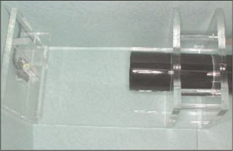


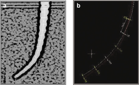
Figure 1
Photograph of a jig that maintains a constant distance between a X-ray tube and a resin block.
Figure 2
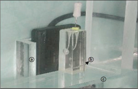
Photograph of a resin block and a digital sensor. (a) Holding part of digital radiographic sensor. (b) Cartesian system. (c) Table for constant position of resin block.
Figure 3
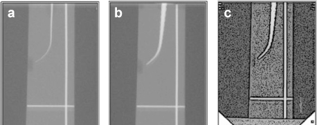
(a) Initial X-ray. (b) X-ray after canal enlargement. (c) Image of the x-ray after canal enlargement processed by Adobe photoshop.
Figure 4
(a) Image of central axis. (b) Measurement of apical transportation using AuotCAD 2000.
Figure 1
Figure 2
Figure 3
Figure 4
Comparison of apical transportation and change of working length in K3, NRT AND PROFILE rotary instruments using transparent resin block
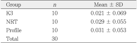
Mean curvature and working length in three groups
W.L, working length; n, sample size.
Change of working length (mm) in three groups
SD, standard deviation; n, sample size.
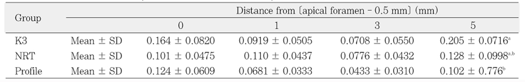
Mean and SD of transportation (mm) at different apical level
a,b, Groups with statistically significant differences (p < 0.05); SD, standard deviation.
Table 1
Mean curvature and working length in three groups
W.L, working length;
Table 2
Change of working length (mm) in three groups
SD, standard deviation;
Table 3
Mean and SD of transportation (mm) at different apical level
a,b, Groups with statistically significant differences (

