
Search
- Page Path
- HOME > Search
Research Articles
- Ex vivo comparative analysis of retrievability among four calcium silicate-based sealers for regaining apical patency
- Darian Shomali, Timothy Kirkpatrick, Sang Won Kwak, Hyeon-Cheol Kim, Ji Wook Jeong
- Restor Dent Endod 2026;51(1):e3. Published online January 14, 2026
- DOI: https://doi.org/10.5395/rde.2026.51.e3
-
 Abstract
 PDF
PDF PubReader
 ePub
ePub - Objectives
Efficient retrievability is a key requirement for endodontic sealers. This study evaluated the retrievability of four different calcium silicate-based sealers (CSS).
Methods
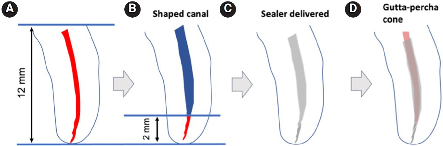
A total of 153 single-rooted human teeth with straight canals were decoronated to a standardized working length of 12 mm. The canals were negotiated to working length using K files up to size 15/.02, followed by rotary instrumentation up to 35/.04, 2 mm short of working length. The teeth were randomly assigned to five groups: NeoSEALER Flo (NEO; Avalon Biomed), Ceraseal (CS; Meta Biomed), Endosequence BC Sealer (BC; Brasseler USA), AH Plus Bioceramic Sealer (AHB; Dentsply Sirona), and a negative control group. Sealer application and obturation with a 35/.04 gutta-percha cone were performed. After incubation at 37°C in 100% humidity for 7 days, retreatment was performed until apical patency was obtained, with retrievability assessed by regaining apical patency. One-way analysis of variance and Tukey contrast test were used to determine whether there was a significant difference among the four different CSS (p < 0.05).
Results
Success rates in regaining apical patency were NEO (79.4%), CS (37.0%), BC (50.0%), and AHB (69.7%). NEO demonstrated the highest retrievability, while CS had the lowest (p < 0.01).
Conclusions
The type of CSS used has a considerable impact on retreatment difficulty. Among the tested sealers, Neo- SEALER Flo showed the highest retrievability, making it the most retrievable CSS in terms of retreatment efficacy.
- 1,777 View
- 159 Download

- Effect of moisture and pH on setting time and microhardness of three premixed calcium silicate-based root canal sealers: an in vitro experimental study
- Sooyoun Kim
- Restor Dent Endod 2025;50(4):e41. Published online November 28, 2025
- DOI: https://doi.org/10.5395/rde.2025.50.e41
-
Abstract
PDFPubReaderePub
- Objectives
The study aimed to investigate how environmental conditions impact the setting time and microhardness of premixed calcium silicate-based sealers.
Methods
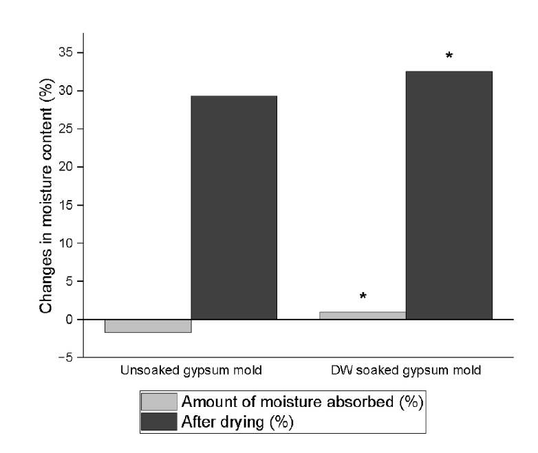
The setting time and microhardness of three sealers (Endoseal MTA [MARUCHI], One-Fil [MEDICLUS], and Well-Root ST [VERICOM]) were evaluated under four environmental conditions: unsoaked, distilled water-soaked, phosphate-buffered saline-soaked, and pH 5-soaked gypsum molds (n = 12/group/condition). The setting time was measured with Gilmore needles, and microhardness was assessed using a Vickers tester after 3 days. Welch’s analysis of variance and Games-Howell post hoc tests were used for statistical analysis.
Results
The sealer type and environmental conditions significantly influenced setting time and microhardness (p < 0.001). The initial and final setting times were the shortest in the unsoaked samples. For Endoseal MTA and One-Fil, the unsoaked condition exhibited significantly shorter setting times than the soaked conditions. Well-Root ST exhibited significantly longer setting times in acidic conditions. Surface microhardness was highest in the unsoaked group (p < 0.001). Among the soaked groups, the phosphate-buffered saline-soaked group had the lowest hardness for Endoseal MTA, whereas the pH 5-soaked group exhibited the lowest hardness for One-Fil and Well-Root ST. Endoseal MTA consistently demonstrated a lower microhardness than the other sealers (p < 0.001).
Conclusions
Moisture, pH, and solution chemistry influenced the setting time and microhardness of premixed calcium silicate sealers. Although acidic conditions generally prolong the setting time and reduce hardness, the effects vary based on the sealers used and the setting environment. -
Citations
Citations to this article as recorded by
- Setting Characteristics, Solubility, Bioactivity and Interaction with Dentin of Four Calcium Silicate-Based Endodontic Sealers
Areti Dimitra Vrochari, Anastasia Agrafioti, Maria Dimitriadi, George Eliades
Journal of Functional Biomaterials.2026; 17(4): 192. CrossRef
- Setting Characteristics, Solubility, Bioactivity and Interaction with Dentin of Four Calcium Silicate-Based Endodontic Sealers
- 1,972 View
- 97 Download
- 1 Web of Science
- 1 Crossref
- Comparison of remineralization in caries-affected dentin using calcium silicate, glass ionomer cement, and resin-modified glass ionomer cement: an in vitro study
- Kwanchanok Youcharoen, Onwara Akkaratham, Papichaya Intajak, Pipop Saikaew, Sirichan Chiaraputt
- Restor Dent Endod 2025;50(4):e37. Published online November 14, 2025
- DOI: https://doi.org/10.5395/rde.2025.50.e37
-
Abstract
PDFPubReaderePub
- Objectives
This study evaluated the ability of calcium silicate cement (CSC) as a remineralizing agent compared with conventional glass ionomer cement (GIC) and resin-modified GIC (RMGIC) to remineralize artificial caries-affected dentin.
Methods
Twenty-five class V cavities were prepared on extracted human third molars. Twenty teeth underwent artificial caries induction. The remaining five teeth with sound dentin serve as the positive control. The twenty demineralized teeth were subdivided into four groups (n = 5): carious dentin without restoration (negative control [NC]), carious dentin restored with CSC (Biodentine, Septodont), carious dentin restored with GI (Fuji IX, GC Corporation), and carious dentin restored with RMGIC (Fuji II LC, GC Corporation). Following restoration, the specimens were stored in artificial saliva for 7 days. The elastic modulus was evaluated by a nanoindentation test. The mineral composition was analyzed by scanning electron microscopy-energy-dispersive X-ray spectroscopy (SEM-EDX), and the mineral composition at the dentin-material interface.
Results
CSC had a higher modulus of elasticity compared to GI, RMGI, and NC groups (p < 0.05). Higher calcium and phosphorus content was observed under CSC restorations, as indicated by SEM-EDX examination, which may lead to better remineralization.
Conclusions
Compared to GI and RMGI, CSC showed the best remineralization and mechanical reinforcement in caries-affected dentin, indicating CSC for use in minimally invasive restorative dentistry. -
Citations
Citations to this article as recorded by- Comparison of mineral precipitation, elemental release, pH change and cytotoxicity of calcium-silicate cements and an experimental resin-modified glass ionomer cement containing bioactive glass
Wisitsin Potiprapanpong , Parichart Naruphontjirakul, Naruporn Monmaturapoj, Siriporn Tanodekaew, Somruethai Channasanon, Arnit Toneluck, Somying Patntirapong, Piyaphong Panpisut
Biomaterial Investigations in Dentistry.2026; 13: 337. CrossRef
- Comparison of mineral precipitation, elemental release, pH change and cytotoxicity of calcium-silicate cements and an experimental resin-modified glass ionomer cement containing bioactive glass
- 2,561 View
- 245 Download
- 1 Crossref
- Does the use of different root canal sealers and adhesive resin cements impact the bond strength of glass fiber posts?
- Ália Regina Neves de Paula Porto, Rudá França Moreira, Felipe Gonçalves Belladonna, Victor Talarico Leal Vieira, Emmanuel João Nogueira Leal da Silva
- Restor Dent Endod 2025;50(3):e29. Published online August 29, 2025
- DOI: https://doi.org/10.5395/rde.2025.50.e29
-
Abstract
PDFPubReaderePub
- Objectives
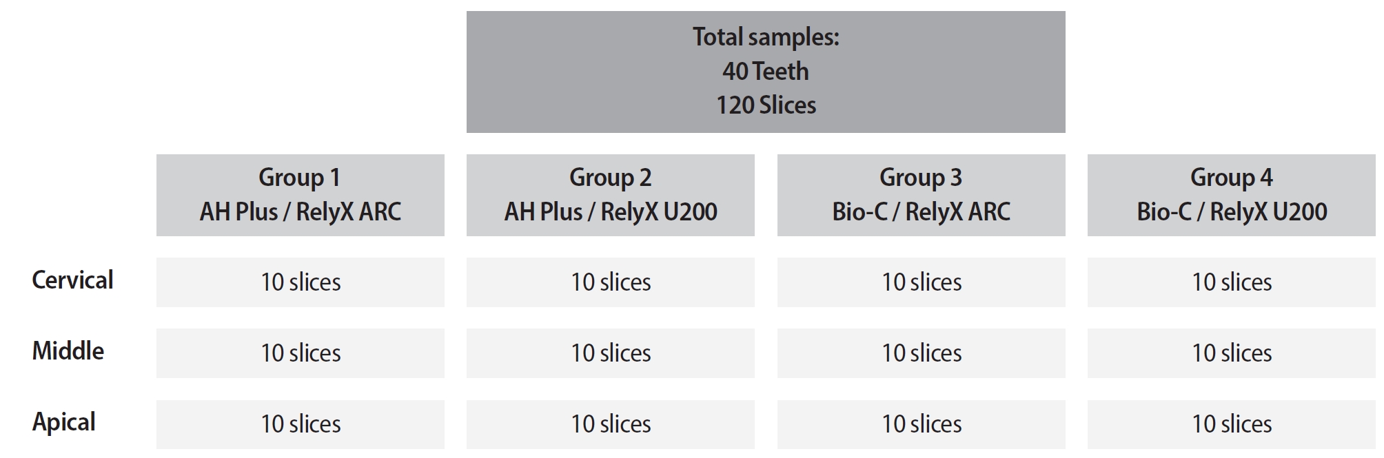
This study aimed to assess the influence of two endodontic sealers on the bond strength of glass fiber posts using conventional and self-adhesive resin cement through a push-out test. Methods: Forty central human incisors were randomly divided into four groups (n = 10) based on sealer (epoxy resin- based or calcium silicate-based) and cement (conventional and self-adhesive resin) types: AH Plus (Dentsply De- Trey)/RelyX ARC (3M ESPE), AH Plus/RelyX U200 (3M ESPE), Bio-C Sealer (Angelus)/RelyX ARC, and Bio-C Sealer/RelyX U200. After canal filling and post cementation, roots were sectioned to obtain one specimen per root third. A pushout test and failure pattern assessment were conducted, with bond strength analyzed using the one-way analysis of variance and Tukey test. Results: AH Plus/RelyX ARC showed the highest bond strength values, with a significant difference in the middle third. The most common failure was mixed (55%), while adhesive failures made up 45%, with 23.5% at the cement/post interface and 21.5% at the cement/dentin interface. Conclusions: AH Plus/RelyX ARC provided the highest bond strength values for glass fiber posts to dentin. -
Citations
Citations to this article as recorded by- Effect of Endodontic Sealers on the Bond Strength of Glass Fibre Posts: A Systematic Review
Thiago Bessa Marconato Antunes, Juliana D. Bronzato, Vanessa Gallego Arias Pecorari, Jennifer Santos Pereira, Talita Tartari, Adriana de Jesus Soares, Brenda P. F. A. Gomes, Marina Angélica Marciano
Australian Endodontic Journal.2026;[Epub] CrossRef
- Effect of Endodontic Sealers on the Bond Strength of Glass Fibre Posts: A Systematic Review
- 2,659 View
- 162 Download
- 1 Web of Science
- 1 Crossref
- Cleaning protocols to enhance bond strength of fiberglass posts on root canals filled with bioceramic sealer: an in vitro comparative study
- Thiago Bessa Marconato Antunes, Juliana Delatorre Bronzato, Joice Graciani, Ana Cristina Padilha Janini, Rocharles Cavalcante Fontenele, Francisco Haiter Neto, Brenda Paula Figueiredo de Almeida Gomes, Marina Angélica Marciano da Silva
- Restor Dent Endod 2025;50(2):e20. Published online May 21, 2025
- DOI: https://doi.org/10.5395/rde.2025.50.e20
-
Abstract
PDFPubReaderePub
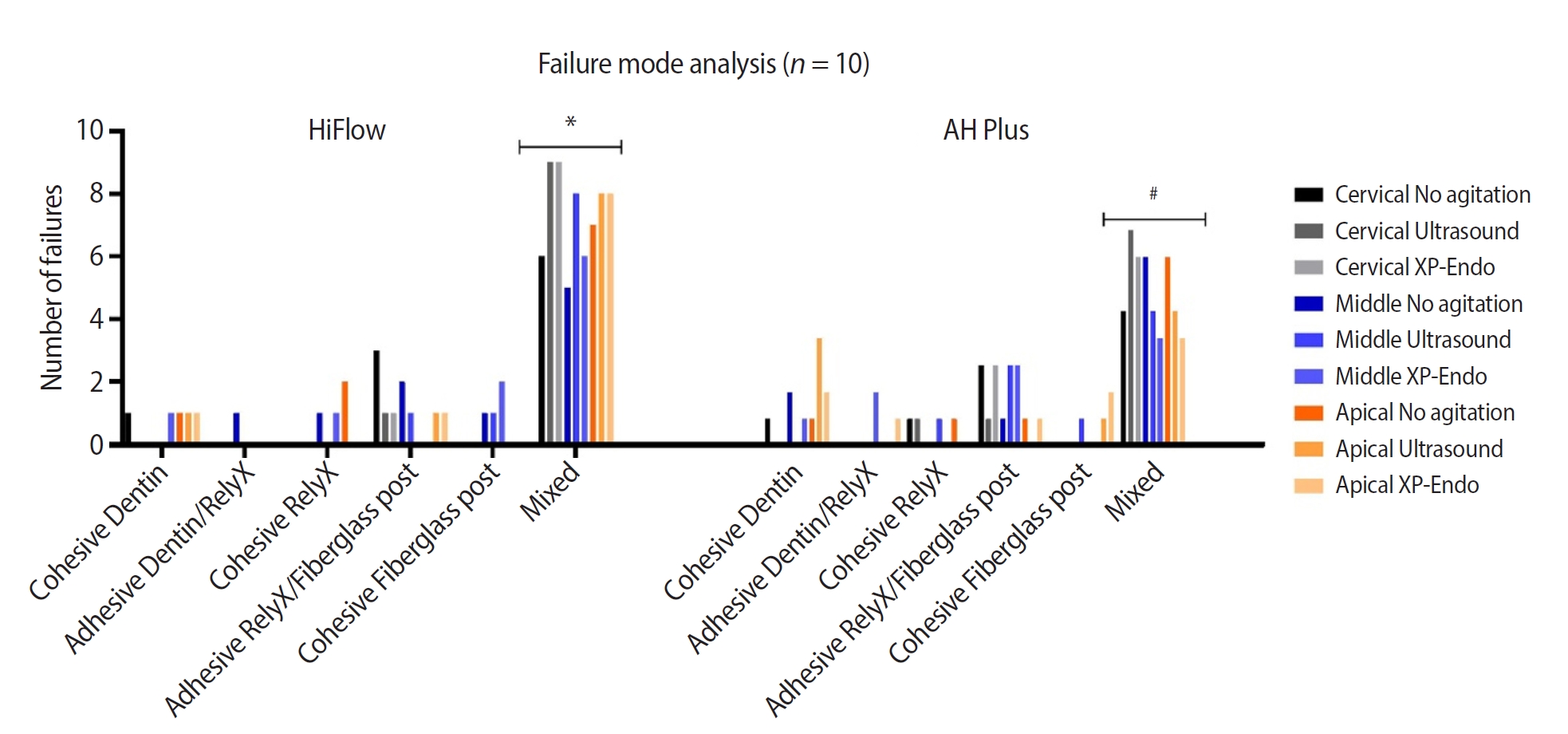
- Objectives
This study aimed to evaluate whether the agitation protocols using ultrasonic inserts or the XP-endo Finisher R file improved the removal of two different endodontic sealer remnants and the bond strength of fiberglass posts to dentin.
Methods
Seventy-two human teeth were selected. The canals were prepared with Reciproc 50 and Easy ProDesign 30/.10 and root filled according to the endodontic sealer groups: AH Plus or EndoSequence BC Sealer HiFlow. The samples were kept at 37ºC and 95% humidity for 28 days. During the post space preparation, the obturation was removed with Largo burs, and the groups were divided according to the irrigant agitation protocols (n = 12): no agitation, agitation with R1-Clearsonic associated with E1-Irrisonic ultrasonic inserts, or agitation with XP-endo Finisher R file. The fiberglass posts were cemented with RelyX ARC. The roots were sectioned into slices and submitted to the push-out test. Micro-computed tomography analysis was used to check the effectiveness of irrigating solution agitation in the elimination of remnants.
Results
The cleaning protocols with agitation were more effective in increasing the bond strength of posts to dentin for both sealer groups compared to non-agitation (p < 0.05). There was no difference between the same cleaning protocols for the different sealers. Among the different thirds, there was no statistical difference for the same sealer in the different cleaning protocols (p > 0.05).
Conclusions
Both agitation protocols effectively clean root-filled canals sealed with resin-based and calcium silicate-based sealers during fiberglass post space preparation. These protocols result in improved bond strength compared to non-agitation methods. -
Citations
Citations to this article as recorded by- Cleaning efficacy and bond interaction of glycine-based air polishing and glass microparticles abrasion on dentin impregnated with premixed bioceramic sealer
Ândresson Aurélio Fernandes Martins, Maria Carolina Sidonio Alves, Bruno Martins Maciel, José Rodolfo Estruc Verbicário, João Felipe Besegato, Wilfredo Gustavo Escalante-Otárola, Milton Carlos Kuga
International Journal of Adhesion and Adhesives.2026; 147: 104277. CrossRef - Effect of Endodontic Sealers on the Bond Strength of Glass Fibre Posts: A Systematic Review
Thiago Bessa Marconato Antunes, Juliana D. Bronzato, Vanessa Gallego Arias Pecorari, Jennifer Santos Pereira, Talita Tartari, Adriana de Jesus Soares, Brenda P. F. A. Gomes, Marina Angélica Marciano
Australian Endodontic Journal.2026;[Epub] CrossRef
- Cleaning efficacy and bond interaction of glycine-based air polishing and glass microparticles abrasion on dentin impregnated with premixed bioceramic sealer
- 4,818 View
- 239 Download
- 2 Web of Science
- 2 Crossref
Review Article
- Success rate of direct pulp capping on permanent teeth using bioactive materials: a systematic review and meta-analysis of randomized clinical trials
- Karem Paula Pinto, Gabriela Ribeiro da Silva, Cláudio Malizia Alves Ferreira, Luciana Moura Sassone, Emmanuel João Nogueira Leal da Silva
- Restor Dent Endod 2024;49(4):e34. Published online September 6, 2024
- DOI: https://doi.org/10.5395/rde.2024.49.e34
-
Abstract
PDF
 Supplementary MaterialPubReaderePub
 This systematic review and meta-analysis aimed to evaluate the success rate of direct pulp capping (DPC) on permanent teeth, comparing the use of MTA with calcium hydroxide and calcium silicate-based cements. A systematic search was carried out in 4 databases until July 2023. The selection was based on PICOS criteria and only randomized clinical trials were included. The risk of bias was assessed using RoB-2 tool, and meta-analyses were performed using RevMan 5.3 software. The overall quality of evidence was determined using the GRADE tool. Thirteen studies were included. Meta-analyses indicated significantly higher success rate for DPC using MTA compared to calcium hydroxide, while no significant difference was observed between MTA and Biodentine, showing a success rate from 80% to 100% even after 3 years of follow-up. Five studies were classified as having high risk of bias and the GRADE assessment revealed low certainty of evidence. DPC is highly effective for permanent teeth when using MTA or Biodentine. There is a need for future well-designed randomized clinical trials to evaluate the efficacy of DPC using newer bioceramic materials.
-
Citations
Citations to this article as recorded by- Physicochemical effects of nano type-B bone substitute on pulp protective cement formulations
Njwan Fadhel SHEHAB
Dental Materials Journal.2026; 45(1): 92. CrossRef - Photobiomodulation-assisted pulp capping using nano-hydroxyapatite and mineral trioxide aggregate: Report of two cases
Priya Pal, Rhythm Bains, Promila Verma, Vivek Kumar Bains
Journal of Healthcare Research and Education.2026; 2: 2. CrossRef - Histological Tissue Response to Calcium Silicate-Based Cements Assessed in Human Tooth Culture Models: A Systematic Review
Alberto Cabrera-Fernández, Hebertt Gonzaga dos Santos Chaves, Aránzazu Díaz-Cuenca, Juan J. Segura-Egea, Jenifer Martín-González, João Peça, Diana B. Sequeira, João Miguel Marques dos Santos
Journal of Functional Biomaterials.2026; 17(2): 78. CrossRef - Translational Pathways for Smart and Bioactive Dental Biomaterials: Biocompatibility Standards, Sterilisation, Sustainability and Regulation
Katarzyna Chojnacka, Marcin Mikulewicz
MedComm – Biomaterials and Applications.2026;[Epub] CrossRef - Decision-ready evidence for vital pulp therapy: a network meta-analysis of bioactive materials in mature permanent teeth
Firas Elmsmari, Reem B. Abdelsayed, Qamar Albasoumi, Tareq Aljafarawi, Swadheena Patro, Ajinkya M. Pawar
Frontiers in Dental Medicine.2026;[Epub] CrossRef - Comparative Evaluation of Platelet-Rich Fibrin and Mineral Trioxide Aggregate in the Direct Pulp Capping of Carious Exposures: A Randomized Clinical Trial
Geeta Asthana, Rajashree Tamuli, Sadhna Manglani, Saloni Mandhare, Ragini Kulkarni, Anooja Mathirat, Parneet Kaur, P Jyothirmayee, Surabhi Landge
Cureus.2026;[Epub] CrossRef - Indian Association of Conservative Dentistry and Endodontics consensus statement on deep caries management
Deepak Kumar Sharma, R. S. Mohan Kumar, Shishir Singh, Suparna Ganguly Saha, Meenal Nithin Gulve, Dipali Y. Shah, Sathish Abraham, Shruthi Nagaraja, Raksha Bhat
Journal of Conservative Dentistry and Endodontics.2025; 28(8): 714. CrossRef
- Physicochemical effects of nano type-B bone substitute on pulp protective cement formulations
- 22,181 View
- 683 Download
- 4 Web of Science
- 7 Crossref
Research Articles
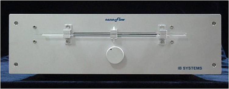
- Nanoleakage of apical sealing using a calcium silicate-based sealer according to canal drying methods
- Yoon-Joo Lee, Kyung-Mo Cho, Se-Hee Park, Yoon Lee, Jin-Woo Kim
- Restor Dent Endod 2024;49(2):e20. Published online April 19, 2024
- DOI: https://doi.org/10.5395/rde.2024.49.e20
-
Abstract
PDFPubReaderePub
Objectives This study investigated the nanoleakage of root canal obturations using calcium silicate-based sealer according to different drying methods.
Materials and Methods Fifty-two extracted mandibular premolars with a single root canal and straight root were selected for this study. After canal preparation with a nickel-titanium rotary file system, the specimens were randomly divided into 4 groups according to canal drying methods (1: complete drying, 2: blot drying/distilled water, 3: blot drying/NaOCl, 4: aspiration only). The root canals were obturated using a single-cone filling technique with a calcium silicate–based sealer. Nanoleakage was evaluated using a nanoflow device after 24 hours, 1 week, and 1 month. Data were collected twice per second at the nanoscale and measured in nanoliters per second. Data were statistically analyzed using the Kruskal-Wallis and Mann–Whitney
U -tests (p < 0.05).Results The mean flow rate measured after 24 hours showed the highest value among the time periods in all groups. However, the difference in the flow rate between 1 week and 1 month was not significant. The mean flow rate of the complete drying group was the highest at all time points. After 1 month, the mean flow rate in the blot drying group and the aspiration group was not significantly different.
Conclusions Within the limitations of this study, the canal drying method had a significant effect on leakage and sealing ability in root canal obturations using a calcium silicate-based sealer. Thus, a proper drying procedure is critical in endodontic treatment.
-
Citations
Citations to this article as recorded by- Effects of interactions between sealers and irrigants on the physicochemical and surface characteristics of endodontic sealers
Hye-In Kim, Yeon-Ju You, Yemi Kim, Jin-Woo Kim, Young-Eun Jang
Clinical Oral Investigations.2026;[Epub] CrossRef - Assessment of Shear Bond Strength of MTA and ApaCal ART Liners to a Universal Bonding Agent: An In Vitro Study
Angel Kurian, Banibrata Lahiri, Archana A Thomas, Gautham Anilkumar, Fawaz Pullishery, Reshma Raju
The Journal of Contemporary Dental Practice.2026; 27(3): 254. CrossRef
- Effects of interactions between sealers and irrigants on the physicochemical and surface characteristics of endodontic sealers
- 3,210 View
- 122 Download
- 1 Web of Science
- 2 Crossref
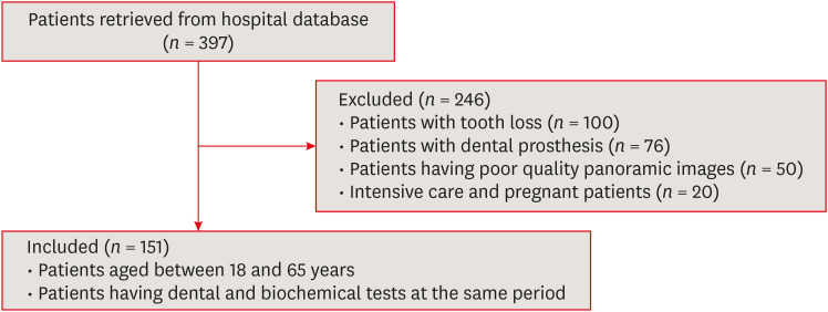
- Pulp stones: any relevance with the levels of serum calcium, parathyroid hormone, vitamin D and uric acid
- Ceyda Gürhan, Ercan Saruhan
- Restor Dent Endod 2024;49(2):e17. Published online March 26, 2024
- DOI: https://doi.org/10.5395/rde.2024.49.e17
-
Abstract
PDFPubReaderePub
Objectives This study evaluated the effect of serum calcium, parathyroid hormone (PTH), vitamin D, and uric acid levels on pulp stone formation.
Materials and Methods Patients who were admitted to the Muğla Sıtkı Koçman University, Faculty of Dentistry, Department of Oral and Maxillofacial Radiology for dental complaints were registered. Among these patients, individuals who had routine biochemical tests at the same period in the Outpatient Clinics of Muğla Sıtkı Koçman University Training and Research Hospital were included in the study. The patients with at least 1 pulp stone on panoramic radiographs recorded as the “pulp stone group” while patients without any pulp stones were the “control group”. Demographic data and serum levels of calcium, PTH, vitamin D, and uric acid were retrospectively evaluated in both groups. Student
t -test or Mann-WhitneyU test was used to evaluate the differences between the groups.Results Among 151 patients, dental pulp stone was detected in 53.6% of patients, and 82.7% of these patients were female. Female sex and pulp stone formation were significantly associated (
p = 0.001). The mean age of the pulp stone group was 43.9, while it was 39.9 in the control group, without any significant correlation between age and pulp stone (p > 0.05). Similarly, there were no significant differences in serum levels of PTH, vitamin D, uric acid and calcium between groups (p > 0.05).Conclusions According to the present study, the effect of dental factors rather than systemic factors should be considered primarily in pulp stone formation.
-
Citations
Citations to this article as recorded by- A novel deep learning-based pipeline architecture for pulp stone detection on panoramic radiographs
Ceyda Gürhan, Hasan Yiğit, Selim Yılmaz, Cihat Çetinkaya
Oral Radiology.2025; 41(2): 285. CrossRef - Vitamin D deficiency and oral health: a systematic review of literature
Saida Ziada, Aws Wishahe, Najet Mabrouk, Souad Sahtout
BMC Oral Health.2025;[Epub] CrossRef - Association between pulp stones and systemic diseases: a retrospective study using digital panoramic radiographs in a Turkish population
Buket Beytaş Alğan, Mustafa Murat Koçak, Sibel Koçak, Baran Can Sağlam
BMC Oral Health.2025;[Epub] CrossRef
- A novel deep learning-based pipeline architecture for pulp stone detection on panoramic radiographs
- 4,504 View
- 116 Download
- 4 Web of Science
- 3 Crossref
Case Report
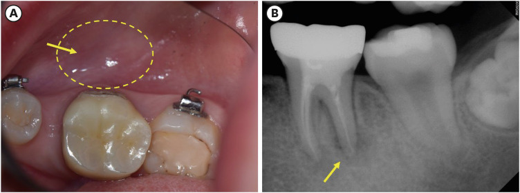
- Garre’s osteomyelitis of the mandible managed by nonsurgical re-endodontic treatment
- Heegyun Kim, Jiyoung Kwon, Hyun-Jung Kim, Soram Oh, Duck-Su Kim, Ji-Hyun Jang
- Restor Dent Endod 2024;49(2):e13. Published online March 18, 2024
- DOI: https://doi.org/10.5395/rde.2024.49.e13
-
Abstract
PDFPubReaderePub
Chronic osteomyelitis with proliferative periostitis, known as Garre’s osteomyelitis, is a type of osteomyelitis characterized by a distinctive gross thickening of the periosteum of bones. Peripheral reactive bone formation can be caused by mild irritation or infection. Garre’s osteomyelitis is usually diagnosed in children and young adults, and the mandible is more affected than the maxilla. The following is a case report of a 12-year-old female patient with Garre’s osteomyelitis of the mandible due to an infection of a root canal-treated tooth. Without surgical intervention, the patient’s symptoms were relieved through nonsurgical root canal re-treatment with long-term calcium hydroxide placement. A cone-beam computed tomography image obtained 6 months after treatment completion displayed complete healing of the periapical lesion and resolution of the peripheral reactive buccal bone. Due to the clinical features of Garre's osteomyelitis, which is characterized by thickening of the periosteum, it can be mistaken for other diseases such as fibrous dysplasia. It is important to correctly diagnose Garre's osteomyelitis based on its distinctive clinical features to avoid unnecessary surgical intervention, and it can lead to minimally invasive treatment options.
-
Citations
Citations to this article as recorded by- Endodontic Intervention in Chronic Osteomyelitis With Proliferative Periostitis: A Rare Case Report and Scoping Review
Gabriel Lima Braz, Ana Paula Neutzling Gomes, Lisandrea Rocha Schardosim, Nadia de Souza Ferreira, Jose Francisco Gomez-Sosa
Case Reports in Dentistry.2026;[Epub] CrossRef - How Clinical and Radiological Findings in Chronic Mandibular Osteomyelitis Do Not Always Correlate: Diagnostic Dilemmas in Dental-Related Bone Inflammations
Kamil Nelke, Ömer Uranbey, Ece Gülbağ, Büşra Ekinci, Burcu Gürsoytrak, Angela Rosa Caso, Michał Gontarz, Maciej Janeczek, Piotr Kuropka, Maciej Dobrzyński
Diagnostics.2026; 16(10): 1427. CrossRef - Focal osteomyelitis with proliferative periostitis
Zarah Yakoob
South African Dental Journal.2025; 79(09): 508. CrossRef - Garré’s osteomyelitis of the mandible in an adolescent: a case report
Wiem Feki, Imen Haddar, Marwa Bahloul, Zeineb Mnif, Thouraya Kammoun, Ines Maaloul
Journal of Medical Case Reports.2025;[Epub] CrossRef - Garré’s Chronic Sclerosing Osteomyelitis: An Overview of Clinical and Radiologic Features
Mohamed Fadil, Ayman Farouki, Rachida Saouab, Hassan En-nouali, Jamal El Fenni, Zakariya Toufga
Oxford Medical Case Reports.2025;[Epub] CrossRef
- Endodontic Intervention in Chronic Osteomyelitis With Proliferative Periostitis: A Rare Case Report and Scoping Review
- 9,904 View
- 220 Download
- 4 Web of Science
- 5 Crossref
Research Articles
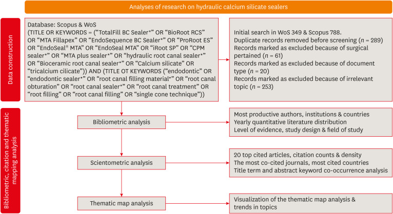
- A scientometric, bibliometric, and thematic map analysis of hydraulic calcium silicate root canal sealers
- Anastasios Katakidis, Konstantinos Kodonas, Anastasia Fardi, Christos Gogos
- Restor Dent Endod 2023;48(4):e41. Published online November 13, 2023
- DOI: https://doi.org/10.5395/rde.2023.48.e41
-
Abstract
PDFPubReaderePub
Objectives This scientometric and bibliometric analysis explored scientific publications related to hydraulic calcium silicate-based (HCSB) sealers used in endodontology, aiming to describe basic bibliometric indicators and analyze current research trends.
Materials and Methods A comprehensive search was conducted in Web of Science and Scopus using specific HCSB sealer and general endodontic-related terms. Basic research parameters were collected, including publication year, authorship, countries, institutions, journals, level of evidence, study design and topic of interest, title terms, author keywords, citation counts, and density.
Results In total, 498 articles published in 136 journals were retrieved for the period 2008–2023. Brazil was the leading country, and the universities of Bologna in Italy and Sao Paolo in Brazil were represented equally as leading institutions. The most frequently occurring keywords were “calcium silicate,” “root canal sealer MTA-Fillapex,” and “biocompatibility,” while title terms such as “calcium,” “sealers,” “root,” “canal,” “silicate based,” and “endodontic” occurred most often. According to the thematic map analysis, “solubility” appeared as a basic theme of concentrated research interest, and “single-cone technique” was identified as an emerging, inadequately developed theme. The co-occurrence analysis revealed 4 major clusters centered on sealers’ biological and physicochemical properties, obturation techniques, retreatability, and adhesion.
Conclusions This analysis presents bibliographic features and outlines changing trends in HCSB sealer research. The research output is dominated by basic science articles scrutinizing the biological and specific physicochemical properties of commonly used HCSB sealers. Future research needs to be guided by studies with a high level of evidence that utilize innovative, sophisticated technologies.
-
Citations
Citations to this article as recorded by- Top 100 Most Cited Articles on Antibiotics in Endodontics: A Bibliometric Analysis
Hajar Albanyan, Mohammed Asseery, Haitham Alahmari, Ikram Ul Haq, Ali Alaqla
Journal of Endodontics.2026; 52(3): 345. CrossRef - Top-cited articles on vital pulp therapy for irreversible pulpitis in permanent teeth: a bibliometric analysis
İkbal Sena Çelebi Keskin, Ferda Karabay
Odontology.2026;[Epub] CrossRef - Agri-Food Sector: Contemporary Trends, Possible Gaps, and Prospective Directions
José Roberto Herrera Cantorani, Meire Ramalho de Oliveira, Luiz Alberto Pilatti, Thales Botelho de Sousa
Metrics.2025; 2(1): 3. CrossRef - Scientific mapping of experimental research on solar cookers: Global trends, evolution, and future directions
Flavio Odoi-Yorke, Bismark Baah, Richard Opoku
Solar Energy Advances.2025; 5: 100093. CrossRef - Bibliometric analysis of the GentleWave system: trends, collaborations, and research gaps
Raimundo Sales de Oliveira Neto, Thais de Moraes Souza, João Vitor Oliveira de Amorim, Thaine Oliveira Lima, Guilherme Ferreira da Silva, Rodrigo Ricci Vivan, Murilo Priori Alcalde, Marco Antonio Hungaro Duarte
Restorative Dentistry & Endodontics.2025; 50(2): e17. CrossRef - A Scientometric Review of Practical Applications in Quantum Natural Language Processing (QNLP): Trends, Gaps, and Research Opportunities
Victor R. Silva, Fábio R. Barbosa, Jasson C. Silva, Francisco J. Santos, Ricardo A. L. Rabelo, Joel J. P. C. Rodrigues
IEEE Access.2025; 13: 210169. CrossRef - A bibliometric analysis of global research trend and progress on Dy doped materials
Sangeeta Kadyan, Manju Nain, Ashima Makhija, Poonam Punia, Anil Ohlan, Sajjan Dahiya, R. Punia, A.S. Maan
Journal of Alloys and Compounds Communications.2024; 3: 100006. CrossRef - Comparative bioactivity and immunomodulatory potential of the new Bioroot Flow and AH Plus Bioceramic sealer: An in vitro study on hPDLSCs
José Luis Sanz, Sergio López-García, David García-Bernal, Francisco Javier Rodríguez-Lozano, Leopoldo Forner, Adrián Lozano, Laura Murcia
Clinical Oral Investigations.2024;[Epub] CrossRef - Analyzing collaboration and impact: A bibliometric review of four highly published authors’ research profiles on collaborative maps
Willy Chou, Julie Chi Chow
Medicine.2024; 103(28): e38686. CrossRef
- Top 100 Most Cited Articles on Antibiotics in Endodontics: A Bibliometric Analysis
- 3,599 View
- 51 Download
- 7 Web of Science
- 9 Crossref
- Influence of access cavity design on calcium hydroxide removal using different cleaning protocols: a confocal laser scanning microscopy study
- Seda Falakaloğlu, Merve Yeniçeri Özata, Betül Güneş, Emmanuel João Nogueira Leal Silva, Mustafa Gündoğar, Burcu Güçyetmez Topal
- Restor Dent Endod 2023;48(3):e25. Published online July 24, 2023
- DOI: https://doi.org/10.5395/rde.2023.48.e25
-
Abstract
PDFPubReaderePub
Objectives The purpose of this study was to evaluate the influence of endodontic access cavities design on the removal of calcium hydroxide medication of the apical third of mandibular incisor root canal walls and dentinal tubules with different cleaning protocols: EDDY sonic activation, Er,Cr:YSGG laser-activated irrigation, or conventional irrigation with IrriFlex.
Materials and Methods Seventy-eight extracted human mandibular incisors were assigned to 6 experimental groups (
n = 13) according to the endodontic access cavity and cleaning protocol for calcium hydroxide removal: traditional access cavity (TradAC)/EDDY; ultraconservative access cavity performed in the incisal edge (UltraAC.Inc)/EDDY; TradAC/Er,Cr:YSGG; UltraAC.Inc/Er,Cr:YSGG; TradAC/IrriFlex; or UltraAC.Inc/IrriFlex. Confocal laser scanning microscopy images were used to measure the non-penetration percentage, maximum residual calcium hydroxide penetration depth, and penetration area at 2 and 4 mm from the apex. Data were statistically analyzed using Shapiro-Wilk and WRS2 package for 2-way comparison of non-normally distributed parameters (depth of penetration, area of penetration, and percentage of non-penetration) according to cavity and cleaning protocol with the significance level set at 5%.Results The effect of cavity and cleaning protocol interactions on penetration depth, penetration area and non-penetration percentage was not found statistically significant at 2 and 4 mm levels (
p > 0.05).Conclusions The present study demonstrated that TradAC or UltraAC.Inc preparations with different cleaning protocols in extracted mandibular incisors did not influence the remaining calcium hydroxide at 2 and 4 mm from the apex.
-
Citations
Citations to this article as recorded by- Retrievability of triple antibiotic paste from simulated grooves in molars: Impact of access design and agitation technique
Oorja Nanda, Vineeta Nikhil
Endodontology.2026; 38(2): 193. CrossRef - Effect of Apical Preparation Size and Preparation Taper on Smear Layer Removal Using Two Different Irrigation Needles: A Scanning Electron Microscopy Study
Rania Lebbos, Naji Kharouf, Deepak Mehta, Jamal Jabr, Cynthia Kamel, Roula El Hachem, Youssef Haikel, Marc Krikor Kaloustian
European Journal of Dentistry.2025; 19(03): 678. CrossRef - Combination of Chitosan Nanoparticles, EDTA, and Irrigation Activation Enhances TGF-β1 Release from Dentin: A Laboratory Study
Sıla Nur Usta, Emre Avcı, Ayşe Nur Oktay, Cangül Keskin
Journal of Endodontics.2025; 51(8): 1081. CrossRef
- Retrievability of triple antibiotic paste from simulated grooves in molars: Impact of access design and agitation technique
- 3,747 View
- 80 Download
- 3 Web of Science
- 3 Crossref
Case Report
- Successful nonsurgical treatment of type II dens invaginatus with 5 root canals using a self-adjusting file: a case report
- George Táccio de Miranda Candeiro, Antônio Sérgio Teixeira de Menezes, Ana Carolina Saldanha de Oliveira, Flávio Rodrigues Ferreira Alves
- Restor Dent Endod 2023;48(2):e17. Published online April 27, 2023
- DOI: https://doi.org/10.5395/rde.2023.48.e17
-
Abstract
PDFPubReaderePub
The present report describes the endodontic treatment of an Oehlers type II dens invaginatus in a maxillary lateral incisor with 5 root canals, an extremely rare condition. Apical periodontitis and related symptoms were noted. Cone-beam computed tomography was used to aid the diagnosis, reveal tooth morphology, and assist in canal location. The pulp chamber was carefully accessed, and the root canals were explored under magnification. All root canals were prepared with an R25 Reciproc Blue system and sodium hypochlorite (NaOCl) irrigation. After initial preparation, a self-adjusting file (SAF) with NaOCl and ethylenediaminetetraacetic acid was used to complement the disinfection. Additionally, calcium hydroxide medication was applied. Vertical compaction was used to fill the canals with a calcium silicate-based endodontic sealer and gutta-percha. After 12 months, the patient exhibited healing of the periapical region, absence of symptoms, and normal dental function. In conclusion, this nonsurgical treatment protocol was successful in promoting the cure of apical periodontitis. Both complementary disinfection with an SAF and use of calcium hydroxide medication should be considered when choosing the best treatment approach for dens invaginatus with very complex anatomy.
-
Citations
Citations to this article as recorded by- Non-surgical endodontic management of dens invaginatus type II in an immature maxillary lateral incisor using a bioceramic apical plug: A 3-year follow-up case report
Yahya Raja Alharbi, Qayed Saad Alharbi, Shaul Hameed Kolarkodi
Saudi Endodontic Journal.2026; 16(1): 115. CrossRef
- Non-surgical endodontic management of dens invaginatus type II in an immature maxillary lateral incisor using a bioceramic apical plug: A 3-year follow-up case report
- 2,691 View
- 80 Download
- 1 Crossref
Research Articles
- Calcium-doped zinc oxide nanocrystals as an innovative intracanal medicament: a pilot study
- Gabriela Leite de Souza, Thamara Eduarda Alves Magalhães, Gabrielle Alves Nunes Freitas, Nelly Xiomara Alvarado Lemus, Gabriella Lopes de Rezende Barbosa, Anielle Christine Almeida Silva, Camilla Christian Gomes Moura
- Restor Dent Endod 2022;47(4):e38. Published online October 4, 2022
- DOI: https://doi.org/10.5395/rde.2022.47.e38
-
Abstract
PDFPubReaderePub
Objectives This study investigated the cytotoxicity, radiopacity, pH, and dentinal tubule penetration of a paste of 1.0% calcium-doped zinc oxide nanocrystals (ZnO:1.0Ca) combined with propylene glycol (PRG) or polyethylene glycol and propylene glycol (PEG-PRG).
Materials and Methods The pastes were prepared by mixing calcium hydroxide [Ca(OH)2] or ZnO:1.0Ca with PRG or a PEG-PRG mixture. The pH was evaluated after 24 and 96 hours of storage in deionized water. Digital radiographs were acquired for radiopacity analysis and bubble counting of each material. The materials were labeled with 0.1% fluorescein and applied to root canals, and images of their dentinal tubule penetration were obtained using confocal laser scanning microscopy. RAW264.7 macrophages were placed in different dilutions of culture media previously exposed to the materials for 24 and 96 hours and tested for cell viability using the MTT assay. Analysis of variance and the Tukey test (
α = 0.05) were performed.Results ZnO:1.0Ca materials showed lower viability at 1:1 and 1:2 dilutions than Ca(OH)2 materials (
p < 0.0001). Ca(OH)2 had higher pH values than ZnO:1.0Ca at 24 and 96 hours, regardless of the vehicle (p < 0.05). ZnO:1.0Ca pastes showed higher radiopacity than Ca(OH)2 pastes (p < 0.01). No between-material differences were found in bubble counting (p = 0.0902). The ZnO:1.0Ca pastes had a greater penetration depth than Ca(OH)2 in the apical third (p < 0.0001).Conclusions ZnO:1.0Ca medicaments presented higher penetrability, cell viability, and radiopacity than Ca(OH)2. Higher values of cell viability and pH were present in Ca(OH)2 than in ZnO:1.0Ca.
-
Citations
Citations to this article as recorded by-
Nano calcium zincate-assisted synthesis of benzo[
d
]thiazol-2-yl phenylisoxazoles: quantum computational,
in silico
molecular docking simulations and DNA interaction
A. K. Smitha, V. Srinivasa Murthy, B. Vinay Kumar, M. Sennappan, A. H. Shridhar, Lohit Naik, K. Yogendra, N. Madhusudhana
Nucleosides, Nucleotides & Nucleic Acids.2026; 45(3): 261. CrossRef - Nanomaterial-Enhanced Dentistry: A Clinical Perspective
Selvam Manoj, Radhakrishnan Sreena, Rajkumar Divya, Starlin Ebinesh, Shenbagaraman Akshaya, Srikumar Sugantha Angel, Arputharaj Joseph Nathanael
ACS Biomaterials Science & Engineering.2025; 11(8): 4671. CrossRef
-
Nano calcium zincate-assisted synthesis of benzo[
d
]thiazol-2-yl phenylisoxazoles: quantum computational,
in silico
molecular docking simulations and DNA interaction
- 2,595 View
- 33 Download
- 2 Web of Science
- 2 Crossref
- Effects of calcium silicate cements on neuronal conductivity
- Derya Deniz-Sungur, Mehmet Ali Onur, Esin Akbay, Gamze Tan, Fügen Daglı-Comert, Taner Cem Sayın
- Restor Dent Endod 2022;47(2):e18. Published online March 7, 2022
- DOI: https://doi.org/10.5395/rde.2022.47.e18
-
Abstract
PDFPubReaderePub
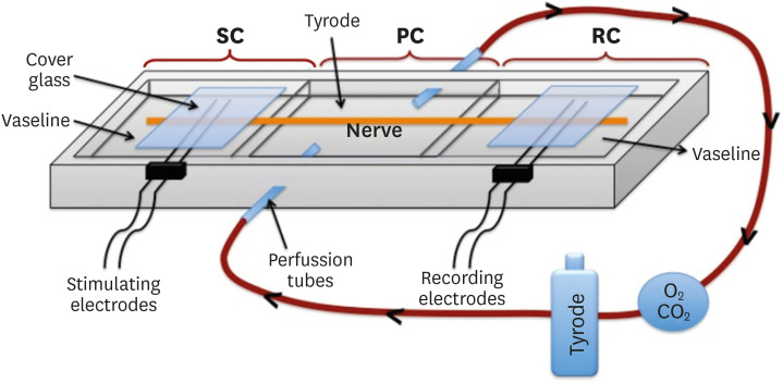
Objectives This study evaluated alterations in neuronal conductivity related to calcium silicate cements (CSCs) by investigating compound action potentials (cAPs) in rat sciatic nerves.
Materials and Methods Sciatic nerves were placed in a Tyrode bath and cAPs were recorded before, during, and after the application of test materials for 60-minute control, application, and recovery measurements, respectively. Freshly prepared ProRoot MTA, MTA Angelus, Biodentine, Endosequence RRM-Putty, BioAggregate, and RetroMTA were directly applied onto the nerves. Biopac LabPro version 3.7 was used to record and analyze cAPs. The data were statistically analyzed.
Results None of the CSCs totally blocked cAPs. RetroMTA, Biodentine, and MTA Angelus caused no significant alteration in cAPs (
p > 0.05). Significantly lower cAPs were observed in recovery measurements for BioAggregate than in the control condition (p < 0.05). ProRoot MTA significantly but transiently reduced cAPs in the application period compared to the control period (p < 0.05). Endosequence RRM-Putty significantly reduced cAPs.Conclusions Various CSCs may alter cAPs to some extent, but none of the CSCs irreversibly blocked them. The usage of fast-setting CSCs during apexification or regeneration of immature teeth seems safer than slow-setting CSCs due to their more favorable neuronal effects.
-
Citations
Citations to this article as recorded by- Endodontic Sealers and Innovations to Enhance Their Properties: A Current Review
Anna Błaszczyk-Pośpiech, Natalia Struzik, Maria Szymonowicz, Przemysław Sareło, Maria Wiśniewska-Wrona, Kamila Wiśniewska, Maciej Dobrzyński, Magdalena Wawrzyńska
Materials.2025; 18(18): 4259. CrossRef
- Endodontic Sealers and Innovations to Enhance Their Properties: A Current Review
- 2,070 View
- 29 Download
- 1 Web of Science
- 1 Crossref
- Comparative analysis of bond strength to root dentin and compression of bioceramic cements used in regenerative endodontic procedures
- Maykely Naara Morais Rodrigues, Kely Firmino Bruno, Ana Helena Gonçalves de Alencar, Julyana Dumas Santos Silva, Patrícia Correia de Siqueira, Daniel de Almeida Decurcio, Carlos Estrela
- Restor Dent Endod 2021;46(4):e59. Published online November 9, 2021
- DOI: https://doi.org/10.5395/rde.2021.46.e59
-
Abstract
PDFPubReaderePub
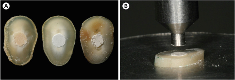
Objectives This study compared the Biodentine, MTA Repair HP, and Bio-C Repair bioceramics in terms of bond strength to dentin, failure mode, and compression.
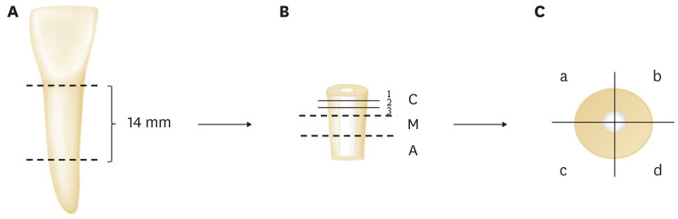
Materials and Methods Fifty-four slices obtained from the cervical third of 18 single-rooted human mandibular premolars were randomly distributed (
n = 18). After insertion of the bioceramic materials, the push-out test was performed. The failure mode was analyzed using stereomicroscopy. Another set of cylindrically-shaped bioceramic samples (n = 10) was prepared for compressive strength testing. The normality of data distribution was analyzed using the Shapiro-Wilk test. The Kruskal-Wallis and Friedman tests were used for the push-out test data, while compressive strength was analyzed with analysis of variance and the Tukey test, considering a significance level of 0.05.Results Biodentine presented a higher median bond strength value (14.79 MPa) than MTA Repair HP (8.84 MPa) and Bio-C Repair (3.48 MPa), with a significant difference only between Biodentine and Bio-C Repair. In the Biodentine group, the most frequent failure mode was mixed (61%), while in the MTA Repair HP and Bio-C Repair groups, it was adhesive (94% and 72%, respectively). Biodentine showed greater resistance to compression (29.59 ± 8.47 MPa) than MTA Repair HP (18.68 ± 7.40 MPa) and Bio-C Repair (19.96 ± 3.96 MPa) (
p < 0.05).Conclusions Biodentine showed greater compressive strength than MTA Repair HP and Bio-C Repair, and greater bond strength than Bio-C Repair. The most frequent failure mode of Biodentine was mixed, while that of MTA Repair HP and Bio-C Repair was adhesive.
-
Citations
Citations to this article as recorded by- Evaluation of marginal adaptation and bond strength of apical root canal plugs using different bioceramic cements
Michel Sena Fernandes Faria Lima, Alberto Nogueira da Gama Antunes, Kênia Maria Pereira Soares de Toubes, Fábio Fernandes Borém Bruzinga, Camila de Sousa Caneschi, Luís Fernando dos Santos Alves Morgan, Frank Ferreira Silveira
BMC Oral Health.2026;[Epub] CrossRef - Assessment of Release of Calcium Ions from Apical Plugs of Different Bioceramic Sealers in Regenerative Endodontics for Immature Permanent Teeth
Alok Dubey, Nischitha Naik, Bhavana Sujanamulk, Mushir Mulla, Munaz Mulla, Alkananda Sahoo, Mohamed T Salama, Murali Patla S Bhat
World Journal of Dentistry.2026; 17(1): 69. CrossRef - Obturation quality analysis of furcation perforations repaired with different magnifications and biomaterials
Mustafa Mert Tulgar, Yağmur Kılıç, Merve Işık Aydın, Ali Keleş
BMC Oral Health.2026;[Epub] CrossRef - Comparación de la resistencia compresiva entre el Agregado Trióxido Mineral y BiodentineTM en perforaciones de furca de molares inferiores permanentes
Jheymy Gerardo Huatuco-Granda, John Paul Torres-Navarro, Rosa Josefina Roncal-Espinoza
Revista Facultad de Odontología.2024;[Epub] CrossRef - Effects of different calcium-silicate based materials on fracture resistance of immature permanent teeth with replacement root resorption and osteoclastogenesis
Gabriela Leite de Souza, Gabrielle Alves Nunes Freitas, Maria Tereza Hordones Ribeiro, Nelly Xiomara Alvarado Lemus, Carlos José Soares, Camilla Christian Gomes Moura
Restorative Dentistry & Endodontics.2023;[Epub] CrossRef - Evaluation the Marginal Adaptation for the Bio C Repair and Other Root end Filling Material by Using Scanning Electron Microscope (A Comparative In Vitro Study)
Fatimah HAMADHİ, Zainab M.
Cumhuriyet Dental Journal.2023; 26(3): 261. CrossRef - Dentin Bond Strength of Calcium Silicate-Based Materials: A Systematic Review of In Vitro Studies
Natalia Radulica, José Luis Sanz, Adrián Lozano
Applied Sciences.2023; 14(1): 104. CrossRef - Evaluation Of The Push-out Bond Strength Of The Bio-C Repair And Compare It With The Mineral Trioxide Aggregate And Amalgam When Used As Root-end Filling Material
Fatimah R. Hammadi, Zainab M Abdul-Ameer
Dental Hypotheses.2023; 14(2): 62. CrossRef - Effect of different root canal irrigants on push-out bond strength of two novel root-end filling materials
Nada Omar, Rasha M. Abdelraouf, Tamer M. Hamdy
BMC Oral Health.2023;[Epub] CrossRef - Effect of irrigation systems on the bond strength of calcium-silicate-based cement used as pulp barrier in regenerative endodontic treatment
Cihan Hascizmeci, Burak Buldur
Journal of Adhesion Science and Technology.2023; 37(23): 3393. CrossRef
- Evaluation of marginal adaptation and bond strength of apical root canal plugs using different bioceramic cements
- 3,962 View
- 84 Download
- 6 Web of Science
- 10 Crossref
- Push-out bond strength and marginal adaptation of apical plugs with bioactive endodontic cements in simulated immature teeth
- Maria Aparecida Barbosa de Sá, Eduardo Nunes, Alberto Nogueira da Gama Antunes, Manoel Brito Júnior, Martinho Campolina Rebello Horta, Rodrigo Rodrigues Amaral, Stephen Cohen, Frank Ferreira Silveira
- Restor Dent Endod 2021;46(4):e53. Published online October 20, 2021
- DOI: https://doi.org/10.5395/rde.2021.46.e53
-
Abstract
PDFPubReaderePub
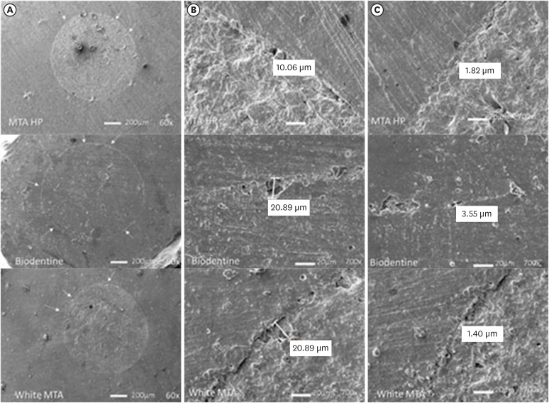
Objectives This study evaluates the bond strength and marginal adaptation of mineral trioxide aggregate (MTA) Repair HP and Biodentine used as apical plugs; MTA was used as reference material for comparison.
Materials and Methods A total of 30 single-rooted teeth with standardized, artificially created open apices were randomly divided into 3 groups (
n = 10 per group), according to the material used to form 6-mm-thick apical plugs: group 1 (MTA Repair HP); group 2 (Biodentine); and group 3 (white MTA). Subsequently, the specimens were transversely sectioned to obtain 2 (cervical and apical) 2.5-mm-thick slices per root. Epoxy resin replicas were observed under a scanning electron microscope to measure the gap size at the material/dentin interface (the largest and smaller gaps were recorded for each replica). The bond strength of the investigated materials to dentin was determined using the push-out test. The variable bond strengths and gap sizes were evaluated independently at the apical and cervical root dentin slices. Data were analyzed using descriptive and analytic statistics.Results The comparison between the groups regarding the variables' bond strengths and gap sizes showed no statistical difference (
p > 0.05) except for a single difference in the smallest gap at the cervical root dentin slice, which was higher in group 3 than in group 1 (p < 0.05).Conclusions The bond strength and marginal adaptation to root canal walls of MTA HP and Biodentine cement were comparable to white MTA.
-
Citations
Citations to this article as recorded by- Evaluation of marginal adaptation and bond strength of apical root canal plugs using different bioceramic cements
Michel Sena Fernandes Faria Lima, Alberto Nogueira da Gama Antunes, Kênia Maria Pereira Soares de Toubes, Fábio Fernandes Borém Bruzinga, Camila de Sousa Caneschi, Luís Fernando dos Santos Alves Morgan, Frank Ferreira Silveira
BMC Oral Health.2026;[Epub] CrossRef - Application of Biodentine for Apexification of Immature Teeth of Children: A Scoping Review
Liz M Gerard, Sumit Gaur
International Journal of Clinical Pediatric Dentistry.2025; 18(5): 573. CrossRef - Evaluation of the root dentin bond strength and intratubular biomineralization of a premixed calcium aluminate-based hydraulic bioceramic endodontic sealer
Yu-Na Lee, Min-Kyeong Kim, Hee-Jin Kim, Mi-Kyung Yu, Kwang-Won Lee, Kyung-San Min
Journal of Oral Science.2024; 66(2): 96. CrossRef - Managing Cracked Teeth with Root Extension: A Prospective Preliminary Study Using Biodentine™ Material
Kênia Maria Soares de Toubes, Isabella Sousa Corrêa, Regina Célia Lopes Valadares, Stephanie Quadros Tonelli, Fábio Fernandes Borém Bruzinga, Frank Ferreira Silveira, Dr Karthikeyan Ramalingam
International Journal of Dentistry.2024;[Epub] CrossRef - Marginal adaptation of customized gutta percha cone with calcium silicate based sealer versus MTA and biodentine apical plugs in simulated immature permanent teeth (an in vitro study)
Mary M. Mina, Sybel M. Moussa, Mahmoud R. Aboelseoud
BMC Oral Health.2024;[Epub] CrossRef - Comparative Evaluation of Push-Out Bond Strength of Conventional Mineral Trioxide Aggregate, Biodentine, a Modified Mineral Trioxide Aggregate, and Two Novel Antibacterial-Enhanced Mineral Trioxide Aggregates
Arokia Rajkumar Shancy Merlin, Vignesh Ravindran, Ganesh Jeevanandan, Rajalakshmanan Eswaramoorthy, Abirami Arthanari
Cureus.2024;[Epub] CrossRef - Push out bond strength of hydraulic cements used at different thicknesses
C. Ruiz Durán, Dra L. Gancedo-Caravia, V. Vera González, C. González Losada
BMC Oral Health.2023;[Epub] CrossRef - Effects of different calcium-silicate based materials on fracture resistance of immature permanent teeth with replacement root resorption and osteoclastogenesis
Gabriela Leite de Souza, Gabrielle Alves Nunes Freitas, Maria Tereza Hordones Ribeiro, Nelly Xiomara Alvarado Lemus, Carlos José Soares, Camilla Christian Gomes Moura
Restorative Dentistry & Endodontics.2023;[Epub] CrossRef
- Evaluation of marginal adaptation and bond strength of apical root canal plugs using different bioceramic cements
- 3,215 View
- 38 Download
- 9 Web of Science
- 8 Crossref
- Micro-computed tomographic evaluation of single-cone obturation with three sealers
- Sahar Zare, Ivy Shen, Qiang Zhu, Chul Ahn, Carolyn Primus, Takashi Komabayashi
- Restor Dent Endod 2021;46(2):e25. Published online April 16, 2021
- DOI: https://doi.org/10.5395/rde.2021.46.e25
-
Abstract
PDFPubReaderePub
Objectives This study used micro-computed tomography (µCT) to compare voids and interfaces in single-cone obturation among AH Plus, EndoSequence BC, and prototype surface pre-reacted glass ionomer (S-PRG) sealers and to determine the percentage of sealer contact at the dentin and gutta-percha (GP) interfaces.
Materials and Methods Fifteen single-rooted human teeth were shaped using ProTaper NEXT size X5 rotary files using 2.5% NaOCl irrigation. Roots were obturated with a single-cone ProTaper NEXT GP point X5 with AH Plus, EndoSequence BC, or prototype S-PRG sealer (
n = 5/group).Results The volumes of GP, sealer, and voids were measured in the region of 0–2, 2–4, 4–6, and 6–8 mm from the apex, using image analysis of sagittal µCT scans. GP volume percentages were: AH Plus (75.5%), EndoSequence BC (87.3%), and prototype S-PRG (94.4%). Sealer volume percentages were less: AH Plus (14.3%), EndoSequence BC (6.8%), and prototype S-PRG (4.6%). Void percentages were AH Plus (10.1%), EndoSequence BC (5.9%), and prototype S-PRG (1.0%). Dentin-sealer contact ratios of AH Plus, EndoSequence BC, and prototype S-PRG groups were 82.4% ± 6.8%, 71.6% ± 25.3%, and 70.2% ± 9.4%, respectively. GP-sealer contact ratios of AH Plus, EndoSequence BC, and prototype S-PRG groups were 65.6% ± 29.1%, 80.7% ± 25.8%, and 87.0% ± 8.6%, respectively.
Conclusions Prototype S-PRG sealer created a low-void obturation, similar to EndoSequence BC sealer with similar dentin-sealer contact (> 70%) and GP-sealer contact (> 80%). Prototype S-PRG sealer presented comparable filling quality to EndoSequence BC sealer.
-
Citations
Citations to this article as recorded by- Assessment of gap areas of root filling techniques in teeth with 3D-printed different configurations of C-shaped root canals: a micro-computed tomography study
Tuba Gok, Adem Gok, Haydar Onur Aciksoz
BMC Oral Health.2025;[Epub] CrossRef - Assessment of the quality of root canal filling using three different sealers: Micro-computed tomography and scanning electron microscope study
Loai Alsofi, Mohammed Yagmoor, Tariq AbuHaimed, Hassan Abed, Ehab Alshouibi, Rafif Mandura, Turki Bakhsh, Hanaa Ashkar, Mey Al-Habib
Saudi Endodontic Journal.2025; 15(2): 152. CrossRef - Comparative evaluation of bioactive calcium silicate coating on functionalized gutta-percha and its effect on bioceramic sealer wettability – An in vitro study
Bollineni Swetha, B. Devi Priya, K. Hanisha Reddy, G. Prasanthi, T. Murali Mohan, Dumpa Tejaswi
Journal of Conservative Dentistry and Endodontics.2025; 28(7): 613. CrossRef - Assessment of isthmus filling using two obturation techniques performed by students with different levels of clinical experience
Yang Yu, Chong-Yang Yuan, Xing-Zhe Yin, Xiao-Yan Wang
Journal of Dental Sciences.2024; 19(1): 169. CrossRef - Micro-CT determination of the porosity of two tricalcium silicate sealers applied using three obturation techniques
Jinah Kim, Kali Vo, Gurmukh S. Dhaliwal, Aya Takase, Carolyn Primus, Takashi Komabayashi
Journal of Oral Science.2024; 66(3): 163. CrossRef - Ex-vivo evaluation of clinically-set hydraulic sealers used with different canal dryness protocols and obturation techniques: a randomized clinical trial
Nawar Naguib Nawar, Mohamed Mohamed Elashiry, Ahmed El Banna, Shehabeldin Mohamed Saber, Edgar Schäfer
Clinical Oral Investigations.2024;[Epub] CrossRef - Hydraulic (Single Cone) Versus Thermogenic (Warm Vertical Compaction) Obturation Techniques: A Systematic Review
Haytham S Jaha
Cureus.2024;[Epub] CrossRef - Sealing ability of various endodontic sealers with or without ethylenediaminetetraacetic acid (EDTA) treatment on bovine root canal
Yusuke AIGAMI, Tomofumi SAWADA, Shunsuke SHIMIZU, Akiko ASANO, Mamoru NODA, Shinji TAKEMOTO
Dental Materials Journal.2024; 43(3): 420. CrossRef - A Literature Review of the Effect of Heat on the Physical-Chemical Properties of Calcium Silicate–Based Sealers
Israa Ashkar, José Luis Sanz, Leopoldo Forner, James Ghilotti, María Melo
Journal of Endodontics.2024; 50(8): 1044. CrossRef - Assessment of the Prevalence of Head Lice Infestation and Parents’ Attitudes Towards Its Management: A School-based Epidemiological Study in İstanbul, Türkiye
Özben Özden, İnci Timur, Hale Ezgi Açma, Duygu Şimşekli, Barış Gülerman, Özgür Kurt
Turkish Journal of Parasitology.2023; 47(2): 112. CrossRef - Calcium-doped zinc oxide nanocrystals as an innovative intracanal medicament: a pilot study
Gabriela Leite de Souza, Thamara Eduarda Alves Magalhães, Gabrielle Alves Nunes Freitas, Nelly Xiomara Alvarado Lemus, Gabriella Lopes de Rezende Barbosa, Anielle Christine Almeida Silva, Camilla Christian Gomes Moura
Restorative Dentistry & Endodontics.2022;[Epub] CrossRef - Micro‐CT assessment of gap‐containing areas along the gutta‐percha‐sealer interface in oval‐shaped canals
Gustavo De‐Deus, Gustavo O. Santos, Iara Zamboni Monteiro, Daniele M. Cavalcante, Marco Simões‐Carvalho, Felipe G. Belladonna, Emmanuel J. N. L. Silva, Erick M. Souza, Raphael Licha, Carla Zogheib, Marco A. Versiani
International Endodontic Journal.2022; 55(7): 795. CrossRef - A critical analysis of research methods and experimental models to study root canal fillings
Gustavo De‐Deus, Erick Miranda Souza, Emmanuel João Nogueira Leal Silva, Felipe Gonçalves Belladonna, Marco Simões‐Carvalho, Daniele Moreira Cavalcante, Marco Aurélio Versiani
International Endodontic Journal.2022; 55(S2): 384. CrossRef - Use of micro-CT to examine effects of heat on coronal obturation
Ivy Shen, Joan Daniel, Kali Vo, Chul Ahn, Carolyn Primus, Takashi Komabayashi
Journal of Oral Science.2022; 64(3): 224. CrossRef - Obturation of Root Canals By Vertical Condensation of Gutta-Percha – Benefits and Pitfalls
Calkovsky Bruno, Slobodnikova Ladislava, Bacinsky Martin, Janickova Maria
Acta Medica Martiniana.2021; 21(3): 103. CrossRef
- Assessment of gap areas of root filling techniques in teeth with 3D-printed different configurations of C-shaped root canals: a micro-computed tomography study
- 2,808 View
- 39 Download
- 11 Web of Science
- 15 Crossref
- Interface between calcium silicate cement and adhesive systems according to adhesive families and cement maturation
- Nelly Pradelle-Plasse, Caroline Mocquot, Katherine Semennikova, Pierre Colon, Brigitte Grosgogeat
- Restor Dent Endod 2021;46(1):e3. Published online December 9, 2020
- DOI: https://doi.org/10.5395/rde.2021.46.e3
-
Abstract
PDFPubReaderePub
Objectives This study aimed to evaluate the interface between a calcium silicate cement (CSC), Biodentine and dental adhesives in terms of sealing ability.
Materials and Methods Microleakage test: 160 standardized class II cavities were prepared on 80 extracted human molars. The cavities were filled with Biodentine and then divided into 2 experimental groups according to the time of restoration: composite resin obturation 15 minutes after Biodentine handling (D0); restoration after 7 days (D7). Each group was then divided into 8 subgroups (
n = 5) according to the adhesive system used: etch-and-rinse adhesive (Prime & Bond); self-etch adhesive 2 steps (Optibond XTR and Clearfil SE Bond); self-etch adhesive 1 step (Xeno III, G-aenial Bond, and Clearfil Tri-S Bond); and universal used as etch-and-rinse or self-etch (ScotchBond Universal ER or SE). After thermocycling, the teeth were immersed in a silver nitrate solution, stained, longitudinally sectioned, and the Biodentine/adhesive percolation was quantified. Scanning electron microscopic observations: Biodentine/adhesive interfaces were observed.Results A tendency towards less microleakage was observed when Biodentine was etched (2.47%) and when restorations were done without delay (D0: 4.31%, D7: 6.78%), but this was not significant. The adhesives containing 10-methacryloyloxydecyl dihydrogen phosphate monomer showed the most stable results at both times studied. All Biodentine/adhesive interfaces were homogeneous and regular.
Conclusions The good sealing of the CSC/adhesive interface is not a function of the system adhesive family used or the cement maturation before restoration. Biodentine can be used as a dentine substitute.
-
Citations
Citations to this article as recorded by- Comparison of compressive strength, surface microhardness, and surface microstructure of different types of bioceramics following varying surface treatments
Zeynep Hale Keleş, Vasfiye Işık, Soner Sismanoglu
Journal of the Australian Ceramic Society.2026; 62(2): 949. CrossRef - Temporal effects of restorative pretreatments on biodentine: an in-vitro study
Narendrakumar Akshaya, Hrudi Sundar Sahoo, Kurinji Amalavathy Ratnakaran, Adisree Ravichandran
Biomaterial Investigations in Dentistry.2026; 13: 109. CrossRef - In vitro evaluation of microleakage in a tricalcium silicate-based cement matured for 12 min or 24 h and covered with glass ionomer cement or resin composite
Wichida Chaweewannakorn, Chanoknan Karuna, Nightingale Kasemsook, Woramet Kittithummaroj, Narinee Chinajitphan, Nirada Dhanesuan, Kwanchanok Youcharoen
Journal of Oral Science.2026; 68(2): 93. CrossRef - Effect of Er Cr YSGG laser etching procedure on the bond strength of different calcium silicate cements
Yesim Sesen Uslu, Hakan Yasin Gönder, Pinar Sesen, Gizem Gunduz Bektaş
Lasers in Dental Science.2024;[Epub] CrossRef - Managing Cracked Teeth with Root Extension: A Prospective Preliminary Study Using Biodentine™ Material
Kênia Maria Soares de Toubes, Isabella Sousa Corrêa, Regina Célia Lopes Valadares, Stephanie Quadros Tonelli, Fábio Fernandes Borém Bruzinga, Frank Ferreira Silveira, Dr Karthikeyan Ramalingam
International Journal of Dentistry.2024;[Epub] CrossRef - In Vitro Resistance of Natural Molars vs. Additive-Manufactured Simulators Treated with Pulpotomy and Endocrown
Marie-Laure Munoz-Sanchez, Alexis Gravier, Olivier Francois, Emmanuel Nicolas, Martine Hennequin, Nicolas Decerle
Journal of Functional Biomaterials.2023; 14(9): 444. CrossRef - Characterisation of the calcium silicate‐based cement–composite interface and the bonding strength with total‐etch or single/two‐stage self‐etch adhesive systems
Abidin Talha Mutluay, Merve Mutluay
Australian Endodontic Journal.2022; 48(3): 501. CrossRef - Bond Strength of Adhesive Systems to Calcium Silicate-Based Materials: A Systematic Review and Meta-Analysis of In Vitro Studies
Louis Hardan, Davide Mancino, Rim Bourgi, Alejandra Alvarado-Orozco, Laura Emma Rodríguez-Vilchis, Abigailt Flores-Ledesma, Carlos Enrique Cuevas-Suárez, Monika Lukomska-Szymanska, Ammar Eid, Maya-Line Danhache, Maryline Minoux, Youssef Haïkel, Naji Kharo
Gels.2022; 8(5): 311. CrossRef
- Comparison of compressive strength, surface microhardness, and surface microstructure of different types of bioceramics following varying surface treatments
- 3,526 View
- 59 Download
- 6 Web of Science
- 8 Crossref
Review Article
- Calcium silicate-based root canal sealers: a literature review
- Miyoung Lim, Chanyong Jung, Dong-Hoon Shin, Yong-bum Cho, Minju Song
- Restor Dent Endod 2020;45(3):e35. Published online June 9, 2020
- DOI: https://doi.org/10.5395/rde.2020.45.e35
-
Abstract
PDFPubReaderePub
Epoxy resin-based sealers are currently widely used, and several studies have considered AH Plus to be the gold-standard sealer. However, it still has limitations, including possible mutagenicity, cytotoxicity, inflammatory response, and hydrophobicity. Drawing upon the advantages of mineral trioxide aggregate, calcium silicate-based sealers were introduced with high levels of biocompatibility and hydrophilicity. Because of the hydrophilic environment in root canals, water resorption and solubility of root canal sealers are important factors contributing to their stability. Sealers displaying lower microleakage and stronger push-out bond strength are also needed to endure the dynamic tooth environment. Although the physical properties of calcium silicate-based sealers meet International Organization for Standardization recommendations, and they have consistently reported to be biocompatible, they have not overcome conventional resin-based sealers in actual practice. Therefore, further studies aiming to improve the physical properties of calcium silicate-based sealers are needed.
-
Citations
Citations to this article as recorded by- Does the Use of a Bioceramic Sealer Reduce Postoperative Pain Compared With an Epoxy Resin‐Based Sealer After Primary Root Canal Treatment and Retreatment?—An Umbrella Review
Lokhasudhan Govindaraju, Rajeswari Kalaiselvam, Mathan Rajan Rajendran, Aleksandar Jakovljevic, Jelena Jacimovic, Henry F. Duncan, Venkateshbabu Nagendrababu
International Endodontic Journal.2026; 59(3): 341. CrossRef - Evidence synthesis of postoperative pain with bioceramic vs. epoxy resin sealers: umbrella review of randomized trials within existing systematic reviews
Mrunali Dahikar, Ashish Mandwe, Kulvinder Singh Banga, Alexander Maniangat Luke, Suraj Arora, Unmesh Khanvilkar, Ajinkya M. Pawar
Frontiers in Dental Medicine.2026;[Epub] CrossRef - Effects of interactions between sealers and irrigants on the physicochemical and surface characteristics of endodontic sealers
Hye-In Kim, Yeon-Ju You, Yemi Kim, Jin-Woo Kim, Young-Eun Jang
Clinical Oral Investigations.2026;[Epub] CrossRef - Physical and mechanical properties of a strontium silicate-based sealer
Shannon Wong, Xiaofei Zhu, Tun-Yi Hsu, Sami Chogle, Russell A. Giordano, Yuwei Fan
Odontology.2026;[Epub] CrossRef - Assessment of Release of Calcium Ions from Apical Plugs of Different Bioceramic Sealers in Regenerative Endodontics for Immature Permanent Teeth
Alok Dubey, Nischitha Naik, Bhavana Sujanamulk, Mushir Mulla, Munaz Mulla, Alkananda Sahoo, Mohamed T Salama, Murali Patla S Bhat
World Journal of Dentistry.2026; 17(1): 69. CrossRef - Push-out bond strength of bioceramic-based sealers following different irrigation activation techniques
Fatma Begüm Peker, Hümeyra Çapkın, Ahsen Narbay
Journal of Applied Biomaterials & Functional Materials.2026;[Epub] CrossRef - In Vitro Assessment of Osteogenic Modulation and Molecular Responses Induced by Contemporary Endodontic Sealers in MC3T3-E1 Pre-Osteoblasts
Yuka Miyamoto, Yuka Kato, Ryan Needle, Julie Yongsook Kim, Jin Koo Kim, Paul H. Krebsbach, Insoon Chang
Dentistry Journal.2026; 14(3): 160. CrossRef - The Versatile Role of Rare Earth Elements in Biomaterials
Alfred Ngowi, Thywill Cephas Dzogbewu, Mohammad Rezwan Habib
Advances in Materials Science and Engineering.2026;[Epub] CrossRef - Thermal stability and setting dynamics of nanozinc oxide and AH Plus sealers: An in vitro study
Maryam Gharechahi, Amirreza Panjtanian, Pooya Ahmadi, Arsalan Shahri
Saudi Endodontic Journal.2026; 16(2): 167. CrossRef - Evaluación in vitro de la solubilidad, radiopacidad y pH de un sellador endodóntico biocerámico experimental preparado con diferentes proporciones de silicato de calcio y radiopacificador // In vitro evaluation of solubility, radiopacity and pH of an exp
Alejandro Leonhardt, Osvaldo Zmener, Nicolás Paduli, Roberto Della Porta, Miguel Chantiri
Revista de la Asociación Odontológica Argentina.2026;[Epub] CrossRef - Physiochemical properties of premixed bioceramic-based root canal sealer reinforced with copper oxide nanoparticles
Prasanti Kumari Pradhan, Neelanjana Majee, Gaurav Patri, Prahlad Saraf, Debkant Jena, Akansha Tilokani, Pratik Agrawal
Journal of Conservative Dentistry and Endodontics.2026; 29(6): 672. CrossRef - Effect of Different Tapered Gutta-Percha Points on Push-Out Bond Strength of Two Root Canal Sealers
Warattama Suksaphar, Pakit Tungsawat, Ninnita Wongwatanasanti, Siripat Lertnantapanya, Prattana Yodmanothum
European Journal of General Dentistry.2025; 14(03): 285. CrossRef - Effect of Electrical Heat Carrier Temperature on Bacterial Leakage of Endodontically Treated Teeth Using a Bioceramic Sealer
Mir Ahmad Nabavi, Mahmood Reza Kalantar Motamedi, Pedram Fattahi, Saber Khazaei
Clinical and Experimental Dental Research.2025;[Epub] CrossRef - Nanoparticles modified bioceramic sealers on solubility, antimicrobial efficacy, pushout bond strength and marginal adaptation at apical-third of canal dentin
Basil Almutairi, Fahad Alkhudhairy
PeerJ.2025; 13: e18840. CrossRef - Assessing the antimicrobial properties of bioceramic sealers enhanced with herbal extracts against E. faecalis
KS Sachin, K Shibani Shetty, KB Jeyalakshmi, S Harishma, S Harshini
Folia Medica.2025;[Epub] CrossRef - Estudio comparativo de la solubilidad de dos selladores endodónticos biocerámicos y un sellador a base de resinas
//Comparative study of the solubility of two bioceramic endodontic sealers and one epoxi-resin based sealer
Alejandro Leonhardt, Nicolás Paduli, Osvaldo Zmener, Miguel Chantiri
Revista de la Asociación Odontológica Argentina.2025; : 1. CrossRef - Enhancing root canal sealing: Exploring the sealing potential of epoxy and calcium silicate-based sealers with chitosan nanoparticle enhancement
S. Harishma, Srilekha Jayakumar, K Shibani Shetty, Barkavi Panchatcharam, Jwaalaa Rajkumar, S. Harshini
Endodontology.2025; 37(3): 306. CrossRef - Evaluation of the Genotoxicity and Cytotoxicity of Bioceramic Endodontic Sealers in HepG2 and V79 Cell Lines: An In Vitro Study Using the Comet and Micronucleus Assays
Antonija Tadin, Marija Badrov, Danijela Juric Kacunic, Nada Galic, Matea Macan, Ivan Kovacic, Davor Zeljezic
Journal of Functional Biomaterials.2025; 16(5): 169. CrossRef - In Vitro Apatite-Forming Ability of Different Root Canal Sealers (A Comparative Study)
Raghad A Al-Askary, Wiaam M. O. Al-Ashou, Sawsan H. Al-Jubori
Journal of International Society of Preventive and Community Dentistry.2025; 15(2): 173. CrossRef - Microstructural and elemental characterization of novel bioactive glass bioceramic sealer using Fourier transform infrared and X-ray diffraction analysis
Poulomi Guha, Pradeep Solete, Delphine Antony, Nishitha Arun, Mohmed Isaqali Karobari, Surendar Ramamoorthi
Journal of Conservative Dentistry and Endodontics.2025; 28(5): 412. CrossRef - Microstructural and Elemental Characterization of Calcium Silicate-Based Sealers
Mateusz Radwanski, Ireneusz Piwonski, Tomasz Szmechtyk, Salvatore Sauro, Monika Lukomska-Szymanska
Nanomaterials.2025; 15(10): 756. CrossRef - Apical negative pressure-enhanced sealer infiltration for obturating long oval-shaped root canals with the single-cone technique
Yaxu Feng, Brian E. Bergeron, Shijin Zhang, Danyang Sun, Kole Fisher, Franklin R. Tay, Bing Fan
Journal of Dentistry.2025; 160: 105909. CrossRef - Effects of different apical preparation sizes and root canal sealers on the fracture resistance of roots aged for 12 months in endodontically retreated mandibular premolars
Dilek Hancerliogullari, Sevda Durust Baris, Ali Turkyilmaz, Ali Erdemir
British Dental Journal.2025;[Epub] CrossRef - Influence of different endodontic treatment protocols on tooth survival: A retrospective cohort study with multistate analysis and group balancing
Ahmed Elmaasarawi, Mohamed Mekhemar, Andreas Bartols
International Endodontic Journal.2025; 58(10): 1529. CrossRef - Evaluation of 2,6-xylidine precipitate on sealer penetration of calcium silicate-based sealer and resin-based sealer: An in vitro study
M. B. Kalpana, Divya Shetty, Rajaram Naik
Endodontology.2025; 37(2): 183. CrossRef - Translational Advances in Regenerative Dentistry: Functional Biomaterials and Emerging Technologies
Seher Yaylacı, Hacer Eberliköse, Hakan Ceylan
Current Oral Health Reports.2025;[Epub] CrossRef - Marginal adaptation of heat and non-heat compatible bioceramic sealers in warm obturation: an in vitro SEM study
Thanomsuk Jearanaiphaisarn, Thanida Leelayuttakarn, Panisara Amatamahuthana, Pinmanus Chenpairojsakul, Keskanya Subbalekha, Pavena Chivatxaranukul
Scientific Reports.2025;[Epub] CrossRef - Influence of irrigating solutions on the hydration of calcium silicate-based dental biomaterials: An in vitro study
Pradeep M. Divya, Amit Jena, Saumyakanta Mohanty, Govind Shashirekha, Rashmi Rekha Mallick, Priyanka Sarangi
Journal of Conservative Dentistry and Endodontics.2025; 28(8): 758. CrossRef - Multispecies Biofilms Treated With Endodontic Sealers or Calcium Hydroxide: Antimicrobial Activity and Changes in Community Composition
Steven K. Uttech, Ronald Ordinola‐Zapata, W. Craig Noblett, Maria Martell, Bruno Lima, Christopher Staley
International Endodontic Journal.2025; 58(11): 1764. CrossRef - A comparative analysis of adhesion abilities between AH Plus® Bioceramic, Ceraseal® and AH Plus® on root canal dentine surfaces
Ike Dwi Maharti, Indira Larasputri, Nendar Herdianto, Anggraini Margono, Riesma Tasomara, Romilda Rosseti
Journal of Conservative Dentistry and Endodontics.2025; 28(9): 881. CrossRef - Clinical and radiographic success of single-cone bioceramic obturation versus traditional techniques: a systematic review and meta-analysis of randomized controlled trials
Firas Elmsmari, Yousef Elsayed, Abdelrahman Aboubakr, Mahdi Kaafarani, Osama Nour, Ajinkya M. Pawar
Journal of Oral Biology and Craniofacial Research.2025; 15(6): 1422. CrossRef - The Effect of Irrigation Solutions on the Setting Time, Solubility, and pH of Three Types of Premixed Bioceramic‐Based Root Canal Sealers
Kitichai Singharat, Ninnita Wongwatanasanti, Warattama Suksaphar, Pakit Tungsawat, Zhengrui Li
International Journal of Dentistry.2025;[Epub] CrossRef - Endodontie – State of the Art von A bis Z
Will Qian, Andreas Bartols
Zahnmedizin up2date.2025; 19(04): 281. CrossRef - Assessing Volume of Two Sealers’ Remnants after Reinstrumentation Using 3D Imaging Technology: An In Vitro Comparative Study
Khalel Mutaz Dawod, Raghad Abdulrazzaq Al-Hashimi
The Journal of Contemporary Dental Practice.2025; 26(8): 743. CrossRef - Functional and Bioactive Performance of Premixed Bioceramic Sealers with Warm Obturation: A Scoping Review
Patryk Wiśniewski, Stanisław Krokosz, Małgorzata Pietruska, Anna Zalewska
Gels.2025; 11(11): 932. CrossRef - Correlation of Bond Strength and Dentinal Tubule Penetration Evaluation of Four Different Endodontic Sealers: AH Plus, MTA Fillapex, Endoseal MTA, and Endoseal TCS (Maruchi): An In Vitro Study
Arezoo Mirzaei Sadeghloo, Seyedali Seyedmajidi, Akam Saeidi, Elham Mahmoudi, Murilo Baena Lopes
International Journal of Dentistry.2025;[Epub] CrossRef - Osteogenic Potential of Various Premixed Hydraulic Calcium Silicate-Based Sealers on Human Bone Marrow Stem Cells
Na-Hyun You, Donghee Lee, Yemi Kim, Sieun Nam, Sin-Young Kim
Materials.2025; 18(23): 5326. CrossRef - Polydopamine‐Functionalized Zinc Oxide Nanoparticles as a Root Canal Sealer: Characterization, Biological, and Physicochemical Properties
Arul Nayagi Raj, Aditya Shetty, Lakshmi Nidhi Rao, Giuseppe Ciccarella
Bioinorganic Chemistry and Applications.2025;[Epub] CrossRef - Management of rarely seen internal tunnelling root resorption associated with a maxillary permanent incisor
Kirsty A. Carney, Thibault N. E. Colloc, Julie K. Kilgariff
British Dental Journal.2024; 236(12): 955. CrossRef - Top tips for treatment planning: tooth-by-tooth prognosis - Part 3: endodontic prognosis
Prashanti Eachempati, Andrew Harris, Guy Lambourn, Tony Francis, Ewen McColl
British Dental Journal.2024; 237(9): 686. CrossRef - Retreatability of calcium silicate-based sealers based on micro-computed tomographic evaluation − A systematic review
Sundus Mohammed Bukhary
The Saudi Dental Journal.2024; 36(10): 1278. CrossRef - Evaluation of Setting Time, Flowability, Film Thickness, and Radiopacity of Experimental Monocalcium Silicate‐Based Root Canal Sealers
Sukanya Juntha, Pakit Tungsawat, Ninnita Wongwatanasanti, Warattama Suksaphar, Siripat Lertnantapanya, Carlos M. Ardila
International Journal of Dentistry.2024;[Epub] CrossRef - Root Canal Treatment and Demand for Continuing Education among Thai Dental Practitioners
Ninnita Wongwatanasanti, Pakit Tungsawat, Warattama Suksaphar, Siripat Lertnantapanya, Prattana Yodmanotham
The Open Dentistry Journal.2024;[Epub] CrossRef - Clinical outcome of non-surgical root canal treatment using different sealers and techniques of obturation in 237 patients: A retrospective study
Mateusz Radwanski, Krystyna Pietrzycka, Tan Fırat Eyüboğlu, Mutlu Özcan, Monika Lukomska-Szymanska
Clinical Oral Investigations.2024;[Epub] CrossRef - Endodontic sealers after exposure to chlorhexidine digluconate: An assessment of physicochemical properties
Vasileios Kapralos, Josette Camilleri, Andreas Koutroulis, Håkon Valen, Dag Ørstavik, Pia Titterud Sunde
Dental Materials.2024; 40(3): 420. CrossRef - Assessment the bioactivity of zinc oxid eugenol sealer after the addition of different concentrations of nano hydroxyapatite-tyrosine amino acid
Rasha M. Al-Shamaa, Raghad A. Al-Askary
Brazilian Journal of Oral Sciences.2024; 23: e243733. CrossRef - Interfacial adaptation of newly prepared nano-tricalcium silicate-58s bioactive glass-based endodontic sealer
Nawal A. Al-Sabawi, Sawsan Hameed Al-Jubori
Journal of Dental Research, Dental Clinics, Dental Prospects.2024; 18(2): 115. CrossRef - Marginal adaptation of customized gutta percha cone with calcium silicate based sealer versus MTA and biodentine apical plugs in simulated immature permanent teeth (an in vitro study)
Mary M. Mina, Sybel M. Moussa, Mahmoud R. Aboelseoud
BMC Oral Health.2024;[Epub] CrossRef - Solubility of Endoseal and AH26 Root Canal Sealers
Nooshin Fakhari, Ali Reza Mirjani, Abbas Bagheri, Jalil Modaresi
Journal of Research in Dental and Maxillofacial Sciences.2024; 9(1): 1. CrossRef - Novel bioactive nanospheres show effective antibacterial effect against multiple endodontic pathogens
Jin Liu, Haoze Wu, Jun Qiu, Sirui Yang, Doudou Xiang, Xinhua Zhang, Jinxin Kuang, Min Xiao, Qing Yu, Xiaogang Cheng
Heliyon.2024; 10(7): e28266. CrossRef - Evaluation of canal patency and cleanliness following retreatment of bioceramic sealer‐obturated root canals using three different irrigant activation protocols
Daiasharailang Lyngdoh, Sharique Alam, Huma Iftekhar, Surendra Kumar Mishra
Australian Endodontic Journal.2024; 50(3): 475. CrossRef - Antibiofilm Efficacy of Calcium Silicate-Based Endodontic Sealers
Matilde Ruiz-Linares, Vsevolod Fedoseev, Carmen Solana, Cecilia Muñoz-Sandoval, Carmen María Ferrer-Luque
Materials.2024; 17(16): 3937. CrossRef - Enhancing the Biological Properties of Organic–Inorganic Hybrid Calcium Silicate Cements: An In Vitro Study
Minji Choi, Jiyoung Kwon, Ji-Hyun Jang, Duck-Su Kim, Hyun-Jung Kim
Journal of Functional Biomaterials.2024; 15(11): 337. CrossRef - Cytotoxicity and cell migration evaluation of a strontium silicate-based root canal sealer on stem cells from rat apical papilla: an in vitro study
Guanglei Zhou, Yu Zhao, Liangjing Cai, Liwei Liu, Xu Li, Lu Sun, Jiayin Deng
BMC Oral Health.2024;[Epub] CrossRef - An In Vitro Comparative Analysis of Physico–Mechanical Properties of Commercial and Experimental Bioactive Endodontic Sealers
Abdulmajeed Kashaf, Faisal Alonaizan, Khalid S. Almulhim, Dana Almohazey, Deemah Abdullah Alotaibi, Sultan Akhtar, Ashwin C. Shetty, Abdul Samad Khan
Bioengineering.2024; 11(11): 1079. CrossRef - Chemical, Antibacterial, and Cytotoxic Properties of Four Different Endodontic Sealer Leachates Over Time
Jo-Hsun Chen, Veksina Raman, Sarah A. Kuehne, Josette Camilleri, Josefine Hirschfeld
Journal of Endodontics.2024; 50(11): 1612. CrossRef - Comparative Analysis of Fracture Resistance of Endodontic Sealer Types and Filling Methods
Yun Song, Kee-Deog Kim, Bock-Young Jung, Wonse Park, Nan-Sim Pang
Materials.2024; 18(1): 40. CrossRef - Comparative Evaluation of Removal of Bioceramic Sealers Using Rotary Retreatment Files Supplemented with Passive Ultrasonic Activation: An In Vitro Study
Anuradha B Patil, Amrut Bambawale, Pooja R Barghare, Sumanthini V Margasahayam, Divya Naik, Jayeeta S Verma
World Journal of Dentistry.2024; 15(4): 292. CrossRef - Nonsurgical Endodontic Management of Nonperforating Internal Root Resorption in a Maxillary Central Incisor: A Case Report with a 4-Year Follow-Up
Paras M. Gehlot, Divya S. Rajkumar, Annapoorna B. Mariswamy, Upendra Natha N. Reddy, Chaitanya Chappidi
Journal of Pharmacy and Bioallied Sciences.2024; 16(Suppl 3): S3005. CrossRef - Evaluating the Sealing Performance of Endodontic Sealers: Insights Into Achieving Complete Sealing
Ajay Chhabra, Ramya K P., Saravana Prathap, Priyanka Yadav, Himani Mehra, Sona J Parvathy
Cureus.2024;[Epub] CrossRef - Effects of vehicles on the physical properties and biocompatibility of premixed calcium silicate cements
Gitae SON, Gyeung Mi SEON, Sang Hoon CHOI, Hyeong-Cheol YANG
Dental Materials Journal.2024; 43(2): 276. CrossRef - Comparative cytotoxicity study of putty- and powder-type calcium silicate cements
Sora Park, Dohyun Cho, Ji Hyeon Yoon, Yeonjoo Kang, Quang Canh Vo, Gitae Son, Hongjoo Park, Hyeong-Cheol Yang
Korean Journal of Dental Materials.2024; 51(4): 259. CrossRef - Physical-chemical properties and acellular bioactivity of newly prepared nano-tricalcium silicate-58s bioactive glass-based endodontic sealer
Nawal A. Al-Sabawi, Sawsan Hameed Al-Jubori
Journal of Oral Biosciences.2023; 65(4): 305. CrossRef - Dentinal Tubule Penetrability and Bond Strength of Two Novel Calcium Silicate-Based Root Canal Sealers
Karissa Shieh, Jack Yang, Elsa Heng Zhu, Ove Andreas Peters, Sepanta Hosseinpour
Materials.2023; 16(9): 3309. CrossRef - Cytotoxicity and Mineralization Activity of Calcium Silicate-Based Root Canal Sealers Compared to Conventional Resin-Based Sealer in Human Gingival Fibroblast Cells
Mohammad Shokrzadeh, Farzaneh Sadat Motafeghi, Anahita Lotfizadeh, Mohammad Ghorbani, Azam Haddadi Kohsar, Cesar Rogério Pucci
International Journal of Dentistry.2023; 2023: 1. CrossRef - Effect of three different photosensitizers in photodynamic therapy on bond strength of a calcium silicate‐based sealer to radicular dentin
Cihan Küden, Seda Nur Karakaş
Australian Endodontic Journal.2023; 49(S1): 265. CrossRef - Effect of endodontic sealer on postoperative pain: a network meta-analysis
Cynthia Maria Chaves Monteiro, Ana Cristina Rodrigues Martins, Alessandra Reis, Juliana Larocca de Geus
Restorative Dentistry & Endodontics.2023;[Epub] CrossRef - Antimicrobial Activity of Five Calcium Silicate Based Root Canal Sealers against a Multispecies Engineered Biofilm: An In Vitro Study
Carla Zogheib, Issam Khalil, Wajih Hage, Dolla Karam Sarkis, Mireille Kallasy, Germain Sfeir, May Mallah, Roula El Hachem
The Journal of Contemporary Dental Practice.2023; 24(9): 707. CrossRef - Calcium silicate sealers in endodontics
Archana Chavan, Nidambur Ballal
Acta stomatologica Naissi.2023; 39(87): 2624. CrossRef - Assessing the Sealing Performance and Clinical Outcomes of Endodontic Treatment in Patients with Chronic Apical Periodontitis Using Epoxy Resin and Calcium Salicylate Seals
Razvan Mihai Horhat, Bogdan Andrei Bumbu, Laura Orel, Oana Velea-Barta, Laura Cirligeriu, Gratiana Nicoleta Chicin, Marius Pricop, Mircea Rivis, Stefania Dinu, Delia Ioana Horhat, Felix Bratosin, Roxana Manuela Fericean, Rodica Anamaria Negrean, Luminita
Medicina.2023; 59(6): 1137. CrossRef -
In Vitro Cytotoxicity and Mineralization Potential of an Endodontic Bioceramic Material
Soumya Sheela, Mohannad Nassar, Fatma M. AlGhalban, Mehmet O. Gorduysus
European Journal of Dentistry.2023; 17(02): 548. CrossRef - Dislodgment Resistance, Adhesive Pattern, and Dentinal Tubule Penetration of a Novel Experimental Algin Biopolymer-Incorporated Bioceramic-Based Root Canal Sealer
Galvin Sim Siang Lin, Norhayati Luddin, Huwaina Abd Ghani, Josephine Chang Hui Lai, Tahir Yusuf Noorani
Polymers.2023; 15(5): 1317. CrossRef - Impact of Final Irrigation Protocol on the Push-Out Bond Strength of Two Types of Endodontic Sealers
Germain Sfeir, Frédéric Bukiet, Wajih Hage, Roula El Hachem, Carla Zogheib
Materials.2023; 16(5): 1761. CrossRef - Clinical Approaches to the Three-Dimensional Endodontic Obturation Protocol for Teeth with Periapical Bone Lesions
Angela Gusiyska, Elena Dyulgerova
Applied Sciences.2023; 13(17): 9755. CrossRef - Evaluating the bioactivity of endodontic sealers with respect to their thermo-nanomechanical properties
Andreea Marica, Luminita Fritea, Florin Banica, Iosif Hulka, Gerlinde Rusu, Cosmin Sinescu, Traian Octavian Costea, Simona Cavalu
Materials Science-Poland.2023; 41(3): 126. CrossRef - Advances and challenges in regenerative dentistry: A systematic review of calcium phosphate and silicate-based materials on human dental pulp stem cells
B. Christie, N. Musri, N. Djustiana, V. Takarini, N. Tuygunov, M.N. Zakaria, A. Cahyanto
Materials Today Bio.2023; 23: 100815. CrossRef - Radiographic Evaluation of Periapical Healing Rates Between Bio-Ceramic Sealer and AH+ Sealer: A Retrospective Study
Dalia Nayil Alharith, Iman T. Mansi, YoumnaElsaid Abdulmotalib, HebaFuad Amous, TagreedSuliman Aljulban, Haifa Mohammed Al Aiban, Sali Mohamad Haffar
Annals of Dental Specialty.2023; 11(2): 124. CrossRef - Obturation canalaire
N. Linas, M.-L. Munoz-Sanchez, N. Decerle, P.-Y. Cousson
EMC - Médecine buccale.2023; 16(5): 1. CrossRef - Biodentine Inhibits the Initial Microbial Adhesion of Oral Microbiota In Vivo
Ali Al-Ahmad, Michael Haendel, Markus Altenburger, Lamprini Karygianni, Elmar Hellwig, Karl Wrbas, Kirstin Vach, Christian Tennert
Antibiotics.2022; 12(1): 4. CrossRef - Pilot Evaluation of Sealer-Based Root Canal Obturation Using Epoxy-Resin-Based and Calcium-Silicate-Based Sealers: A Randomized Clinical Trial
Minju Song, Min-Gyu Park, Sang-Won Kwak, Ruben H. Kim, Jung-Hong Ha, Hyeon-Cheol Kim
Materials.2022; 15(15): 5146. CrossRef - The antibacterial activity of mineral trioxide aggregate containing calcium fluoride
Miyoung Lim, Seunghoon Yoo
Journal of Dental Sciences.2022; 17(2): 836. CrossRef - Physicochemical and Mechanical Properties of Premixed Calcium Silicate and Resin Sealers
Naji Kharouf, Salvatore Sauro, Ammar Eid, Jihed Zghal, Hamdi Jmal, Anta Seck, Valentina Macaluso, Frédéric Addiego, Francesco Inchingolo, Christine Affolter-Zbaraszczuk, Florent Meyer, Youssef Haikel, Davide Mancino
Journal of Functional Biomaterials.2022; 14(1): 9. CrossRef - Comparison of Fracture Resistance between Single-cone and Warm Vertical Compaction Technique Using Bio-C Sealer® in Mandibular Incisors: An In Vitro Study
Raphael Lichaa, George Deeb, Rami Mhanna, Carla Zogheib
The Journal of Contemporary Dental Practice.2022; 23(2): 143. CrossRef - In vitro physicochemical characterization of five root canal sealers and their influence on an ex vivo oral multi‐species biofilm community
Flavia M. Saavedra, Lauter E. Pelepenko, William S. Boyle, Anqi Zhang, Christopher Staley, Mark C. Herzberg, Marina A. Marciano, Bruno P. Lima
International Endodontic Journal.2022; 55(7): 772. CrossRef - Premixed Calcium Silicate-Based Root Canal Sealer Reinforced with Bioactive Glass Nanoparticles to Improve Biological Properties
Min-Kyung Jung, So-Chung Park, Yu-Jin Kim, Jong-Tae Park, Jonathan C. Knowles, Jeong-Hui Park, Khandmaa Dashnyam, Soo-Kyung Jun, Hae-Hyoung Lee, Jung-Hwan Lee
Pharmaceutics.2022; 14(9): 1903. CrossRef - A critical analysis of research methods and experimental models to study root canal fillings
Gustavo De‐Deus, Erick Miranda Souza, Emmanuel João Nogueira Leal Silva, Felipe Gonçalves Belladonna, Marco Simões‐Carvalho, Daniele Moreira Cavalcante, Marco Aurélio Versiani
International Endodontic Journal.2022; 55(S2): 384. CrossRef - Bioactivity Potential of Bioceramic-Based Root Canal Sealers: A Scoping Review
Mauro Schmitz Estivalet, Lucas Peixoto de Araújo, Felipe Immich, Adriana Fernandes da Silva, Nadia de Souza Ferreira, Wellington Luiz de Oliveira da Rosa, Evandro Piva
Life.2022; 12(11): 1853. CrossRef - The influence of humidity on bond strength of AH Plus, BioRoot RCS, and Nanoseal-S sealers
Sunanda Laxman Gaddalay, Damini Vilas Patil, Ramchandra Kabir
Endodontology.2022; 34(3): 202. CrossRef - The Effect of Bioceramic HiFlow and EndoSequence Bioceramic Sealers on Increasing the Fracture Resistance of Endodontically Treated Teeth: An In Vitro Study
Mohamad Khir Abdulsamad Alskaf, Hassan Achour, Hasan Alzoubi
Cureus.2022;[Epub] CrossRef - Unravelling the effects of ibuprofen-acetaminophen infused copper-bioglass towards the creation of root canal sealant
Chitra S, Riju Chandran, Ramya R, Durgalakshmi D, Balakumar S
Biomedical Materials.2022; 17(3): 035001. CrossRef - A Micro-CT Analysis of Initial and Long-Term Pores Volume and Porosity of Bioactive Endodontic Sealers
Mateusz Radwanski, Michal Leski, Adam K. Puszkarz, Jerzy Sokolowski, Louis Hardan, Rim Bourgi, Salvatore Sauro, Monika Lukomska-Szymanska
Biomedicines.2022; 10(10): 2403. CrossRef - A comprehensive in vitro comparison of the biological and physicochemical properties of bioactive root canal sealers
Sabina Noreen Wuersching, Christian Diegritz, Reinhard Hickel, Karin Christine Huth, Maximilian Kollmuss
Clinical Oral Investigations.2022; 26(10): 6209. CrossRef - Stability and solubility test of endodontic materials
Ivan Matovic, Jelena Vucetic
Stomatoloski glasnik Srbije.2022; 69(4): 169. CrossRef - Antimicrobial effectiveness of root canal sealers againstEnterococcus faecalis
Paola Castillo-Villagomez, Elizabeth Madla-Cruz, Fanny Lopez-Martinez, Idalia Rodriguez-Delgado, Jorge Jaime Flores-Treviño, Guadalupe Ismael Malagon-Santiago, Myriam Angelica de La Garza-Ramos
Biomaterial Investigations in Dentistry.2022; 9(1): 47. CrossRef - Tricalcium silicate cement sealers
Anita Aminoshariae, Carolyn Primus, James C. Kulild
The Journal of the American Dental Association.2022; 153(8): 750. CrossRef - Influence of variations in the environmental pH on the solubility and water sorption of a calcium silicate‐based root canal sealer
E. J. N. L. Silva, C. M. Ferreira, K. P. Pinto, A. F. A. Barbosa, M. V. Colaço, L. M. Sassone
International Endodontic Journal.2021; 54(8): 1394. CrossRef - Calcium Silicate-Based Root Canal Sealers: A Narrative Review and Clinical Perspectives
Germain Sfeir, Carla Zogheib, Shanon Patel, Thomas Giraud, Venkateshbabu Nagendrababu, Frédéric Bukiet
Materials.2021; 14(14): 3965. CrossRef - Development of A Nano-Apatite Based Composite Sealer for Endodontic Root Canal Filling
Angelica Bertacci, Daniele Moro, Gianfranco Ulian, Giovanni Valdrè
Journal of Composites Science.2021; 5(1): 30. CrossRef - Bone repair in defects filled with AH Plus sealer and different concentrations of MTA: a study in rat tibiae
Jessica Emanuella Rocha Paz, Priscila Oliveira Costa, Albert Alexandre Costa Souza, Ingrid Macedo de Oliveira, Lucas Fernandes Falcão, Carlos Alberto Monteiro Falcão, Maria Ângela Area Leão Ferraz, Lucielma Salmito Soares Pinto
Restorative Dentistry & Endodontics.2021;[Epub] CrossRef - Characterization, Antimicrobial Effects, and Cytocompatibility of a Root Canal Sealer Produced by Pozzolan Reaction between Calcium Hydroxide and Silica
Mi-Ah Kim, Vinicius Rosa, Prasanna Neelakantan, Yun-Chan Hwang, Kyung-San Min
Materials.2021; 14(11): 2863. CrossRef - Synthesis and Characterization of Novel Calcium-Silicate Nanobioceramics with Magnesium: Effect of Heat Treatment on Biological, Physical and Chemical Properties
Konstantina Kazeli, Ioannis Tsamesidis, Anna Theocharidou, Lamprini Malletzidou, Jonathan Rhoades, Georgia K. Pouroutzidou, Eleni Likotrafiti, Konstantinos Chrissafis, Theodoros Lialiaris, Lambrini Papadopoulou, Eleana Kontonasaki, Evgenia Lymperaki
Ceramics.2021; 4(4): 628. CrossRef - Calcium Silicate Cements vs. Epoxy Resin Based Cements: Narrative Review
Mario Dioguardi, Cristian Quarta, Diego Sovereto, Giuseppe Troiano, Khrystyna Zhurakivska, Maria Bizzoca, Lorenzo Lo Muzio, Lucio Lo Russo
Oral.2021; 1(1): 23. CrossRef - In Vitro Microleakage Evaluation of Bioceramic and Zinc-Eugenol Sealers with Two Obturation Techniques
Francesco De Angelis, Camillo D’Arcangelo, Matteo Buonvivere, Rachele Argentino, Mirco Vadini
Coatings.2021; 11(6): 727. CrossRef - Efficacy Of Calcium Silicate-Based Sealers In Root Canal Treatment: A Systematic Review
Hattan Mohammed Omar Baismail, Mohammed Ghazi Moiser Albalawi, Alaa Mofareh Thoilek Alanazi, Muhannad Atallah Saleem Alatawi, Badr Soliman Alhussain
Annals of Dental Specialty.2021; 9(1): 87. CrossRef - Apical Sealing Ability of Two Calcium Silicate-Based Sealers Using a Radioactive Isotope Method: An In Vitro Apexification Model
Inês Raquel Pereira, Catarina Carvalho, Siri Paulo, José Pedro Martinho, Ana Sofia Coelho, Anabela Baptista Paula, Carlos Miguel Marto, Eunice Carrilho, Maria Filomena Botelho, Ana Margarida Abrantes, Manuel Marques Ferreira
Materials.2021; 14(21): 6456. CrossRef
- Does the Use of a Bioceramic Sealer Reduce Postoperative Pain Compared With an Epoxy Resin‐Based Sealer After Primary Root Canal Treatment and Retreatment?—An Umbrella Review
- 15,883 View
- 305 Download
- 105 Crossref
Research Articles
- Calcium hydroxide intracanal medication effects on pain and flare-up: a systematic review and meta-analysis
- Ahmed Mohamed Ibrahim, Siza Yacoub Zakhary, Suzan Abdul Wanees Amin
- Restor Dent Endod 2020;45(3):e26. Published online May 22, 2020
- DOI: https://doi.org/10.5395/rde.2020.45.e26
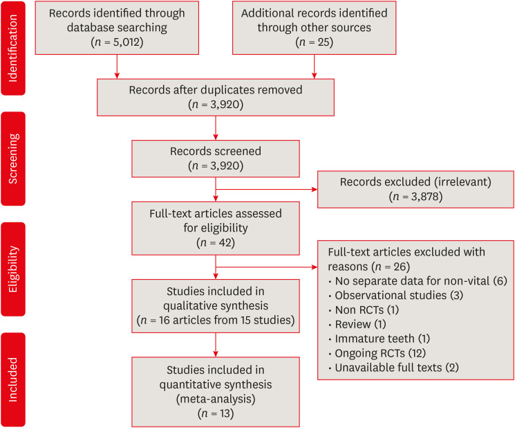
-
Abstract
PDFSupplementary MaterialPubReaderePub
Objectives This study aimed to systematically review the pain and flare-up effects of calcium hydroxide (CH) as intracanal medication (ICM) in non-vital mature teeth.
Materials and Methods Electronic-databases searching for published and grey literature and manual searching were conducted. Only randomized clinical trials (RCTs) were included comparing CH to other ICMs in non-vital mature teeth. The risk of bias was assessed using the RoB 2.0 Cochrane tool. The main outcomes were pain and flare-up. Qualitative and quantitative analysis, wherever applicable, was performed. The certainty of evidence (CoE) was assessed using Grading of Recommendations Assessment, Development and Evaluation (GRADE).
Results Sixteen articles were included in 6 comparisons at different time points for different outcomes. CH reduced pain risk than no ICM within the 1–14-days interval (
p < 0.05) and than triple-antibiotic paste within the first day (p < 0.05) and was similar to corticosteroid/antibiotics combination (p > 0.05). Chlorhexidine (CHX) or CH/CHX, however, reduced pain levels than CH alone (p < 0.05). CH showed higher flare-up risk than CHX (p < 0.05). CoE, however, ranged from very low to moderate.Conclusion Most comparisons for different outcomes are based on very few studies, mostly low-powered, with an overall low CoE. Thus, the available evidence is considered insufficient to either support or refute CH effectiveness or to recommend one ICM over another. Therefore, further well-designed, larger RCTs are required.
Trial Registration PROSPERO database Identifier:
CRD42016041953 -
Citations
Citations to this article as recorded by- Garre’s osteomyelitis of the mandible managed by nonsurgical re-endodontic treatment
Heegyun Kim, Jiyoung Kwon, Hyun-Jung Kim, Soram Oh, Duck-Su Kim, Ji-Hyun Jang
Restorative Dentistry & Endodontics.2024;[Epub] CrossRef - Interpretation by literature review of the use of calcium hydroxide as an intra-ductal medication
María Belén Muñoz Padilla, Verónica Alicia Vega Martínez, Camila Alejandra Villafuerte Moya
Salud, Ciencia y Tecnología.2024; 4: 924. CrossRef - ProTaper Ultimate: The Evolution of Rotary Systems and Their Clinical Applicability
Mateo-Castillo JF, Siqueira-Sandrin VDS, Moreira RA, Brandelero Junior S, Olano-Dextre TL, Claudia R Pinheiro
Journal of Operative Dentistry & Endodontics.2024; 9(1): 16. CrossRef - Comparative Evaluation of Bacterial Reduction by Laser-Activated Irrigation Technique (LAI) With Conventional Needle Irrigation (CNI) in Single-Rooted Teeth With Pulpal Necrosis: A Single-Blinded Randomized Controlled Trial
Keerthika Rajamanickam, Sandhya Raghu, J Vijayashree Priyadharsini, Delphine Priscilla Antony, Nivedhitha Malli Sureshbabu
Cureus.2023;[Epub] CrossRef - Post-Operative Endodontic Pain Management: An Overview of Systematic Reviews on Post-Operatively Administered Oral Medications and Integrated Evidence-Based Clinical Recommendations
Federica Di Spirito, Giuseppe Scelza, Roberto Fornara, Francesco Giordano, Donato Rosa, Alessandra Amato
Healthcare.2022; 10(5): 760. CrossRef - Management of External Inflammatory Root Resorption following Tooth Autotransplantation Using a Modified Combination of Triple Antibiotics
Saeed Asgary, Ardavan Parhizkar, Paulo J. Palma
Case Reports in Dentistry.2022;[Epub] CrossRef - Calcium hydroxide as an intracanal medication for postoperative pain during primary root canal therapy: A systematic review and meta-analysis with trial sequential analysis of randomised controlled trials
Muhammad Zubair Ahmad, Durre Sadaf, Khalid A Merdad, Amani Almohaimeed, Igho J. Onakpoya
Journal of Evidence-Based Dental Practice.2022; 22(1): 101680. CrossRef - Post-operative Pain and Antibacterial Efficacy of Silver Nanoparticles Formulations Intracanal Medication: A Randomized Controlled Clinical Study
Nermine Hassan, Alaa Diab, Geraldine Ahmed
Open Access Macedonian Journal of Medical Sciences.2021; 9(D): 248. CrossRef
- Garre’s osteomyelitis of the mandible managed by nonsurgical re-endodontic treatment
- 6,520 View
- 96 Download
- 8 Crossref
- A micro-computed tomographic evaluation of root canal filling with a single gutta-percha cone and calcium silicate sealer
- Jong Cheon Kim, Maung Maung Kyaw Moe, Sung Kyo Kim
- Restor Dent Endod 2020;45(2):e18. Published online February 12, 2020
- DOI: https://doi.org/10.5395/rde.2020.45.e18
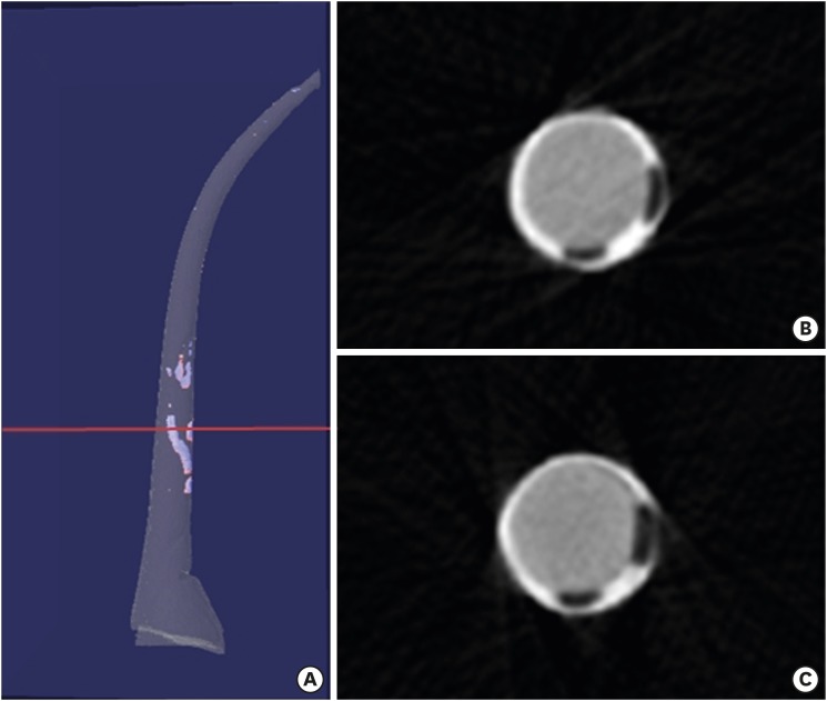
-
Abstract
PDFPubReaderePub
Objectives The purpose of this study was to evaluate the void of root canal filling over time when a calcium silicate sealer was used in the single gutta-percha cone technique.
Materials and Methods Twenty-four J-shaped simulated root canals and twenty-four palatal root canals from extracted human maxillary molars were instrumented with ProFile Ni-Ti rotary instruments up to size 35/0.06 or size 40/0.06, respectively. Half of the canals were filled with Endoseal MTA and the other half were with AH Plus Jet using the single gutta-percha cone technique. Immediately after and 4 weeks after the root canal filling, the samples were scanned using micro-computed tomography at a resolution of 12.8 μm. The scanned images were reconstructed using the NRecon software and the void percentages were calculated using the CTan software, and statistically analyzed by 1-way analysis of variance, paired
t- test and Tukeypost hoc test.Results After 4 weeks, there were no significant changes in the void percentages at all levels in both material groups (
p > 0.05), except at the apical level of the AH Plus Jet group (p < 0.05) in the simulated root canal showing more void percentage compared to other groups. Immediately after filling the extracted human root canals, the Endoseal MTA group showed significantly less void percentage compared to the AH Plus Jet group (p < 0.05).Conclusions Under the limitations of this study, the Endoseal MTA does not seem to reduce the voids over time.
-
Citations
Citations to this article as recorded by- Comparative analysis between resin-based root canal sealer and recent bioceramic-based root canal sealers using MicroCT, film thickness, and solubility
Amira Galal Ismail, Manar M. Galal, Tamer M. Hamdy
Journal of Oral Biology and Craniofacial Research.2026; 16(2): 101400. CrossRef - Evaluation of various obturation techniques with bioceramic sealers in 3D-printed C-shaped canals
Maryam Gharechahi, Melika Hoseinzadeh, Saeed Moradi, Mina Mehrjouei
BMC Oral Health.2024;[Epub] CrossRef - Comparison of obturation quality in natural and replica teeth root-filled using different sealers and techniques
Chuta Kooanantkul, Richard M Shelton, Josette Camilleri
Clinical Oral Investigations.2023; 27(5): 2407. CrossRef - Obturation canalaire
N. Linas, M.-L. Munoz-Sanchez, N. Decerle, P.-Y. Cousson
EMC - Médecine buccale.2023; 16(5): 1. CrossRef - The Effect of Sealer Application Methods on Voids Volume after Aging of Three Calcium Silicate-Based Sealers: A Micro-Computed Tomography Study
Amre R. Atmeh, Rakan Alharbi, Ibrahim Aljamaan, Abdulrahman Alahmari, Ashwin C. Shetty, Ahmed Jamleh, Imran Farooq
Tomography.2022; 8(2): 778. CrossRef - Clinical Efficacy of Sealer-based Obturation Using Calcium Silicate Sealers: A Randomized Clinical Trial
Ji-hyung Kim, Sin-Yeon Cho, Yoonwoo Choi, Do-hyun Kim, Su-Jung Shin, Il-Young Jung
Journal of Endodontics.2022; 48(2): 144. CrossRef - A critical analysis of research methods and experimental models to study root canal fillings
Gustavo De‐Deus, Erick Miranda Souza, Emmanuel João Nogueira Leal Silva, Felipe Gonçalves Belladonna, Marco Simões‐Carvalho, Daniele Moreira Cavalcante, Marco Aurélio Versiani
International Endodontic Journal.2022; 55(S2): 384. CrossRef - Calcium Silicate Cements vs. Epoxy Resin Based Cements: Narrative Review
Mario Dioguardi, Cristian Quarta, Diego Sovereto, Giuseppe Troiano, Khrystyna Zhurakivska, Maria Bizzoca, Lorenzo Lo Muzio, Lucio Lo Russo
Oral.2021; 1(1): 23. CrossRef - Physico-Chemical Properties of Calcium-Silicate vs. Resin Based Sealers—A Systematic Review and Meta-Analysis of Laboratory-Based Studies
Viresh Chopra, Graham Davis, Aylin Baysan
Materials.2021; 15(1): 229. CrossRef - Micro-computed tomography in preventive and restorative dental research: A review
Mehrsima Ghavami-Lahiji, Reza Tayefeh Davalloo, Gelareh Tajziehchi, Paria Shams
Imaging Science in Dentistry.2021; 51(4): 341. CrossRef - Main and Accessory Canal Filling Quality of a Premixed Calcium Silicate Endodontic Sealer According to Different Obturation Techniques
Su-Yeon Ko, Hae Won Choi, E-Deun Jeong, Vinicius Rosa, Yun-Chan Hwang, Mi-Kyung Yu, Kyung-San Min
Materials.2020; 13(19): 4389. CrossRef
- Comparative analysis between resin-based root canal sealer and recent bioceramic-based root canal sealers using MicroCT, film thickness, and solubility
- 2,867 View
- 24 Download
- 11 Crossref
- A micro-computed tomography evaluation of voids using calcium silicate-based materials in teeth with simulated internal root resorption
- Vildan Tek, Sevinç Aktemur Türker
- Restor Dent Endod 2020;45(1):e5. Published online November 29, 2019
- DOI: https://doi.org/10.5395/rde.2020.45.e5
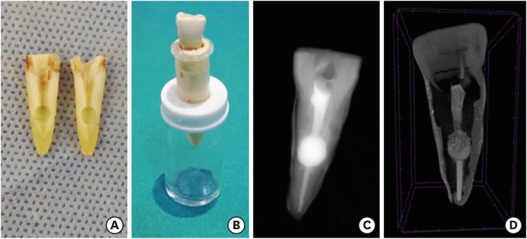
-
Abstract
PDFPubReaderePub
Objectives The obturation quality of MTA, Biodentine, Total Fill BC root canal sealer (RCS), and warm gutta-percha (WGP) in teeth with simulated internal root resorption (IRR) was evaluated by using micro-computed tomography.
Materials and Methods Standardized IRR cavities were created using 40 extracted maxillary central incisor teeth and randomly assigned into 4 groups (
n = 10). IRR cavities were filled with MTA, Biodentine, Total Fill BC RCS (bulk-fill form) and WGP + Total Fill BC RCS. Percentage of voids between resorptive cavity walls and obturation material (external void), and inside the filling materials (internal voids) were measured.Results Total Fill BC sealer in the bulk-fill form presented significantly highest values of external and internal void percentages (
p < 0.05). Biodentine showed a significantly lowest external void percentage (p < 0.05). WGP + Total Fill BC RCS presented significantly lower values of internal void percentages than all groups (p < 0.05), except Biodentine (p > 0.05).Conclusion None of the filling materials were created void-free obturation in resorption cavities. Biodentine may favor its application in teeth with IRR over Angelus MTA and bulk-fill form of Total Fill BC.
-
Citations
Citations to this article as recorded by- The role of calcium silicate cements in endodontics: from material science to clinical success
Takwa E. Ellakwa, Ayman Ellakwa, Doha El-Sayed Ellakwa
Discover Materials.2026;[Epub] CrossRef - Micro-CT Assessment of Hydraulic Calcium Silicate Cements for Perforating Internal Resorption in 3D-printed Tooth Replicas at Different Root Thirds: An In Vitro Study
Angelo José Sócrates Torres-Carrillo, Jardel Francisco Mazzi-Chaves, Gustavo Creazzo, Rodrigo E. Salazar-Gamarra, Helena Cristina de Assis, Ronald Ordinola-Zapata, Manoel D. Sousa-Neto, Fabiane Carneiro Lopes-Olhê
Journal of Endodontics.2026;[Epub] CrossRef - Removal of AH Plus Bioceramic Sealer from Artificial Internal Resorption Cavities Using Different Irrigation Activation Systems
Mine Büker, Meltem Sümbüllü, Emine Şimşek, Fadime Sena Sezer
Cumhuriyet Dental Journal.2025; 28(3): 383. CrossRef - Functional and Bioactive Performance of Premixed Bioceramic Sealers with Warm Obturation: A Scoping Review
Patryk Wiśniewski, Stanisław Krokosz, Małgorzata Pietruska, Anna Zalewska
Gels.2025; 11(11): 932. CrossRef - Evaluation of the effectiveness of different supplemental cleaning techniques in the retreatment of roots with small simulated internal resorption cavities: an in vitro comparative study
Sine Güngör Us, Özgür Uzun, Nazlı Merve Güngör
BMC Oral Health.2025;[Epub] CrossRef - Evaluation of Different Techniques and Materials for Filling in 3-dimensional Printed Teeth Replicas with Perforating Internal Resorption by Means of Micro–Computed Tomography
Angelo J.S. Torres-Carrillo, Helena C. Assis, Rodrigo E. Salazar-Gamarra, Leonardo Moreira Teodosio, Alice C. Silva-Sousa, Jardel F. Mazzi-Chaves, Priscila B. Ferreira-Soares, Manoel D. Sousa-Neto, Fabiane C. Lopes-Olhê
Journal of Endodontics.2024; 50(2): 205. CrossRef - Three-Dimensional Measurement of Obturation Quality of Bioceramic Materials in Filling Artificial Internal Root Resorption Cavities Using Different Obturation Techniques: An In Vitro Comparative Study
Ammar M. Sharki, Ahmed H. Ali
Journal of Endodontics.2024; 50(7): 997. CrossRef - Evaluation of calcium hydroxide root canal filling materials by cone beam computed tomography and three-dimensional modeling
Asel Usdat Ozturk, Ekin Dogan, Venus Seyedoskuyi, Berk Senguler, Asli Topaloglu-Ak
Folia Medica.2024; 66(2): 250. CrossRef - Clinical applications of calcium silicate‐based materials: a narrative review
S Küçükkaya Eren
Australian Dental Journal.2023;[Epub] CrossRef - A critical analysis of research methods and experimental models to study root canal fillings
Gustavo De‐Deus, Erick Miranda Souza, Emmanuel João Nogueira Leal Silva, Felipe Gonçalves Belladonna, Marco Simões‐Carvalho, Daniele Moreira Cavalcante, Marco Aurélio Versiani
International Endodontic Journal.2022; 55(S2): 384. CrossRef - An Updated Review on Properties and Indications of Calcium Silicate‐Based Cements in Endodontic Therapy
Fateme Eskandari, Alireza Razavian, Rozhina Hamidi, Khadije Yousefi, Susan Borzou, Zohaib Khurshid
International Journal of Dentistry.2022;[Epub] CrossRef - Efficacy Of Calcium Silicate-Based Sealers In Root Canal Treatment: A Systematic Review
Hattan Mohammed Omar Baismail, Mohammed Ghazi Moiser Albalawi, Alaa Mofareh Thoilek Alanazi, Muhannad Atallah Saleem Alatawi, Badr Soliman Alhussain
Annals of Dental Specialty.2021; 9(1): 87. CrossRef
- The role of calcium silicate cements in endodontics: from material science to clinical success
- 3,009 View
- 40 Download
- 12 Crossref
- Bioactivity of endodontic biomaterials on dental pulp stem cells through dentin
- Bahar Javid, Narges Panahandeh, Hassan Torabzadeh, Hamid Nazarian, Ardavan Parhizkar, Saeed Asgary
- Restor Dent Endod 2020;45(1):e3. Published online November 4, 2019
- DOI: https://doi.org/10.5395/rde.2020.45.e3
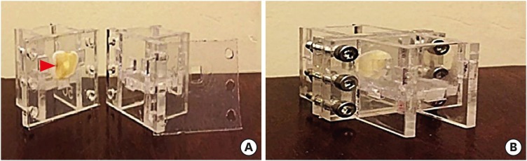
-
Abstract
PDFPubReaderePub
Objectives This study investigated the indirect effect of calcium-enriched mixture (CEM) cement and mineral trioxide aggregate (MTA), as 2 calcium silicate-based hydraulic cements, on human dental pulp stem cells (hDPSCs) through different dentin thicknesses.
Materials and Methods Two-chamber setups were designed to simulate indirect pulp capping (IPC). Human molars were sectioned to obtain 0.1-, 0.3-, and 0.5-mm-thick dentin discs, which were placed between the 2 chambers to simulate an IPC procedure. Then, MTA and CEM were applied on one side of the discs, while hDPSCs were cultured on the other side. After 2 weeks of incubation, the cells were removed, and cell proliferation, morphology, and attachment to the discs were evaluated under scanning electron microscopy (SEM). Energy-dispersive X-ray (EDXA) spectroscopy was performed for elemental analysis. Alkaline phosphatase (ALP) activity was assessed quantitatively. The data were analyzed using the Kruskal-Wallis and Mann-Whitney tests.
Results SEM micrographs revealed elongated cells, collagen fibers, and calcified nucleations in all samples. EDXA verified that the calcified nucleations consisted of calcium phosphate. The largest calcifications were seen in the 0.1-mm-thick dentin subgroups. There was no significant difference in ALP activity across the CEM subgroups; however, ALP activity was significantly lower in the 0.1-mm-thick dentin subgroup than in the other MTA subgroups (
p < 0.05).Conclusions The employed capping biomaterials exerted biological activity on hDPSCs, as shown by cell proliferation, morphology, and attachment and calcific precipitations, through 0.1- to 0.5-mm-thick layers of dentin. In IPC, the bioactivity of these endodontic biomaterials is probably beneficial.
-
Citations
Citations to this article as recorded by- Dental pulp capping materials: modulators of stem cell behavior and regenerative potential
Ali Cheayto, Sara Ayoub, Sarah Ayad Al-Tameemi, Mohammad Fayyad-Kazan
Biomedical Physics & Engineering Express.2025; 11(6): 062004. CrossRef - Effect of pulp capping materials on odontogenic differentiation of human dental pulp stem cells: An in vitro study
Mahmoud M. Bakr, Mohamed Shamel, Shereen N. Raafat, Robert M. Love, Mahmoud M. Al‐Ankily
Clinical and Experimental Dental Research.2024;[Epub] CrossRef - Effects of Growth Factors on the Differentiation of Dental Stem Cells: A
Systematic Review and Meta-analysis (Part I)
Sayna Shamszadeh, Armin Shirvani, Hassan Torabzadeh, Saeed Asgary
Current Stem Cell Research & Therapy.2024; 19(4): 523. CrossRef - The Role of Growth Factor Delivery Systems on Cellular Activities of Dental
Stem Cells: A Systematic Review (Part II)
Sayna Shamszadeh, Armin Shirvani, Saeed Asgary
Current Stem Cell Research & Therapy.2024; 19(4): 587. CrossRef - Comprehensive review of composition, properties, clinical applications, and future perspectives of calcium-enriched mixture (CEM) cement: a systematic analysis
Saeed Asgary, Mahtab Aram, Mahta Fazlyab
BioMedical Engineering OnLine.2024;[Epub] CrossRef - Evaluation of dental pulp stem cells behavior after odontogenic differentiation induction by three different bioactive materials on two different scaffolds
Basma Ahmed, Mai H. Ragab, Rania A. Galhom, Hayam Y. Hassan
BMC Oral Health.2023;[Epub] CrossRef - Characterization of Dental Pulp Stem Cell Responses to Functional Biomaterials Including Mineralized Trioxide Aggregates
Sejin Bae, Bueonguk Kang, Hyungbin Lee, Harrison Luu, Eric Mullins, Karl Kingsley
Journal of Functional Biomaterials.2021; 12(1): 15. CrossRef - Incorporation of amoxicillin-loaded microspheres in mineral trioxide aggregate cement: an in vitro study
Fábio Rocha Bohns, Vicente Castelo Branco Leitune, Isadora Martini Garcia, Bruna Genari, Nélio Bairros Dornelles, Silvia Stanisçuaski Guterres, Fabrício Aulo Ogliari, Mary Anne Sampaio de Melo, Fabrício Mezzomo Collares
Restorative Dentistry & Endodontics.2020;[Epub] CrossRef
- Dental pulp capping materials: modulators of stem cell behavior and regenerative potential
- 2,224 View
- 17 Download
- 8 Crossref
Review Article
- Effect of calcium hydroxide on inflammatory root resorption and ankylosis in replanted teeth compared with other intracanal materials: a review
- Maryam Zare Jahromi, Mahmood Reza Kalantar Motamedi
- Restor Dent Endod 2019;44(3):e32. Published online August 1, 2019
- DOI: https://doi.org/10.5395/rde.2019.44.e32
-
Abstract
PDFPubReaderePub
Calcium hydroxide (CH) is the gold-standard intracanal dressing for teeth subjected to traumatic avulsion. A common complication after the replantation of avulsed teeth is root resorption (RR). The current review was conducted to compare the effect of CH with that of other intracanal medications and filling materials on inflammatory RR and replacement RR (ankylosis) in replanted teeth. The PubMed and Scopus databases were searched through June 2018 using specific keywords related to the title of the present article. The materials that were compared to CH were in 2 categories: 1) mineral trioxide aggregate (MTA) and endodontic sealers as permanent filling materials for single-visit treatment, and 2) Ledermix, bisphosphonates, acetazolamide, indomethacin, gallium nitrate, and enamel matrix-derived protein (Emdogain) as intracanal medicaments for multiple-visit management of avulsed teeth prior to the final obturation. MTA can be used as a single-visit root filling material; however, there are limited data on its efficacy due to a lack of clinical trials. Ledermix and acetazolamide were comparable to CH in reducing RR. Emdogain seems to be an interesting material, but the data supporting its use as an intracanal medication remain very limited. The conclusions drawn in this study were limited by the insufficiency of clinical trials.
-
Citations
Citations to this article as recorded by- Endodontic Intracanal Medicaments and Agents
Anu Priya Guruswamy Pandian, Depti Bellani, Ritya Mary Jibu, Varsha Agnihotri
Dental Clinics of North America.2026; 70(1): 45. CrossRef - Comparison of 2 kinds of endodontic treatments for pulp necrosis and infection in immature teeth following avulsion: A retrospective study
Weiwei Zhou, Xin Zhang, Meng Wang, Liang Xue, Lili Zhang
Medicine.2026; 105(14): e48250. CrossRef - Efficacy of Simvastatin in Inhibiting Bone Resorption and Promoting Healing in Delayed Tooth Avulsion: A Case Series
Rajesh Kumar, Supraja N Atluri, Alekhya Achanta, Chittaranjan Bogishetty, Tejaswini R Chunduri, Tejaswini PSS, Ramakrishna Ravi
Cureus.2025;[Epub] CrossRef - Interdisziplinäre Lösung nach dentalem Trauma mit Avulsion und Wurzelresorption
Eva Maier, Julia Lubauer, Kerstin M. Galler
Oralprophylaxe & Kinderzahnmedizin.2025; 47(3): 161. CrossRef - Bioactive potential of Bio-C Temp demonstrated by systemic mineralization markers and immunoexpression of bone proteins in the rat connective tissue
Camila Soares Lopes, Mateus Machado Delfino, Mário Tanomaru-Filho, Estela Sasso-Cerri, Juliane Maria Guerreiro-Tanomaru, Paulo Sérgio Cerri
Journal of Materials Science: Materials in Medicine.2024;[Epub] CrossRef - The use of mineral trioxide aggregate for treatment of children with complications of dental trauma
L.Yu. Kharkova, M.V. Korolenkova
Stomatology.2024; 103(4): 59. CrossRef - Instant Re-Implantation of Avulsed Teeth
Smita Paul, Sambarta Das, Nirmal Debbarma, Barun Dasgupta, Bidyut Seal, Ayesha Satapathy
Journal of Pharmacy and Bioallied Sciences.2024; 16(Suppl 4): S3461. CrossRef - Interpretation by literature review of the use of calcium hydroxide as an intra-ductal medication
María Belén Muñoz Padilla, Verónica Alicia Vega Martínez, Camila Alejandra Villafuerte Moya
Salud, Ciencia y Tecnología.2024; 4: 924. CrossRef - Evaluation of the physicochemical properties of intracanal medications used in traumatized teeth
Patricia Almeida da Silva de Macedo, Walbert de Andrade Vieira, Paulo Henrique Gabriel, Karla de Faria Vasconcelos, Francisco Haiter Neto, Ana Carolina Correia Laurindo de Cerqueira Neto, Brenda Paula Figueiredo de Almeida Gomes, Marcos Roberto dos Santo
Brazilian Journal of Oral Sciences.2024; 23: e242997. CrossRef - Treatment of Teeth with Root Resorptions: A Case Report and Systematic Review
Damla Erkal, Abdullah Başoğlu, Damla Kırıcı, Nezahat Arzu Kayar, Simay Koç, Kürşat Er
Galician Medical Journal.2023;[Epub] CrossRef - Successful outcome of permanent maxillary incisor reimplanted after 30 hours of extra‐oral time—a case report with 5‐year follow‐up
Ibadat Preet Kaur, Ashok Kumar, Mukul Kumar, Kanistika Jha
Clinical Case Reports.2023;[Epub] CrossRef - Replantation of an Avulsed Tooth: A Case Report
Nishad Kadulkar, Rubi Kataki, Adrija Deka, Salouno Thonai
Cureus.2023;[Epub] CrossRef - Avulsion of Permanent Mandibular Incisors: A Report of Two Cases with Pertinent Literature
Ibadat Preet Kaur, Jitendra Sharan, Pallawi Sinha, Ashok Kumar, Anand Marya, Leandro Napier de Souza
Case Reports in Dentistry.2023; 2023: 1. CrossRef - The Impact of Autologous Platelet Concentrates on the Periapical Tissues and Root Development of Replanted Teeth: A Systematic Review
Zohaib Khurshid, Faris Yahya I. Asiri, Shariq Najeeb, Jithendra Ratnayake
Materials.2022; 15(8): 2776. CrossRef
- Endodontic Intracanal Medicaments and Agents
- 4,916 View
- 104 Download
- 14 Crossref
Case Report
- Surgical management of an accessory canal in a maxillary premolar: a case report
- Hee-Jin Kim, Mi-Kyung Yu, Kwang-Won Lee, Kyung-San Min
- Restor Dent Endod 2019;44(3):e30. Published online July 29, 2019
- DOI: https://doi.org/10.5395/rde.2019.44.e30
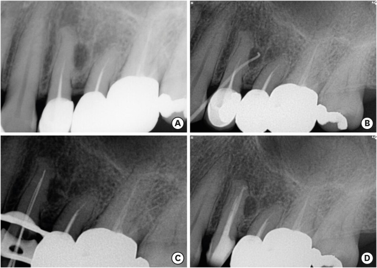
-
Abstract
PDFPubReaderePub
We report the surgical endodontic treatment of a maxillary first premolar with a lateral lesion that originated from an accessory canal. Although lesions originating from accessory canals frequently heal with simple conventional endodontic therapy, some lesions may need additional and different treatment. In the present case, conventional root canal retreatment led to incomplete healing with the need for further treatment (
i.e. , surgery). Surgical endodontic management with a fast-setting calcium silicate cement was performed on the accessory canal using a dental operating microscope. At the patient's 9-month recall visit, the lesion was resolved upon radiography.-
Citations
Citations to this article as recorded by- Predictive analysis of root canal morphology in relation to root canal treatment failures: a retrospective study
Mohmed Isaqali Karobari, Vishnu Priya Veeraraghavan, P. J. Nagarathna, Sudhir Rama Varma, Jayaraj Kodangattil Narayanan, Santosh R. Patil
Frontiers in Dental Medicine.2025;[Epub] CrossRef - Endodontic management of internal replacement resorption of two maxillary central incisors with the aid of cone-beam computed tomography as the diagnostic tool: a case report and review of literature
Fatemeh Eskandari, Safoora Sahebi, Negar Ghorbani Jahandizi, Hossein Mofidi
Journal of Medical Case Reports.2025;[Epub] CrossRef - The Impact of the Preferred Reporting Items for Case Reports in Endodontics (PRICE) 2020 Guidelines on the Reporting of Endodontic Case Reports
Sofian Youssef, Phillip Tomson, Amir Reza Akbari, Natalie Archer, Fayjel Shah, Jasmeet Heran, Sunmeet Kandhari, Sandeep Pai, Shivakar Mehrotra, Joanna M Batt
Cureus.2023;[Epub] CrossRef - Main and Accessory Canal Filling Quality of a Premixed Calcium Silicate Endodontic Sealer According to Different Obturation Techniques
Su-Yeon Ko, Hae Won Choi, E-Deun Jeong, Vinicius Rosa, Yun-Chan Hwang, Mi-Kyung Yu, Kyung-San Min
Materials.2020; 13(19): 4389. CrossRef
- Predictive analysis of root canal morphology in relation to root canal treatment failures: a retrospective study
- 2,164 View
- 18 Download
- 4 Crossref
Research Articles
-
Coronal tooth discoloration induced by regenerative endodontic treatment using different scaffolds and intracanal coronal barriers: a 6-month
ex vivo study - Noushin Shokouhinejad, Hassan Razmi, Maryam Farbod, Marzieh Alikhasi, Josette Camilleri
- Restor Dent Endod 2019;44(3):e25. Published online July 16, 2019
- DOI: https://doi.org/10.5395/rde.2019.44.e25
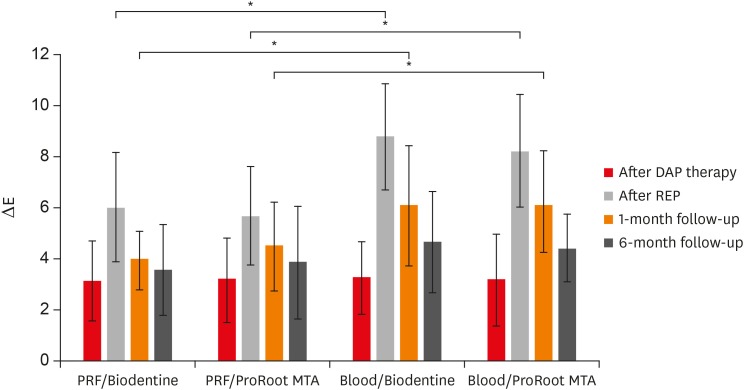
-
Abstract
PDFPubReaderePub
Objective The aim of this study was to evaluate discoloration of teeth undergoing regenerative endodontic procedures (REPs) using blood clot or platelet-rich fibrin (PRF) as the scaffolds and different calcium silicate-based materials as the intracanal coronal barriers in an
ex vivo model.Materials and Methods Forty-eight bovine incisors were prepared and disinfected using 1 mg/mL double antibiotic paste (DAP). The specimens were then randomly divided into 2 groups (
n = 24) according to the scaffolds (blood or PRF). After placement of scaffolds each group was divided into 2 subgroups (n = 12) according to the intracanal coronal barriers (ProRoot MTA or Biodentine). The pulp chamber walls were sealed with dentin bonding agent before placement of DAP and before placement of scaffolds. The color changes (∆E) were measured at different steps. The data were analyzed using 2-way analysis of variance.Results Coronal discoloration induced by DAP was not clinically perceptible (ΔE ≤ 3.3). Regarding the type of the scaffold, coronal discoloration was significantly higher in blood groups compared with PRF groups at the end of REP and after 1 month (
p < 0.05). However, no significant difference was found between PRF and blood clot after 6 months (p > 0.05). Considering the type of intracanal coronal barrier, no significant difference existed between ProRoot MTA and Biodentine (p > 0.05).Conclusions With sealing the dentinal tubules of pulp chamber with a dentin bonding agent and application of DAP as an intracanal medicament, coronal color change of the teeth following the use of PRF and blood sealed with either ProRoot MTA or Biodentine was not different at 6-month follow-up.
-
Citations
Citations to this article as recorded by- Effect of Addition of Zinc Oxide on Color Stability of a Calcium Silicate–Based Cement Containing Bismuth Oxide in the Presence of Blood and Sodium Hypochlorite
Mehrfam Khoshkhounejad, Noushin Shokouhinejad, Pegah Sarraf, MohammadHossein Mirisiahi, Hannah Wesley
International Journal of Dentistry.2026;[Epub] CrossRef - Calcium Silicate-Based Cements for Vital Pulp Therapy: Integrated Assessment of Radiopacity, Elemental Composition, and 24-h Pulp Cell Responses
Belen Şirinoğlu Çapan, Vasfiye Işık, Tugba Elgün, Zeynep Hale Keleş, Soner Şişmanoğlu
Biomimetics.2026; 11(4): 280. CrossRef - Effect of output intensity of light curing unit on coronal tooth discoloration induced by silicate calcium-based materials: an ex vivo study
Fatemeh Farshad, Noushin Shokouhinejad, Parham Pedram, Mohammad Javad KharaziFard, Elham Ahmadi
Biomaterial Investigations in Dentistry.2026; 13: 379. CrossRef - Impact of Biodentine Placement on Fracture Resistance and its Influence on Discoloration with Different Scaffolds
Evren Sarıyılmaz, Öznur Sarıyılmaz, Burak Çarıkçıoğlu, Gülşah Uslu, Raif Alan
Journal of Endodontics.2025; 51(9): 1199. CrossRef - Effect of pH on the solubility and volumetric change of ready-to-use Bio-C Repair bioceramic material
Luana Raphael da SILVA, Jader Camilo PINTO, Juliane Maria GUERREIRO-TANOMARU, Mário TANOMARU-FILHO
Brazilian Oral Research.2024;[Epub] CrossRef - Efficacy of Potassium Iodide and Glutathione for Correlation of Dentin Discoloration Caused by Silver Diamine Fluoride
Mahsa Samani, Hamid Majzoub, Faramarz Zakavi, Ayyub Mojaddami
Cureus.2024;[Epub] CrossRef - Intracanal medicaments and coronal sealing materials influence on root fracture resistance and coronal discoloration: An in vitro study
Rasoul Sahebalam, Marzie Boskabady, Maryam Naghavi, Samira Dehghanitafti
Saudi Endodontic Journal.2024; 14(2): 199. CrossRef - Potential Crown Discoloration Induced by the Combination of Various Intracanal Medicaments and Scaffolds Applied in Regenerative Endodontic Therapy
NB Altun, A Turkyilmaz
Nigerian Journal of Clinical Practice.2024; 27(7): 897. CrossRef - Evaluation of the effectiveness of different treatment approaches in preventing coronal discoloration caused by regenerative endodontic treatment
Melis Oya Ateş, Zeliha Uğur Aydın
Clinical Oral Investigations.2023; 27(8): 4595. CrossRef - Evaluation of the Effectiveness of Laser‐Assisted Bleaching of the Teeth Discolored due to Regenerative Endodontic Treatment
Noushin Shokouhinejad, Mehrfam Khoshkhounejad, Fatemeh Hamidzadeh, Murilo Baena Lopes
International Journal of Dentistry.2022;[Epub] CrossRef - Effectiveness of Teeth Whitening after Regenerative Endodontics Procedures: An In Vitro Study
Irini Fagogeni, Joanna Metlerska, Tomasz Falgowski, Maciej Górski, Mariusz Lipski, Alicja Nowicka
Journal of Clinical Medicine.2022; 11(23): 7016. CrossRef - Microstructure and color stability of calcium silicate-based dental materials exposed to blood or platelet-rich fibrin
Noushin Shokouhinejad, Ibrahim Abu Tahun, Shima Saber Tahan, Fatemeh Mohandes, Mohammad H. Nekoofar, Paul M. H. Dummer
Clinical Oral Investigations.2022; 27(3): 1193. CrossRef - Spectrophotometric analysis of internal bleaching of traumatized teeth with coronal discoloration following regenerative endodontic procedures
Jaqueline Lazzari, Walbert Vieira, Vanessa Pecorari, Brenda Paula Figueiredo de Almeida Gomes, José Flávio Affonso de Almeida, Adriana De-Jesus-Soares
Brazilian Journal of Oral Sciences.2021;[Epub] CrossRef - Biological parameters, discolouration and radiopacity of calcium silicate‐based materials in a simulated model of partial pulpotomy
Lilian Vieira Oliveira, Gabriela Leite de Souza, Gisele Rodrigues da Silva, Thamara Eduarda Alves Magalhães, Gabrielle Alves Nunes Freitas, Ana Paula Turrioni, Gabriella Lopes de Rezende Barbosa, Camilla Christian Gomes Moura
International Endodontic Journal.2021; 54(11): 2133. CrossRef - Effect of hydrogel-based antibiotic intracanal medicaments on crown discoloration
Rayan B. Yaghmoor, Jeffrey A. Platt, Kenneth J. Spolnik, Tien Min Gabriel Chu, Ghaeth H. Yassen
Restorative Dentistry & Endodontics.2021;[Epub] CrossRef - The effect of different calcium silicate-based pulp capping materials on tooth discoloration: an in vitro study
Ahmad S. Al-Hiyasat, Dana M. Ahmad, Yousef S. Khader
BMC Oral Health.2021;[Epub] CrossRef - Knowledge, attitudes, and practices of undergraduate students concerning Regenerative Endodontics
Ligia B. da Silva, Mariana Gabriel, Márcia M. Marques, Fernanda C. Carrer, Flávia Gonçalves, Giovanna Sarra, Giovanna L. Carvalho, Ana Armas-Vega, Maria S. Moreira
Minerva Stomatologica.2020;[Epub] CrossRef - Coronal Discoloration Related to Bioceramic and Mineral Trioxide Aggregate Coronal Barrier in Non-vital Mature Teeth Undergoing Regenerative Endodontic Procedures
Mazen Doumani, Mohammad Yaman Seirawan, Kinda Layous, Mohammad Kinan Seirawan
World Journal of Dentistry.2020; 11(1): 52. CrossRef
- Effect of Addition of Zinc Oxide on Color Stability of a Calcium Silicate–Based Cement Containing Bismuth Oxide in the Presence of Blood and Sodium Hypochlorite
- 2,789 View
- 26 Download
- 18 Crossref
- Effect of intracanal medications on the interfacial properties of reparative cements
- Andrea Cardoso Pereira, Mariana Valerio Pallone, Marina Angélica Marciano, Karine Laura Cortellazzi, Marcos Frozoni, Brenda P. F. A. Gomes, José Flávio Affonso de Almeida, Adriana de Jesus Soares
- Restor Dent Endod 2019;44(2):e21. Published online May 9, 2019
- DOI: https://doi.org/10.5395/rde.2019.44.e21
-
Abstract
PDFPubReaderePub
Objectives The purpose of the present study was to evaluate the effect of calcium hydroxide with 2% chlorhexidine gel (HCX) or distilled water (HCA) compared to triple antibiotic paste (TAP) on push-out bond strength and the cement/dentin interface in canals sealed with White MTA Angelus (WMTA) or Biodentine (BD).
Materials and Methods A total of 70 extracted human lower premolars were endodontically prepared and randomly divided into 4 groups according to the intracanal medication, as follows: group 1, HCX; group 2, TAP; group 3, HCA; and group 4, control (without intracanal medication). After 7 days, the medications were removed and the cervical third of the specimens was sectioned into five 1-mm sections. The sections were then sealed with WMTA or BD as a reparative material. After 7 days in 100% humidity, a push-out bond strength test was performed. Elemental analysis was performed at the interface, using energy-dispersive spectroscopy. The data were statistically analyzed using analysis of variance and the Tukey test (
p < 0.05).Results BD presented a higher bond strength than WMTA (
p < 0.05). BD or WMTA in canals treated with calcium hydroxide intracanal medications had the highest bond strength values, with a statistically significant difference compared to TAP in the WMTA group (p < 0.05). There were small amounts of phosphorus in samples exposed to triple antibiotic paste, regardless of the coronal sealing.Conclusions The use of intracanal medications did not affect the bond strength of WMTA and BD, except when TAP was used with WMTA.
-
Citations
Citations to this article as recorded by- Endodontic treatment of teeth with a wide apical opening – a clinical case
V. A. Popov, E. S. Pestova, S. A. Anisimova
Endodontics Today.2025; 23(2): 282. CrossRef - Effects of calcium hydroxide intracanal medicament on push‐out bond strength of endodontic sealers: A systematic review and meta‐analysis
Mohammed Nasser Alhajj, Fadhilah Daud, Sadeq Ali Al‐Maweri, Yanti Johari, Zuryati Ab‐Ghani, Mariatti Jaafar, Yoshihito Naito, Widyasri Prananingrum, Zaihan Ariffin
Journal of Esthetic and Restorative Dentistry.2022; 34(8): 1166. CrossRef - Evaluation of a Novel Tool for Apical Plug Formation during Apexification of Immature Teeth
Yasser Alsayed Tolibah, Line Droubi, Saleh Alkurdi, Mohammad Tamer Abbara, Nada Bshara, Thuraya Lazkani, Chaza Kouchaji, Ibrahim Ali Ahmad, Ziad D. Baghdadi
International Journal of Environmental Research and Public Health.2022; 19(9): 5304. CrossRef - Spectrophotometric analysis of internal bleaching of traumatized teeth with coronal discoloration following regenerative endodontic procedures
Jaqueline Lazzari, Walbert Vieira, Vanessa Pecorari, Brenda Paula Figueiredo de Almeida Gomes, José Flávio Affonso de Almeida, Adriana De-Jesus-Soares
Brazilian Journal of Oral Sciences.2021;[Epub] CrossRef - Do intracanal medications used in regenerative endodontics affect the bond strength of powder-to-liquid and ready-to-use cervical sealing materials?
MarinaCarvalho Prado, Kevillin Martiniano, AndreaCardoso Pereira, KarineL Cortellazzi, MarinaA Marciano, Gabriel Abuna, Adriana de-Jesus-Soares
Journal of Conservative Dentistry.2021; 24(5): 464. CrossRef
- Endodontic treatment of teeth with a wide apical opening – a clinical case
- 2,019 View
- 17 Download
- 5 Crossref
- Discoloration of teeth due to different intracanal medicaments
- Farzaneh Afkhami, Sadaf Elahy, Alireza Mahmoudi Nahavandi, Mohamad Javad Kharazifard, Aidin Sooratgar
- Restor Dent Endod 2019;44(1):e10. Published online February 12, 2019
- DOI: https://doi.org/10.5395/rde.2019.44.e10
-
Abstract
PDFPubReaderePub
Objectives The objective of this study was to assess coronal discoloration induced by the following intracanal medicaments: calcium hydroxide (CH), a mixture of CH paste and chlorhexidine gel (CH/CHX), and triple antibiotic paste (3Mix).
Materials and Methods Seventy extracted single-canal teeth were selected. Access cavities were prepared and each canal was instrumented with a rotary ProTaper system. The specimens were randomly assigned to CH, CH/CHX, and 3Mix paste experimental groups (
n = 20 each) or a control group (n = 10). Each experimental group was randomly divided into 2 subgroups (A and B). In subgroup A, medicaments were only applied to the root canals, while in subgroup B, the root canals were completely filled with medicaments and a cotton pellet dipped in medicament was also placed in the pulp chamber. Spectrophotometric readings were obtained from the mid-buccal surface of the tooth crowns immediately after placing the medicaments (T1) and at 1 week (T2), 1 month (T3), and 3 months (T4) after filling. The ∆E was then calculated. Data were analyzed using 2-way analysis of variance (ANOVA), 3-way ANOVA, and the Scheffépost hoc test.Results The greatest color change (ΔE) was observed at 3 months (
p < 0.0001) and in 3Mix subgroup B (p = 0.0057). No significant color change occurred in the CH (p = 0.7865) or CH/CHX (p = 0.1367) groups over time, but the 3Mix group showed a significant ΔE (p = 0.0164).Conclusion Intracanal medicaments may induce tooth discoloration. Use of 3Mix must be short and it must be carefully applied only to the root canals; the access cavity should be thoroughly cleaned afterwards.
-
Citations
Citations to this article as recorded by- Time-dependent Tooth Color Changes Following Conventional, Silver-based, and Photodynamic Root Canal Irrigants: An In Vitro Study
Laila Mohamed Mohamed Kenawi, Mohamed Fattouh, Khaled Abid Althaqafi, Abla Arafa
The Open Dentistry Journal.2026;[Epub] CrossRef - Comparative analysis of tooth discoloration induced by different intracanal medicaments in regenerative endodontics: A systematic review and network meta-analysis
Ashlesha Nageshwar Madankar, Sulabha Radke, Shanmuga Priya, Darshan Dakshindas
Endodontology.2026; 38(1): 8. CrossRef - Effects of Intra-canal Medicaments on Infrared Light Energy Transmission Through Enamel and Dentin During Photobiomodulation: An In Vitro Study
Sachin Kulkarni, Laurence J. Walsh, Yash Bhurani, Roy George
Journal of Endodontics.2025; 51(5): 616. CrossRef - Tooth discoloration caused by nanographene oxide as an irrigant and intracanal medicament in the endodontic treatment of extracted single-rooted teeth: An ex-vivo study
Abbas Abbaszadegan, Zeinab Rafiee, Bahar Asheghi, Ahmad Gholami, Mohmed Isaqali Karobari
PLOS One.2025; 20(6): e0325430. CrossRef - Investigation of Discoloration of Anterior Teeth With Three Types of Substances Used in Endodontic Treatment
Sahar Soltani, Eshagh Ali Saberi, Nazanin Shahradnia, Pedram Abdollahzade Sangrodi, Elham Majidi
Clinical and Experimental Dental Research.2025;[Epub] CrossRef - A New Disinfection Approach Using a Chitosan-Based Endodontic Irrigant
Alejandra Itzel Lopez-Flores, Ulises Velazquez-Enriquez, Rogelio Jose Scougall-Vilchis, Laura Susana Acosta-Torres, Laura Emma Rodriguez-Vilchis, Rosalía Contreras-Bulnes, Paloma Netzayeli Serrano-Diaz, Rene Garcia-Contreras
Materials.2025; 18(24): 5552. CrossRef - Spectrophotometric Analysis of Intracoronal Bleaching on Crown Discoloration Induced by Various Antibiotic Pastes: An In Vitro Study
Avneet Kaur, Dileep Soni, Poorva R Sharma
International Journal of Clinical Pediatric Dentistry.2025; 18(12): 1443. CrossRef - Effect of Calcium Hydroxide Versus Double Antibiotic Paste on Endodontic Treatment Outcomes in Teeth With Large Periapical Lesions: A Triple‐Blind Randomized Clinical Trial
Afsaneh Rahmati, Farshad Seyedein, Omid Dianat, Sara Saedi, Golriz Rostami, Alireza Akbarzadeh Baghban, Shima Sabertahan, Majid Kazem, Kee Y. Kum
International Journal of Dentistry.2024;[Epub] CrossRef - The effect of different intracanal irrigants on the push-out bond strength of dentin in damaged anterior primary teeth
Leila Bassir, Shirin Taravati, Farzad Nouri, Saeide Rahimi
Journal of Medicine and Life.2024; 17(5): 536. CrossRef - In Vıtro Evaluatıon of Dıscoloratıon Caused by Root Canal Sealers and Color Changes after Bleachıng
Emre Bodrumlu, Esma Dinger
Annals of Dental Specialty.2024; 12(1): 77. CrossRef - Assessment of Discoloration Induced by Root Canal Sealers and Color Alterations Post-Bleaching
T.P. Van der Burgt, T.P. Mullaney, A.J.M. Plasschaert
International Journal of Dental Research and Allied Sciences.2024; 4(1): 1. CrossRef - The effect of four different intracanal medicaments on the push-out bond strength of root canal sealers
Shalu Maan, Vijaya Dhar Bhatt, Rohit Singh, Sayak Gupta, Syed Alay Noorain, Aashna Gill, Pradeep Kumar, Sushil Yadav, Preeti Sharma
Journal of Medicine and Life.2022; 15(4): 448. CrossRef - Effect of hydrogel-based antibiotic intracanal medicaments on crown discoloration
Rayan B. Yaghmoor, Jeffrey A. Platt, Kenneth J. Spolnik, Tien Min Gabriel Chu, Ghaeth H. Yassen
Restorative Dentistry & Endodontics.2021;[Epub] CrossRef
- Time-dependent Tooth Color Changes Following Conventional, Silver-based, and Photodynamic Root Canal Irrigants: An In Vitro Study
- 3,790 View
- 68 Download
- 13 Crossref
- Effects of the exposure site on histological pulpal responses after direct capping with 2 calcium-silicate based cements in a rat model
- Panruethai Trongkij, Supachai Sutimuntanakul, Puangwan Lapthanasupkul, Chitpol Chaimanakarn, Rebecca Wong, Danuchit Banomyong
- Restor Dent Endod 2018;43(4):e36. Published online August 22, 2018
- DOI: https://doi.org/10.5395/rde.2018.43.e36
-
Abstract
PDFPubReaderePub
Objectives Direct pulp capping is a treatment for mechanically exposed pulp in which a biocompatible capping material is used to preserve pulpal vitality. Biocompatibility tests in animal studies have used a variety of experimental protocols, particularly with regard to the exposure site. In this study, pulp exposure on the occlusal and mesial surfaces of molar teeth was investigated in a rat model.
Materials and Methods A total of 58 maxillary first molars of Wistar rats were used. Forty molars were mechanically exposed and randomly assigned according to 3 factors: 1) the exposure site (occlusal or mesial), 2) the pulp-capping material (ProRoot White MTA or Bio-MA), and 3) 2 follow-up periods (1 day or 7 days) (
n = 5 each). The pulp of 6 intact molars served as negative controls. The pulp of 12 molars was exposed without a capping material (n = 3 per exposure site for each period) and served as positive controls. Inflammatory cell infiltration and reparative dentin formation were histologically evaluated at 1 and 7 days using grading scores.Results At 1 day, localized mild inflammation was detected in most teeth in all experimental groups. At 7 days, continuous/discontinuous calcified bridges were formed at exposure sites with no or few inflammatory cells. No significant differences in pulpal response according to the exposure site or calcium-silicate cement were observed.
Conclusions The location of the exposure site had no effect on rat pulpal healing. However, mesial exposures could be performed easily, with more consistent results. The pulpal responses were not significantly different between the 2 capping materials.
-
Citations
Citations to this article as recorded by- Bioactivity and biocompatibility of bioceramic-based pulp capping materials in laboratory and animal models
Rafiqul Islam, Md. Refat Readul Islam, Kenta Tsuchiya, Yu Toida, Hidehiko Sano, Monica Yamauti, Hany Mohamed Aly Ahmed, Atsushi Tomokiyo
Journal of Materials Science: Materials in Medicine.2025;[Epub] CrossRef - The road map to proper dental pulp experiments in animal models
Nuha A Elmubarak
International Dental Journal of Student's Research.2024; 11(4): 163. CrossRef - Treatment outcomes of root perforations repaired by calcium silicate-based cements with or without an accelerator: A randomized controlled trial
Kanyarat Tungputsa, Danuchit Banomyong, Sittichoke Osiri, Supachai Sutimuntanakul
Endodontology.2024; 36(4): 315. CrossRef - Biological evaluation of novel phosphorylated pullulan‐based calcium hydroxide formulations as direct pulp capping materials: An in vivo study on a rat model
Md Refat Readul Islam, Rafiqul Islam, Yunqing Liu, Yu Toida, Yasuhiro Yoshida, Hidehiko Sano, Hany Mohamed Aly Ahmed, Atsushi Tomokiyo
International Endodontic Journal.2024; 57(9): 1247. CrossRef - 3D-printed microgels supplemented with dentin matrix molecules as a novel biomaterial for direct pulp capping
Diana Cunha, Nayara Souza, Manuela Moreira, Nara Rodrigues, Paulo Silva, Cristiane Franca, Sivaporn Horsophonphong, Ashley Sercia, Ramesh Subbiah, Anthony Tahayeri, Jack Ferracane, Pamela Yelick, Vicente Saboia, Luiz Bertassoni
Clinical Oral Investigations.2022; 27(3): 1215. CrossRef - Calcium silicate and calcium aluminate cements for dentistry reviewed
Carolyn Primus, James L. Gutmann, Franklin R. Tay, Anna B. Fuks
Journal of the American Ceramic Society.2022; 105(3): 1841. CrossRef - Pulpal response to mineral trioxide aggregate containing phosphorylated pullulan-based capping material
Yu TOIDA, Shimpei KAWANO, Rafiqul ISLAM, Fu JIALE, AFM A CHOWDHURY, Shuhei HOSHIKA, Yasushi SHIMADA, Junji TAGAMI, Masahiro YOSHIYAMA, Satoshi INOUE, Ricardo M. CARVALHO, Yasuhiro YOSHIDA, Hidehiko SANO
Dental Materials Journal.2022; 41(1): 126. CrossRef - The Effect of Calcium-Silicate Cements on Reparative Dentinogenesis Following Direct Pulp Capping on Animal Models
Mihai Andrei, Raluca Paula Vacaru, Anca Coricovac, Radu Ilinca, Andreea Cristiana Didilescu, Ioana Demetrescu
Molecules.2021; 26(9): 2725. CrossRef - Histological evaluation of a novel phosphorylated pullulan‐based pulp capping material: An in vivo study on rat molars
Rafiqul Islam, Yu Toida, Fei Chen, Toru Tanaka, Satoshi Inoue, Tetsuya Kitamura, Yasuhiro Yoshida, Abu Faem Mohammad Almas Chowdhury, Hany Mohamed Aly Ahmed, Hidehiko Sano
International Endodontic Journal.2021; 54(10): 1902. CrossRef - Effectiveness of Direct Pulp Capping Bioactive Materials in Dentin Regeneration: A Systematic Review
Ermin Nie, Jiali Yu, Rui Jiang, Xiangzhen Liu, Xiang Li, Rafiqul Islam, Mohammad Khursheed Alam
Materials.2021; 14(22): 6811. CrossRef - A strontium and amorphous calcium phosphate dipped premixed injectable calcium silicate-based ceramic for dental root canal sealing
Huimin Jin, Yuzhu Li, Qingqing Wang, Menglu Dong, Mengmeng Yang, Wendy Chen, Shengrui Wang, Heng Zhang, Shunli Zheng, Chris Ying Cao, Zheng Zhou, Quan-Li Li
Ceramics International.2021; 47(23): 33738. CrossRef - Bioactive tri/dicalcium silicate cements for treatment of pulpal and periapical tissues
Carolyn M. Primus, Franklin R. Tay, Li-na Niu
Acta Biomaterialia.2019; 96: 35. CrossRef
- Bioactivity and biocompatibility of bioceramic-based pulp capping materials in laboratory and animal models
- 2,843 View
- 24 Download
- 12 Crossref
-
Retention of BioAggregate and MTA as coronal plugs after intracanal medication for regenerative endodontic procedures: an
ex vivo study - Suzan Abdul Wanees Amin, Shaimaa Ismail Gawdat
- Restor Dent Endod 2018;43(3):e18. Published online April 26, 2018
- DOI: https://doi.org/10.5395/rde.2018.43.e18
-
Abstract
PDFPubReaderePub
Objectives This study compared the retention of BioAggregate (BA; Innovative BioCeramix) and mineral trioxide aggregate (MTA; Angelus) as coronal plugs after applying different intracanal medications (ICMs) used in regenerative endodontics.
Materials and Methods One-hundred human maxillary central incisors were used. The canals were enlarged to a diameter of 1.7 mm. Specimens were divided into 5 groups (
n = 20) according to the ICM used: calcium hydroxide (CH), 2% chlorhexidine (CHX), triple-antibiotic paste (TAP), double-antibiotic paste (DAP), and no ICM (control; CON). After 3 weeks of application, ICMs were removed and BA or MTA were placed as the plug material (n = 10). The push-out bond strength and the mode of failure were assessed. The data were analyzed using 2-way analysis of variance, the Tukey's test, and the χ2 test;p values < 0.05 indicated statistical significance.Results The type of ICM and the type of plug material significantly affected bond strength (
p < 0.01). Regardless of the type of ICM, BA showed a lower bond strength than MTA (p < 0.05). For MTA, CH showed a higher bond strength than CON, TAP and DAP; CHX showed a higher bond strength than DAP (p < 0.01). For BA, CH showed a higher bond strength than DAP (p < 0.05). The mode of failure was predominantly cohesive for BA (p < 0.05).Conclusions MTA may show better retention than BA. The mode of bond failure with BA can be predominantly cohesive. BA retention may be less affected by ICM type than MTA retention.
-
Citations
Citations to this article as recorded by- An in vitro comparative evaluation of the effect of three intracanal medicaments – chlorhexidine gel, triple antibiotic paste, and calcium hydroxide paste on the push-out bond strength of MTA Plus, Biodentine, and calcium-enriched mixture
Gouthami Datta, Ramya Raghu, Ashish Shetty, Gautham P Manjunath, Dishant Patel, Subhashini Rajasekhara
Endodontology.2023; 35(1): 60. CrossRef - Effects of calcium hydroxide intracanal medicament on push‐out bond strength of endodontic sealers: A systematic review and meta‐analysis
Mohammed Nasser Alhajj, Fadhilah Daud, Sadeq Ali Al‐Maweri, Yanti Johari, Zuryati Ab‐Ghani, Mariatti Jaafar, Yoshihito Naito, Widyasri Prananingrum, Zaihan Ariffin
Journal of Esthetic and Restorative Dentistry.2022; 34(8): 1166. CrossRef - A Breakthrough in the Era of Calcium Silicate-Based Cements: A Critical Review
Payal S Chaudhari, Manoj G Chandak, Akshay A Jaiswal, Nikhil P Mankar, Priyanka Paul
Cureus.2022;[Epub] CrossRef - Modern Medicaments for Endodontic Treatment in Children
Н.В. Шаковец, О.С. Романова
Стоматология. Эстетика. Инновации.2021; (4): 408. CrossRef - Do intracanal medications used in regenerative endodontics affect the bond strength of powder-to-liquid and ready-to-use cervical sealing materials?
MarinaCarvalho Prado, Kevillin Martiniano, AndreaCardoso Pereira, KarineL Cortellazzi, MarinaA Marciano, Gabriel Abuna, Adriana de-Jesus-Soares
Journal of Conservative Dentistry.2021; 24(5): 464. CrossRef - In vivo Biocompatibility and Bioactivity of Calcium Silicate-Based Bioceramics in Endodontics
Wencheng Song, Wei Sun, Lili Chen, Zhenglin Yuan
Frontiers in Bioengineering and Biotechnology.2020;[Epub] CrossRef - Effect of Hydrogel-Based Antibiotic Intracanal Medicaments on Push-Out Bond Strength
Rayan B. Yaghmoor, Jeffrey A. Platt, Kenneth J. Spolnik, Tien Min Gabriel Chu, Ghaeth H. Yassen
European Journal of Dentistry.2020; 14(04): 575. CrossRef - Regenerative Endodontic Procedures, Disinfectants and Outcomes: A Systematic Review
Adam S. Kharchi, Nara Tagiyeva-Milne, Shalini Kanagasingam
Primary Dental Journal.2020; 9(4): 65. CrossRef
- An in vitro comparative evaluation of the effect of three intracanal medicaments – chlorhexidine gel, triple antibiotic paste, and calcium hydroxide paste on the push-out bond strength of MTA Plus, Biodentine, and calcium-enriched mixture
- 2,412 View
- 10 Download
- 8 Crossref
Case Report
- Periodontal healing following non-surgical repair of an old perforation with pocket formation and oral communication
- Saeed Asgary, Prashant Verma, Ali Nosrat
- Restor Dent Endod 2018;43(2):e17. Published online April 13, 2018
- DOI: https://doi.org/10.5395/rde.2018.43.e17
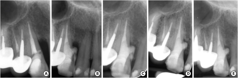
-
Abstract
PDFPubReaderePub
Iatrogenic perforations negatively impact the outcome of endodontic treatments. Studies on prognostic factors showed that perforations in the coronal third of the root with periodontal pocket formation have an unfavorable prognosis. A 36-year-old female was referred for endodontic evaluation of tooth #13 with a history of an iatrogenic perforation, happened 3 years ago. There was a sinus tract associated with perforation, 10 mm probing on the mesial and mesio-palatal, bleeding on probing, radiolucent lesion adjacent to the perforation and complete resorption of the interdental bone between teeth #13 and #12. After the treatment options were discussed, she chose to save the tooth. The tooth was accessed under rubber dam isolation, the perforation site was cleaned and disinfected using 0.5% sodium hypochlorite and sealed with calcium-enriched mixture cement. Eighteen months after treatment the tooth was functional and asymptomatic. The probing depths were normal without bleeding on probing. Radiographically, the interdental crestal bone formed between teeth #13 and #12. Despite all negative prognostic factors in this case (
i.e. , perforations in the coronal third, pocket formation, and radiolucent lesion), healing was unexpectedly achieved via non-surgical repair of the perforation. Further research on biological aspects of healing in the periodontium following iatrogenic perforations are recommended.-
Citations
Citations to this article as recorded by- Factors Influencing the Outcome of Iatrogenic Perforation Repair in Permanent Teeth—A Narrative Review
Saravanan Poorni, Srividhya Srinivasan, Thomas Clauder, Manali Ramakrishnan Srinivasan, Paul M. H. Dummer, Venkateshbabu Nagendrababu
Australian Endodontic Journal.2026;[Epub] CrossRef - Managing Internal Inflammatory Root Resorption and Perforation of a Mandibular Primary Molar: A Case Report With 15 Months Follow‐Up
Mana Mowji, Motahareh Khosrojerdi
Clinical Case Reports.2025;[Epub] CrossRef - Nonsurgical Management of Furcation Defects Using Cervical Sealing With Calcium–Silicate Cements: A Clinical Case Series
Saeed Asgary, Shamimul Hasan
Case Reports in Medicine.2025;[Epub] CrossRef - Nonsurgical Management of Simultaneous Double Lateral Root Perforations in Adjacent Teeth Using CBCT and MTA: A Case Report
Beyhan Başkan, Hatice Kübra Başkan, Beyza Güler, Ricardo Faria Ribeiro
Case Reports in Dentistry.2025;[Epub] CrossRef - Comprehensive review of composition, properties, clinical applications, and future perspectives of calcium-enriched mixture (CEM) cement: a systematic analysis
Saeed Asgary, Mahtab Aram, Mahta Fazlyab
BioMedical Engineering OnLine.2024;[Epub] CrossRef - External Cervical Resorption: A Volumetric Analysis on Evolution of Defects over Time
Ali Nosrat, Omid Dianat, Prashant Verma, Martin D. Levin, Jeffery B. Price, Anita Aminoshariae, Fabio Antonio Piola Rizzante
Journal of Endodontics.2023; 49(1): 36. CrossRef - Comparative evaluation of mineral trioxide aggregate, endoseal, and biodentine in furcation perforation repair
Udita Khare Baralay, Srinidhi Surya Raghavendra
Endodontology.2022; 34(1): 22. CrossRef - Calcium Silicate Cements Application in Lateral Root Perforation Repair: A Case Report with 16-Month Follow-Up
Juan G. Robledo, Pablo A. Rodríguez
Open Journal of Stomatology.2021; 11(08): 317. CrossRef - Vital Pulp Therapy as a Conservative Approach for Management of Invasive Cervical Root Resorption: A Case Series
Saeed Asgary, Mahdieh Nourzadeh, Prashant Verma, M. Lamar Hicks, Ali Nosrat
Journal of Endodontics.2019; 45(9): 1161. CrossRef
- Factors Influencing the Outcome of Iatrogenic Perforation Repair in Permanent Teeth—A Narrative Review
- 3,476 View
- 16 Download
- 9 Crossref
Research Articles
-
Removal efficacy and cytotoxicity of a calcium hydroxide paste using
N -2-methyl-pyrrolidone as a vehicle - Myung-Jin Lim, Hyun-Jin Jang, Mi-Kyung Yu, Kwang-Won Lee, Kyung-San Min
- Restor Dent Endod 2017;42(4):290-300. Published online October 20, 2017
- DOI: https://doi.org/10.5395/rde.2017.42.4.290
-
Abstract
PDFPubReaderePub
Objectives This study investigated the removal efficacy and cytotoxicity of a newly developed calcium hydroxide paste (cleaniCal, Maruchi) using
N -2-methyl-pyrrolidone (NMP) as a vehicle in comparison with ApexCal (Ivoclar Vivadent) and Calcipex II (Nishika), which use different vehicles such as polyethylene glycol and propylene glycol, respectively.Materials and Methods Thirty maxillary premolars with oval-shaped canals were divided into 3 groups and the teeth were filled with one of the pastes. After removal of the paste, micro-computed tomographic (μ-CT) imaging was obtained to assess the volume of residual paste in the root canal of each tooth. The teeth were then split longitudinally and the area of the paste-coated surface was evaluated by stereomicroscopy. The cytotoxicity of each product was assessed using an agar overlay assay. The effect of each vehicle on cell viability was evaluated using the 3-(4,5-dimethylthiazol-2-yl)-2,5-diphenyltetrazolium bromide (MTT) assay. The data were analyzed using one-way analysis of variance and Tukey's tests to detect any significance (
p < 0.05).Results In the μ-CT and stereomicroscopic analysis, cleaniCal exhibited less remnants of medicament than ApexCal and Calcipex. cleaniCal showed a higher cytotoxicity than the other pastes in the agar overlay assay. Furthermore, NMP exhibited lower cell viability compared to the other vehicles.
Conclusions cleaniCal showed better removal efficacy compared to the other products. However, clinicians should be aware of the higher cytotoxicity of the NMP-based material and consider its possible adverse effects on periradicular tissue when it is overfilled.
-
Citations
Citations to this article as recorded by- Removal of Calcium Hydroxide Paste Leaked Into the Maxillary Sinus
Dohee Kim, Young Kim, Jeong Joon Han
Ear, Nose & Throat Journal.2025;[Epub] CrossRef - Synergistic effects of reduced graphene oxide on the antibacterial activity of calcium hydroxide-based intracanal medicaments containing different vehicles
Mi-Ah Kim, Min-Kyeong Kim, Eun-Sook Kang, Kyung-San Min
Journal of Oral Science.2025; 67(1): 35. CrossRef - Lipoteichoic Acid from Lacticaseibacillus rhamnosus GG as a Novel Intracanal Medicament Targeting Enterococcus faecalis Biofilm Formation
Ji-Young Yoon, Somin Park, Dongwook Lee, Ok-Jin Park, WooCheol Lee, Seung Hyun Han
Journal of Microbiology.2024; 62(10): 897. CrossRef - Rheological properties and handling characteristics of four injectable calcium hydroxide pastes
Min-Jung KIM, In-Bog LEE
Dental Materials Journal.2024; 43(6): 796. CrossRef - Role of vehicles on antimicrobial efficacy of calcium hydroxide
Dikshya Purohit, Shronika, Pradyumna Misra, Gaurav Jain, Preeti Shukla
Asian Journal of Oral Health and Allied Sciences.2023; 13: 9. CrossRef - Conservative Management of Molar Incisor Hypomineralization Using Biomimetic Material in a 9-Year-Old Boy
Sahili Mungekar-Markandey, Ashwin Jawdekar
Journal of Dental Research and Review.2022; 9(4): 320. CrossRef - Sonic irrigation for removal of calcium hydroxide in the apical root canal: A micro-CT and light-coupled tracking analysis
Wonjoon Moon, Shin Hye Chung, Juhea Chang, Zhaoqiang Zhang
PLOS ONE.2022; 17(6): e0268791. CrossRef - Effect of N-2-methyl-pyrrolidone on Enterococcus faecalis biofilms
Mi-Ah KIM, Prasanna NEELAKANTAN, Kyung-San MIN
Dental Materials Journal.2022; 41(5): 774. CrossRef - Characterization, Antimicrobial Effects, and Cytocompatibility of a Root Canal Sealer Produced by Pozzolan Reaction between Calcium Hydroxide and Silica
Mi-Ah Kim, Vinicius Rosa, Prasanna Neelakantan, Yun-Chan Hwang, Kyung-San Min
Materials.2021; 14(11): 2863. CrossRef - Synthesis, structure, and theoretical studies of a calcium complex of a unique dianion derived from 1-methylpyrrolidin-2-one
Ray J. Butcher, Andrew P. Purdy, Paul A. Brown, Daniel Gunlycke
Acta Crystallographica Section E Crystallographic Communications.2021; 77(1): 70. CrossRef - Effect of a calcium hydroxide-based intracanal medicament containing N-2-methyl pyrrolidone as a vehicle against Enterococcus faecalis biofilm
Taegun KIM, Mi-Ah KIM, Yun-Chan HWANG, Vinicius ROSA, Massimo DEL FABBRO, Kyung-San MIN
Journal of Applied Oral Science.2020;[Epub] CrossRef
- Removal of Calcium Hydroxide Paste Leaked Into the Maxillary Sinus
- 2,777 View
- 16 Download
- 11 Crossref
- White mineral trioxide aggregate mixed with calcium chloride dihydrate: chemical analysis and biological properties
- Hany Mohamed Aly Ahmed, Norhayati Luddin, Thirumulu Ponnuraj Kannan, Khairani Idah Mokhtar, Azlina Ahmad
- Restor Dent Endod 2017;42(3):176-187. Published online April 17, 2017
- DOI: https://doi.org/10.5395/rde.2017.42.3.176
-
Abstract
PDFPubReaderePub
Objectives This study aimed to evaluate the chemical and biological properties of fast-set white mineral trioxide aggregate (FS WMTA), which was WMTA combined with calcium chloride dihydrate (CaCl2·2H2O), compared to that of WMTA.
Materials and Methods Surface morphology, elemental, and phase analysis were examined using scanning electron microscope (SEM), energy dispersive X-ray microanalysis (EDX), and X-ray diffraction (XRD), respectively. The cytotoxicity and cell attachment properties were evaluated on human periodontal ligament fibroblasts (HPLFs) using methyl-thiazol-diphenyltetrazolium (MTT) assay and under SEM after 24 and 72 hours, respectively.
Results Results showed that the addition of CaCl2·2H2O to WMTA affected the surface morphology and chemical composition. Although FS WMTA exhibited a non-cytotoxic profile, the cell viability values of this combination were lesser than WMTA, and the difference was significant in 7 out of 10 concentrations at the 2 time intervals (
p < 0.05). HPLFs adhered over the surface of WMTA and at the interface, after 24 hours of incubation. After 72 hours, there were increased numbers of HPLFs with prominent cytoplasmic processes. Similar findings were observed with FS WMTA, but the cells were not as confluent as with WMTA.Conclusions The addition of CaCl2·2H2O to WMTA affected its chemical properties. The favorable biological profile of FS WMTA towards HPLFs may have a potential impact on its clinical application for repair of perforation defects.
-
Citations
Citations to this article as recorded by- The effect of three additives on properties of mineral trioxide aggregate cements: a systematic review and meta-analysis of in vitro studies
Behnam Bolhari, Faranak Noori, Hadi Assadian, Amir Raee, Sholeh Ghabraei, Ahmad-Reza Shamshiri, Artak Heboyan
BMC Oral Health.2024;[Epub] CrossRef - Evaluation of sorption and solubility of materials based on calcium aluminate
Renata Josipovic, Violeta Petrovic, Marijana Popovic-Bajic, Irena Kuzmanovic-Radman, Mirjana Umicevic-Davidovic, Aleksandra Djeri, Slavoljub Zivkovic
Stomatoloski glasnik Srbije.2023; 70(1): 26. CrossRef - Chitosan-Based Accelerated Portland Cement Promotes Dentinogenic/Osteogenic Differentiation and Mineralization Activity of SHED
Hasan Subhi, Adam Husein, Dasmawati Mohamad, Nik Rozainah Nik Abdul Ghani, Asma-Abdullah Nurul
Polymers.2021; 13(19): 3358. CrossRef - Chemical modification of MTA and CEM cement to decrease setting time and improve bioactivity properties by adding alkaline salts
Faeze Jamali Zavare, Hanieh Nojehdehian, Maryam Moezizadeh, Mehdi Daneshpooya
Journal of Dental Research, Dental Clinics, Dental Prospects.2020; 14(1): 1. CrossRef - Biological effects of acid-eroded MTA Repair HP and ProRoot MTA on human periodontal ligament stem cells
Mar Collado-González, Sergio López-García, David García-Bernal, Ricardo E. Oñate-Sánchez, Christopher J. Tomás-Catalá, Jose M. Moraleda, Adrián Lozano, Leopoldo Forner, Francisco J. Rodríguez-Lozano
Clinical Oral Investigations.2019; 23(10): 3915. CrossRef - Comparative Cytocompatibility and Mineralization Potential of Bio-C Sealer and TotalFill BC Sealer
Sergio López-García, Miguel R. Pecci-Lloret, Julia Guerrero-Gironés, María P. Pecci-Lloret, Adrián Lozano, Carmen Llena, Francisco Javier Rodríguez-Lozano, Leopoldo Forner
Materials.2019; 12(19): 3087. CrossRef - Evaluation of changes in ion release and biological properties of NeoMTA‐Plus and Endocem‐MTA exposed to an acidic environment
F. J. Rodríguez‐Lozano, M. Collado‐González, S. López‐García, D. García‐Bernal, J. M. Moraleda, A. Lozano, L. Forner, L. Murcia, R. E. Oñate‐Sánchez
International Endodontic Journal.2019; 52(8): 1196. CrossRef
- The effect of three additives on properties of mineral trioxide aggregate cements: a systematic review and meta-analysis of in vitro studies
- 2,203 View
- 9 Download
- 7 Crossref
- Effect of three nanobiomaterials on microhardness of bleached enamel
- Maryam Khoroushi, Farinaz Shirban, Sara Kaveh, Samaneh Doustfateme
- Restor Dent Endod 2016;41(3):196-201. Published online July 14, 2016
- DOI: https://doi.org/10.5395/rde.2016.41.3.196
-
Abstract
PDFPubReaderePub
Objectives The aim of this
in vitro study was to evaluate the effect of incorporating three different nanobiomaterials into bleaching material on microhardness of bleached enamel.Materials and Methods The crowns of 24 extracted sound human molars were sectioned. Sixty enamel specimens (2 × 3 × 4 mm) were selected and divided into five groups (
n = 12): Group 1 received no bleaching procedure (control); Group 2 underwent bleaching with a 40% hydrogen peroxide (HP) gel; Groups 3, 4, and 5 were bleached with a 40% HP gel modified by incorporation of bioactive glass (BAG), amorphous calcium phosphate (ACP) and hydroxyapatite (HA), respectively. The enamel microhardness was evaluated. The differences in Knoop microhardness data of each group were analyzed by one-way ANOVA, followed bypost hoc Tukey tests.Results Significant differences were observed between the study groups. The enamel microhardness changes in Groups 1, 3, 4, and 5 were significantly lower than that of Group 2 (
p < 0.001).Conclusions Within the limitations of this study, it can be concluded that incorporation of each one of the three tested biomaterials as remineralizing agents might be effective in decreasing enamel microhardness changes subsequent to in-office bleaching.
-
Citations
Citations to this article as recorded by- Effects of bleaching agents containing nano-hydroxyapatite on sound and demineralized enamel: an in vitro study
Merve Haberal, Çiğdem Çelik
Odontology.2026;[Epub] CrossRef - Influence of 10% propolis extract on morphology, microhardness, chemical composition, and color of bleached enamel
Roberta Pimentel de Oliveira, Cíntia de Melo Silva, Elma Vieira Takeuchi, Geovanni Pereira Mitre, Lindalva Maria de Meneses Costa Ferreira, Jesuina Lamartine Nogueira Araújo, Hilton Túlio Costi, Maria Sueli da Silva Kataoka, Roseane Maria Ribeiro Costa, C
Odontology.2026;[Epub] CrossRef - Protective role of calcium-based agents in dental bleaching gels: insights from a systematic review and meta-analysis of clinical and laboratory evidence
Gabriel Pereira Nunes, Renata de Oliveira Alves, Geórgia Rondó Peres, Matheus Henrique Faccioli Ragghianti, Priscila Toninatto Alves de Toledo, Alexandre Henrique dos Reis Prado, Carla Ferreira-Baptista, Alberto Carlos Botazzo Delbem
Clinical Oral Investigations.2025;[Epub] CrossRef - Analysis of Enamel Surface and Shear Bond Strength with Orthodontic Bracket at Different Time Intervals After Bleaching Treatment
Yu-Jin Lee, Ji-Yeon Hong, Hye-Min Ku, Song-Yi Yang
Journal of Dental Hygiene Science.2025; 25(2): 130. CrossRef - Effect of strontium fluorophosphate bioactive glass on color, microhardness and surface roughness of bleached enamel
Shiza Yezdani, Monisha Khatri, Sampath Vidhya, Sekar Mahalaxmi
Technology and Health Care.2024; 32(1): 285. CrossRef - In vitro evaluation of the effect of addition of biomaterials to carbamide peroxide on the bleaching efficacy and microhardness of enamel
Sowmya Kavoor, M. A. Ranjini, Naval Abdul Aziz, H. K. Ashok, Roopa R. Nadig
Journal of Conservative Dentistry and Endodontics.2024; 27(3): 310. CrossRef - Effect of hydrogen peroxide and its combination with nano-hydroxyapatite or nano-bioactive glass on the enamel demineralization and tooth color: An in vitro study
Elham Kheradmand, Alirea Daneshkazemi, Abdolrahim Davari, Maede Kave, Solmaz Ghanbarnejad
Dental Research Journal.2023;[Epub] CrossRef - Over‐the‐counter bleaching agents can help with tooth whitening maintenance
Olívia Santana Jorge, Carolina Noronha Ferraz de Arruda, Rafaella Tonani Torrieri, Rocio Geng Vivanco, Fernanda de Carvalho Panzeri Pires‐de‐Souza
Journal of Esthetic and Restorative Dentistry.2022; 34(2): 328. CrossRef - Effect of Indigenously Developed Nano-Hydroxyapatite Crystals from Chicken Egg Shell on the Surface Hardness of Bleached Human Enamel
Divya Kunam, Vidhya Sampath, Sujatha Manimaran, Mahalaxmi Sekar
Contemporary Clinical Dentistry.2019; 10(3): 489. CrossRef - Influence of Time Intervals between Bleaching Procedures on Enamel Microhardness and Surface Roughness
Roberta Pimentel de Oliveira, Juliana Costa Pereira Baia, Mara Eliane Soares Ribeiro, Mario Honorato da Silva e Souza Junior, Sandro Cordeiro Loretto
The Open Dentistry Journal.2018; 12(1): 555. CrossRef
- Effects of bleaching agents containing nano-hydroxyapatite on sound and demineralized enamel: an in vitro study
- 2,117 View
- 8 Download
- 10 Crossref
Case Report
- Accidental injury of the inferior alveolar nerve due to the extrusion of calcium hydroxide in endodontic treatment: a case report
- Yooseok Shin, Byoung-Duck Roh, Yemi Kim, Taehyeon Kim, Hyungjun Kim
- Restor Dent Endod 2016;41(1):63-67. Published online January 6, 2016
- DOI: https://doi.org/10.5395/rde.2016.41.1.63
-
Abstract
PDFPubReaderePub
During clinical endodontic treatment, we often find radiopaque filling material beyond the root apex. Accidental extrusion of calcium hydroxide could cause the injury of inferior alveolar nerve, such as paresthesia or continuous inflammatory response. This case report presents the extrusion of calcium hydroxide and treatment procedures including surgical intervention. A 48 yr old female patient experienced Calcipex II extrusion in to the inferior alveolar canal on left mandibular area during endodontic treatment. After completion of endodontic treatment on left mandibular first molar, surgical intervention was planned under general anesthesia. After cortical bone osteotomy and debridement, neuroma resection and neurorrhaphy was performed, and prognosis was observed. But no improvement in sensory nerve was seen following surgical intervention after 20 mon. A clinician should be aware of extrusion of intracanal medicaments and the possibility of damage on inferior alveolar canal. Injectable type of calcium hydroxide should be applied with care for preventing nerve injury. The alternative delivery method such as lentulo spiral was suggested on the posterior mandibular molar.
-
Citations
Citations to this article as recorded by- Nicolau syndrome in endodontics: A narrative review on calcium hydroxide extrusion and its therapeutic risks
Manisha Chaudhary, Akash Kumar Giri, Ashok Ayer
Saudi Endodontic Journal.2026; 16(1): 12. CrossRef - Inferior alveolar nerve injury following calcium hydroxide extrusion: a case report with preventive insights and three-year follow-up
Sofia Drouri, Chaimae Ghouni, Khaoula Sadel, Hafsa El Merini
Frontiers in Dental Medicine.2026;[Epub] CrossRef - Invasion of Calcium Hydroxide Preparations Leading to Severe Chemical Nerve Injury Treated Through Nerve Repair Using Artificial Nerve Conduit: A Case Report
Akihiro Nishiyama, Andreas Neff, Takahiro Nakada, Takaharu Ariizumi, Akira Iwasaki, Keisuke Sugahara, Akira Katakura, Hannah Wesley
Case Reports in Dentistry.2025;[Epub] CrossRef - Automatic localization of inferior alveolar nerve canal in panoramic dental images
Uma Maheswari Pandyan, Banumathi Arumugam, Ulaganathan Gurunathan, Shahul Hameed Kopuli Ashkar Ali
Signal, Image and Video Processing.2022; 16(5): 1389. CrossRef - Inferior alveolar nerve injury due to the extrusion of calcium hydroxide during endodontic treatment: A case report
Metin Berk Kasapoğlu, Gülce Ecem Doğancalı
Australian Endodontic Journal.2022; 48(2): 342. CrossRef - Inferior alveolar nerve canal segmentation by local features based neural network model
P. Uma Maheswari, A. Banumathi, G. Ulaganathan, R. Yoganandha
IET Image Processing.2022; 16(3): 703. CrossRef - Microsurgical Repair of Inferior Alveolar Nerve Injuries Associated With Endodontic Treatment: Results on Sensory Function and Relief of Pain
Keith A. Sonneveld, Kristopher L. Hasstedt, Roger A. Meyer, Shahrokh C. Bagheri
Journal of Oral and Maxillofacial Surgery.2021; 79(7): 1434. CrossRef - The significance of diagnosis and treatment planning in periapical lesion overfilled with calcium hydroxide paste
Kyoung-Hwa Jung, Eun-Young Kwon, Youn-Kyung Choi, So-Yeun Kim, Hye-Mi Jeon, Jeong-Kil Park
Journal of Dental Rehabilitation and Applied Science.2021; 37(2): 95. CrossRef - The anatomical relationship between the roots of erupted permanent teeth and the mandibular canal: a systematic review
Michał Puciło, Mariusz Lipski, Magdalena Sroczyk-Jaszczyńska, Aleksandra Puciło, Alicja Nowicka
Surgical and Radiologic Anatomy.2020; 42(5): 529. CrossRef - Massive extrusion of calcium hydroxide paste containing barium sulphate during endodontic treatment
Jéssica Montenegro Fonsêca, Natália Rangel Palmier, Gleyson Kleber Amaral‐Silva, Lady Paola Aristizabal Arboleda, José Flávio Affonso Almeida, Mario Fernando de Goes, Pablo Agustin Vargas, Marcio Ajudarte Lopes, Alan Roger Santos‐Silva
Australian Endodontic Journal.2020; 46(2): 257. CrossRef - The double-edged sword of calcium hydroxide in endodontics
Alan H. Gluskin, Gordon Lai, Christine I. Peters, Ove A. Peters
The Journal of the American Dental Association.2020; 151(5): 317. CrossRef - Endodontic-related inferior alveolar nerve injuries: A review and a therapeutic flow chart
R. Castro, M. Guivarc'h, J.M. Foletti, J.H. Catherine, C. Chossegros, L. Guyot
Journal of Stomatology, Oral and Maxillofacial Surgery.2018; 119(5): 412. CrossRef - Relationship between Root Apices and the Mandibular Canal: A Cone-beam Computed Tomographic Comparison of 3 Populations
Alex Lvovsky, Shir Bachrach, Hyeon-Cheol Kim, Ajinkya Pawar, Oleg Levinzon, Joe Ben Itzhak, Michael Solomonov
Journal of Endodontics.2018; 44(4): 555. CrossRef - A case of high density abnormality in x-ray findings of mandible caused by leakage of root canal filling paste
Haruko Kashiwamura, Kyoko Oka, Yoko Tuchihashi, Hanako Yoshioka, Mayumi Kato, Atsuko Baba, Toyohiro Kagawa, Kazuhiko Okamura, Masao Ozaki
Pediatric Dental Journal.2017; 27(3): 162. CrossRef - Oral dysesthesia
Christopher J. Spencer, Gary D. Klasser
The Journal of the American Dental Association.2017; 148(12): 941. CrossRef - Microsurgical Decompression of Inferior Alveolar Nerve After Endodontic Treatment Complications
Bernardo Bianchi, Andrea Ferri, Andrea Varazzani, Michela Bergonzani, Enrico Sesenna
Journal of Craniofacial Surgery.2017; 28(5): 1365. CrossRef - Birefringence Detection of Calcipex II Endodontic Material in Chronic Granulomatous Lesion of Periapical Cyst
Soung Min Kim, Young Jae Yoo, Suk Keun Lee
The Korean Journal of Oral and Maxillofacial Pathology.2016; 40(2): 775. CrossRef
- Nicolau syndrome in endodontics: A narrative review on calcium hydroxide extrusion and its therapeutic risks
- 4,480 View
- 58 Download
- 17 Crossref
Research Articles
- Effect of dentin treatment on proliferation and differentiation of human dental pulp stem cells
- Minjeong Park, Nan-Sim Pang, Il-Young Jung
- Restor Dent Endod 2015;40(4):290-298. Published online September 23, 2015
- DOI: https://doi.org/10.5395/rde.2015.40.4.290
-
Abstract
PDFPubReaderePub
Objectives Sodium hypochlorite (NaOCl) is an excellent bactericidal agent, but it is detrimental to stem cell survival, whereas intracanal medicaments such as calcium hydroxide (Ca[OH]2) promote the survival and proliferation of stem cells. This study evaluated the effect of sequential NaOCl and Ca[OH]2 application on the attachment and differentiation of dental pulp stem cells (DPSCs).
Materials and Methods DPSCs were obtained from human third molars. All dentin specimens were treated with 5.25% NaOCl for 30 min. DPSCs were seeded on the dentin specimens and processed with additional 1 mg/mL Ca[OH]2, 17% ethylenediaminetetraacetic acid (EDTA) treatment, file instrumentation, or a combination of these methods. After 7 day of culture, we examined DPSC morphology using scanning electron microscopy and determined the cell survival rate with 3-(4,5-dimethylthiazol-2-yl)-2,5-diphenyltetrazolium bromide assay. We measured cell adhesion gene expression levels after 4 day of culture and odontogenic differentiation gene expression levels after 4 wk using quantitative real-time polymerase chain reaction.
Results DPSCs did not attach to the dentin in the NaOCl-treated group. The gene expression levels of fibronectin-1 and secreted phosphoprotein-1 gene in both the Ca[OH]2- and the EDTA-treated groups were significantly higher than those in the other groups. All Ca[OH]2-treated groups showed higher expression levels of dentin matrix protein-1 than that of the control. The dentin sialophosphoprotein level was significantly higher in the groups treated with both Ca[OH]2 and EDTA.
Conclusions The application of Ca[OH]2 and additional treatment such as EDTA or instrumentation promoted the attachment and differentiation of DPSCs after NaOCl treatment.
-
Citations
Citations to this article as recorded by- Effect of Intracanal Medications on the Viability of Human Periodontal Ligament‐Derived Mesenchymal Stem Cells
Arian Braido, Walbert de Andrade Vieira, Bruno Cazotti Pereira, Karina Gonzales Silvério, Paulo Henrique Gabriel, Aline Cristine Gomes Matta, Emerson Alves Martins, Adriana de Jesus Soares
Australian Endodontic Journal.2026; 52(1): 35. CrossRef - Biological, antimicrobial, and apical tissue dissolution effects of ambroxol hydrochloride as an irrigant for regenerative endodontics: an in vitro study
Letycia Accioly Simoes Coelho, Ester Alves Ferreira Bordini, Fernanda Balestrero Cassiano, Mirela Cesar Barros, Renato Menezes Silva, Flaviana Bombarda de Andrade, Diana Gabriela Soares, Marco Antonio Hungaro Duarte
BMC Oral Health.2026;[Epub] CrossRef - An injectable κ-carrageenan hydrogel carrier maintains antibiofilm activity while supporting dental pulp-derived stem cell viability in a dentin-based model
Muneera A. Alajlan, Rhodanne Nicole A. Lambarte, Terrence S. Sumague, Abdurahman A. Niazy
Journal of Dentistry.2026; 171: 106713. CrossRef - Effect of Intracanal Medications on Cell Adhesion, Viability, and Differentiation During Regenerative Endodontic Procedures: A Systematic Review of In Vitro Studies
Arian Braido, Luisa Coutinho Cardim, Aline Cristine Gomes Matta, Karina Gonzales Silvério, Paulo Henrique Gabriel, Emerson Alves Martins, Adriana de Jesus Soares, Walbert A. Vieira
Journal of Endodontics.2026;[Epub] CrossRef - Effect of erbium yttrium aluminium garnet laser dentin conditioning on dental pulp stem cells viability
Aryan Jafari, Mehdi Vatanpour, Nooshin Barikrow, Pouyan Razavi, Sohrab Tour Savadkouhi
Heliyon.2024; 10(5): e26954. CrossRef - Comparison of the effect of NaOCL, curcumin, and EDTA on differentiation, proliferation, and adhesion of dental pulp stem cells
Vahid Zand, Amin Salem Milani, Carolyn Primus, Marzie Aghazade, Hadi Mokhtari, Sabete Bagheri Sabzevar, Pardis Tehranchi
Journal of Oral Biology and Craniofacial Research.2023; 13(2): 347. CrossRef - Differential Effects of Extracellular Matrix Glycoproteins Fibronectin and Laminin-5 on Dental Pulp Stem Cell Phenotypes and Responsiveness
Hyungbin Lee, Allen Bae, John Kim, Karl Kingsley
Journal of Functional Biomaterials.2023; 14(2): 91. CrossRef - Assessment of the Influence of Various Concentrations of Sodium Hypochlorite on Stem Cell Derived From Human Exfoliated Deciduous Teeth (SHED) Proliferation and Differentiation
Viral Maru, Ashwini KB, Manisha Madkaikar, R K Sarada Devi, Ashita Gada, Salil Bapat
Cureus.2022;[Epub] CrossRef - Effects of sodium hypochlorite and ethylenediaminetetraacetic acid on proliferation, osteogenic/odontogenic differentiation, and mechanosensitive gene expression of human dental pulp stem cells
Yuejun Li, Changlong Jin, Shouliang Zhao, Han Xie
Tissue and Cell.2022; 79: 101955. CrossRef - Influence of ethylenediaminetetraacetic acid on regenerative endodontics: A systematic review
Alexandre H. dos Reis‐Prado, Lucas G. Abreu, Rogéria R. Fagundes, Sabrina de C. Oliveira, Marco C. Bottino, Antônio P. Ribeiro‐Sobrinho, Francine Benetti
International Endodontic Journal.2022; 55(6): 579. CrossRef - An in-vitro Comparative Evaluation of Quantitative Release of Transforming Growth Factor β-1 from Dentin upon the Action of Endodontic Irrigants, Medicaments, Ultrasonic Activation, and Low-Level Laser Irradiation
Anilkumar Akhila, V. P. Prabath Singh, Kerala R. Varma, Senthil V. Vasudevan, V. Sukhithasri, Salu Sasikumar
Amrita Journal of Medicine.2021; 17(2): 34. CrossRef - Non-Thermal Atmospheric Pressure Plasma-Conditioned Root Dentin Promotes Attraction and Attachment of Primary Human Dental Pulp Stem Cells in Real-Time Ex Vivo
Yeon-Jee Yoo, Min-Ji Kang, Hiran Perinpanayagam, Joo-Cheol Park, Seung-Ho Baek, Kee-Yeon Kum
Applied Sciences.2021; 11(15): 6836. CrossRef - The Effects of Intracanal Irrigants and Medicaments on Dental-Derived Stem Cells Fate in Regenerative Endodontics: An update
Sara Ayoub, Ali Cheayto, Sanaa Bassam, Mehdi Najar, Antoine Berbéri, Mohammad Fayyad-Kazan
Stem Cell Reviews and Reports.2020; 16(4): 650. CrossRef - An Immunofluorescence Study to Analyze Wound Healing Outcomes of Regenerative Endodontics in an Immature Premolar with Chronic Apical Abscess
Jeen Nee Lui, Wen Yi Lim, Domenico Ricucci
Journal of Endodontics.2020; 46(5): 627. CrossRef - Dynamic Irrigation Promotes Apical Papilla Cell Attachment in an Ex Vivo Immature Root Canal Model
Sanupong Prompreecha, Thanapat Sastraruji, Phumisak Louwakul, Tanida Srisuwan
Journal of Endodontics.2018; 44(5): 744. CrossRef - Odontoblast-like differentiation and mineral formation of pulpsphere derived cells on human root canal dentin in vitro
Jörg Neunzehn, Sandra Pötschke, Christian Hannig, Hans-Peter Wiesmann, Marie-Theres Weber
Head & Face Medicine.2017;[Epub] CrossRef - Effects of Epigallocatechin Gallate, an Antibacterial Cross-linking Agent, on Proliferation and Differentiation of Human Dental Pulp Cells Cultured in Collagen Scaffolds
Young-Sun Kwon, Hee-Jin Kim, Yun-Chan Hwang, Vinicius Rosa, Mi-Kyung Yu, Kyung-San Min
Journal of Endodontics.2017; 43(2): 289. CrossRef
- Effect of Intracanal Medications on the Viability of Human Periodontal Ligament‐Derived Mesenchymal Stem Cells
- 2,359 View
- 6 Download
- 17 Crossref
- Evaluation of the effects of two novel irrigants on intraradicular dentine erosion, debris and smear layer removal
- Melahat Görduysus, Selen Küçükkaya, Nursel Pekel Bayramgil, Mehmet Ömer Görduysus
- Restor Dent Endod 2015;40(3):216-222. Published online July 3, 2015
- DOI: https://doi.org/10.5395/rde.2015.40.3.216
-
Abstract
PDFPubReaderePub
Objectives To evaluate the effects of copolymer of acrylic acid and maleic acid (Poly[AA-
co -MA]) and calcium hypochlorite (Ca(OCl)2) on root canal dentin using scanning electron microscope (SEM).Materials and Methods Twenty-four single-rooted teeth were instrumented and the apical and coronal thirds of each root were removed, leaving the 5 mm middle thirds, which were then separated into two pieces longitudinally. The specimens were randomly divided into six groups and subjected to each irrigant for 5 min as follows: G1, Ca(OCl)2; G2, Poly(AA-
co -MA); G3, Ca(OCl)2 + Poly(AA-co -MA); G4, sodium hypochlorite (NaOCl); G5, ethylenediaminetetraacetic acid (EDTA); G6, NaOCl+EDTA. The specimens were prepared for SEM evaluation. Smear layer, debris and erosion scores were recorded by two blinded examiners. One image from G3 was analyzed with energy dispersive spectroscopy (EDS) on suspicion of precipitate formation. Data were analyzed using the Kruskal-Wallis and Dunn tests.Results G1 and G4 showed the presence of debris and smear layer and they were statistically different from G2, G3, G5 and G6 where debris and smear layer were totally removed (
p < 0.05). In G1 and G4, erosion evaluation could not be done because of debris and smear layer. G2, G3 and G5 showed no erosion, and there was no significant difference between them. G6 showed severe erosion and was statistically different from G2, G3 and G5 (p < 0.05). EDS microanalysis showed the presence of Na, P, and Ca elements on the surface.Conclusions Poly(AA-
co -MA) is effective in removing the smear layer and debris without causing erosion either alone or with Ca(OCl)2.-
Citations
Citations to this article as recorded by- Effect of Heat and Irrigation Agitation on the Smear Layer Removal Ability of Calcium Hypochlorite: A Scanning Electron Microscope Study
Damla Erkal, Kürşat Er
Microscopy Research and Technique.2026; 89(1): 65. CrossRef - Effect of sodium and calcium hypochlorite with or without surfactant on the adhesion of epoxy resin-based endodontic sealer
Guilherme Pauletto, Natália Franco Brum, Israel Bangel Carlotto, Lucas Saldanha da Rosa, Carlos Alexandre Souza Bier
Brazilian Journal of Oral Sciences.2025; 24: e254080. CrossRef - Bonding and Cleaning Effects of Irrigation Protocols Using Calcium Hypochlorite on the Post-space Radicular Dentin
JF Besegato, GR Bravo, JF Zaniboni, LG Belizário, ENM de Almeida, MB Gelio, WG Escalante-Otárola, MC Kuga
Operative Dentistry.2024; 49(6): E1. CrossRef - Effect of sodium hypochlorite and calcium hypochlorite on the apical sealing ability of endodontic sealers
Israel Bangel Carlotto, Natália Franco Brum, Guilherme Pauletto, Lucas Saldanha da Rosa, Carlos Alexandre Souza Bier
Brazilian Journal of Oral Sciences.2024; 23: e242700. CrossRef - Comparative evaluation of antifungal activity of Sodium Hypochlorite, Calcium Hypochlorite and modified Salt Solution associated with passive ultrasonic irrigation against Candida albicans - An In-Vitro study
Helen Thomas, D. N. Nirupama, Mohan Thomas Nainan, D. N. Naveen, C. Y. Ranjini, R. Vijay
Journal of Conservative Dentistry and Endodontics.2024; 27(2): 159. CrossRef - Effect of calcium hypochlorite as an irrigant alternative in the removal of methylene blue after photodynamic therapy under the post-space adhesive interface
João Felipe Besegato, Joatan Lucas de Sousa Gomes Costa, Joissi Ferrari Zaniboni, Giovanna Righetti Bravo, Jéssika Mayhara Pereira Morais, Wilfredo Gustavo Escalante-Otárola, Milton Carlos Kuga
Laser Physics.2023; 33(5): 055601. CrossRef - Effectiveness of Different Irrigation Techniques on Post Space Smear Layer Removal: SEM Evaluation
Alfredo Iandolo, Massimo Pisano, Dina Abdellatif, Alessandra Amato, Francesco Giordano, Alessio Buonavoglia, Giuseppe Sangiovanni, Mario Caggiano
Prosthesis.2023; 5(2): 539. CrossRef - The dynamic interplay of dietary acid pH and concentration during early-stage human enamel and dentine erosion
J. Pattem, J. Field, P. J. Waterhouse, M. J. German
Frontiers in Dental Medicine.2022;[Epub] CrossRef - Effect of EDTA, sodium, and calcium hypochlorite on the inorganic component of root canal dentin: A SEM analysis
Luana Roleto Cardoso, Flávia Emi Razera Baldasso, Débora Delai, Francisco Montagner, Patrícia Maria Poli Kopper
Microscopy Research and Technique.2019; 82(2): 128. CrossRef - Comparative Analysis of Manual, Rotary and Reciprocal Systems on Primary Teeth Root Canals: An In Vitro Scanning Electron Microscopy Study
İpek ARSLAN, Sema AYDINOĞLU, Ozgul BAYGIN, Tamer TÜZÜNER, Murat ŞİRİN
Cumhuriyet Dental Journal.2019; 22(3): 299. CrossRef - Regenerating a monoblock to obturate root canalsvia a mineralising strategy
Le Zhang, Quan-Li Li, Ying Cao, Yun Wang
Scientific Reports.2018;[Epub] CrossRef - Antibacterial Efficacy of Calcium Hypochlorite with Vibringe Sonic Irrigation System onEnterococcus faecalis: An In Vitro Study
Aysin Dumani, Hatice Korkmaz Guvenmez, Sehnaz Yilmaz, Oguz Yoldas, Zeliha Gonca Bek Kurklu
BioMed Research International.2016; 2016: 1. CrossRef
- Effect of Heat and Irrigation Agitation on the Smear Layer Removal Ability of Calcium Hypochlorite: A Scanning Electron Microscope Study
- 2,253 View
- 8 Download
- 12 Crossref
- Effect of intracanal medicaments used in endodontic regeneration procedures on microhardness and chemical structure of dentin
- Ghaeth Hamdon Yassen, George Joseph Eckert, Jeffrey Allen Platt
- Restor Dent Endod 2015;40(2):104-112. Published online December 24, 2014
- DOI: https://doi.org/10.5395/rde.2015.40.2.104
-
Abstract
PDFPubReaderePub
Objectives This study was performed to investigate the effects of different intracanal medicaments on chemical structure and microhardness of dentin.
Materials and Methods Fifty human dentin discs were obtained from intact third molars and randomly assigned into two control groups and three treatment groups. The first control group received no treatment. The second control group (no medicament group) was irrigated with sodium hypochlorite (NaOCl), stored in humid environment for four weeks and then irrigated with ethylenediaminetetraacetic acid (EDTA). The three treatment groups were irrigated with NaOCl, treated for four weeks with either 1 g/mL triple antibiotic paste (TAP), 1 mg/mL methylcellulose-based triple antibiotic paste (DTAP), or calcium hydroxide [Ca(OH)2] and finally irrigated with EDTA. After treatment, one half of each dentin disc was subjected to Vickers microhardness (
n = 10 per group) and the other half was used to evaluate the chemical structure (phosphate/amide I ratio) of treated dentin utilizing attenuated total reflection Fourier transform infrared spectroscopy (n = 5 per group). One-way ANOVA followed by Fisher's least significant difference were used for statistical analyses.Results Dentin discs treated with different intracanal medicaments and those treated with NaOCl + EDTA showed significant reduction in microhardness (
p < 0.0001) and phosphate/amide I ratio (p < 0.05) compared to no treatment control dentin. Furthermore, dentin discs treated with TAP had significantly lower microhardness (p < 0.0001) and phosphate/amide I ratio (p < 0.0001) compared to all other groups.Conclusions The use of DTAP or Ca(OH)2 medicaments during endodontic regeneration may cause significantly less microhardness reduction and superficial demineralization of dentin compared to the use of TAP.
-
Citations
Citations to this article as recorded by- The Effects of Sodium Dichloroisocyanurate and Calcium Hydroxide as Intracanal Medicaments on Microhardness and Fracture Resistance of Dentin: An In Vitro Study
Fereshte Sobhnamayan, Alireza Adl, Negin Firouzi, Saeed Moravej, Samina Gavahianjahromi
Clinical and Experimental Dental Research.2026;[Epub] CrossRef - Assessment of the dentine microhardness following the application of different intracanal medicaments. An in-vitro study
Sara Gamal Elgamal, Kareem Hamdy Ahmed Aly, Nadia Saeed Hosny
BDJ Open.2026;[Epub] CrossRef - Antibiotic approaches to endodontic regeneration
Samiya Riaz, Asmak Binti Abdul Samat, Muhammad Amber Fareed, Muhammad Sohail Zafar
Biomaterial Investigations in Dentistry.2026; 13: 271. CrossRef - Effect of allicin-incorporated graphene oxide hydrogel on dentin microhardness
Rathna Piriyanga, Manish Ranjan, Anand Sherwood, Mohammad Fareed, Mohmed Isaqali Karobari
BMC Oral Health.2025;[Epub] CrossRef - Recent Advances in Injectable Hydrogel Biotherapeutics for Regenerative Dental Medicine
Renan Dal‐Fabbro, Arwa Daghrery, Caroline Anselmi, Igor Paulino M. Soares, Alexandre Henrique dos Reis‐Prado, Pedro Henrique Chaves de Oliveira, Marco C. Bottino
Macromolecular Bioscience.2025;[Epub] CrossRef - Ex vivo analysis of clindamycin’s impact on dentin microhardness and surface chemistry
Mandana Naseri, Farshid Gholami, Kamyar Khosravi, Mohammadreza Vatankhah, Mohammad Atai, Omid Dianat
Journal of Conservative Dentistry and Endodontics.2025; 28(10): 982. CrossRef - Comparative evaluation of effect of modified triple antibiotic paste and calcium hydroxide as intracanal medicament on microhardness of root dentin: An in vitro study
Aparna Palekar, Piyush Mantri, Minal Awinashe, Basawaraj Biradar, Mukund Singh
Endodontology.2024; 36(2): 143. CrossRef - The Use of Nanofibers in Regenerative Endodontic Therapy—A Systematic Review
Sebastian Candrea, Alexandrina Muntean, Anida-Maria Băbțan, Antonia Boca, Claudia Nicoleta Feurdean, Ioana Roxana Bordea, Adina Bianca Boșca, Aranka Ilea
Fibers.2024; 12(5): 42. CrossRef - Effect of sodium hypochlorite and ethylenediaminotetraacetic acid activated by laser and ultrasonic energy on surface morphology and chemical composition of intracanal dentin
Adriana Katunarić, Sandra Flinčec Grgac, Dragana Gabrić, Božidar Pavelić, Ivona Bago
Microscopy Research and Technique.2024; 87(4): 818. CrossRef - Development of a Thermoresponsive Core–Shell Hydrogel for Sequential Delivery of Antibiotics and Growth Factors in Regenerative Endodontics
Sayna Shamszadeh, Saeed Asgary, Mohammad Akrami, Fatemeh Mashhadiabbas, Alireza Akbarzadeh Baghban, Forough Shams
Frontiers in Bioscience-Elite.2024;[Epub] CrossRef - Intracanal medicaments and coronal sealing materials influence on root fracture resistance and coronal discoloration: An in vitro study
Rasoul Sahebalam, Marzie Boskabady, Maryam Naghavi, Samira Dehghanitafti
Saudi Endodontic Journal.2024; 14(2): 199. CrossRef - Final irrigation with bioglass solution in regenerative endodontic procedure induces tissue formation inside the root canals, collagen maturation, proliferation cell and presence of osteocalcin
Kiani dos Santos de Paula, Alexandre Henrique dos Reis‐Prado, Witalo Pereira de Jesus, Juliana Goto, Lara Cancella de Arantes, Marina Verçosa, Luciano Tavares Angelo Cintra, Edilson Ervolino, Raphael Escorsim Szawka, Murilo Camuri Crovace, Ricardo Alves d
International Endodontic Journal.2024; 57(5): 586. CrossRef - In vitro analysis of compressive strength of root dentin on application of intracanal medicaments for different time periods
Kushal Kumar Ghosh, Sayantan Mukherjee, Paromita Mazumdar, Sahil Ali, Lovely Das
Journal of Conservative Dentistry and Endodontics.2024; 27(12): 1289. CrossRef - Effects of endodontic root canal irrigants on tooth dentin revealed by infrared spectroscopy: a systematic literature review
Hamza Elfarraj, Franco Lizzi, Kerstin Bitter, Paul Zaslansky
Dental Materials.2024; 40(8): 1138. CrossRef - Comparative evaluation of antimicrobial efficacy of triple antibiotic paste and propolis extract using three different vehicles as intracanal medicament: An in vitro study
Gunde Veronica, B. V Thimma Reddy, Uday K. Chowdary Birapu, Raghavendra K. Jadadoddi, R Hemanth Kumar, Kanamarlapudi V. Saikiran
Journal of Dr. NTR University of Health Sciences.2024; 13(4): 323. CrossRef - Outcome of Regenerative Endodontic Procedures in Nonvital Immature Permanent Teeth Using 2 Intracanal Medications: A Prospective Randomized Clinical Study
Aladdin Al-Qudah, Mohammad Almomani, Layla Hassoneh, Lama Awawdeh
Journal of Endodontics.2023; 49(7): 776. CrossRef - The Effect of Calcium Hydroxide, Triple Antibiotic Paste and Chlorhexidine on Pain in Teeth with Symptomatic Apical Periodontitis: A Randomised Controlled Trial
Asma Munir Khan, Irfana Khursheed Ahmed Gangoo, Naila Amir Ali, Mansoor Khan, Muhammad Qasim Javed, Mustafa Hussein AlAttas, Ayman M. Abulhamael, Hammam Ahmed Bahammam, Loai Alsofi, Rayan Suliman Al Yahya
International Journal of Environmental Research and Public Health.2023; 20(4): 3091. CrossRef - Comparative evaluation of the depth of penetration and postoperative pain associated with the use of continuous chelation using HEBP and standard irrigation protocol in the endodontic treatment of adult permanent nonvital teeth: A randomized controlled tr
Janhvi Samir Parekh, Mrunalini J. Vaidya, Vibha R. Hegde
Endodontology.2023; 35(4): 344. CrossRef - Regenerative Endodontics and Minimally Invasive Dentistry: Intertwining Paths Crossing Over Into Clinical Translation
Hisham Elnawam, Menatallah Abdelmougod, Ahmed Mobarak, Mai Hussein, Hamdy Aboualmakarem, Michael Girgis, Rania El Backly
Frontiers in Bioengineering and Biotechnology.2022;[Epub] CrossRef - Effect of calcium hydroxide on fracture resistance and microhardness of dentin in human teeth
Simar Sethi, Alpa Gupta, Ansy Hanna Kurian, Dax Abraham, Parul Chauhan, Kritika Aneja, Sucheta Jala, Arundeep Singh
Endodontology.2022; 34(4): 223. CrossRef - Evaluation of Effects of Various Irrigating Solutions on Chemical Structure of Root Canal Dentin Using FTIR, SEM, and EDS: An In Vitro Study
Indu Padmakumar, Dharam Hinduja, Abdul Mujeeb, Raghu Kachenahalli Narasimhaiah, Ashwini Kumar Saraswathi, Mubashir Baig Mirza, Ali Robaian, Syed Nahid Basheer, Mohmed Isaqali Karobari, Giuseppe Alessandro Scardina
Journal of Functional Biomaterials.2022; 13(4): 197. CrossRef - The Effects of Double Antibiotic Paste and Amoxicillin-clavulanate Paste Used in Endodontic Regeneration on Microhardness of Radicular Dentine: An In vitro Study
Meenu Madhukumar, Praveena Geetha, K. Radhakrishnan Nair, Manu Unnikrishnan
Journal of Pharmacy and Bioallied Sciences.2021; 13(Suppl 1): S510. CrossRef - The effect of intracanal medication variations on microhardness of simulated immature root dentin
Pınar Serdar Eymirli, Ayhan Eymirli, Emel Uzunoğlu Özyürek
Australian Endodontic Journal.2021; 47(3): 616. CrossRef - Antibacterial Effect and Bioactivity of Innovative and Currently Used Intracanal Medicaments in Regenerative Endodontics
Sarah Alfadda, Theeb Alquria, Eda Karaismailoglu, Hacer Aksel, Adham A. Azim
Journal of Endodontics.2021; 47(8): 1294. CrossRef - Evaluation of the Effect of Nitrofurantoin Paste as an Intracanal Medicament on the Chemical Structure of Radicular Dentine
Mewan Abdulrahman, Bestoon Faraj, Kawa Dizaye
Sulaimani Dental Journal.2021; 8(2): 8. CrossRef - Effect of hydrogel-based antibiotic intracanal medicaments on crown discoloration
Rayan B. Yaghmoor, Jeffrey A. Platt, Kenneth J. Spolnik, Tien Min Gabriel Chu, Ghaeth H. Yassen
Restorative Dentistry & Endodontics.2021;[Epub] CrossRef - Microhardness and Fracture Resistance of Radicular Dentin Treated with Different Concentrations of Calcium Hydroxide in Endodontic Regeneration Procedures
Sara N. Hashem, Maha Adel Elhousiny
Open Access Macedonian Journal of Medical Sciences.2021; 9(D): 324. CrossRef - Effect of medicaments used in endodontic regeneration on the morphological characteristics of bovine radicular dentin: Experimental immature tooth model
Maira C. Conte, Cleonice da Silveira Teixeira, Eduardo A. Bortoluzzi, Wilson T. Felippe, Luciane G. P. dos Santos, Mariana T. Pandolfo, Patrícia da Agostim Cancelier, Lucas da Fonseca Roberti Garcia
Microscopy Research and Technique.2020; 83(4): 354. CrossRef - Effect of intracanal medicaments on radicular dentine: An attenuated total reflection-Fourier-transform infrared spectroscopy analysis
Promila Verma, Afsana Ansari, Aseem Prakash Tikku, Anil Chandra, Rakesh Kumar Yadav, Ramesh Bharti, Rhythm Bains
Asian Journal of Oral Health and Allied Sciences.2020; 10: 3. CrossRef - Effect of Hydrogel-Based Antibiotic Intracanal Medicaments on Push-Out Bond Strength
Rayan B. Yaghmoor, Jeffrey A. Platt, Kenneth J. Spolnik, Tien Min Gabriel Chu, Ghaeth H. Yassen
European Journal of Dentistry.2020; 14(04): 575. CrossRef - Antimicrobial Susceptibility of Enterococcus faecalis Isolated From Root Canal: An In Vitro Study
Nazanin Zargar, Mohammad J Nasiri, Hengameh Ashraf, Bahareh Hajikhani, Shirin Etminani Esfahani, Maryam Etminani Esfahani
Avicenna Journal of Pharmaceutical Research.2020; 1(2): 60. CrossRef - The Effect of Calcium Hydroxide and Nano–calcium Hydroxide on Microhardness and Superficial Chemical Structure of Root Canal Dentin: An Ex Vivo Study
Mandana Naseri, Leila Eftekhar, Farshid Gholami, Mohammad Atai, Omid Dianat
Journal of Endodontics.2019; 45(9): 1148. CrossRef - Antimicrobial efficacy of clindamycin and triple antibiotic paste as root canal medicaments on tubular infection: An in vitro study
Nazanin Zargar, Motahare Rayat Hosein Abadi, Mohammad Sabeti, Zahra Yadegari, Alireza Akbarzadeh Baghban, Omid Dianat
Australian Endodontic Journal.2019; 45(1): 86. CrossRef - Influence of silver nanoparticle solution on the mechanical properties of resin cements and intrarradicular dentin
Thaís Yumi Umeda Suzuki, Juno Gallego, Wirley Gonçalves Assunção, André Luiz Fraga Briso, Paulo Henrique dos Santos, Chun-Pin Lin
PLOS ONE.2019; 14(6): e0217750. CrossRef - Triple antibiotic paste: momentous roles and applications in endodontics: a review
Ardavan Parhizkar, Hanieh Nojehdehian, Saeed Asgary
Restorative Dentistry & Endodontics.2018;[Epub] CrossRef - Retention of BioAggregate and MTA as coronal plugs after intracanal medication for regenerative endodontic procedures: an ex vivo study
Suzan Abdul Wanees Amin, Shaimaa Ismail Gawdat
Restorative Dentistry & Endodontics.2018;[Epub] CrossRef - Fourier transform infrared spectroscopy (FTIR) application chemical characterization of enamel, dentin and bone
Camila de Carvalho Almança Lopes, Pedro Henrique Justino Oliveira Limirio, Veridiana Resende Novais, Paula Dechichi
Applied Spectroscopy Reviews.2018; 53(9): 747. CrossRef - Chlorhexidine Prevents Root Dentine Mineral Loss and Fracture Caused by Calcium Hydroxide over Time
Michael Ranniery Garcia Ribeiro, Érika Bárbara Abreu Fonseca Thomaz, Darlon Martins Lima, Tarcísio Jorge Leitão, José Bauer, Soraia De Fátima Carvalho Souza
International Journal of Dentistry.2017; 2017: 1. CrossRef - Attachment and proliferation of dental pulp stem cells on dentine treated with different regenerative endodontic protocols
M. A. Alghilan, L. J. Windsor, J. Palasuk, G. H. Yassen
International Endodontic Journal.2017; 50(7): 667. CrossRef - Second-generation Platelet Concentrate (Platelet-rich Fibrin) as a Scaffold in Regenerative Endodontics: A Case Series
Hengameh Bakhtiar, Shahram Esmaeili, Setareh Fakhr Tabatabayi, Mohammad Reza Ellini, Mohammad Hossein Nekoofar, Paul M.H. Dummer
Journal of Endodontics.2017; 43(3): 401. CrossRef - Antibacterial Effects of Antimicrobials Used in Regenerative Endodontics against Biofilm Bacteria Obtained from Mature and Immature Teeth with Necrotic Pulps
Jordon C. Jacobs, Alex Troxel, Ygal Ehrlich, Kenneth Spolnik, Josef S. Bringas, Richard L. Gregory, Ghaeth H. Yassen
Journal of Endodontics.2017; 43(4): 575. CrossRef - Triple Antibiotic Polymer Nanofibers for Intracanal Drug Delivery: Effects on Dual Species Biofilm and Cell Function
Divya Pankajakshan, Maria T.P. Albuquerque, Joshua D. Evans, Malgorzata M. Kamocka, Richard L. Gregory, Marco C. Bottino
Journal of Endodontics.2016; 42(10): 1490. CrossRef - Inhibitory effect of gels loaded with a low concentration of antibiotics against biofilm formation by Enterococcus faecalis and Porphyromonas gingivalis
Amnah A. Algarni, Ghaeth H. Yassen, Richard L. Gregory
Journal of Oral Science.2015; 57(3): 213. CrossRef
- The Effects of Sodium Dichloroisocyanurate and Calcium Hydroxide as Intracanal Medicaments on Microhardness and Fracture Resistance of Dentin: An In Vitro Study
- 3,072 View
- 35 Download
- 43 Crossref
Review Article
-
Antimicrobial effect of calcium hydroxide as an intracanal medicament in root canal treatment: a literature review - Part II.
in vivo studies - Dohyun Kim, Euiseong Kim
- Restor Dent Endod 2015;40(2):97-103. Published online December 9, 2014
- DOI: https://doi.org/10.5395/rde.2015.40.2.97
-
Abstract
PDFPubReaderePub
The first part of this study reviewed the characteristics of calcium hydroxide (Ca(OH)2) and summarized the results of
in vitro studies related to its antimicrobial effects. The second part of this review coversin vivo studies including human clinical studies and animal studies. The use of Ca(OH)2 as an intracanal medicament represented better histological results in animal studies. However, human clinical studies showed limited antimicrobial effects that microorganisms were reduced but not eliminated through the treatment, and that some species had resistance to Ca(OH)2. Most of clinical outcome studies supported that there is no improvement in healing of periapical lesions when Ca(OH)2 was applied between appointments. Further studies are required for the antimicrobial effects of Ca(OH)2, and search for the ideal material and technique to completely clean infected root canals should be continued.-
Citations
Citations to this article as recorded by- Endodontic Intracanal Medicaments and Agents
Anu Priya Guruswamy Pandian, Depti Bellani, Ritya Mary Jibu, Varsha Agnihotri
Dental Clinics of North America.2026; 70(1): 45. CrossRef - Exploratory Evaluation of a Sodium Iodide-Based Root Canal Filling Formulation in a Canine Model of Enterococcus faecalis-Induced Periapical Inflammation
Saeromi Jun, Sak Lee, Jong-Soo Kim, Min-Cheol Song, Ji-Sun Shin, Yu-Jin Kim, Jung-Wook Kim, Jung-Hwan Lee
Pharmaceutics.2026; 18(4): 493. CrossRef - Antibacterial Efficacy of Graphene Nanoparticles against Enterococcus faecalis: In Vitro Study
Omer Sheriff Sultan, Preena Sidhu, Kiran Rehman, Thiagrajan Madheswaran, Amalraj Fabian Davamani
European Journal of Dentistry.2025; 19(01): 103. CrossRef - Evaluation of antibacterial efficacy of propolis as an intercanal medicament against Enterococcus faecalis (a randomized controlled in vitro study)
Alaa Almahameed, Joul Kassis, Magd Aboud, Kinda Layous
Heliyon.2025; 11(1): e41733. CrossRef - Neoangiogenetic potential of Nd:YAG 1064 nm photobiomodulation in non-surgical healing of trauma induced periapical bone defects: a clinicalprospective pilot study
Jagruti Mutalikdesai, Rhythm Bains, Aseem P. Tikku, Ramesh Bharti, Vijay Kumar Shakya, Sukriti Kumar, Promila Verma
Lasers in Medical Science.2025;[Epub] CrossRef - Jasminum-based Nano-reinforced Calcium Hydroxide Reduces Postoperative Pain in Symptomatic Apical Periodontitis: A Randomized Controlled Trial
Nehal Amir, Emaan Mansoor, Nabiha Eeman, Muhammad Nouman Ahmed, Ezza Mansoor, Efrah Mansoor, Khadim Hussain, Vera Afreixo, Afsheen Mansoor, João Filipe Brochado Martins, Paulo J. Palma
Journal of Endodontics.2025; 51(8): 996. CrossRef - Management of Enterococcus faecalis associated endodontic infection using gold nanogel: An in-vitro study
Omer Sheriff Sultan, Goh Chong Ming Jonathan, Seow Liang Lin, Fabian Davamani, Preena Sidhu, Minati Choudhury
The Saudi Dental Journal.2025;[Epub] CrossRef - Nano-sized Calcium Hydroxide-Zinc Oxide Intracanal Medicament: Physicochemical Characterization and Potential Endodontic Applications
Kashmiri Chowdhury, S. Delphine Priscilla Antony, Praveen Kumar Elango, Pradeep Solete, Shreshtha Muskan
Journal of International Oral Health.2025; 17(3): 196. CrossRef - Comparative Evaluation of Tooth Discoloration Induced by an Experimental Antibiotic Paste Modified with Nano Chitosan: An In Vitro Study
Mohamed Ahmed Elsayed, Md Sofiqul Islam, Safiya Ali, Zainab Hussain, Muhammed Mustahsen Rahman, Okba Mahmoud
Dentistry Journal.2025; 13(7): 307. CrossRef - An in vitro study on the antimicrobial efficacy of a calcium hydroxide versus a calcium silicate-based endodontic medicament
Dheepthi Jana, Eda Dzinovic, Ahmed Almaroof, Dipti Mehta, Sherif Elsharkawy, Sanjukta Deb, Sadia Niazi
Clinical Oral Investigations.2025;[Epub] CrossRef - Effect of canal medicaments triple antibiotic paste, Bio-C Temp, and Nano-silver gel activated by visible blue light on canal dentin microhardness and extrusion bond strength of AH plus sealer: A SEM and EDX analysis
Ahoud Alshamrani, Laila AlDeeb, Thamer Almohareb, Khold Alahdal, Ahmed Maawadh, Ali Alrahlah
Photodiagnosis and Photodynamic Therapy.2024; 47: 104088. CrossRef - Garre’s osteomyelitis of the mandible managed by nonsurgical re-endodontic treatment
Heegyun Kim, Jiyoung Kwon, Hyun-Jung Kim, Soram Oh, Duck-Su Kim, Ji-Hyun Jang
Restorative Dentistry & Endodontics.2024;[Epub] CrossRef - Hydrogel-Based Intracanal Medicaments in Endodontics: A Systematic Review of Development and Antibacterial Efficacy
Rathna Piriyanga, Manish Ranjan, Anand Sherwood, Swathi Priyadharshini
Journal of International Oral Health.2024; 16(6): 449. CrossRef - The advancement in irrigation solution within the field of endodontics, A Review
Fatima Fahad , Raghad A Al-Hashimi , Munther J Hussain
Journal of Baghdad College of Dentistry.2024; 36(1): 54. CrossRef - Antimicrobial Action of Novel Panax ginseng Paste, Calcium Hydroxide and Bio-C Temp as an Intracanal Medicament Against Enterococcus faecalis and Streptococcus mutans
Zadeno Kithan, Sonali Taneja, Abhik Mukherjee
Journal of Natural Remedies.2024; : 2705. CrossRef - Antimicrobial Efficacy of Two Different Calcium Hydroxide Endodontic Dressings on the Eradication of Enterococcus faecalis in Single-Rooted Canals: An In Vitro Study
Paola G Rumhein, Kinda J Layous, Hassan Achour, Mudar Mohammad Mousa, Haya Deeb, Mohammad Y Hajeer
Cureus.2023;[Epub] CrossRef - Influence of access cavity design on calcium hydroxide removal using different cleaning protocols: a confocal laser scanning microscopy study
Seda Falakaloğlu, Merve Yeniçeri Özata, Betül Güneş, Emmanuel João Nogueira Leal Silva, Mustafa Gündoğar, Burcu Güçyetmez Topal
Restorative Dentistry & Endodontics.2023;[Epub] CrossRef - Antimicrobial potential of new diclofenac hydrogels for disinfection in regenerative endodontics: An in vitro and ex vivo study
Matilde Ruiz‐Linares, Javier F. Monroy‐Rojas, Carmen Solana, Pilar Baca, Beatriz Aguado, Ana Soriano‐Lerma, María Teresa Arias‐Moliz, Carmen María Ferrer‐Luque
International Endodontic Journal.2023; 56(1): 103. CrossRef - Comparative evaluation of the antibacterial effect ofAllium sativum, calcium hydroxide and their combination as intracanal medicaments in infected mature anterior teeth: A randomized clinical trial
Shaimaa Mohamed Mahfouz Omer, Dalia Abd‐Allah Mohamed, Reham Mohamed Ali Abdel Latif
International Endodontic Journal.2022; 55(10): 1010. CrossRef - Antibacterial efficacy of antibiotic pastes versus calcium hydroxide intracanal dressing: A systematic review and meta-analysis of ex vivo studies
Mohammadreza Vatankhah, Kamyar Khosravi, Nazanin Zargar, Armin Shirvani, MohammadHossein Nekoofar, Omid Dianat
Journal of Conservative Dentistry.2022; 25(5): 463. CrossRef - Evaluation of the antibacterial activity of Lactobacilli probiotics supernatants against Enterococcus faecalis (in-vitro study)
Shymaa Shaaban, Gamal M. Hamad, Salma Genena, Marwa A. Meheissen, Sybel Moussa
BMC Oral Health.2022;[Epub] CrossRef - Superior Antibacterial Efficacy of Calcium Hydroxide–Propolis Paste against Enterococcus faecalis in Infected Root Canals: A Randomized Controlled In Vitro Study
Alessandro Marino, Luca De Santis, Paolo Romano
Interdisciplinary Research in Medical Sciences Specialty.2022; 2(1): 49. CrossRef - Post-operative Pain and Antibacterial Efficacy of Silver Nanoparticles Formulations Intracanal Medication: A Randomized Controlled Clinical Study
Nermine Hassan, Alaa Diab, Geraldine Ahmed
Open Access Macedonian Journal of Medical Sciences.2021; 9(D): 248. CrossRef - EFFICACY OF VARIOUS LASER-ASSISTED IRRIGATION ACTIVATION TECHNIQUES ON CALCIUM HYDROXIDE REMOVAL
Ezgi DOĞANAY YILDIZ, Fatma DURNA YURTSEVEN, Dilek HANÇERLİOĞULLARI
Atatürk Üniversitesi Diş Hekimliği Fakültesi Dergisi.2021; : 1. CrossRef - Experimental study the anti-inflammatory and osteo-regenerative qualities of the paste based on symphytum officinale tincture and calcium hydroxide
Iryna Kostyiuk, Victor Kostiuk, Halyna Kimak, Yuriy Oktysyuk, Lilia Tarnavska
Pharmacia.2021; 68(3): 585. CrossRef - Complete Obturation—Cold Lateral Condensation vs. Thermoplastic Techniques: A Systematic Review of Micro-CT Studies
Shilpa Bhandi, Mohammed Mashyakhy, Abdulaziz S. Abumelha, Mazen F. Alkahtany, Mohamed Jamal, Hitesh Chohan, A. Thirumal Raj, Luca Testarelli, Rodolfo Reda, Shankargouda Patil
Materials.2021; 14(14): 4013. CrossRef - Preserving pulp vitality: part two - vital pulp therapies
David Edwards, Simon Stone, Oliver Bailey, Phillip Tomson
British Dental Journal.2021; 230(3): 148. CrossRef - Evaluation of the Antibacterial Efficacy of Daptomycin, Gentamicin, and Calcium Hydroxide—Antibiotic Combinations on Enterococcus faecalis Dentinal Biofilm: An In Vitro Study
Arunajatesan Subbiya, Suresh Mitthra, Kesavaram Padmavathy, Krishnan Mahalakshmi, Alagarsamy Venkatesh, Kotishwaran Gayathri
The Journal of Contemporary Dental Practice.2021; 22(2): 128. CrossRef - Evaluation of the influence of sprinkling powdered slaked lime on microorganisms for the prevention of domestic animal infectious diseases
Miho Mori, Yoshikazu Sakagami, Yousuke Hamazaki, Toru Jojima
Environmental Technology.2019; 40(23): 3094. CrossRef - Efficacy of XP-Endo finisher in removal of calcium hydroxide from root canal system: A systematic review
Shruti Kamath, Rajesh Shetty, Soumya Shetty, Nikhil Nighot, Karuna Ramnani, Dhananjay Bhujbal
Journal of the International Clinical Dental Research Organization.2019; 11(2): 54. CrossRef - Calcium Hydroxide as Intracanal Medicament in Pulp Necrosis with Periapical Lesion : A Case Report
Elvi Sahara, Rahmi Alma Farah, Myrna Nurlatifah Zakaria
Key Engineering Materials.2019; 829: 226. CrossRef - Antimicrobial efficacy of clindamycin and triple antibiotic paste as root canal medicaments on tubular infection: An in vitro study
Nazanin Zargar, Motahare Rayat Hosein Abadi, Mohammad Sabeti, Zahra Yadegari, Alireza Akbarzadeh Baghban, Omid Dianat
Australian Endodontic Journal.2019; 45(1): 86. CrossRef - 2016 ASE undergraduate essay competition candidate information
Alice Chen
Australian Endodontic Journal.2018; 44(1): 7. CrossRef - Efficacy of self‐adjusting file, XP‐endo finisher and passive ultrasonic irrigation on the removal of calcium hydroxide paste from an artificial standardized groove
Anda Kfir, Nuphar Blau‐Venezia, Tomer Goldberger, Itzhak Abramovitz, Ronald Wigler
Australian Endodontic Journal.2018; 44(1): 26. CrossRef - Local drug delivery in endodontics: A literature review
Shin Hye Chung, Young-Seok Park
Journal of Drug Delivery Science and Technology.2017; 39: 334. CrossRef - Antimicrobial activity of Annona crassiflora Mart. against Candida albicans
de Mendonça Cavalcante Amaro, Antonio Lisboa Ribeiro Junior Karlos, CameloPessoa de Azevedo Ximenes Eulália, Porfirio Silva Zenaldo, Ivo Limeira dos Reis José, Euzebio Goulart de Santana Antonio
Journal of Medicinal Plants Research.2017; 11(13): 253. CrossRef - Antifungal effects of synthetic human β-defensin 3-C15 peptide
Sang-Min Lim, Ki-Bum Ahn, Christine Kim, Jong-Won Kum, Hiran Perinpanayagam, Yu Gu, Yeon-Jee Yoo, Seok Woo Chang, Seung Hyun Han, Won-Jun Shon, Woocheol Lee, Seung-Ho Baek, Qiang Zhu, Kee-Yeon Kum
Restorative Dentistry & Endodontics.2016; 41(2): 91. CrossRef - Microbiology of Root Canal Infections
Marjut Sakko, Leo TjÄDerhane, Riina Rautemaa-Richardson
Primary Dental Journal.2016; 5(2): 84. CrossRef
- Endodontic Intracanal Medicaments and Agents
- 3,937 View
- 46 Download
- 38 Crossref
Research Article
- Calcium hydroxide dressing residues after different removal techniques affect the accuracy of Root-ZX apex locator
- Emel Uzunoglu, Ayhan Eymirli, Mehmet Özgür Uyanik, Semra Çalt, Emre Nagas
- Restor Dent Endod 2015;40(1):44-49. Published online November 5, 2014
- DOI: https://doi.org/10.5395/rde.2015.40.1.44
-
Abstract
PDFPubReaderePub
Objectives This study compared the ability of several techniques to remove calcium hydroxide (CH) from the root canal and determined the influence of CH residues on the accuracy of the electronic apex locator.
Materials and Methods Root canals of 90 human maxillary lateral incisors with confirmed true working length (TWL) were prepared and filled with CH. The teeth were randomly assigned to one of the experimental groups according to the CH removal technique (
n = 14): 0.9% saline; 0.9% saline + master apical file (MAF); 17% ethylenediamine tetraacetic acid (EDTA); 17% EDTA + MAF; 5.25% sodium hypochlorite (NaOCl); 5.25% NaOCl + MAF. Six teeth were used as negative control. After CH removal, the electronic working length was measured using Root-ZX (Morita Corp.) and compared with TWL to evaluate Root-ZX accuracy. All specimens were sectioned longitudinally, and the area of remaining CH (CH) and total canal area were measured using imaging software.Results The EDTA + MAF and NaOCl + MAF groups showed better CH removal than other groups (
p < 0.05). Root-ZX reliability to prevent overestimated working length to be > 85% within a tolerance of ± 1.0 mm (p < 0.05). There was strong negative correlation between amount of CH residues and EAL accuracy (r = -0.800 for ± 0.5 mm;r = -0.940 for ± 1.0 mm).Conclusions The mechanical instrumentation improves the CH removal of irrigation solutions although none of the techniques removed the dressing completely. Residues of CH medication in root canals affected the accuracy of Root-ZX adversely.
-
Citations
Citations to this article as recorded by- Evaluation of the Effect of Calcium Hydroxide Residues Including Different Vehicles on the Accuracy of Electronic Apex Locators
Simay Koç, Damla Erkal, Dide Tekinarslan, Kürs¸at Er
Journal of Advanced Oral Research.2025; 16(1): 54. CrossRef - Evaluation of heated sodium hypochlorite’s effect on the accuracy of contemporary electronic apex locators: an in vitro study
İkbal Sena Çelebi Keskin, Turgut Yağmur Yalçın
BMC Oral Health.2025;[Epub] CrossRef - Factors influencing the accuracy of electronic apex locators: A scoping review
Shayan Golkar, Abbasali Khademi, Amin Saatchi, Amir Ghorani, Pedram Iranmanesh
Dental Research Journal.2025;[Epub] CrossRef - Effectiveness of a new irrigation solution -RISA- on removing calcium hydroxide from artificial standardized grooves in root canals - an in vitro study
İpek Eraslan Akyüz, Salih Düzgün, Hüseyin Sinan Topçuoğlu
BMC Oral Health.2025;[Epub] CrossRef - Comparative evaluation of the accuracy of electronic apex locators and cone-beam computed tomography in detection of root canal perforation and working length during endodontic retreatment
Simay Koç, Hatice Harorlı, Alper Kuştarcı
BMC Oral Health.2024;[Epub] CrossRef - Effects of Intracanal Medicaments on the Measurement Accuracy of Four Apex Locators: An In Vitro Study
Hamza Cudal, Tuğrul Aslan, Bertan Kesim
Meandros Medical and Dental Journal.2023; 24(3): 215. CrossRef - Electronic Apex Locators and their Implications in Contemporary Clinical Practice: A Review
Zainab Shirazi, Anas Al-Jadaa, Abdul Rahman Saleh
The Open Dentistry Journal.2023;[Epub] CrossRef - Influence of Apical Patency, Coronal Preflaring and Calcium Hydroxide on the Accuracy of Root ZX Apex Locator for Working Length Determination: An In Vitro Study
Mostafa Godiny, Reza Hatam, Roya Safari-Faramani, Atefeh Khavid, Mohammad Reza Rezaei
Journal of Advanced Oral Research.2022; 13(1): 38. CrossRef - Endodontic cement penetration after removal of calcium hydroxide dressing using XP-endo finisher
Alyssa Sales dos Santos, Maria Aparecida Barbosa de Sá, Marco Antônio Húngaro Duarte, Martinho Campolina Rebello Horta, Frank Ferreira Silveira, Eduardo Nunes
Brazilian Oral Research.2022;[Epub] CrossRef - Efficacy of glycolic acid for the removal of calcium hydroxide from simulated internal Resorption cavities
Cangül Keskin, Ali Keleş, Öznur Sarıyılmaz
Clinical Oral Investigations.2021; 25(7): 4407. CrossRef - Accuracy of electronic apex locator in the presence of different irrigating solutions
Padmanabh Jha, Vineeta Nikhil, Shalya Raj, Rohit Ravinder, Preeti Mishra
Endodontology.2021; 33(4): 232. CrossRef - Farklı Kanal İçi Ortamların Apeks Bulucuların Doğruluğu Üzerine Etkisi
Asena OKUR, Tuğrul ASLAN, Burak SAĞSEN
Selcuk Dental Journal.2021; 8(3): 859. CrossRef - Evaluation of the accuracy of different apex locators in determiningthe working length during root canal retreatment
Pelin Tufenkci, Aylin Kalaycı
Journal of Dental Research, Dental Clinics, Dental Prospects.2020; 14(2): 125. CrossRef - Influence of calcium hydroxide residues after using different irrigants on the accuracy of two electronic apex locators: An in vitro study
NooshinSadat Shojaee, Zahra Zaeri, MohammadMehdi Shokouhi, Fereshteh Sobhnamayan, Alireza Adl
Dental Research Journal.2020; 17(1): 48. CrossRef - The Effect of Calcium Hydroxide and File Sızes on the Accuracy of the Electronic Apex Locator in Simulated Immature Teeth
Leyla AYRANCİ, Ahmet ÇETİNKAYA, Serkan ÖZKAN
Middle Black Sea Journal of Health Science.2019; 5(3): 273. CrossRef - The Effect of File Size and Type and Irrigation Solutions on the Accuracy of Electronic Apex Locators: AnIn VitroStudy on Canine Teeth
Maciej Janeczek, Piotr Kosior, Dagmara Piesiak-Pańczyszyn, Krzysztof Dudek, Aleksander Chrószcz, Agnieszka Czajczyńska-Waszkiewicz, Małgorzata Kowalczyk-Zając, Aleksandra Gabren-Syller, Karol Kirstein, Aleksandra Skalec, Ewelina Bryła, Maciej Dobrzyński
BioMed Research International.2016; 2016: 1. CrossRef
- Evaluation of the Effect of Calcium Hydroxide Residues Including Different Vehicles on the Accuracy of Electronic Apex Locators
- 2,337 View
- 10 Download
- 16 Crossref
Review Articles
- Current perspectives of bio-ceramic technology in endodontics: calcium enriched mixture cement - review of its composition, properties and applications
- Shivani Utneja, Ruchika Roongta Nawal, Sangeeta Talwar, Mahesh Verma
- Restor Dent Endod 2015;40(1):1-13. Published online November 3, 2014
- DOI: https://doi.org/10.5395/rde.2015.40.1.1
-
Abstract
PDFPubReaderePub
Advancements in bio-ceramic technology has revolutionised endodontic material science by enhancing the treatment outcome for patients. This class of dental materials conciliates excellent biocompatibility with high osseoconductivity that render them ideal for endodontic care. Few recently introduced bio-ceramic materials have shown considerable clinical success over their early generations in terms of good handling characteristics. Calcium enriched mixture (CEM) cement, Endosequence sealer, and root repair materials, Biodentine and BioAggregate are the new classes of bio-ceramic materials. The aim of this literature review is to present investigations regarding properties and applications of CEM cement in endodontics. A review of the existing literature was performed by using electronic and hand searching methods for CEM cement from January 2006 to December 2013. CEM cement has a different chemical composition from that of mineral trioxide aggregate (MTA) but has similar clinical applications. It combines the biocompatibility of MTA with more efficient characteristics, such as significantly shorter setting time, good handling characteristics, no staining of tooth and effective seal against bacterial leakage.
-
Citations
Citations to this article as recorded by- CBCT‐Assisted Microsurgical Management of Dual Periapical Lesions Involving Vital and Previously Endodontically Treated Maxillary Molars: A Case Report
Saeed Asgary
Clinical Case Reports.2026;[Epub] CrossRef - Comparative evaluation of calcium-enriched mixture and mineral trioxide aggregate in vital pulp therapy of molars: A systematic review
Aarti Ravishankar Lamb, Sheetal Ghivari, Rishikesh Meshram, Ambar Raut, Maithilee Sapkal, Saniya Rege
Journal of Conservative Dentistry and Endodontics.2026; 29(2): 127. CrossRef - Management of Furcal Perforation/Involvement in a Primary Molar With Irreversible Pulpitis Using Tampon Pulpotomy: A 18‐Month Healing Case Report
Saeed Asgary, Fatemeh Shekarchi
Clinical Case Reports.2026;[Epub] CrossRef - Antibacterial Efficacy of Graphene Nanoparticles against Enterococcus faecalis: In Vitro Study
Omer Sheriff Sultan, Preena Sidhu, Kiran Rehman, Thiagrajan Madheswaran, Amalraj Fabian Davamani
European Journal of Dentistry.2025; 19(01): 103. CrossRef - Biomineralization reaction from nanosized calcium silicate: A new method for reducing dentin hypersensitivity
Mi-Jeong Jeon, Yu-Sung Choi, Jeong-Kil Park, Jin-Soo Ahn, Yu-Chih Chiang, Deog-Gyu Seo
Journal of Dental Sciences.2025; 20(1): 428. CrossRef - How to Deal with Pulpitis: An Overview of New Approaches
Jakub Fiegler-Rudol, Wojciech Niemczyk, Katarzyna Janik, Anna Zawilska, Małgorzata Kępa, Marta Tanasiewicz
Dentistry Journal.2025; 13(1): 25. CrossRef - Effect of Manipulation Methods and Storage Environments on the Microstructural, Chemical, and Mechanical Properties of Calcium‐Enriched Mixture Cement
Leyla Roghanizadeh, Hassan Torabzadeh, Ardavan Parhizkar, Alireza Akbarzadeh Baghban, Saeed Asgary, Luca Fiorillo
International Journal of Biomaterials.2025;[Epub] CrossRef - Advances in 3D Bioprinting of Scaffolds for Dental Tissue Engineering and Regeneration
Senyao Chen, Jianwei Sun, Wenzhi Wu, Zhuo Chen
Advanced Functional Materials.2025;[Epub] CrossRef - Effect of Vital Pulp Therapy Biomaterials on Tooth Discolouration: A Review of the Literature
Maedeh Gilvari Sarshari, Kiana Shakeri, Ardavan Parhizkar, Naresh Kasoju
International Journal of Biomaterials.2025;[Epub] CrossRef - Vital Pulpa Tedavilerinde Biyoseramik Materyallerin Kullanımı: Sistematik Derleme
Duygu Bal, Gül Keskin
Türk Diş Hekimliği Araştırma Dergisi.2025; 4(2): 103. CrossRef - Bioactive Materials in Pediatric Endodontics: Current Applications and Future Directions
Abdulrahman S Alshalan, Fai A Almutiri, Ali H Al-battat, Abdulrahman M Alqahtani, Khalid A Binzamil, Reem M Alabdan, Khalidah K Alrabghi, Asma M Aldohailan, Eman A Alshammari, Abdulrahman S Khurayniq, Mazen T Alshahrani
Cureus.2025;[Epub] CrossRef - Comparative clinical success of direct pulp capping materials: A network meta-regression of randomized clinical trials
Ömer Hatipoğlu, Elif Varlı Tekingür, Fatma Pertek Hatipoğlu
Journal of Dentistry.2025; 162: 106073. CrossRef - Pharmacopée intracanalaire
J. Davril, R. Balthazard, R. Giess, M. Vincent, E. Mortier
EMC - Odontologie.2025; 41(4): 1. CrossRef - Comparative evaluation of four different calcium-based medicaments as an indirect pulp capping agent: An in vivo study
Ray Anuja Awadhesh, Nitin Kararia, Deepak Kumar Sharma, Shyam Agrawal, Rachit Mathur, Jyotirmoyee Bhanja
Journal of Conservative Dentistry and Endodontics.2025; 28(10): 1013. CrossRef - Calcium Phosphate Incorporated Polymeric Fibrous Scaffolds for Bone Tissue Engineering: A Comprehensive Review
Parvathy GH, Nidhish Kumar P, Swapna YV, Mathew CT, Jijimon K Thomas
Journal of Macromolecular Science, Part B.2025; : 1. CrossRef - Investigation of the crystal formation from calcium silicate in human dentinal tubules and the effect of phosphate buffer saline concentration
Mi-Jeong Jeon, Jin-Soo Ahn, Jeong-Kil Park, Deog-Gyu Seo
Journal of Dental Sciences.2024; 19(4): 2278. CrossRef - Comprehensive review of composition, properties, clinical applications, and future perspectives of calcium-enriched mixture (CEM) cement: a systematic analysis
Saeed Asgary, Mahtab Aram, Mahta Fazlyab
BioMedical Engineering OnLine.2024;[Epub] CrossRef - Dentinal tubule penetration following ultrasonic, sonic, and single-cone technique of a biosealer: An ex vivo study
Dina Abdellatif, Massimo Pisano, Renato Gullà, Giuseppe Sangiovanni, Shishir Singh, Francesco Giordano, Alessio Buonavoglia, Alfredo Iandolo
Journal of Conservative Dentistry and Endodontics.2024; 27(3): 331. CrossRef - Physicochemical properties of silicate tricalcium-based cement for use as pulp capping or repair material
Suyane Maria LUNA-CRUZ, Bernardo Almeida AGUIAR, Pierre Basílio Almeida FECHINE, Marco Antônio Húngaro DUARTE, Bruno Carvalho de VASCONCELOS, Juliano Sartori MENDONÇA
Brazilian Oral Research.2024;[Epub] CrossRef - Successful Tampon Pulpotomy in a Molar With an Endodontic Lesion: A Case Report
Saeed Asgary
Cureus.2024;[Epub] CrossRef - Comparative evaluations of shear bond strength of mineral trioxide aggregate, Biodentine, and calcium-enriched mixture to bulk-fill flowable composite using three different adhesive systems: An in vitro study
Asmat Fatima, Huma İftekhar, Sharique Alam, Rajendra Kumar Tewari, Mukhtar Un Nisar Andrabi
Journal of Conservative Dentistry and Endodontics.2024; 27(7): 706. CrossRef - Comparative in vitro analysis of the antifungal activity of different calcium silicate-based endodontic sealers
Luiz Felipe Nunes Moreira, Fernando Peña-Bengoa, Sven Eric Niklander, Carlos Eduardo da Silveira Bueno, Alexandre Sigrist de Martin, Daniel Guimarães Pedro Rocha
Brazilian Journal of Oral Sciences.2024; 23: e243355. CrossRef - Enhancing pH Modulation and Calcium Ions Release in External Resorption Artificial Defects
Azadeh Kheradyar, Mamak Adel, Majid Sirati-Sabet, Alireza Kolahdouzan, Sahar Shafagh, Lucas da Fonseca Roberti Garcia
International Journal of Dentistry.2024;[Epub] CrossRef - Efficacy of pulpotomy for permanent teeth with carious pulp exposure: A systematic review and meta-analysis of randomized controlled trials
Wenjun Li, Bo Yang, Jing Shi, Carlos Alberto Antunes Viegas
PLOS ONE.2024; 19(7): e0305218. CrossRef - Comparative evaluation of cervical pulpotomy and pulpectomy for primary molars with irreversible pulpitis: a multicentre randomised controlled trial
S. Sabbagh, Z. Bahrololoomi, A. Sarraf Shirazi, F. Zarebidoki, S. Salajegheh, F. Fotouhi, A. Akbarzadeh Baghban, S. Asgary
European Archives of Paediatric Dentistry.2024; 25(2): 255. CrossRef - Bioceramic Materials: A Boon in Pediatric Dentistry: A Literature Review
Sheenam Ayub, Sonal Gupta, Menia Gumro
Journal of Primary Care Dentistry and Oral Health.2024; 5(1): 3. CrossRef - Peptide KN-17-Loaded Supramolecular Hydrogel Induces the Regeneration of the Pulp-Dentin Complex
Borui Zhao, Qian Zhang, Houzhi Yang, Shuipeng Yu, Rui Fu, Shurui Shi, Yuanyuan Wang, Wei Zhou, Yange Cui, Qingxiang Guo, Xi Zhang
ACS Biomaterials Science & Engineering.2024; 10(4): 2523. CrossRef - Cimentos biocerâmicos na endodontia: atualizações sobre as propriedades regenerativas e antibacterianas
Víctor Lucas Ribeiro Lopes, Even Herlany Pereira Alves, Hélio Mateus Silva Nascimento, Maria de Fátima Leal de Sousa, Daniel Fernando Pereira Vasconcelos, Francisca Meire Soares de Freitas Portela
Cuadernos de Educación y Desarrollo.2024; 16(8): e5259. CrossRef - ZrO2 and ZnO nanoparticles effect on setting time, microhardness, and compressive strength of calcium-enriched-mixture cement
Faezeh Sadat Razavi, Fatemeh Mahmoudi Afsah, Alireza Akbarzadeh Baghban, Hasan Torabzadeh, Saeed Asgary
Brazilian Journal of Oral Sciences.2024; 23: e244482. CrossRef - Radiographic Evaluation of Periapical Healing Rates Between Bio-Ceramic Sealer and AH+ Sealer: A Retrospective Study
Dalia Nayil Alharith, Iman T. Mansi, YoumnaElsaid Abdulmotalib, HebaFuad Amous, TagreedSuliman Aljulban, Haifa Mohammed Al Aiban, Sali Mohamad Haffar
Annals of Dental Specialty.2023; 11(2): 124. CrossRef - Bioceramics in endodontics – A review
Chris Cherian Geogi, Ananya Rawat, Sandeep Dubey, Palak Singh
IP Indian Journal of Conservative and Endodontics.2023; 7(4): 163. CrossRef - Exploring the Most Effective Apical Seal for Contemporary Bioceramic and Conventional Endodontic Sealers Using Three Obturation Techniques
Hira Akhtar, Farah Naz, Arshad Hasan, Anum Tanwir, Danish Shahnawaz, Umair Wahid, Fariha Irfan, Muhammad Adeel Ahmed, Khalid H. Almadi, Mazen F. Alkahtany, Tariq Abduljabbar, Fahim Vohra
Medicina.2023; 59(3): 567. CrossRef - Tissue Response to a Heat Resistant Silicate-Based and an Epoxy Resin-Based Endodontic Sealer Implanted in Rat Tibias
Osvaldo Zmener, Cornelis H. Pameijer, Roberto Della Porta, Romina de Lucca
Applied Sciences.2023; 13(18): 10075. CrossRef - Autotransplantation of a Third Molar to Replace an Adjacent Unrestorable Tooth: A Case Report
Saeed Asgary
Cureus.2023;[Epub] CrossRef - Fracture Resistance of Molars With Simulated Strip Perforation Repaired With Different Calcium Silicate-Based Cements
Alaa Kabtoleh, Ossama Aljabban, Yasser Alsayed Tolibah
Cureus.2023;[Epub] CrossRef - Outcomes of Endodontic-Treated Teeth Obturated with Bioceramic Sealers in Combination with Warm Gutta-Percha Obturation Techniques: A Prospective Clinical Study
Denise Irene Karin Pontoriero, Edoardo Ferrari Cagidiaco, Valerio Maccagnola, Daniele Manfredini, Marco Ferrari
Journal of Clinical Medicine.2023; 12(8): 2867. CrossRef - An in vitro comparative evaluation of the effect of three intracanal medicaments – chlorhexidine gel, triple antibiotic paste, and calcium hydroxide paste on the push-out bond strength of MTA Plus, Biodentine, and calcium-enriched mixture
Gouthami Datta, Ramya Raghu, Ashish Shetty, Gautham P Manjunath, Dishant Patel, Subhashini Rajasekhara
Endodontology.2023; 35(1): 60. CrossRef - Effects of CEM cement and emdogain on proliferation and differentiation of human stem cells from the apical papilla: a comparative in vitro study
Elham Khoshbin, Leila Ghasemi, Rezvan Najafi, Hamed Karkehabadi
Biotechnology Letters.2023; 45(1): 69. CrossRef - Ceramic nanomaterials: Preparation and applications in osteoporosis and bone tissue regeneration
Anish John, Apurva M. Shetty, Kshema Salian, Samantha Neha Sequeria, P. R. Sumukh, Dewi Sukmawati, Gowtham Menon, Shajan Abraham, Jayachandran Venkatesan, V. Anoop Narayanan
Journal of Materials Research.2023; 38(17): 4023. CrossRef - Recent Advances in Endodontic Diagnosis and Modern Treatment Plans
Alfredo Iandolo
Diagnostics.2023; 13(17): 2786. CrossRef - Outcome of pulpotomy in permanent teeth with irreversible pulpitis: a systematic review and meta-analysis
Amber Ather, Biraj Patel, Jonathan A. L. Gelfond, Nikita B. Ruparel
Scientific Reports.2022;[Epub] CrossRef - Comparison of Coronal Discoloration Induced by White MTA and CEM Cement
Mamak Adel, Sareh Aflaki, Mohammad Jafar Eghbal, Alireza Darvish, Amanda Mandana Golshiri, Nima Moradi Majd, Rodolfo Reda, Maryam Tofangchiha, Alessio Zanza, Luca Testarelli
Journal of Composites Science.2022; 6(12): 371. CrossRef - Current trends and future perspectives on dental nanomaterials – An overview of nanotechnology strategies in dentistry
Vidhya Rekha Umapathy, Prabhu Manickam Natarajan, C. SumathiJones, Bhuminathan Swamikannu, W.M.S. Johnson, V. Alagarsamy, Ashequr Rahman Milon
Journal of King Saud University - Science.2022; 34(7): 102231. CrossRef - Evaluation of the Sealing Ability and Bond Strength of Two Endodontic Root Canal Sealers: An In Vitro Study
Manuel Marques Ferreira, José Pedro Martinho, Inês Duarte, Diogo Mendonça, Ana Catarina Craveiro, Maria Filomena Botelho, Eunice Carrilho, Carlos Miguel Marto, Ana Coelho, Anabela Paula, Siri Paulo, Nuno Chichorro, Ana Margarida Abrantes
Dentistry Journal.2022; 10(11): 201. CrossRef - Outcomes of root canal therapy or full pulpotomy using two endodontic biomaterials in mature permanent teeth: a randomized controlled trial
Saeed Asgary, Mohammad Jafar Eghbal, Arash Shahravan, Eshaghali Saberi, Alireza Akbarzadeh Baghban, Ardavan Parhizkar
Clinical Oral Investigations.2022; 26(3): 3287. CrossRef - Trends of calcium silicate biomaterials in medical research and applications: A bibliometric analysis from 1990 to 2020
Hua Yin, Xiaoli Yang, Lisi Peng, Chuanchao Xia, Deyu Zhang, Fang Cui, Haojie Huang, Zhaoshen Li
Frontiers in Pharmacology.2022;[Epub] CrossRef - Different types of bioceramics as dental pulp capping materials: A systematic review
Sotoudeh Davaie, Tabassom Hooshmand, Sajjad Ansarifard
Ceramics International.2021; 47(15): 20781. CrossRef - Effect of MTA versus CEM apical plugs on fracture resistance of endodontically treated simulated immature teeth restored with cast metal posts: an in-vitro study
Ensieh Grayli, Abbas Dashtban, Leyla Shadan, Naser Behnampour, Elham Afshari
BMC Oral Health.2021;[Epub] CrossRef - Effectiveness of Direct Pulp Capping Bioactive Materials in Dentin Regeneration: A Systematic Review
Ermin Nie, Jiali Yu, Rui Jiang, Xiangzhen Liu, Xiang Li, Rafiqul Islam, Mohammad Khursheed Alam
Materials.2021; 14(22): 6811. CrossRef - Pediatric Endodontic Treatment of Adolescent Patients
Adriana Modesto Vieira, Herbert L. Ray
Dental Clinics of North America.2021; 65(4): 775. CrossRef - Management of primary molars with irreversible pulpitis employing tampon pulpotomy: Report of three cases with 34‐month mean follow‐up
Saeed Asgary, Alireza Sarraf Shirazi, Sedigheh Sabbagh
Clinical Case Reports.2021; 9(4): 2289. CrossRef - Effects of various liquid-to-powder ratios on the compressive strength of calcium enriched mixture: Original research
Mohammad Forough Reyhani, Sheida Hosseinian Ahangarnezhad, Negin Ghasemi, Amin Salem Milani
Journal of Dental Research, Dental Clinics, Dental Prospects.2021; 15(2): 129. CrossRef - Evaluation of the Use of Bioceramics in Endodontic Management, Literature Review
Wejdan Ali Alkaabinah, Bashayr Faisal Alanazi, Amlak Munahi Albaqami, Bashayer Mohammed Almutiry, Maram Saleh A Alkhamis, Ali Abdullah Alhejailan, Ibrahim Owaidh M Almutairi, Bassel Hamad Aldahman, Alhanoof Falah Alanazi
Pharmacophore.2021; 12(3): 87. CrossRef - Bioactive Glass Modified Calcium Phosphate Cement with Improved Bioactive Properties: A Potential Material for Dental Pulp-Capping Approaches
Sotoudeh Davaie, Sima Shahabi, Marjan Behroozibakhsh, Sanaz Vali, Farhood Najafi
Journal of Biomimetics, Biomaterials and Biomedical Engineering.2021; 51: 1. CrossRef - Intratubular penetration of endodontic sealers depends on the fluorophore used for CLSM assessment
Taiane Correa Furtado, Igor Abreu de Bem, Lucas Silveira Machado, Jefferson Ricardo Pereira, Marcus Vinícius Reis Só, Ricardo Abreu da Rosa
Microscopy Research and Technique.2021; 84(2): 305. CrossRef - From the Desk of the Editor: The New-Age Bioceramic Root Canal Sealers
Shishir Singh
Journal of Conservative Dentistry.2021; 24(5): 413. CrossRef - Influence of Blood Contamination on Push-Out Bond Strength of Three Calcium Silicate-Based Materials to Root Dentin
Cristina Rodrigues Paulo, Joana A. Marques, Diana B. Sequeira, Patrícia Diogo, Rui Paiva, Paulo J. Palma, João Miguel Santos
Applied Sciences.2021; 11(15): 6849. CrossRef - Comparison of the Success Rate of Mineral Trioxide Aggregate, Endosequence Bioceramic Root Repair Material, and Calcium Hydroxide for Apexification of Immature Permanent Teeth: Systematic Review and Meta-Analysis
Izaz Shaik, Bhargavi Dasari, Rashmi Kolichala, Mina Doos, Fida Qadri, Jenefer Loveline Arokiyasamy, Rahul Vinay Chandra Tiwari
Journal of Pharmacy and Bioallied Sciences.2021; 13(Suppl 1): S43. CrossRef - Local Drug Delivery Systems for Vital Pulp Therapy: A New Hope
Ardavan Parhizkar, Saeed Asgary, Carlo Galli
International Journal of Biomaterials.2021; 2021: 1. CrossRef - Toughening of Bioceramic Composites for Bone Regeneration
Zahid Abbas, Massimiliano Dapporto, Anna Tampieri, Simone Sprio
Journal of Composites Science.2021; 5(10): 259. CrossRef - Performance of Bioceramic-based Root Filling Material with Artifact Reduction Properties in the Detection of Vertical Root Fractures Using Cone-beam Computed Tomography
Ali Bahmani, Hamed Karkehabadi, Abbas Shokri, Maryam Farhadian
The Open Dentistry Journal.2021; 15(1): 170. CrossRef - The effect of partial pulpotomy with iRoot BP Plus in traumatized immature permanent teeth: A randomized prospective controlled trial
YingTing Yang, Bin Xia, Zheng Xu, Guili Dou, Yue Lei, Wei Yong
Dental Traumatology.2020; 36(5): 518. CrossRef - Effect of ultrasonic cleaning on the bond strength of fiber posts in oval canals filled with a premixed bioceramic root canal sealer
Fernando Peña Bengoa, Maria Consuelo Magasich Arze, Cristobal Macchiavello Noguera, Luiz Felipe Nunes Moreira, Augusto Shoji Kato, Carlos Eduardo Da Silveira Bueno
Restorative Dentistry & Endodontics.2020;[Epub] CrossRef - Silico-Aluminophosphate and Alkali-Aluminosilicate Geopolymers: A Comparative Review
Yan-Shuai Wang, Yazan Alrefaei, Jian-Guo Dai
Frontiers in Materials.2019;[Epub] CrossRef - Combination of mineral trioxide aggregate and propolis promotes odontoblastic differentiation of human dental pulp stem cells through ERK signaling pathway
Jae-Hwan Kim, Soo-Yung Kim, Su-Mi Woo, Ha-Na Jeong, Ji-Yeon Jung, Seon-Mi Kim, Hae-Soon Lim
Food Science and Biotechnology.2019; 28(6): 1801. CrossRef - Effectiveness of a Novel Calcium-enriched Mixture Root Cement to Decelerate Replacement Resorption in Replanted Teeth: A Case Report
Nasil Sakkir, Tony Francis, Sonal B Joshi
World Journal of Dentistry.2019; 10(6): 457. CrossRef - Which procedures and materials could be applied for full pulpotomy in permanent mature teeth? A systematic review
M. Zanini, M. Hennequin, PY. Cousson
Acta Odontologica Scandinavica.2019; 77(7): 541. CrossRef - Microstructure and chemical analysis of four calcium silicate-based cements in different environmental conditions
K. Ashofteh Yazdi, Sh. Ghabraei, B. Bolhari, M. Kafili, N. Meraji, M. H. Nekoofar, P. M. H. Dummer
Clinical Oral Investigations.2019; 23(1): 43. CrossRef - Treatment Outcomes of 4 Vital Pulp Therapies in Mature Molars
Saeed Asgary, Raheleh Hassanizadeh, Hassan Torabzadeh, Mohammad Jafar Eghbal
Journal of Endodontics.2018; 44(4): 529. CrossRef - Sectional Fixed Orthodontic Extrusion Technique in Management of Teeth with Complicated Crown-Root Fractures: Report of Two Cases
S. Nagarajan M. P. Sockalingam, Katherine Kong Loh Seu, Halimah Mohamed Noor, Ahmad Shuhud Irfani Zakaria
Case Reports in Dentistry.2018; 2018: 1. CrossRef - Evaluation of Physicochemical Properties of New Calcium Silicate-Based Sealer
Aline Teixeira Mendes, Paula Barcellos da Silva, Bruna Barcelos Só, Lina Naomi Hashizume, Rodrigo Ricci Vivan, Ricardo Abreu da Rosa, Marco Antonio Húngaro Duarte, Marcus Vinícius Reis Só
Brazilian Dental Journal.2018; 29(6): 536. CrossRef - Periodontal healing following non-surgical repair of an old perforation with pocket formation and oral communication
Saeed Asgary, Prashant Verma, Ali Nosrat
Restorative Dentistry & Endodontics.2018;[Epub] CrossRef - Nonsurgical Management and 2-year Follow-up by means of Cone Beam Computed Tomography of an Invasive Cervical Resorption in a Molar
Esam Halboub, Hemant R Chourasia, Rafael A Roges
The Journal of Contemporary Dental Practice.2018; 19(9): 1152. CrossRef - Management of merged external/internal root resorption using CEM cement: a case report.
Hesam Mirmohammadi, Saeed Asgary
Journal of Oral Research.2018; 7(8): 318. CrossRef - Maturogenesis of an Immature Dens Evaginatus Nonvital Premolar with an Apically Placed Bioceramic Material (EndoSequence Root Repair Material®): An Unexpected Finding
S. Nagarajan M. P. Sockalingam, Mohd Safwani Affan Alli Awang Talip, Ahmad Shuhud Irfani Zakaria
Case Reports in Dentistry.2018; 2018: 1. CrossRef - The implications and applications of nanotechnology in dentistry: A review
Rawan N. AlKahtani
The Saudi Dental Journal.2018; 30(2): 107. CrossRef - Calcium silicate‐based cements: composition, properties, and clinical applications
Alaa E. Dawood, Peter Parashos, Rebecca H.K. Wong, Eric C. Reynolds, David J. Manton
Journal of Investigative and Clinical Dentistry.2017;[Epub] CrossRef - Biocompatibility of three new calcium silicate‐based endodontic sealers on human periodontal ligament stem cells
M. Collado‐González, D. García‐Bernal, R. E. Oñate‐Sánchez, P. S. Ortolani‐Seltenerich, A. Lozano, L. Forner, C. Llena, F. J. Rodríguez‐Lozano
International Endodontic Journal.2017; 50(9): 875. CrossRef - Cytotoxicity and bioactivity of various pulpotomy materials on stem cells from human exfoliated primary teeth
M. Collado‐González, D. García‐Bernal, R. E. Oñate‐Sánchez, P. S. Ortolani‐Seltenerich, T. Álvarez‐Muro, A. Lozano, L. Forner, C. Llena, J. M. Moraleda, F. J. Rodríguez‐Lozano
International Endodontic Journal.2017;[Epub] CrossRef - The Effect of Three Different Biomaterials on Proliferation and Viability of Human Dental Pulp Stem Cells (In-vitro Study)
Dalia A. Mohamed, Maha I. Abdelfattah, Eman H. A. Aboulezz
Open Access Macedonian Journal of Medical Sciences.2017; 5(5): 657. CrossRef - Bioactive-glass in Endodontic Therapy and Associated Microsurgery
Andrea Corrado Profeta, Gian Marco Prucher
The Open Dentistry Journal.2017; 11(1): 164. CrossRef - Comparison of mineral trioxide aggregate and calcium hydroxide for apexification of immature permanent teeth: A systematic review and meta-analysis
Jia-Cheng Lin, Jia-Xuan Lu, Qian Zeng, Wei Zhao, Wen-Qing Li, Jun-Qi Ling
Journal of the Formosan Medical Association.2016; 115(7): 523. CrossRef - Cytotoxic effects of mineral trioxide aggregate, calcium enrichedmixture cement, Biodentine and octacalcium pohosphate onhuman gingival fibroblasts
Eshagh A. Saberi, Narges Farhadmollashahi, Faroogh Ghotbi, Hamed Karkeabadi, Roholla Havaei
Journal of Dental Research, Dental Clinics, Dental Prospects.2016; 10(2): 75. CrossRef - Influence of Biodentine® - A Dentine Substitute - On Collagen Type I Synthesis in Pulp Fibroblasts In Vitro
Frangis Nikfarjam, Kim Beyer, Anke König, Matthias Hofmann, Manuel Butting, Eva Valesky, Stefan Kippenberger, Roland Kaufmann, Detlef Heidemann, August Bernd, Nadja Nicole Zöller, Dimitrios Karamichos
PLOS ONE.2016; 11(12): e0167633. CrossRef - Challenges in developing valid techniques for equine endodontic treatment of apically infected cheek teeth
R. M. Baratt
Equine Veterinary Education.2016; 28(11): 609. CrossRef - Regenerative Endodontic Procedure in Korean Children and Adolescents: A Case Report
So-Youn An, Jin-Kyoung Kim, Youn-Soo Shim
Journal of dental hygiene science.2016; 16(4): 317. CrossRef
- CBCT‐Assisted Microsurgical Management of Dual Periapical Lesions Involving Vital and Previously Endodontically Treated Maxillary Molars: A Case Report
- 4,565 View
- 43 Download
- 86 Crossref
-
Antimicrobial effect of calcium hydroxide as an intracanal medicament in root canal treatment: a literature review - Part I.
In vitro studies - Dohyun Kim, Euiseong Kim
- Restor Dent Endod 2014;39(4):241-252. Published online August 20, 2014
- DOI: https://doi.org/10.5395/rde.2014.39.4.241
-
Abstract
PDFPubReaderePub
The goal of endodontic treatment is the prevention and control of pulpal and periradicular infections. Calcium hydroxide (Ca(OH)2) has been widely used in endodontics as an intracanal medicament to eliminate the remaining microorganisms after chemomechanical preparation. The purpose of this article is to review the antimicrobial properties of Ca(OH)2 as an intracanal medicament in root canal treatment. The first part of this review details the characteristics of Ca(OH)2 and summarizes the results of
in vitro studies related to its antimicrobial effect. The antimicrobial effect of Ca(OH)2 results from the release of hydroxyl ions when it comes into contact with aqueous fluids. Ca(OH)2 has a wide range of antimicrobial effects against common endodontic pathogens, but is less effective againstEnterococcus faecalis andCandida albicans . The addition of vehicles or other agents might contribute to the antimicrobial effect of Ca(OH)2.-
Citations
Citations to this article as recorded by- Evaluating antimicrobial, anti-biofilm, and cytotoxic effects of titanium dioxide (TiO2) nanoparticles-coated calcium hydroxide for intracanal use
Khaled Beshr, Hisham M. Elhalabi, Heba Yehia
Advances in Traditional Medicine.2026; 26(1): 163. CrossRef - Endodontic Intracanal Medicaments and Agents
Anu Priya Guruswamy Pandian, Depti Bellani, Ritya Mary Jibu, Varsha Agnihotri
Dental Clinics of North America.2026; 70(1): 45. CrossRef - Sustained-release medicament incorporated with cetylpyridinium chloride: An in vitro assessment of Enterococcus faecalis disinfection
Sankar Vishwanath, Sadasiva Kadandale, Revathy Parthasarathy, Srividhya Srinivasan, Sangita Ilango, Nikesh Shakthivel
Saudi Endodontic Journal.2026; 16(1): 66. CrossRef - Influence of Different Types of Intracanal Medicaments and Root Canal Sealers on Root Dentine Fracture Resistance at 2 Different Time Intervals
Damla Akkaya, Tugba Kosar
Journal of Endodontics.2026; 52(5): 783. CrossRef - Long-term Outcome of Vital Pulp Therapy and Regenerative Endodontic Procedures in Immature Permanent Maxillary Incisors: A 7-year Follow-up Case Report
Mulham Almaliki, Motaz Almaghraby, Abdulaziz Bakhsh
International Journal of Clinical Pediatric Dentistry.2026; 19(3): 403. CrossRef - The conundrum of current endodontic disinfection strategies in microbial load reduction: a scoping review
Shymaa Shaaban, Abdelrahman Thabet, Semha Elsayed Elnaggar, Farah Tarek Barakat, Rana Hegazi, Hams Abdelrahman, Rania Elbackly, Hisham Elnawam
BMC Oral Health.2026;[Epub] CrossRef - Quantitative Evaluation of the Inhibitory Effects of Commercially Available Probiotics on Dual-Species Biofilms in Root Canals: A qPCR-Based Short-Term In Vitro Study
Beyza Yalçıntaş, Yakup Üstün, Nurbanu Yaşar, Seda Tezcan Ülger, Gönül Aslan, Bertan Kesim
Antibiotics.2026; 15(4): 354. CrossRef - Effectiveness of Intracanal Medicament Removal in Root Canals Using Four Irrigation Techniques
Diatri N Ratih, Rahmadani Puspitasari, Sri B Barunawati, Agrippina Hadi
The Journal of Contemporary Dental Practice.2026; 27(2): 179. CrossRef - Effect of Different Obturation Techniques on Sealer Penetration into Dentinal Tubules after Final Irrigation with XP-Endo Finisher File
Erhan Demir, Selin Göker Kamalı, Dilek Türkaydın
Odovtos - International Journal of Dental Sciences.2026; 25(2): 30. CrossRef - A Comparative Evaluation of the Antimicrobial Properties of 50% Grape Seed Extract, N-acetyl Cysteine and 5.25% Sodium Hypochlorite against Enterococcus faecalis (ATCC 19433) – An In vitro Study
Nikita Vishweshwar Kurtkoti, Madhura Vivek Pawar, Vaishnavi Ketan Mathawala, Shraddha Mahadeo Shirsat
Advances in Human Biology.2025; 15(2): 237. CrossRef - Evaluation of Antimicrobial Performance of Calcium Dihydroxide (Ca(OH)2) Coating on Ti for Potential Metallic Orthopedic Implant Applications
Harald Holeczek, Michael de Wild, Jasmine Ruegg, Philipp Gruner, Walter Moser, Olivier Braissant
Antibiotics.2025; 14(1): 91. CrossRef - Synthesis of uniform core‐shell calcium hydroxide‐calcium carbonate biocidal particles via encapsulation into dry ice
Noora Darwish, Mehdi Mohammadi Ashani, Ahmed Mehairi, Ian A. Lewis, Maen M. Husein
The Canadian Journal of Chemical Engineering.2025; 103(10): 4774. CrossRef - PROANTHOCYANIDIN-PLGA NANOPARTICLE INFUSED CALCIUM HYDROXIDE SEALER: ADVANCING MATERIAL PERFORMANCE AND CLINICAL STABILITY IN ENDODONTICS
VANDANA SADANANDA, GOWRISH S., R. NARAYANA CHARYULU, MITHRA N. HEGDE
International Journal of Applied Pharmaceutics.2025; : 126. CrossRef - Antimicrobial Assessment of Modified Triple Antibiotic Paste, Calcium Hydroxide, Chitosan-loaded Modified Triple Antibiotic Paste against Enterococcus faecalis: An In Vitro Study
Asiya Mujawar, Varsha Pandit, Sumaiyya Shaikh, Bilal A Shaikh
World Journal of Dentistry.2025; 16(2): 142. CrossRef - Recent advances in antibacterial nanoformulations for endodontic applications
Tiago Dionísio, Pedro Brandão, Vanessa Machado, João Botelho, José João Mendes, Pedro Fonte
Expert Opinion on Drug Delivery.2025; 22(8): 1117. CrossRef - Effectiveness of Novel Calcium Hydroxide Nanoparticles in the Different Vehicles against Mixed-species Biofilm: An In Vitro and Ex Vivo Study
Parinthron Rattanakijkamol, Patarawadee Promta, Phenphichar Wanachantararak, Warat Leelapornpisid
The Journal of Contemporary Dental Practice.2025; 26(3): 265. CrossRef - Comparison of Efficacy of Trunatomy Irrigation Needle, Sonic Irrigation Technique, and Ultrasonic Irrigation Technique in Retrievability of Rc Cal, Metapex, and Bio C Temp from Root Canals: An In-Vitro CBCT Analysis
Ajay Nevil, Faisal M. A. Gaffoor, Rethi Gopakumar, C. Sabari Girish, N. C. Sajeena, V. N. Anoop
Journal of Pharmacy and Bioallied Sciences.2025; 17(Suppl 2): S1827. CrossRef - Non-surgical Management of Immature Permanent Anterior Tooth followed by Single-step Mineral Trioxide Aggregate Apexification
Harsha Haridas, K. Surya, V. Vanitha
Kerala Dental Journal.2025; 48(2): 95. CrossRef - In Vitro Effectiveness of Endodontic Triple Antibiotic Paste Associated With Daptomycin
Sabrina S Azevedo, Gabriela C Chianca, Bruna A Thurler, Raiane C Chamon, Helvécio C Corrêa Póvoa, Leonardo S Antunes, Natalia L Pontes Póvoa Iorio
Cureus.2025;[Epub] CrossRef - Enhanced penetration and antibacterial efficacy of calcium hydroxide modified with titanium dioxide nanoparticles
Teena Sheethal Dsouza, Lakshmi Nidhi Rao, Ashma Dorothy Monteiro, Heeresh Shetty
Scientific Reports.2025;[Epub] CrossRef - Endodontie – State of the Art von A bis Z
Will Qian, Andreas Bartols
Zahnmedizin up2date.2025; 19(04): 281. CrossRef - An in vitro study on the antimicrobial efficacy of a calcium hydroxide versus a calcium silicate-based endodontic medicament
Dheepthi Jana, Eda Dzinovic, Ahmed Almaroof, Dipti Mehta, Sherif Elsharkawy, Sanjukta Deb, Sadia Niazi
Clinical Oral Investigations.2025;[Epub] CrossRef - Antibacterial Effect of Cannabinoids on Bacteria Associated with Persistent Endodontic Infections
Cassandra Wieczerza, Haoyan Zhai, Mazin Askar, Zheng Zhou, Susan Paurazas
International Journal of Molecular Sciences.2025; 26(24): 11936. CrossRef - Effect of niobium pentoxide incorporated calcium hydroxide as an intracanal medicament on fracture resistance of root canal dentin
Nadimpalli Teja Varma, Venkatappan Sujatha, Kittappa Karthikeyan, Sekar Mahalaxmi
Journal of Conservative Dentistry and Endodontics.2025; 28(12): 1211. CrossRef - Anterior Palatal Radicular Cyst: A Case Report
Prasanna R Sonar, Aarati Panchbhai, Ankita Pathak, Aachal N Lande, Sandeep Kalisipudi, Osama Ahmed
Cureus.2024;[Epub] CrossRef - Comparative Evaluation of Penetration Depth and Antimicrobial Efficacy of Calcium Hydroxide, MAS Paste, Nitrofurantoin, and Levonadifloxacin against Enterococcus faecalis: A Confocal Laser Scanning Microscopic Study
Nishitha Gunnam, Swathi Aravelli, Nimeshika Ramachandruni, Mounika Gandla, Swetha Kasam, Uday Kumar Podugu
Journal of International Oral Health.2024; 16(5): 394. CrossRef - The Evaluation of Anti-Osteoclastic Activity of the Novel Calcium Hydroxide Biodegradable Nanoparticles as an Intracanal Medicament
Patarawadee Promta, Patcharaporn Chaiyosang, Aussara Panya, Pongrapee Laorodphun, Warat Leelapornpisid, Napatsorn Imerb
Journal of Endodontics.2024; 50(5): 667. CrossRef - Garre’s osteomyelitis of the mandible managed by nonsurgical re-endodontic treatment
Heegyun Kim, Jiyoung Kwon, Hyun-Jung Kim, Soram Oh, Duck-Su Kim, Ji-Hyun Jang
Restorative Dentistry & Endodontics.2024;[Epub] CrossRef - The Effect of Glycyrrhizin on the Viability and Proliferation of Dental Pulp Stem Cells Compared to Intracanal Medicaments
Mohamed G Elbeltagy, Manal F Badawi, Amany E Badr, Mohammad A Alrashidi
The Journal of Contemporary Dental Practice.2024; 25(3): 267. CrossRef - Impact of Calcium Hydroxide Particle Size on the Intracanal Medicament Penetration Efficacy
Atia Nurul Sidiqa, Achdi Afidi, Mutiara Sukma Suntana, Myrna Nurlatifah Zakaria, I Made Joni, Ani Melani Maskoen
Diffusion Foundations and Materials Applications.2024; 37: 29. CrossRef - Antibiofilm efficacy of a calcium silicate-based intracanal medicament against Fusobacterium nucleatum strains
Hanan Balto, Reem Barakat, Sumaya Basudan, Ghazal Fakeeha, Sarah R. Alharbi, Rahaf Almohareb
Scientific Reports.2024;[Epub] CrossRef - Non-surgical management of mandibular first premolar with vertucci type v root canal configuration: A case report
Mohd Faisal Azeez, Neelam Mittal, Shelly Sharma
IP Indian Journal of Conservative and Endodontics.2024; 9(3): 146. CrossRef - Autotransplantation of an impacted migrated mandibular canine using platelet-rich fibrin and physio-dispenser system: a report of two cases
Rajmohan Shetty, Vabitha Shetty, Nikhitha Aswath, Kavitha Rai
Dental Update.2024; 51(3): 203. CrossRef - Enterococcus Phage vB_EfaS_HEf13 as an Anti-Biofilm Agent Against Enterococcus faecalis
Dongwook Lee, Jintaek Im, A Reum Kim, Woohyung Jun, Cheol-Heui Yun, Seung Hyun Han
Journal of Microbiology.2024; 62(8): 683. CrossRef - In-vitro and In-silico evaluation of antimicrobial and antibiofilm effect of Neem oil and Calcium hydroxide nanoparticles against Mutans Streptococci and Enterococcus faecalis isolated from endodontic infections
Wedad M. Nageeb, Sherouk Hussein Adam, Nasr Hashem, Nelly Abdelsalam
Scientific Reports.2024;[Epub] CrossRef - Antimicrobial Effects of Formulations of Various Nanoparticles and Calcium Hydroxide as Intra-canal Medications Against Enterococcus faecalis: A Systematic Review
Seema H Bukhari, Dax Abraham, Shakila Mahesh
Cureus.2024;[Epub] CrossRef - A Comparative Evaluation of Antimicrobial Efficacy of Various Intracanal Medicaments (Curcuma longa, Honey, Nitrofurantoin, and Calcium Hydroxide) on Enterococcus faecalis: An in vitro Study
M. S. Rangareddy, Shanti Priya P., Basa Srinivas Karteek, Chigurupati Swetha, B. Sravan Kumar, Sumaiya Waheed, Jagrati Agrawal
Journal of Pharmacology and Pharmacotherapeutics.2024; 15(1): 19. CrossRef - A Novel Control Method of Enterococcus faecalis by Co-Treatment with Protamine and Calcium Hydroxide
Yu Abe, Michiyo Honda
Pharmaceutics.2023; 15(6): 1629. CrossRef - Periapical Pathologies Treated with Conservative Approach
Mohammad Imran Khan, Khushboo Arif, Abhisheik Khare, Pradyumna Mishra
The Traumaxilla.2023; 5(1-3): 64. CrossRef - Effect of different activation methods on the intratubular penetration of CaOH2 paste: Ex‐vivo analysis by confocal laser scanning microscopy
João Pedro Gasparin Tadano, Carlos Eduardo da Silveira Bueno, Cláudia Fernandes de Magalhães Silveira, Tainara Bielecki Yamanaka, Gabriela Gonçalez Piai, Marco Antônio Húngaro Duarte, Rodrigo Ricci Vivan, Alexandre Sigrist De Martin
Australian Endodontic Journal.2023; 49(S1): 18. CrossRef - Comparative evaluation of postoperative pain with continuous ultrasonic irrigation, laser-activated irrigation, and laser irradiation: A randomized clinical trial
Karishma Krishnakumar, Anita Sanap Tandale, Twinkle Talreja, Ridhi Dube
Endodontology.2023; 35(3): 267. CrossRef - Therapeutic Potential of Chlorhexidine-Loaded Calcium Hydroxide-Based Intracanal Medications in Endo-Periodontal Lesions: An Ex Vivo and In Vitro Study
Kadiatou Sy, Charlène Chevalier, Mickaël Maton, Ilham Mokbel, Séverine Mahieux, Isabelle Houcke, Christel Neut, Brigitte Grosgogeat, Etienne Deveaux, Kerstin Gritsch, Kevimy Agossa
Antibiotics.2023; 12(9): 1416. CrossRef - Non surgical management of a large periapical cyst like lesion using metapex, a three year followup - A case report
Subhashini Ayodhi, Ashok Laburu, Madhuram Krishnamurthy, Naveen Kumar
IP Indian Journal of Conservative and Endodontics.2023; 7(4): 186. CrossRef - Single versus multiple visits endodontic therapy on healing rate of periapical lesions: A systematic review and meta-analysis
Athanasios Theodoridis, Nikolaos Economides
Balkan Journal of Dental Medicine.2023; 27(3): 140. CrossRef - Effect of Irrigation Solution of Sodium Hypochlorite, Ethylenediamine Tetraacetic Acid, Chitosan Oligosaccharide and Agitation Techniques on Calcium Hydroxide Removal in Root Canal: In Vitro Study
Melia Heptania, Trimurni Abidin, Widi Prasetia, Basri A. Gani
Research Journal of Pharmacy and Technology.2023; : 6008. CrossRef - Cytotoxicity and genotoxicity of various types of endodontic sealers in Chinese hamster ovary (CHO-K1) cells
Mi-Jeong JEON, Hyunjung KO, Su-Jung SHIN, Miri KIM
Dental Materials Journal.2023; 42(6): 774. CrossRef - Comparative Evaluation of Antimicrobial Efficacy of Various Antibiotic Pastes and Calcium Hydroxide Using Chitosan as a Carrier Against Enterococcus faecalis: An In Vitro Study
Srinidhi S R, Sania Singh, Ajit Hindlekar, Niranjan Desai, Nishant Vyavahare
Cureus.2023;[Epub] CrossRef - Antimicrobial Irrigation Solutions in Root Canal Treatment: A Glance at the Past, the Present, and the Future
Abubaker Qutieshat, Nutayla Al Harthy, Shima Al Busaidi, Ahmed Al Sadoon, Dima Al Sayahien, Maryam Sedqi, Sumaiya Al Rashdi, Samiya Al Ghammari
The Open Dentistry Journal.2023;[Epub] CrossRef - The effect of different intracanal medicaments on the dislodgement resistance of mineral trioxide aggregate
Farzaneh Afkhami, Shahrzad Razavi, Sholeh Ghabraei
BMC Oral Health.2022;[Epub] CrossRef - Inhibitory Effect of Medicament Camphorated Parachlorophenol to Bacteria in Chronic Apical Abscess
Diani Prisinda, Yuti Malinda
Applied Mechanics and Materials.2022; 910: 9. CrossRef - The Assessment of Quality of the Root Canal Filling and the Number of Visits Needed for Completing Primary Root Canal Treatment by Operators with Different Experience
Krystyna Pietrzycka, Mateusz Radwanski, Louis Hardan, Rim Bourgi, Davide Mancino, Youssef Haikel, Monika Lukomska-Szymanska
Bioengineering.2022; 9(9): 468. CrossRef - Evaluation of antimicrobial action and push-out bond strength and compressive strength using mineral trioxide aggregate and triple antibiotic medicament combination as root-end filling material
Rahul S. Halkai, Raeesunisa Begum, Kiran R. Halkai, Kiran Ghatole, Ashwini Hambire, Amaan Ahmed
Endodontology.2022; 34(1): 61. CrossRef - Propionate Attenuates Growth of Oral Streptococci through Enhancing Methionine Biosynthesis
Taehwan Park, Jintaek Im, A Reum Kim, Dongwook Lee, Sungho Jeong, Cheol-Heui Yun, Seung Hyun Han
Journal of Microbiology and Biotechnology.2022; 32(10): 1234. CrossRef - Cytotoxicity of two different intercanal medicaments on human gingival fibroblasts - A Laboratory study
Behnaz Barakatein, Alireza Farhad, Elham Shadmehr, Hamidreza Mohammad Sharifi, Masoud Mohammadi Hamidreza Mohamad Sharif, Amin Davoudi
Endodontology.2022; 34(2): 76. CrossRef - The efficacy of EndoActivator, passive ultrasonic irrigation, and Ultra X in removing calcium hydroxide from root canals: an in-vitro study
Alireza Adl, Alireza Razavian, Fateme Eskandari
BMC Oral Health.2022;[Epub] CrossRef - Application of Calcium Hydroxide with Vehicles Relate to the pH Change, Calcium Ion Diffusion, Roughness, and Frequency of Chemical Compound in Root Canal
Dwi Yani Sastika, Trimurni Abidin, Harry Agusnar, Basri A. Gani
Research Journal of Pharmacy and Technology.2022; : 2976. CrossRef - Influence of different calcium hydroxide removal protocols on the bond strength of epoxy resin‐based sealer in long oval root canals
Patrícia Maria Escobar, Fabiane Carneiro Lopes, Kleber Carvalho, Vicente Fretes, Gabriela Gavilán Hadid, Jardel Francisco Mazzi‐Chaves, Manoel Damião Sousa‐Neto
Microscopy Research and Technique.2022; 85(2): 781. CrossRef - Antibacterial efficacy of antibiotic pastes versus calcium hydroxide intracanal dressing: A systematic review and meta-analysis of ex vivo studies
Mohammadreza Vatankhah, Kamyar Khosravi, Nazanin Zargar, Armin Shirvani, MohammadHossein Nekoofar, Omid Dianat
Journal of Conservative Dentistry.2022; 25(5): 463. CrossRef - EFFICACY OF VARIOUS LASER-ASSISTED IRRIGATION ACTIVATION TECHNIQUES ON CALCIUM HYDROXIDE REMOVAL
Ezgi DOĞANAY YILDIZ, Fatma DURNA YURTSEVEN, Dilek HANÇERLİOĞULLARI
Atatürk Üniversitesi Diş Hekimliği Fakültesi Dergisi.2021; : 1. CrossRef - Antibacterial activity, cytocompatibility and effect of Bio‐C Temp bioceramic intracanal medicament on osteoblast biology
J. C. M. Guerreiro, V. M. Ochoa‐Rodrígez, E. M. Rodrigues, G. M. Chavez‐Andrade, M. Tanomaru‐Filho, J. M. Guerreiro‐Tanomaru, G. Faria
International Endodontic Journal.2021; 54(7): 1155. CrossRef - Comparison of Antibacterial Effects of Heracleum persicum and Ziziphora tenuior L. Extracts, Calcium Hydroxide and Chlorhexidine on Enterococcus faecalis as Intracanal Medicaments in Root Canal Therapy – An In Vitro Study
Aida Mehdipour, Maryam Akbarzadeh, Somayeh Kermani, Saeed Shams, Alireza Karimi
Journal of Evolution of Medical and Dental Sciences.2021; 10(38): 3395. CrossRef - Interdisciplinary management of a maxillary central incisor with a palato‐radicular groove: A case report with 27 years follow‐up
David P. Mathews, David E. Hansen
Journal of Esthetic and Restorative Dentistry.2021; 33(8): 1077. CrossRef - How Adding Chlorhexidine or Metallic Nanoparticles Affects the Antimicrobial Performance of Calcium Hydroxide Paste as an Intracanal Medication: An In Vitro Study
Kadiatou Sy, Kevimy Agossa, Mickaël Maton, Henry Chijcheapaza-Flores, Bernard Martel, Florence Siepmann, Etienne Deveaux, Nicolas Blanchemain, Christel Neut
Antibiotics.2021; 10(11): 1352. CrossRef - Selenium intracanal dressing: effects on the periapical immune response
Marcela Carvalho Espaladori, Julia Mourão Braga Diniz, Luciana Carla Neves de Brito, Warley Luciano Fonseca Tavares, Toshihisa Kawai, Leda Quercia Vieira, Antônio Paulino Ribeiro Sobrinho
Clinical Oral Investigations.2021; 25(5): 2951. CrossRef - Post-operative Pain and Antibacterial Efficacy of Silver Nanoparticles Formulations Intracanal Medication: A Randomized Controlled Clinical Study
Nermine Hassan, Alaa Diab, Geraldine Ahmed
Open Access Macedonian Journal of Medical Sciences.2021; 9(D): 248. CrossRef - Comparative Assessment of Role of Intracanal Medicaments in Pain Reduction during Endodontic Treatment
Sadashiv G. Daokar, Aishwarya Rajesh Mantri, Kalpana S. Patil, Kapil D. Wahane, Suraj V. Rathi, Shivangi Shashikant Sharma
Journal of Interdisciplinary Dentistry.2021; 11(2): 73. CrossRef - Comparative evaluation of antimicrobial efficacy of Asphaline Temp, Triple antibiotic Paste and Ultracal XS against Enterococcus faecalis – An in vitro study
Siddhesh Bandekar, Aditi Amin, Shirin Kshirsagar, N Vathsala, Vyas Chinmay, Anjum Sayyad
Endodontology.2021; 33(1): 6. CrossRef - Antibacterial evaluation of guava leaves extract and its effect on reactive oxygen species formed by calcium hydroxide and chlorhexidine mixture
KarmaDeepak Chandran, Kittappa Karthikeyan, Venkatappan Sujatha, Sekar Mahalaxmi
Journal of Conservative Dentistry.2021; 24(4): 389. CrossRef - The Effects of Intracanal Irrigants and Medicaments on Dental-Derived Stem Cells Fate in Regenerative Endodontics: An update
Sara Ayoub, Ali Cheayto, Sanaa Bassam, Mehdi Najar, Antoine Berbéri, Mohammad Fayyad-Kazan
Stem Cell Reviews and Reports.2020; 16(4): 650. CrossRef - Nonsurgical management of persistent periapical lesions in the anterior region - A systematic review
SayaliAnil Maral, AnamikaChetan Borkar, AnitaBabasaheb Tandale, NikhilBabaji Nighot, SanchitVilas Mujumdar, ShrutiSudhakar Khade
Journal of the International Clinical Dental Research Organization.2020; 12(1): 8. CrossRef - Nonsurgical Removal of Overextended Gutta-Percha Root Canal Filling in a Permanent Maxillary Central Incisor with Apical Root Resorption - A Case Report
Gaurav Umesh Chaudhari, Sumanthini Venkatsubramanyam Margasahayam, Vanitha Umesh Shenoy, Akash Kiran More, Anuradha Bhausaheb Patil
Journal of Evolution of Medical and Dental Sciences.2020; 9(42): 3159. CrossRef - Oroactive dental biomaterials and their use in endodontic therapy
Ebrahim Patel, Priyamvada Pradeep, Pradeep Kumar, Yahya E. Choonara, Viness Pillay
Journal of Biomedical Materials Research Part B: Applied Biomaterials.2020; 108(1): 201. CrossRef - The Use of Calcium Hydroxide as an Intracanal Medicament in the Treatment of Large Periapical Lesions. A Review
Timea Dako, Mihai Pop, Julia Fulop, Janos Kantor, Monica Monea
Acta Medica Transilvanica.2020; 25(2): 58. CrossRef - Facile synthesis of highly tunable monodispersed calcium hydroxide composite particles by using a two-step ion exchange reaction
Chih-Hui Yang, Ya-Chin Wang, Ta-Chen Wang, Yi-Ching Chang, Yun-Chul Lin, Pei-Fan Chen, Wei-Jie Huang, Hsin-Yi Wen, Yu-Mei Lin, Wen-Shuo Kuo, Yi-Ting Wang, Keng-Shiang Huang
RSC Advances.2020; 10(23): 13700. CrossRef - The Comparison of Different Irrigation Systems to Remove Calcium Hydroxide from the Root Canal: A Systematic Review and Meta-Analysis
Samira Jamali, Golchin Jabbari, Elnaz Mousavi, Hashem Ahmadizadeh, Mohammad Khorram, Azad Jamee
Pesquisa Brasileira em Odontopediatria e Clínica Integrada.2020;[Epub] CrossRef - Detection, treatment and prevention of endodontic biofilm infections: what’s new in 2020?
Sumaya Abusrewil, Om Alkhir Alshanta, Khawlah Albashaireh, Saeed Alqahtani, Christopher J. Nile, James Alun Scott, William McLean
Critical Reviews in Microbiology.2020; 46(2): 194. CrossRef - Antimicrobial efficacy of clindamycin and triple antibiotic paste as root canal medicaments on tubular infection: An in vitro study
Nazanin Zargar, Motahare Rayat Hosein Abadi, Mohammad Sabeti, Zahra Yadegari, Alireza Akbarzadeh Baghban, Omid Dianat
Australian Endodontic Journal.2019; 45(1): 86. CrossRef - Antibacterial activity of various calcium hydroxide solvents against Fusobacterium nucleatum and Enterococcus faecalis
Siti Rusdiana Puspa Dewi, Riki Agung Santoso, Billy Sujatmiko, Ickman Seto Wibowo
Journal of Physics: Conference Series.2019; 1246(1): 012010. CrossRef - Efficacy of XP-Endo finisher in removal of calcium hydroxide from root canal system: A systematic review
Shruti Kamath, Rajesh Shetty, Soumya Shetty, Nikhil Nighot, Karuna Ramnani, Dhananjay Bhujbal
Journal of the International Clinical Dental Research Organization.2019; 11(2): 54. CrossRef - Antibacterial power effectiveness of calcium hydroxide and propolis mixture on Fusobacterium nucleatum bacteria
Ira Widjiastuti, S. Sukaton, Agnes Melinda Wong, Nanik Zubaidah
Conservative Dentistry Journal.2019; 9(1): 1. CrossRef - Antimicrobial Potential of Calcium Hydroxide Chlorhexidine, Octenidol, Endoseptone and Combination of Calcium Hydroxide and Chlorhexidine against Enterococcus faecalis as Intracanal Medicament
Aakriti Aakriti, Dildeep Bali, Preeti Sharma, Vijaya Dhar Bhatt, Prashant Bhasin, Era Arora, Suhrab Singh, Pradeep Kumar
Journal of Pure and Applied Microbiology.2019; 13(3): 1725. CrossRef - Efficacy of self‐adjusting file, XP‐endo finisher and passive ultrasonic irrigation on the removal of calcium hydroxide paste from an artificial standardized groove
Anda Kfir, Nuphar Blau‐Venezia, Tomer Goldberger, Itzhak Abramovitz, Ronald Wigler
Australian Endodontic Journal.2018; 44(1): 26. CrossRef - Triple antibiotic paste: momentous roles and applications in endodontics: a review
Ardavan Parhizkar, Hanieh Nojehdehian, Saeed Asgary
Restorative Dentistry & Endodontics.2018;[Epub] CrossRef - Antimicrobial properties of calcium hydroxide dressing when used for long‐term application: A systematic review
Garima Sharma, Hany Mohamed Aly Ahmed, Peter S. Zilm, Giampiero Rossi‐Fedele
Australian Endodontic Journal.2018; 44(1): 60. CrossRef - Influence of dental materials on cells of the equine periodontium
H. Ringeisen, A. Pöschke, B. Krähling, C. Schröck, M. Stoll, J. Vogelsberg, K. Failing, C. Staszyk
Equine Veterinary Journal.2018; 50(3): 363. CrossRef - Effect of Calcium Hydroxide Dressing on the Dentinal Tubule Penetration of 2 Different Root Canal Sealers: A Confocal Laser Scanning Microscopic Study
Emel Uzunoglu-Özyürek, Özge Erdoğan, Sevinç Aktemur Türker
Journal of Endodontics.2018; 44(6): 1018. CrossRef - Synthesis and Characterization of Calcium Hydroxide from Indonesian Limestone as Endodontic Intracanal Medicament
Myrna Nurlatifah Zakaria, Atia Nurul Sidiqa, Ira Artilia, Arief Cahyanto
Key Engineering Materials.2018; 782: 268. CrossRef - Slightly acidic electrolyzed water combined with chemical and physical treatments to decontaminate bacteria on fresh fruits
Charles Nkufi Tango, Imran Khan, Paul-François Ngnitcho Kounkeu, Rubab Momna, Mohammad Shakhawat Hussain, Deog-Hwan Oh
Food Microbiology.2017; 67: 97. CrossRef - Antimicrobial activity of Annona crassiflora Mart. against Candida albicans
de Mendonça Cavalcante Amaro, Antonio Lisboa Ribeiro Junior Karlos, CameloPessoa de Azevedo Ximenes Eulália, Porfirio Silva Zenaldo, Ivo Limeira dos Reis José, Euzebio Goulart de Santana Antonio
Journal of Medicinal Plants Research.2017; 11(13): 253. CrossRef - Comparison of ozone gas and sodium hypochlorite/chlorhexidine two-visit disinfection protocols in treating apical periodontitis: a randomized controlled clinical trial
Stefan Kist, Maximilian Kollmuss, Jette Jung, Sören Schubert, Reinhard Hickel, Karin Christine Huth
Clinical Oral Investigations.2017; 21(4): 995. CrossRef - Cytotoxicities and genotoxicities of cements based on calcium silicate and of dental formocresol
Hyunjung Ko, Youngdan Jeong, Miri Kim
Mutation Research/Genetic Toxicology and Environmental Mutagenesis.2017; 815: 28. CrossRef - Local drug delivery in endodontics: A literature review
Shin Hye Chung, Young-Seok Park
Journal of Drug Delivery Science and Technology.2017; 39: 334. CrossRef - Comparative Evaluation of Mixture of Calcium Hydroxide and Chlorhexidine, with Triple Antibiotic Paste and Combination of Calcium Hydroxide, Chlorhexidine, and Lycopene on Incidence of Interappointment Flare-up: An in vivo Study
Priyanka S Bilgi, Jash Mehta
International Journal of Clinical Dentistry and Research.2017; 1(1): 10. CrossRef - Antibacterial effectiveness in vitro of different formulations of calcium hydroxide paste
Israel Alexandre De Araujo SENA, Isaac Jordão De Souza ARAÚJO, Marquiony Marques Dos SANTOS, Isabela Pinheiro Cavalcanti LIMA
RGO - Revista Gaúcha de Odontologia.2017; 65(4): 293. CrossRef - Challenges in developing valid techniques for equine endodontic treatment of apically infected cheek teeth
R. M. Baratt
Equine Veterinary Education.2016; 28(11): 609. CrossRef - Discuss the role of microorganisms in the aetiology and pathogenesis of periapical disease
Vincent Aw
Australian Endodontic Journal.2016; 42(2): 53. CrossRef - Lesion Sterilization and Tissue Repair (LSTR): A Review
Anila B, Murali H, Cheranjeevi J, Kapil RS
Journal of Scientific Dentistry.2014; 4(2): 49. CrossRef
- Evaluating antimicrobial, anti-biofilm, and cytotoxic effects of titanium dioxide (TiO2) nanoparticles-coated calcium hydroxide for intracanal use
- 9,372 View
- 135 Download
- 97 Crossref
Research Articles
- Comparative efficacy of photo-activated disinfection and calcium hydroxide for disinfection of remaining carious dentin in deep cavities: a clinical study
- Sidhartha Sharma, Ajay Logani, Naseem Shah
- Restor Dent Endod 2014;39(3):195-200. Published online June 26, 2014
- DOI: https://doi.org/10.5395/rde.2014.39.3.195
-
Abstract
PDFPubReaderePub
Objectives To comparatively evaluate the efficacy of photo-activated disinfection (PAD), calcium hydroxide (CH) and their combination on the treatment outcome of indirect pulp treatment (IPT).
Materials and Methods Institutional ethical clearance and informed consent of the patients were taken. The study was also registered with clinical registry of India. Sixty permanent molars exhibiting deep occlusal carious lesion in patients with the age range of 18 - 22 yr were included. Clinical and radiographic evaluation and set inclusion and exclusion criteria's were followed. Gross caries excavation was accomplished. In group I (
n = 20) PAD was applied for sixty seconds. In group II (n = 20), CH was applied to the remaining carious dentin, while in group III (n = 20), PAD application was followed by CH placement. The teeth were permanently restored. They were clinically and radiographically followed-up at 45 day, 6 mon and 12 mon. Relative density of the remaining affected dentin was measured by 'Radiovisiography (RVG) densitometric' analysis.Results Successful outcome with an increase in radiographic grey values were observed in all three groups. However, on inter-group comparison, this change was not significant (
p > 0.05).Conclusions PAD and CH both have equal disinfection efficacy in the treatment of deep carious dentin. PAD alone is as effective for treatment of deep carious lesion as calcium hydroxide and hence can be used as an alternative to CH. They can be used independently in IPT, since combining both does not offer any additional therapeutic benefits.
-
Citations
Citations to this article as recorded by- Bioactive and Ion-releasing materials in minimum intervention dentistry: a clinical pathway from prevention to restorative treatment
Hervé Tassery, Salvatore Sauro, Amel Slimani
Frontiers in Dental Medicine.2026;[Epub] CrossRef - Clinical and Radiographic Evaluation between Conventional Mineral Trioxide Aggregate and Gel-based Mineral Trioxide Aggregate in Indirect Pulp Therapy: A Randomized Clinical Trial
BK Vanishree, Supriya S Dighe, Rooposhi Saha, Yusuf Chunawala
International Journal of Clinical Pediatric Dentistry.2025; 17(12): 1383. CrossRef - Potentialities of photoactivated disinfection in dentistry
E.I. Utkina, M.A. Gorbatova, A.M. Grjibovski, L.N. Gorbatova, A.A. Simakova
Stomatology.2023; 102(2): 84. CrossRef - Clinical and radiographic evaluation of diode laser and chemical disinfection in comparison to selective caries removal in management of patients with deep carious lesions
Mohamed Bahgat AbdelHamid, Ahmed Fawzy Abo Elezz, Ola M. Ibrahim Fahmy
Lasers in Dental Science.2022; 6(4): 219. CrossRef - Commercially Available Ion-Releasing Dental Materials and Cavitated Carious Lesions: Clinical Treatment Options
Amel Slimani, Salvatore Sauro, Patricia Gatón Hernández, Sevil Gurgan, Lezize Sebnem Turkun, Ivana Miletic, Avijit Banerjee, Hervé Tassery
Materials.2021; 14(21): 6272. CrossRef - Radiological Appraisal of Biodentine and Pulpotec Individually or in Combination with Photo-activated Disinfection as Pulp-capping Cements in Mature Teeth
Pratik Agrawal, Gaurav Patri, Surabhi Soumya, Prasanti K Pradhan, Vijeta Patri
The Journal of Contemporary Dental Practice.2021; 22(9): 1014. CrossRef - Clinical and radiographic evaluation of indirect pulp treatment of young permanent molars using photo-activated oral disinfection versus calcium hydroxide: a randomized controlled pilot trial
Marwa Aly Elchaghaby, Dalia Mohamed Moheb, Osama Ibrahim El Shahawy, Ahmed Mohamed Abd Alsamad, Mervat Abdel Moniem Rashed
BDJ Open.2020;[Epub] CrossRef - Guidelines for the selection, use, and maintenance of LED light-curing units - Part 1
A. C. Shortall, R. B. Price, L. MacKenzie, F. J. T. Burke
British Dental Journal.2016; 221(8): 453. CrossRef
- Bioactive and Ion-releasing materials in minimum intervention dentistry: a clinical pathway from prevention to restorative treatment
- 2,290 View
- 6 Download
- 8 Crossref
-
In vitro cytotoxicity of four calcium silicate-based endodontic cements on human monocytes, a colorimetric MTT assay - Sedigheh Khedmat, Somayyeh Dehghan, Jamshid Hadjati, Farimah Masoumi, Mohammad Hossein Nekoofar, Paul Michael Howell Dummer
- Restor Dent Endod 2014;39(3):149-154. Published online April 30, 2014
- DOI: https://doi.org/10.5395/rde.2014.39.3.149
-
Abstract
PDFPubReaderePub
Objectives This study was performed to evaluate the cytotoxicity of four calcium silicate-based endodontic cements at different storage times after mixing.
Materials and Methods Capillary tubes were filled with Biodentine (Septodont), Calcium Enriched Mixture (CEM cement, BioniqueDent), Tech Biosealer Endo (Tech Biosealer) and ProRoot MTA (Dentsply Tulsa Dental). Empty tubes and tubes containing Dycal were used as negative and positive control groups respectively. Filled capillary tubes were kept in 0.2 mL microtubes and incubated at 37℃. Each material was divided into 3 groups for testing at intervals of 24 hr, 7 day and 28 day after mixing. Human monocytes were isolated from peripheral blood mononuclear cells and cocultered with 24 hr, 7 day and 28 day samples of different materials for 24 and 48 hr. Cell viability was evaluated using an MTT assay.
Results In all groups, the viability of monocytes significantly improved with increasing storage time regardless of the incubation time (
p < 0.001). After 24 hr of incubation, there was no significant difference between the materials regarding monocyte viability. However, at 48 hr of incubation, ProRoot MTA and Biodentine were less cytotoxic than CEM cement and Biosealer (p < 0.01).Conclusions Biodentine and ProRoot MTA had similar biocompatibility. Mixing ProRoot MTA with PBS in place of distilled water had no effect on its biocompatibility. Biosealer and CEM cement after 48 hr of incubation were significantly more cytotoxic to on monocyte cells compared to ProRoot MTA and Biodentine.
-
Citations
Citations to this article as recorded by- Comprehensive review of composition, properties, clinical applications, and future perspectives of calcium-enriched mixture (CEM) cement: a systematic analysis
Saeed Asgary, Mahtab Aram, Mahta Fazlyab
BioMedical Engineering OnLine.2024;[Epub] CrossRef - Biocompatibility of mineral trioxide aggregate and biodentine as root-end filling materials: an in vivo study
Mohamed Nabeel, Ashraf M. Abu-Seida, Abeer A. Elgendy, Hossam M. Tawfik
Scientific Reports.2024;[Epub] CrossRef - Apoptotic effects of biodentine, calcium-enriched mixture (CEM) cement, ferric sulfate, and mineral trioxide aggregate (MTA) on human mesenchymal stem cells isolated from the human pulp of exfoliated deciduous teeth
Bahareh NAZEMI SALMAN, Mahshid MOHEBBI RAD, Ehsan SABURI
Minerva Dental and Oral Science.2024;[Epub] CrossRef - Evaluation of the Effects of Retro-Cavity Preconditioning with or Without Ethylenediaminetetraacetic Acid on Root Surface pH and Dislodgement Resistance of NeoMTA2 and Mineral Trioxide Aggregate Flow Retro-Fills: An Ex Vivo Investigation
Sedigheh Khedmat, Seyyed Ali Abaee, Hadi Assadian, Antonio Signore, Stefano Benedicenti
Journal of Functional Biomaterials.2024; 16(1): 3. CrossRef - Bone morphogenetic proteins in biomineralization of two endodontic restorative cements
Tamara A. Souza, Mirna M. Bezerra, Paulo G. B. Silva, José J. N. Costa, Rayssa F. L. A. Carneiro, Janice O. F. Barcelos, Bruno C. Vasconcelos, Hellíada V. Chaves
Journal of Biomedical Materials Research Part B: Applied Biomaterials.2021; 109(3): 348. CrossRef - Cytotoxicity and Bioactivity of Mineral Trioxide Aggregate and Bioactive Endodontic Type Cements: A Systematic Review
Uma Dixit, Rucha Shivajirao Bhise Patil, Rupanshi Parekh
International Journal of Clinical Pediatric Dentistry.2021; 14(1): 30. CrossRef - Physicochemical and Biological Properties of Mg-Doped Calcium Silicate Endodontic Cement
Kyung-Hyeon Yoo, Yong-Il Kim, Seog-Young Yoon
Materials.2021; 14(8): 1843. CrossRef - Comparative evaluation of the effect of cold ceramic and MTA-Angelus on cell viability, attachment and differentiation of dental pulp stem cells and periodontal ligament fibroblasts: an in vitro study
Sedigheh Khedmat, Pegah Sarraf, Ehsan Seyedjafari, Parisa Sanaei-rad, Faranak Noori
BMC Oral Health.2021;[Epub] CrossRef - Cytotoxicity of universal dental adhesive systems: Assessment in vitro assays on human gingival fibroblasts
Stefano Pagano, Guido Lombardo, Stefania Balloni, Maria Bodo, Stefano Cianetti, Antonella Barbati, Azadeh Montaseri, Lorella Marinucci
Toxicology in Vitro.2019; 60: 252. CrossRef - The effect of desiccation on water sorption, solubility and hygroscopic volumetric expansion of dentine replacement materials
Ruba Mustafa, Ruwaida Z. Alshali, Nick Silikas
Dental Materials.2018; 34(8): e205. CrossRef - Evaluation of the Viability of Rat Periodontal Ligament Cells after Storing at 0℃/2 MPa Condition up to One Week: In Vivo MTT Method
Sun Mi Jang, Sin-Yeon Cho, Eui-Seong Kim, Il-Young Jung, Seung Jong Lee
Journal of Korean Dental Science.2016; 9(1): 1. CrossRef - Cytotoxic effects of one‐step self‐etching adhesives on an odontoblast cell line
Yoon Lee, So‐Youn An, Yoon‐Jung Park, Frank H. Yu, Joo‐Cheol Park, Deog‐Gyu Seo
Scanning.2016; 38(1): 36. CrossRef - Influence of Biodentine® - A Dentine Substitute - On Collagen Type I Synthesis in Pulp Fibroblasts In Vitro
Frangis Nikfarjam, Kim Beyer, Anke König, Matthias Hofmann, Manuel Butting, Eva Valesky, Stefan Kippenberger, Roland Kaufmann, Detlef Heidemann, August Bernd, Nadja Nicole Zöller, Dimitrios Karamichos
PLOS ONE.2016; 11(12): e0167633. CrossRef - Evaluation of cytotoxicity and gelatinases activity in 3T3 fibroblast cell by root repair materials
Varol Basak, Tuna Elif Bahar, Karsli Emine, Kasimoglu Yelda, Koruyucu Mine, Seymen Figen, Nurten Rustem
Biotechnology & Biotechnological Equipment.2016; 30(5): 984. CrossRef - In VitroCytotoxicity of Calcium Silicate-Based Endodontic Cement as Root-End Filling Materials
Selen Küçükkaya, Mehmet Ömer Görduysus, Naciye Dilara Zeybek, Sevda Fatma Müftüoğlu
Scientifica.2016; 2016: 1. CrossRef - Cytotoxic effects of mineral trioxide aggregate, calcium enrichedmixture cement, Biodentine and octacalcium pohosphate onhuman gingival fibroblasts
Eshagh A. Saberi, Narges Farhadmollashahi, Faroogh Ghotbi, Hamed Karkeabadi, Roholla Havaei
Journal of Dental Research, Dental Clinics, Dental Prospects.2016; 10(2): 75. CrossRef - Cytotoxicity and Initial Biocompatibility of Endodontic Biomaterials (MTA and Biodentine™) Used as Root-End Filling Materials
Diana María Escobar-García, Eva Aguirre-López, Verónica Méndez-González, Amaury Pozos-Guillén
BioMed Research International.2016; 2016: 1. CrossRef - Cytotoxicity and osteogenic potential of silicate calcium cements as potential protective materials for pulpal revascularization
Eduardo A. Bortoluzzi, Li-na Niu, Chithra D. Palani, Ahmed R. El-Awady, Barry D. Hammond, Dan-dan Pei, Fu-cong Tian, Christopher W. Cutler, David H. Pashley, Franklin R. Tay
Dental Materials.2015; 31(12): 1510. CrossRef
- Comprehensive review of composition, properties, clinical applications, and future perspectives of calcium-enriched mixture (CEM) cement: a systematic analysis
- 2,586 View
- 6 Download
- 18 Crossref
Review Article
- Biologic response of local hemostatic agents used in endodontic microsurgery
- Youngjune Jang, Hyeon Kim, Byoung-Duck Roh, Euiseong Kim
- Restor Dent Endod 2014;39(2):79-88. Published online March 21, 2014
- DOI: https://doi.org/10.5395/rde.2014.39.2.79
-
Abstract
PDFPubReaderePub
Appropriate use of local hemostatic agent is one of the important factors on the prognosis of endodontic microsurgery. However, most investigations to date focus on the hemostatic efficacy of the agents, whereas their biologic characteristics have not received enough attention. The purpose of this paper was to review the biologic response of local hemostatic agents, and to provide clinical guidelines on their use during endodontic microsurgery. Electronic database (PUBMED) was screened to search related studies from 1980 to 2013, and 8 clinical studies and 18 animal studies were identified. Among the materials used in these studies, most widely-investigated and used materials, epinephrine, ferric sulfate (FS) and calcium sulfate (CS), were thoroughly discussed. Influence of these materials on local tissue and systemic condition, such as inflammatory and foreign body reaction, local ischemia, dyspigmentation, delayed or enhanced bone and soft tissue healing, and potential cardiovascular complications were assessed. Additionally, biological property of their carrier materials, cotton pellet and absorbable collagen, were also discussed. Clinicians should be aware of the biologic properties of local hemostatic agents and their carrier materials, and should pay attention to the potential complications when using them in endodontic microsurgery.
-
Citations
Citations to this article as recorded by- Hemostatic Agents in Periapical Surgery: Systematic Review and Meta-analysis
Fabio Genchi, Juan Antonio Blaya Tárraga, Cristina Palma Carrió, Raquel Estévez Llorens, Isabel Menéndez Nieto
Journal of Endodontics.2026; 52(6): 850. CrossRef - Expert consensus on apical microsurgery
Hanguo Wang, Xin Xu, Zhuan Bian, Jingping Liang, Zhi Chen, Benxiang Hou, Lihong Qiu, Wenxia Chen, Xi Wei, Kaijin Hu, Qintao Wang, Zuhua Wang, Jiyao Li, Dingming Huang, Xiaoyan Wang, Zhengwei Huang, Liuyan Meng, Chen Zhang, Fangfang Xie, Di Yang, Jinhua Yu
International Journal of Oral Science.2025;[Epub] CrossRef - A drug-carrying, multiscene, absorbable biological suture from fish swim bladder
Peng Sun, Hao Cui, Jinwei Zhang, Jingan Li, Changwei Ren, Yongqiang Lai
International Journal of Surgery.2025; 111(10): 6663. CrossRef - Assessing the efficacy of apicoectomy without retrograde filling in treating periapical inflammatory cysts
Jeong-Kui Ku, Woo-Young Jeon, Seung-O Ko, Ji-Young Yoon
Journal of the Korean Association of Oral and Maxillofacial Surgeons.2024; 50(3): 140. CrossRef - Functional and structural neurodegenerative activities of Ankaferd BloodStopper in a mouse sciatic nerve model
Ramazan Üstün, Elif Oğuz, Ayşe Şeker, Filiz Taspinar
Experimental and Therapeutic Medicine.2024;[Epub] CrossRef - Local and Systemic Hemostatic Agents: A Comprehensive Review
Bardia Jamali, Saeed Nouri, Salimeh Amidi
Cureus.2024;[Epub] CrossRef - PLGA Nanoparticle Rapamycin- or Necrostatin-1-Coated Sutures Inhibit Inflammatory Reactions after Arterial Closure in Rats
Liwei Zhang, Wang Wang, Boao Xie, Peng Sun, Shunbo Wei, Haoliang Wu, Cong Zhang, Jingan Li, Zhuo Li, Hualong Bai
ACS Applied Bio Materials.2022; 5(4): 1501. CrossRef - COMPARING THE CLINICAL AND RADIOGRAPHIC OUTCOMES OF PULPOTOMIES IN PRIMARY MOLARS USING BIOACTIVE ENDODONTIC MATERIALS AND FERRIC SULFATE – A SYSTEMATIC REVIEW AND META-ANALYSIS OF RANDOMIZED CLINICAL TRIALS
VELLORE KANNAN GOPINATH, SHAJU JACOB PULIKKOTIL, SAJESH K VEETTIL, LALLI DHARMARAJAN, PONNUDURAI SAMUEL GNANA PRAKASH, VINEET DHAR, JAYAKUMAR JAYARAMAN
Journal of Evidence-Based Dental Practice.2022; 22(4): 101770. CrossRef - Perioperative Antiplatelet and Anticoagulant Management with Endodontic Microsurgical Techniques
Anita Aminoshariae, Mark Donaldson, Michael Horan, James C. Kulild, Dale Baur
Journal of Endodontics.2021; 47(10): 1557. CrossRef - Effect of blood contamination and various hemostatic procedures on the push-out bond strength of Biodentine when used for furcation perforation repair
Shanthana Reddy, Ramya Shenoy, LohithReddy Mandadi, Ishani Saluja, ManuelS Thomas
Journal of Conservative Dentistry.2021; 24(3): 260. CrossRef - Endodontic Perforation Closure by Five Mineral Oxides Silicate-Based Cement with/without Collagen Sponge Matrix
Talal Al-Nahlawi, Maisour Ala Rachi, Amjad Abu Hasna, Zohaib Khurshid
International Journal of Dentistry.2021; 2021: 1. CrossRef - Hemostatic agents in periapical surgery: The systematic review
Z. S. Khabadze, D. A. Nazarova, E. S. Shilyaeva, A. P. Kotelnikova, Yu. A. Bakayev, S. M. Abdulkerimova, Kh. O. Omarova
Endodontics Today.2021; 19(3): 184. CrossRef - An Innovative Bioceramic Bone Graft Substitute for Bone Defect Treatment: In Vivo Evaluation of Bone Healing
Syamsiah Syam, Yung-Chieh Cho, Chung-Ming Liu, Mao-Suan Huang, Wen-Chien Lan, Bai-Hung Huang, Takaaki Ueno, Chi-Hsun Tsai, Takashi Saito, May-Show Chen, Keng-Liang Ou
Applied Sciences.2020; 10(22): 8303. CrossRef - Trial finds better haemostasis with aluminium chloride during periapical surgery
Niall Mc Goldrick, Carly Ross, James Nelson
Evidence-Based Dentistry.2017; 18(2): 50. CrossRef - Comparison of the Hemostatic Activity of Quercus persica Jaub. & Spach. (Oak) With Ferric Sulfate in Bony Crypts
Mohammad Reza Nabavizadeh, Arman Zargaran, Fariborz Moazami, Fatemeh Askari, Safoora Sahebi, Alireza Farhadpoor, Pouya Faridi
Journal of Evidence-Based Complementary & Alternative Medicine.2016; 21(1): 34. CrossRef - Effect of the plant-based hemostatic agent Ankaferd Blood Stopper® on the biocompatibility of mineral trioxide aggregate
Muzaffer Emir Dinçol, Hakan Ozbas, Bulent Yılmaz, Handan Ersev, Selcuk Gokyay, Vakur Olgac
BMC Oral Health.2016;[Epub] CrossRef
- Hemostatic Agents in Periapical Surgery: Systematic Review and Meta-analysis
- 3,440 View
- 25 Download
- 16 Crossref
Case Reports
- Surgical management of a failed internal root resorption treatment: a histological and clinical report
- Saeed Asgary, Mohammad Jafar Eghbal, Leili Mehrdad, Sanam Kheirieh, Ali Nosrat
- Restor Dent Endod 2014;39(2):137-142. Published online March 21, 2014
- DOI: https://doi.org/10.5395/rde.2014.39.2.137
-
Abstract
PDFPubReaderePub
This article presents the successful surgical management of a failed mineral trioxide aggregate (MTA) orthograde obturation of a tooth with a history of impact trauma and perforated internal root resorption. A symptomatic maxillary lateral incisor with a history of perforation due to internal root resorption and nonsurgical repair using MTA was referred. Unintentional overfill of the defect with MTA had occurred 4 yr before the initial visit. The excess MTA had since disappeared, and a radiolucent lesion adjacent to the perforation site was evident radiographically. Surgical endodontic retreatment was performed using calcium enriched mixture (CEM) cement as a repair material. Histological examination of the lesion revealed granulation tissue with chronic inflammation, and small fragments of MTA encapsulated within fibroconnective tissue. At the one and two year follow up exams, all signs and symptoms of disease had resolved and the tooth was functional. Complete radiographic healing of the lesion was observed two years after the initial visit. This case report illustrates how the selection of an appropriate approach to treatment of a perforation can affect the long term prognosis of a tooth. In addition, extrusion of MTA into a periradicular lesion should be avoided.
-
Citations
Citations to this article as recorded by- Multidisciplinary Management of Severe Iatrogenic Injury to the Maxillary Incisors Following Mesiodens Extraction
Prabhleen Brar, Sudhir Bhandari, Raj K Verma
Cureus.2026;[Epub] CrossRef - Managing Internal Inflammatory Root Resorption and Perforation of a Mandibular Primary Molar: A Case Report With 15 Months Follow‐Up
Mana Mowji, Motahareh Khosrojerdi
Clinical Case Reports.2025;[Epub] CrossRef - Endodontic management of internal replacement resorption of two maxillary central incisors with the aid of cone-beam computed tomography as the diagnostic tool: a case report and review of literature
Fatemeh Eskandari, Safoora Sahebi, Negar Ghorbani Jahandizi, Hossein Mofidi
Journal of Medical Case Reports.2025;[Epub] CrossRef - Removal of AH Plus Bioceramic Sealer from Artificial Internal Resorption Cavities Using Different Irrigation Activation Systems
Mine Büker, Meltem Sümbüllü, Emine Şimşek, Fadime Sena Sezer
Cumhuriyet Dental Journal.2025; 28(3): 383. CrossRef - Evaluation of the effectiveness of different supplemental cleaning techniques in the retreatment of roots with small simulated internal resorption cavities: an in vitro comparative study
Sine Güngör Us, Özgür Uzun, Nazlı Merve Güngör
BMC Oral Health.2025;[Epub] CrossRef - Comprehensive review of composition, properties, clinical applications, and future perspectives of calcium-enriched mixture (CEM) cement: a systematic analysis
Saeed Asgary, Mahtab Aram, Mahta Fazlyab
BioMedical Engineering OnLine.2024;[Epub] CrossRef - The various forms of tooth resorption
Jordan Samuel Blum
Australian Endodontic Journal.2024; 50(2): 191. CrossRef - Bioceramics in Endodontics: Updates and Future Perspectives
Xu Dong, Xin Xu
Bioengineering.2023; 10(3): 354. CrossRef - Imaging techniques and various treatment modalities used in the management of internal root resorption: A systematic review
R. S Digholkar, S D Aggarwal, P S Kurtarkar, P. B Dhatavkar, V L Neil, D N Agarwal
Endodontology.2023; 35(2): 85. CrossRef - Treatment of Teeth with Root Resorptions: A Case Report and Systematic Review
Damla Erkal, Abdullah Başoğlu, Damla Kırıcı, Nezahat Arzu Kayar, Simay Koç, Kürşat Er
Galician Medical Journal.2023;[Epub] CrossRef - Effects of calcium silicate cements on neuronal conductivity
Derya Deniz-Sungur, Mehmet Ali Onur, Esin Akbay, Gamze Tan, Fügen Daglı-Comert, Taner Cem Sayın
Restorative Dentistry & Endodontics.2022;[Epub] CrossRef - Mineral trioxide aggregate and other bioactive endodontic cements: an updated overview – part II: other clinical applications and complications
M. Torabinejad, M. Parirokh, P. M. H. Dummer
International Endodontic Journal.2018; 51(3): 284. CrossRef - Periodontal healing following non-surgical repair of an old perforation with pocket formation and oral communication
Saeed Asgary, Prashant Verma, Ali Nosrat
Restorative Dentistry & Endodontics.2018;[Epub] CrossRef - Conservative Management of Class 4 Invasive Cervical Root Resorption Using Calcium-enriched Mixture Cement
Saeed Asgary, Ali Nosrat
Journal of Endodontics.2016; 42(8): 1291. CrossRef - Importance of CBCT in the management plan of upper canine with internal resorption
Roberto Fornara, Dario Re Cecconi
Giornale Italiano di Endodonzia.2015; 29(2): 70. CrossRef
- Multidisciplinary Management of Severe Iatrogenic Injury to the Maxillary Incisors Following Mesiodens Extraction
- 2,895 View
- 22 Download
- 15 Crossref
- Conservative approach of a symptomatic carious immature permanent tooth using a tricalcium silicate cement (Biodentine): a case report
- Cyril Villat, Brigitte Grosgogeat, Dominique Seux, Pierre Farge
- Restor Dent Endod 2013;38(4):258-262. Published online November 12, 2013
- DOI: https://doi.org/10.5395/rde.2013.38.4.258
-
Abstract
PDFPubReaderePub
The restorative management of deep carious lesions and the preservation of pulp vitality of immature teeth present real challenges for dental practitioners. New tricalcium silicate cements are of interest in the treatment of such cases. This case describes the immediate management and the follow-up of an extensive carious lesion on an immature second right mandibular premolar. Following anesthesia and rubber dam isolation, the carious lesion was removed and a partial pulpotomy was performed. After obtaining hemostasis, the exposed pulp was covered with a tricalcium silicate cement (Biodentine, Septodont) and a glass ionomer cement (Fuji IX extra, GC Corp.) restoration was placed over the tricalcium silicate cement. A review appointment was arranged after seven days, where the tooth was asymptomatic with the patient reporting no pain during the intervening period. At both 3 and 6 mon follow up, it was noted that the tooth was vital, with normal responses to thermal tests. Radiographic examination of the tooth indicated dentin-bridge formation in the pulp chamber and the continuous root formation. This case report demonstrates a fast tissue response both at the pulpal and root dentin level. The use of tricalcium silicate cement should be considered as a conservative intervention in the treatment of symptomatic immature teeth.
-
Citations
Citations to this article as recorded by- Bioceramic Pulpotomy and Direct Pulp
Capping on Complicated Fractures
Reattachment of Young Mature
Permanent Teeth: Case Series
A. Lavanya
The Traumaxilla.2025;[Epub] CrossRef - Clinical and radiographic outcomes of pulpotomy materials in permanent teeth: a systematic review of calcium hydroxide, MTA, biodentine, and iRoot BP plus
Anggi Putri Riandani, Arief Cahyanto, Rana Abdelbaset Lotfy Diab, Ratih Widyasari, Atia Nurul Sidiqa, Hendra Dian Adhita Dharsono, Myrna Nurlatifah Zakaria
BMC Oral Health.2025;[Epub] CrossRef - How Does Ethylenediaminetetraacetic Acid Irrigation Affect Biodentine? A Multimethod Ex Vivo Study
Katarzyna Dąbrowska, Aleksandra Palatyńska-Ulatowska, Leszek Klimek
Materials.2024; 17(6): 1230. CrossRef - Evaluation of Biodentine Tricalcium Silicate-Based Cement after Chlorhexidine Irrigation
Katarzyna Dąbrowska, Aleksandra Palatyńska-Ulatowska, Leszek Klimek
Applied Sciences.2024; 14(19): 8702. CrossRef - Evaluation of the chemical, physical, and biological properties of a newly developed bioceramic cement derived from cockle shells: an in vitro study
Monthip Wannakajeepiboon, Chankhrit Sathorn, Chatvadee Kornsuthisopon, Busayarat Santiwong, Thanakorn Wasanapiarnpong, Pairoj Linsuwanont
BMC Oral Health.2023;[Epub] CrossRef - Protección pulpar directa y posterior apexogénesis. Informe de un caso clínico / Direct pulp capping followed by apexogenesis. A clinical case report
Osvaldo Zmener, Ana C. Boetto
Revista de la Asociación Odontológica Argentina.2022;[Epub] CrossRef - The Effect of Irrigation with Citric Acid on Biodentine Tricalcium Silicate-Based Cement: SEM-EDS In Vitro Study
Katarzyna Dąbrowska, Aleksandra Palatyńska-Ulatowska, Leszek Klimek
Materials.2022; 15(10): 3467. CrossRef - The Immunomodulatory and Regenerative Effect of Biodentine™ on Human THP‐1 Cells and Dental Pulp Stem Cells: In Vitro Study
Duaa Abuarqoub, Nazneen Aslam, Rand Zaza, Hanan Jafar, Suzan Zalloum, Renata Atoom, Walhan Alshaer, Mairvat Al-Mrahleh, Abdalla Awidi, Bruna Sinjari
BioMed Research International.2022;[Epub] CrossRef - Biodentine: Material of choice for apexification
Himanshu Aeran, Mahema Sharma, Avantika Tuli
International Journal of Oral Health Dentistry.2021; 7(1): 54. CrossRef - Minimal Intervention in Dentistry: A Literature Review on Biodentine as a Bioactive Pulp Capping Material
Naji Ziad Arandi, Mohammad Thabet, Mona Abbassy
BioMed Research International.2021;[Epub] CrossRef - Biodentine Pulpotomies on Permanent Traumatized Teeth with Complicated Crown Fractures
Léa Haikal, Beatriz Ferraz dos Santos, Duy-Dat Vu, Marina Braniste, Basma Dabbagh
Journal of Endodontics.2020; 46(9): 1204. CrossRef - Influence of sodium hypochlorite and ultrasounds on surface features and chemical composition of Biodentine tricalcium silicate-based material
Aleksandra PALATYŃSKA-ULATOWSKA, Katarzyna BUŁA, Leszek KLIMEK
Dental Materials Journal.2020; 39(4): 587. CrossRef - Effects of two fast-setting pulp-capping materials on cell viability and osteogenic differentiation in human dental pulp stem cells: An in vitro study
Yan Sun, Jun Liu, Tao Luo, Ya Shen, Ling Zou
Archives of Oral Biology.2019; 100: 100. CrossRef - Healing Capacity of Autologous Bone Marrow–derived Mesenchymal Stem Cells on Partially Pulpotomized Dogs' Teeth
Mona H. El-Zekrid, Salah H. Mahmoud, Fawzy A. Ali, Mohamed E. Helal, Mohammed E. Grawish
Journal of Endodontics.2019; 45(3): 287. CrossRef - Large Periapical or Cystic Lesions in Association with Roots Having Open Apices Managed Nonsurgically Using 1-step Apexification Based on Platelet-rich Fibrin Matrix and Biodentine Apical Barrier: A Case Series
Sarang Sharma, Vivek Sharma, Deepak Passi, Dhirendra Srivastava, Shibani Grover, Shubha Ranjan Dutta
Journal of Endodontics.2018; 44(1): 179. CrossRef - Microleakage and Shear Bond Strength of Biodentine at Different Setting Time
Yong Ho Song, Nanyoung Lee, Sangho Lee, Myeongkwan Jih
THE JOURNAL OF THE KOREAN ACADEMY OF PEDTATRIC DENTISTRY.2018; 45(3): 344. CrossRef - Biodentine™ material characteristics and clinical applications: a 3 year literature review and update
S. Rajasekharan, L. C. Martens, R. G. E. C. Cauwels, R. P. Anthonappa
European Archives of Paediatric Dentistry.2018; 19(1): 1. CrossRef - Case Report: Immediate pain relief after partial pulpotomy of cariously exposed young permanent molar using mineral trioxide aggregate and root maturation, with two years follow-up
Passant Nagi, Nevine Waly, Adel Elbardissy, Mohammed Khalifa
F1000Research.2018; 7: 1616. CrossRef - Factors affecting the outcomes of direct pulp capping using Biodentine
Mariusz Lipski, Alicja Nowicka, Katarzyna Kot, Lidia Postek-Stefańska, Iwona Wysoczańska-Jankowicz, Lech Borkowski, Paweł Andersz, Anna Jarząbek, Katarzyna Grocholewicz, Ewa Sobolewska, Krzysztof Woźniak, Agnieszka Droździk
Clinical Oral Investigations.2018; 22(5): 2021. CrossRef - Mineral trioxide aggregate and other bioactive endodontic cements: an updated overview – part I: vital pulp therapy
M. Parirokh, M. Torabinejad, P. M. H. Dummer
International Endodontic Journal.2018; 51(2): 177. CrossRef - Effect of iRoot Fast Set root repair material on the proliferation, migration and differentiation of human dental pulp stem cells in vitro
Yan Sun, Tao Luo, Ya Shen, Markus Haapasalo, Ling Zou, Jun Liu, Gianpaolo Papaccio
PLOS ONE.2017; 12(10): e0186848. CrossRef - Dislodgement resistance of calcium silicate‐based materials from root canals with varying thickness of dentine
Ö. İ. Ulusoy, Y. N. Paltun, N. Güven, B. Çelik
International Endodontic Journal.2016; 49(12): 1188. CrossRef - Expression of Mineralization Markers during Pulp Response to Biodentine and Mineral Trioxide Aggregate
Mariana O. Daltoé, Francisco Wanderley G. Paula-Silva, Lúcia H. Faccioli, Patrícia M. Gatón-Hernández, Andiara De Rossi, Léa Assed Bezerra Silva
Journal of Endodontics.2016; 42(4): 596. CrossRef - Biodentine Reduces Tumor Necrosis Factor Alpha–induced TRPA1 Expression in Odontoblastlike Cells
Ikhlas A. El Karim, Maelíosa T.C. McCrudden, Mary K. McGahon, Tim M. Curtis, Charlotte Jeanneau, Thomas Giraud, Chris R. Irwin, Gerard J. Linden, Fionnuala T. Lundy, Imad About
Journal of Endodontics.2016; 42(4): 589. CrossRef - Coronal Pulpotomy Technique Analysis as an Alternative to Pulpectomy for Preserving the Tooth Vitality, in the Context of Tissue Regeneration: A Correlated Clinical Study across 4 Adult Permanent Molars
Raji Viola Solomon, Umrana Faizuddin, Parupalli Karunakar, Grandhala Deepthi Sarvani, Sevvana Sree Soumya, Jiiang H. Jeng
Case Reports in Dentistry.2015;[Epub] CrossRef - A Review on Biodentine, a Contemporary Dentine Replacement and Repair Material
Özlem Malkondu, Meriç Karapinar Kazandağ, Ender Kazazoğlu
BioMed Research International.2014; 2014: 1. CrossRef - The use of platelet rich plasma in the treatment of immature tooth with periapical lesion: a case report
Günseli Güven Polat, Ceren Yıldırım, Özlem Martı Akgün, Ceyhan Altun, Didem Dinçer, Cansel Köse Özkan
Restorative Dentistry & Endodontics.2014; 39(3): 230. CrossRef - Biodentine-a novel dentinal substitute for single visit apexification
Gurudutt Nayak, Mohammad Faiz Hasan
Restorative Dentistry & Endodontics.2014; 39(2): 120. CrossRef
- Bioceramic Pulpotomy and Direct Pulp
Capping on Complicated Fractures
Reattachment of Young Mature
Permanent Teeth: Case Series
- 2,708 View
- 5 Download
- 28 Crossref
Research Articles
- A preliminary report on histological outcome of pulpotomy with endodontic biomaterials vs calcium hydroxide
- Ali Nosrat, Ali Peimani, Saeed Asgary
- Restor Dent Endod 2013;38(4):227-233. Published online November 12, 2013
- DOI: https://doi.org/10.5395/rde.2013.38.4.227
-
Abstract
PDFPubReaderePub
Objectives The purpose of the study was to evaluate human dental pulp response to pulpotomy with calcium hydroxide (CH), mineral trioxide aggregate (MTA), and calcium enriched mixture (CEM) cement.
Materials and Methods A total of nine erupted third molars were randomly assigned to each pulpotomy group. The same clinician performed full pulpotomies and coronal restorations. The patients were followed clinically for six months; the teeth were then extracted and prepared for histological assessments. The samples were blindly assessed by an independent observer for pulp vitality, pulp inflammation, and calcified bridge formation.
Results All patients were free of clinical signs/symptoms of pulpal/periradicular diseases during the follow up period. In CH group, one tooth had necrotic radicular pulp; other two teeth in this group had vital uninflamed pulps with complete dentinal bridge formation. In CEM cement and MTA groups all teeth had vital uninflamed radicular pulps. A complete dentinal bridge was formed beneath CEM cement and MTA in all roots. Odontoblast-like cells were present beneath CEM cement and MTA in all samples.
Conclusions This study revealed that CEM cement and MTA were reliable endodontic biomaterials in full pulpotomy treatment. In contrast, the human dental pulp response to CH might be unpredictable.
-
Citations
Citations to this article as recorded by- Long-term Outcome of Vital Pulp Therapy and Regenerative Endodontic Procedures in Immature Permanent Maxillary Incisors: A 7-year Follow-up Case Report
Mulham Almaliki, Motaz Almaghraby, Abdulaziz Bakhsh
International Journal of Clinical Pediatric Dentistry.2026; 19(3): 403. CrossRef - A meta‐analysis of calcium silicate‐based cements and calcium hydroxide as promoters of hard tissue bridge formation
Emmanuel J. N. L. Silva, Karem P. Pinto, Fernanda N. S. J. Riche, Maristela G. H. Carestiato, Jorge N. R. Martins, Henry F. Duncan, Marco A. Versiani, Gustavo De‐Deus
International Endodontic Journal.2025; 58(5): 685. CrossRef - Clinical and radiographic outcomes of pulpotomy materials in permanent teeth: a systematic review of calcium hydroxide, MTA, biodentine, and iRoot BP plus
Anggi Putri Riandani, Arief Cahyanto, Rana Abdelbaset Lotfy Diab, Ratih Widyasari, Atia Nurul Sidiqa, Hendra Dian Adhita Dharsono, Myrna Nurlatifah Zakaria
BMC Oral Health.2025;[Epub] CrossRef - Comprehensive review of composition, properties, clinical applications, and future perspectives of calcium-enriched mixture (CEM) cement: a systematic analysis
Saeed Asgary, Mahtab Aram, Mahta Fazlyab
BioMedical Engineering OnLine.2024;[Epub] CrossRef - Hard tissue formation in pulpotomized primary teeth in dogs with nanomaterials MCM-48 and MCM-48/hydroxyapatite: an in vivo animal study
Sahar Talebi, Nosrat Nourbakhsh, Ardeshir Talebi, Amir Abbas Nourbakhsh, Abbas Haghighat, Maziar Manshayi, Hamid Reza Bakhsheshi, Razieh Karimi, Rahman Nazeri, Kenneth J.D. Mackenzie
BMC Oral Health.2024;[Epub] CrossRef - The Role of Growth Factor Delivery Systems on Cellular Activities of Dental
Stem Cells: A Systematic Review (Part II)
Sayna Shamszadeh, Armin Shirvani, Saeed Asgary
Current Stem Cell Research & Therapy.2024; 19(4): 587. CrossRef - Calcium Silicate-Based Cements in Restorative Dentistry: Vital Pulp Therapy Clinical, Radiographic, and Histological Outcomes on Deciduous and Permanent Dentition—A Systematic Review and Meta-Analysis
Maria Teresa Xavier, Ana Luísa Costa, João Carlos Ramos, João Caramês, Duarte Marques, Jorge N. R. Martins
Materials.2024; 17(17): 4264. CrossRef - Pulpotomy: An alternative treatment modality to conventional root canal treatment
Günther Streit, Martin Vorster, Peet J Van der Vyver
South African Dental Journal.2023; 78(06): 309. CrossRef - A Comparative Histological Analysis of Human Pulp Following Direct Pulp Capping with Propolis or Biodentine
Nehad A Ahmad, Nevin A. Gad, Marwa H. Abdulmonaem
Journal of Nature and Science of Medicine.2022; 5(3): 281. CrossRef - Expression of Matrix Metalloproteinases-8 and Myeloperoxidase in Pulp Tissue after Pulpotomy with Calcium Silicate Cements
Nayara Nery de Oliveira Cunha, Marina Azevedo Junqueira, Leopoldo Cosme-Silva, Laís da Silveira Terra Santos, George Augusto Veloso de Oliveira, Rafael Tobias Moretti Neto, Denismar Alves Nogueira, Maísa Ribeiro Pereira Lima Brigagão, Ana Beatriz da Silve
Pesquisa Brasileira em Odontopediatria e Clínica Integrada.2021;[Epub] CrossRef - Long-term Outcomes of Full Pulpotomy in Permanent Molars for Patients Treated in a Single, Short Session under Special Conditions
Natacha Linas, Nicolas Decerle, Marie-Laure Munoz-Sanchez, Denise Faulks, Valérie Collado, Emmanuel Nicolas, Martine Hennequin, Pierre-Yves Cousson
Journal of Endodontics.2020; 46(11): 1597. CrossRef - Quantitative Assessment of Root Development after Regenerative Endodontic Therapy: A Systematic Review and Meta-Analysis
Teng Kai Ong, Ghee Seong Lim, Maharaj Singh, Alissa V. Fial
Journal of Endodontics.2020; 46(12): 1856. CrossRef - Dexamethasone- loaded polymeric porous sponge as a direct pulp capping agent
Amjad Alagha, Abdulwahab Nourallah, Sahar Alhariri
Journal of Biomaterials Science, Polymer Edition.2020; 31(13): 1689. CrossRef - Postendodontic Pain after Pulpotomy or Root Canal Treatment in Mature Teeth with Carious Pulp Exposure: A Multicenter Randomized Controlled Trial
Mohammad Jafar Eghbal, Ali Haeri, Arash Shahravan, Ali Kazemi, Fariborz Moazami, Mohammad Ali Mozayeni, Eshaghali Saberi, Mohammad Samiei, Mehdi Vatanpour, Alireza Akbarzade Baghban, Mahta Fazlyab, Ardavan Parhizkar, Mahboobe Ahmadi, Nazila Akbarian Rad,
Pain Research and Management.2020; 2020: 1. CrossRef - Vital Pulp Therapy as a Conservative Approach for Management of Invasive Cervical Root Resorption: A Case Series
Saeed Asgary, Mahdieh Nourzadeh, Prashant Verma, M. Lamar Hicks, Ali Nosrat
Journal of Endodontics.2019; 45(9): 1161. CrossRef - Which procedures and materials could be applied for full pulpotomy in permanent mature teeth? A systematic review
M. Zanini, M. Hennequin, PY. Cousson
Acta Odontologica Scandinavica.2019; 77(7): 541. CrossRef - Periodontal healing following non-surgical repair of an old perforation with pocket formation and oral communication
Saeed Asgary, Prashant Verma, Ali Nosrat
Restorative Dentistry & Endodontics.2018;[Epub] CrossRef - Mineral trioxide aggregate and other bioactive endodontic cements: an updated overview – part I: vital pulp therapy
M. Parirokh, M. Torabinejad, P. M. H. Dummer
International Endodontic Journal.2018; 51(2): 177. CrossRef - Treatment Outcomes of 4 Vital Pulp Therapies in Mature Molars
Saeed Asgary, Raheleh Hassanizadeh, Hassan Torabzadeh, Mohammad Jafar Eghbal
Journal of Endodontics.2018; 44(4): 529. CrossRef - A Review of Criteria for the Evaluation of Pulpotomy Outcomes in Mature Permanent Teeth
Marjorie Zanini, Martine Hennequin, Pierre-Yves Cousson
Journal of Endodontics.2016; 42(8): 1167. CrossRef - Calcium-Enriched Mixture Pulpotomy of Primary Molar Teeth with Irreversible Pulpitis. A Clinical Study
Mahtab Memarpour, Soleiman Fijan, Saeed Asgary, Marzieh Keikhaee
The Open Dentistry Journal.2016; 10(1): 43. CrossRef - Endodontie in der unreifen bleibenden Dentition — Maßnahmen zur Vitalerhaltung, Apexifikation und Regeneration der Pulpa
Martin Jung
Oralprophylaxe & Kinderzahnheilkunde.2016; 38(1): 29. CrossRef - Conservative Management of Class 4 Invasive Cervical Root Resorption Using Calcium-enriched Mixture Cement
Saeed Asgary, Ali Nosrat
Journal of Endodontics.2016; 42(8): 1291. CrossRef - Permanent teeth pulpotomy survival analysis: retrospective follow-up
Gustavo Golgo Kunert, Itaborai Revoredo Kunert, Luiz Cesar da Costa Filho, José Antônio Poli de Figueiredo
Journal of Dentistry.2015; 43(9): 1125. CrossRef - Cytocompatibility and Antibacterial Properties of Capping Materials
Claudio Poggio, Carla Renata Arciola, Riccardo Beltrami, Annachiara Monaco, Alberto Dagna, Marco Lombardini, Livia Visai
The Scientific World Journal.2014; 2014: 1. CrossRef - Surgical management of a failed internal root resorption treatment: a histological and clinical report
Saeed Asgary, Mohammad Jafar Eghbal, Leili Mehrdad, Sanam Kheirieh, Ali Nosrat
Restorative Dentistry & Endodontics.2014; 39(2): 137. CrossRef - Effect of Mineral Trioxide Aggregate Surface Treatments on Morphology and Bond Strength to Composite Resin
Joo-Hee Shin, Ji-Hyun Jang, Sang Hyuk Park, Euiseong Kim
Journal of Endodontics.2014; 40(8): 1210. CrossRef
- Long-term Outcome of Vital Pulp Therapy and Regenerative Endodontic Procedures in Immature Permanent Maxillary Incisors: A 7-year Follow-up Case Report
- 2,754 View
- 15 Download
- 27 Crossref
- Evaluation of the effect of blood contamination on the compressive strength of MTA modified with hydration accelerators
- Kaveh Oloomi, Eshaghali Saberi, Hadi Mokhtari, Hamid Reza Mokhtari Zonouzi, Ali Nosrat, Mohammad Hossein Nekoofar, Paul Michael Howell Dummer
- Restor Dent Endod 2013;38(3):128-133. Published online August 23, 2013
- DOI: https://doi.org/10.5395/rde.2013.38.3.128
-
Abstract
PDFPubReaderePub
Objectives This study was performed to evaluate the effect of blood contamination on the compressive strength (CS) of Root MTA (RMTA) modified with Calcium chloride (CaCl2) and Disodium hydrogen phosphate (Na2HPO4) as setting accelerators over time.
Materials and Methods A total of 110 cylindrical specimens of RMTA were divided into 6 experimental groups as follows: Group1, RMTA; Group 2, RMTA modified with CaCl2 (RMTA-C); Group 3, RMTA modified with Na2HPO4 (RMTA-N); Group 4, RMTA contaminated with blood; Group 5, RMTA-C contaminated with blood; Group 6, RMTA-N contaminated with blood. The CS of specimens in all groups was evaluated after 3 hr, 24 hr, and 1 wk. In the modified groups (groups 2, 3, 5, and 6) the CS of five specimens per group was also evaluated after 1 hr.
Results Blood contamination significantly reduced the CS of all materials at all time intervals (
p < 0.05). After 3 hr, the CS of specimens in the RMTA groups (with and without blood contamination) was significantly lower than those in the RMTA-C and RMTA-N groups (p < 0.05). The CS values were not significantly different at the other time intervals. In all groups, the CS of specimens significantly increased over time (p < 0.05).Conclusions Blood contamination decreased the CS of both original and accelerated RMTA.
-
Citations
Citations to this article as recorded by- A comparative evaluation of setting time in the presence of blood contamination of four root end filling biomaterials (MTA angelus, biodentine, endosequence root repair material, bio-C repair) An in-vitro study
Damini Kesarwani, Sunandan Mittal, Tarun Kumar, Vanita Keshav, Vidushi Gilhotra, Arisha Jain
Endodontology.2026; 38(1): 50. CrossRef - An Updated Classification of Hydraulic Cements Used in Dentistry
Josette Camilleri
International Endodontic Journal.2026;[Epub] CrossRef - The effect of three additives on properties of mineral trioxide aggregate cements: a systematic review and meta-analysis of in vitro studies
Behnam Bolhari, Faranak Noori, Hadi Assadian, Amir Raee, Sholeh Ghabraei, Ahmad-Reza Shamshiri, Artak Heboyan
BMC Oral Health.2024;[Epub] CrossRef - Push‐out bond strength of the calcium silicate‐based endodontic cements in the presence of blood: A systematic review and meta‐analysis of in vitro studies
Mahdieh Alipour, Leili Faraji Gavgani, Negin Ghasemi
Clinical and Experimental Dental Research.2022; 8(2): 571. CrossRef - Effect of bioactive glass addition on the physical properties of mineral trioxide aggregate
Jei Kim, Hyun-Jung Kim, Seok Woo Chang, Soram Oh, Sun-Young Kim, Kyoung-Kyu Choi, Duck-Su Kim, Ji-Hyun Jang
Biomaterials Research.2021;[Epub] CrossRef - Effect of the addition of nanoparticles of CaCO3 and different water‐to‐powder ratios on the physicochemical properties of white Portland cement
Cleonice da Silveira Teixeira, Jessica Coelho Wasielewsky, Giovanna Slongo dos Santos, Anarela Bernardi, Eduardo Antunes Bortoluzzi, Lucas da Fonseca Roberti Garcia
Microscopy Research and Technique.2021; 84(4): 592. CrossRef - Assessment of the interaction of Portland cement-based materials with blood and tissue fluids using an animal model
P. Schembri Wismayer, C. Y. K. Lung, F. Rappa, F. Cappello, J. Camilleri
Scientific Reports.2016;[Epub] CrossRef - Effect of the plant-based hemostatic agent Ankaferd Blood Stopper® on the biocompatibility of mineral trioxide aggregate
Muzaffer Emir Dinçol, Hakan Ozbas, Bulent Yılmaz, Handan Ersev, Selcuk Gokyay, Vakur Olgac
BMC Oral Health.2016;[Epub] CrossRef - Surface microhardness of three thicknesses of mineral trioxide aggregate in different setting conditions
Noushin Shokouhinejad, Leila Jafargholizadeh, Mehrfam Khoshkhounejad, Mohammad Hossein Nekoofar, Maryam Raoof
Restorative Dentistry & Endodontics.2014; 39(4): 253. CrossRef - Surgical management of a failed internal root resorption treatment: a histological and clinical report
Saeed Asgary, Mohammad Jafar Eghbal, Leili Mehrdad, Sanam Kheirieh, Ali Nosrat
Restorative Dentistry & Endodontics.2014; 39(2): 137. CrossRef
- A comparative evaluation of setting time in the presence of blood contamination of four root end filling biomaterials (MTA angelus, biodentine, endosequence root repair material, bio-C repair) An in-vitro study
- 2,429 View
- 4 Download
- 10 Crossref
Review Article
- Chemical characteristics of mineral trioxide aggregate and its hydration reaction
- Seok-Woo Chang
- Restor Dent Endod 2012;37(4):188-193. Published online November 21, 2012
- DOI: https://doi.org/10.5395/rde.2012.37.4.188
-
Abstract
PDFPubReaderePub
Mineral trioxide aggregate (MTA) was developed in early 1990s and has been successfully used for root perforation repair, root end filling, and one-visit apexification. MTA is composed mainly of tricalcium silicate and dicalcium silicate. When MTA is hydrated, calcium silicate hydrate (CSH) and calcium hydroxide is formed. Formed calcium hydroxide interacts with the phosphate ion in body fluid and form amorphous calcium phosphate (ACP) which finally transforms into calcium deficient hydroxyapatite (CDHA). These mineral precipitate were reported to form the MTA-dentin interfacial layer which enhances the sealing ability of MTA. Clinically, the use of zinc oxide euginol (ZOE) based materials may retard the setting of MTA. Also, the use of acids or contact with excessive blood should be avoided before complete set of MTA, because these conditions could adversely affect the hydration reaction of MTA. Further studies on the chemical nature of MTA hydration reaction are needed.
-
Citations
Citations to this article as recorded by- Discoloration, radiopacity, and push-out bond strength of bismuth-based radiopacifiers in endodontic sealers
Mohammad Ali Saghiri, Mina Shekarian, Amir Abdolmaleki, Michael Conte, Steven M. Morgano
Odontology.2026;[Epub] CrossRef - Push-out bond strength and fracture modes of MTA and bioactive glass-based sealers using different final irrigants: an in-vitro study
A. Ajay, K. Hari, B. Joy, R. R, B. S. Babu, D. Kamath
Endodontics Today.2026; 24(1): 116. CrossRef - Injectable Bone Cements: A Generational Framework for Bioactivity, Porosity, and Mechanobiological Design at the Nanoscale
Frank Fei, Yogambha Ramaswamy, Young Jung No
Advanced NanoBiomed Research.2026;[Epub] CrossRef - Dental Glass Ionomer Cement for Root Perforation Management: Physicochemical Characteristics and In Vitro Cell Response
Alexandra Popa, Radu-Vasile Radulescu, Florentina Rus, Vlad Gabriel Vasilescu, Lucian Toma Ciocan, Monica Musteanu, Marina Imre, Silviu Pituru, Ana Cernega, Alexandra Ripszky, Ecaterina Andronescu
Dentistry Journal.2026; 14(5): 284. CrossRef - Retrieval of AH Plus Bioceramic and Ceraseal Versus AH Plus in Endodontic Retreatment
Eurok Shim, Jee Woo Son, Jiyoung Kwon, Hyun-Jung Kim, Ji-Hyun Jang, Seok Woo Chang, Soram Oh
Journal of Clinical Medicine.2025; 14(6): 1826. CrossRef - Inducing Osteogenesis in Human Pulp Stem Cells Cultured on Nano-Hydroxyapatite and Naringin-Coated 3D-Printed Poly Lactic Acid Scaffolds
Reem Mones Dawood, Anas Falah Mahdee
Polymers.2025; 17(5): 596. CrossRef - Enhancing the Physical Properties of Calcium Silicate Cement Modified with Elastin-like Polypeptides and Bioactive Glass
Jiyoung Kwon, Hyun-Jung Kim
Journal of Functional Biomaterials.2025; 16(5): 188. CrossRef - Physicochemical properties and apatite precipitation behavior of experimental calcium silicate-based cements doped with phosphate compounds
Kenta Tsuchiya, Salvatore Sauro, Hidehiko Sano, Jukka P. Matinlinna, Monica Yamauti, Shuhei Hoshika, Yu Toida, Rafiqul Islam, Atsushi Tomokiyo
Dental Materials.2025; 41(9): 1058. CrossRef - Microstructural Evaluation of the Mineralized Apical Barrier Induced by a Calcium Hydroxide Paste Containing Iodoform: A Case Report
Motoki Okamoto, Katsuaki Naito, Henry Fergus Duncan, Yoshifumi Kinomoto, Nanako Kuriki, Jiro Miura, Manabu Mizuhira, Maiko Suzuki, Mikako Hayashi
Journal of Endodontics.2024; 50(2): 243. CrossRef - Comparative Evaluation of Shear Bond Strength of Resin-modified Glass Ionomer Cement with ProRoot MTA and MTA Angelus
Siddharth Anand, Ravi Prakash, Nimish Tyagi, Chandrakar Chaman, Anjali Dhull, Himanshu Tomar
The Journal of Contemporary Dental Practice.2024; 25(1): 35. CrossRef - Analysis of the bond strength between conventional, putty or resin‐modified calcium silicate cement and bulk fill composites
İ Ipek, B Karaağaç Eskibağlar, Ş Yildiz, O Ataş, M Ünal
Australian Dental Journal.2023; 68(4): 265. CrossRef - Calcium hydroxide and niobium pentoxide treatment effects before MTA placement
Kolli Sankeerthana, Kittappa Karthikeyan, Sekar Mahalaxmi
Australian Endodontic Journal.2023; 49(1): 48. CrossRef - Effect of different chelating agents on the shear bond strength of calcium silicate‐based cements to coronal dentin
Mohamed Ahmed Elsayed, Md Sofiqul Islam, Karim Elbeltagy, Mohannad Nassar
Australian Endodontic Journal.2023; 49(S1): 426. CrossRef - Phosphoric acid treatment enhances adaptation of glass-ionomer cement to bioceramic sealer-conditioned dentin
Nandini Suresh, Sooriaprakas Chandrasekaran, M. C. V. Ashritha, Mohammed Abdul Raoufe, Aishwarya Vasudevan, Velmurugan Natanasabapathy
Dental Research Journal.2023;[Epub] CrossRef - Surface Morphological Analysis and Evaluation of the Sealing Ability of a Nanoparticle-Incorporated Hydraulic Root-End Cement
Teena Sheethal Dsouza, Aditya Shetty
International Journal of Nanoscience.2022;[Epub] CrossRef - Evaluation of the Shear Bond Strength of Four Bioceramic Materials with Different Restorative Materials and Timings
Abeer S. Alqahtani, Ayman M. Sulimany, Abdullah S. Alayad, Abdulaziz S. Alqahtani, Omar A. Bawazir
Materials.2022; 15(13): 4668. CrossRef - Alkalizing Properties of Six Calcium-Silicate Endodontic Biomaterials
Katarzyna Kot, Łukasz Kucharski, Ewa Marek, Krzysztof Safranow, Mariusz Lipski
Materials.2022; 15(18): 6482. CrossRef - Physicochemical and biological properties of four calcium silicate-based endodontic cements
Seok Woo Chang, Alexis Gaudin, Mirek Tolar, Soram Oh, Su-Young Moon, Ove A. Peters
Journal of Dental Sciences.2022; 17(4): 1586. CrossRef - A micro-computed tomographic study using a novel test model to assess the filling ability and volumetric changes of bioceramic root repair materials
Fernanda Ferrari Esteves Torres, Jader Camilo Pinto, Gabriella Oliveira Figueira, Juliane Maria Guerreiro-Tanomaru, Mario Tanomaru-Filho
Restorative Dentistry & Endodontics.2021;[Epub] CrossRef - Effect of bioactive glass addition on the physical properties of mineral trioxide aggregate
Jei Kim, Hyun-Jung Kim, Seok Woo Chang, Soram Oh, Sun-Young Kim, Kyoung-Kyu Choi, Duck-Su Kim, Ji-Hyun Jang
Biomaterials Research.2021;[Epub] CrossRef - Antimicrobial Efficacy of Different Pulp-Capping Materials against Enterococcus faecalis: An In vitro Study
Jenny Atom, Ningthoukhongjam Rati Devi, Ronel Lairenlakpam, Mohammed Hussain Dafer Al Wadei, Abdulrahim R. Hakami, Abdulkarim S. BinShaya
Journal of Pharmacy and Bioallied Sciences.2021; 13(Suppl 1): S608. CrossRef - Cytotoxicity and Bioactivity of Mineral Trioxide Aggregate and Bioactive Endodontic Type Cements: A Systematic Review
Uma Dixit, Rucha Shivajirao Bhise Patil, Rupanshi Parekh
International Journal of Clinical Pediatric Dentistry.2021; 14(1): 30. CrossRef - A Silk Fibroin Based Hydration Accelerator for Root Canal Filling Materials
Ching-Shuan Huang, Sung-Chih Hsieh, Nai-Chia Teng, Wei-Fang Lee, Poonam Negi, Wendimi Fatimata Belem, Hsuan-Chen Wu, Jen-Chang Yang
Polymers.2020; 12(4): 994. CrossRef - Synthesis and characterization of novel calcium phosphate glass-derived cements for vital pulp therapy
Jerry Howard, Levi Gardner, Zahra Saifee, Aladdin Geleil, Isaac Nelson, John S. Colombo, Steven E. Naleway, Krista Carlson
Journal of Materials Science: Materials in Medicine.2020;[Epub] CrossRef - The effects of mineral trioxide aggregate on osteo/odontogenic potential of mesenchymal stem cells: a comprehensive and systematic literature review
Danial Babaki, Sanam Yaghoubi, Maryam M. Matin
Biomaterial Investigations in Dentistry.2020; 7(1): 175. CrossRef - APICAL MICROLEAKAGE OF VARIOUS BIOMATERIALS IN SIMULATED IMMATURE APICES
Fatih TULUMBACI, Volkan ARIKAN, Aylin AKBAY OBA, İşıl SÖNMEZ ŞAROĞLU
Selcuk Dental Journal.2019; 6(3): 247. CrossRef - Effects of the exposure site on histological pulpal responses after direct capping with 2 calcium-silicate based cements in a rat model
Panruethai Trongkij, Supachai Sutimuntanakul, Puangwan Lapthanasupkul, Chitpol Chaimanakarn, Rebecca Wong, Danuchit Banomyong
Restorative Dentistry & Endodontics.2018;[Epub] CrossRef - Histopathological evaluation of periodontium after repairing furcation perforation with MTA and biodentine
Nehal Youssef Youssef Abdelati, Ibrahim Hassan Elkalla, Salwa Mohamed Awad, Hanaa Mahmoud Shalan
Pediatric Dental Journal.2018; 28(1): 33. CrossRef - Comparative evaluation of the antibacterial activity of two Biocompatible materials i.e. Biodentine and MTA when used as a direct pulp capping agent against streptococcus mutans and Enterococcus faecalis- An in vitro study
Aditi Subodh Jain, Asmita Singh Gupta, Rupika Agarwal
Endodontology.2018; 30(1): 66. CrossRef - Superfast Set, Strong and Less Degradable Mineral Trioxide Aggregate Cement
Abdullah Alqedairi, Carlos A. Muñoz-Viveros, Eugene A. Pantera, Marc Campillo-Funollet, Hussam Alfawaz, Ensanya Ali Abou Neel, Tariq S. Abuhaimed
International Journal of Dentistry.2017; 2017: 1. CrossRef - Evaluation of physicochemical properties of root-end filling materials using conventional and Micro-CT tests
Fernanda Ferrari Esteves TORRES, Roberta BOSSO-MARTELO, Camila Galletti ESPIR, Joni Augusto CIRELLI, Juliane Maria GUERREIRO-TANOMARU, Mario TANOMARU-FILHO
Journal of Applied Oral Science.2017; 25(4): 374. CrossRef - Effect of acidic solutions on the microhardness of dentin and set OrthoMTA and their cytotoxicity on murine macrophage
Soram Oh, Hiran Perinpanayagam, Yoon Lee, Jae-Won Kum, Yeon-Jee Yoo, Sang-Min Lim, Seok Woo Chang, Won-Jun Shon, Woocheol Lee, Seung-Ho Baek, Kee-Yeon Kum
Restorative Dentistry & Endodontics.2016; 41(1): 12. CrossRef - In vivo evaluation of the effects of hydraulic calcium silicate dental cements on plasma and liver aluminium levels in rats
Kadriye Demirkaya, Birsen Can Demirdöğen, Zeynep Öncel Torun, Onur Erdem, Serdar Çetinkaya, Cemal Akay
European Journal of Oral Sciences.2016; 124(1): 75. CrossRef - Healing of Large Periapical with Tricalcium Silicate-based Root End Filling Material
Snehal S Sonarkar, Rucheet Purba, Rajesh Podar
Conservative Dentistry and Endodontic Journal.2016; 1(2): 41. CrossRef - The Effect of Blood Contamination on the Chemical Characterization of Hydrated Mineral Trioxide Aggregates and Their Dentin-Interface: A Comparative Study
Sawsan T. Abu Zeid, Lubna A. Shafie, Abeer A. Mokeem Saleh, Monazah G. Khafagi
Spectroscopy Letters.2015; 48(9): 631. CrossRef - A review of the physical, chemical properties of MTA
Yong-Bum Cho
Korean Journal of Dental Materials.2015; 42(1): 51. CrossRef - Comparative analysis of physicochemical properties of root perforation sealer materials
Maura Cristiane Gonçales Orçati Dorileo, Fábio Luis Miranda Pedro, Matheus Coelho Bandeca, Orlando Aguirre Guedes, Ricardo Dalla Villa, Alvaro Henrique Borges
Restorative Dentistry & Endodontics.2014; 39(3): 201. CrossRef - Cytotoxicity and physical properties of tricalcium silicate-based endodontic materials
Young-Eun Jang, Bin-Na Lee, Jeong-Tae Koh, Yeong-Joon Park, Nam-Eok Joo, Hoon-Sang Chang, In-Nam Hwang, Won-Mann Oh, Yun-Chan Hwang
Restorative Dentistry & Endodontics.2014; 39(2): 89. CrossRef - Study on Biocompatibility and Mineralization Potential of Capseal
Kwang Shik Bae, Seok Woo Chang, Kee Yeon Kum, Woo Cheol Lee
Journal of Korean Dental Science.2014; 7(1): 1. CrossRef - Surface microhardness of three thicknesses of mineral trioxide aggregate in different setting conditions
Noushin Shokouhinejad, Leila Jafargholizadeh, Mehrfam Khoshkhounejad, Mohammad Hossein Nekoofar, Maryam Raoof
Restorative Dentistry & Endodontics.2014; 39(4): 253. CrossRef - Biodentine-a novel dentinal substitute for single visit apexification
Gurudutt Nayak, Mohammad Faiz Hasan
Restorative Dentistry & Endodontics.2014; 39(2): 120. CrossRef - Tooth Discoloration after the Use of New Pozzolan Cement (Endocem) and Mineral Trioxide Aggregate and the Effects of Internal Bleaching
Ji-Hyun Jang, Minji Kang, Soyeon Ahn, Soyeon Kim, Wooksung Kim, Yaelim Kim, Euiseong Kim
Journal of Endodontics.2013; 39(12): 1598. CrossRef - Evaluation of the effect of blood contamination on the compressive strength of MTA modified with hydration accelerators
Kaveh Oloomi, Eshaghali Saberi, Hadi Mokhtari, Hamid Reza Mokhtari Zonouzi, Ali Nosrat, Mohammad Hossein Nekoofar, Paul Michael Howell Dummer
Restorative Dentistry & Endodontics.2013; 38(3): 128. CrossRef - Is stopping of anticoagulant therapy really required in a minor dental surgery? - How about in an endodontic microsurgery?
Yong-Wook Cho, Euiseong Kim
Restorative Dentistry & Endodontics.2013; 38(3): 113. CrossRef - Washout resistance of fast-setting pozzolan cement under various root canal irrigants
Ga-Yeon Jang, Su-Jung Park, Seok-Mo Heo, Mi-Kyung Yu, Kwang-Won Lee, Kyung-San Min
Restorative Dentistry & Endodontics.2013; 38(4): 248. CrossRef - A preliminary report on histological outcome of pulpotomy with endodontic biomaterials vs calcium hydroxide
Ali Nosrat, Ali Peimani, Saeed Asgary
Restorative Dentistry & Endodontics.2013; 38(4): 227. CrossRef - Conservative approach of a symptomatic carious immature permanent tooth using a tricalcium silicate cement (Biodentine): a case report
Cyril Villat, Brigitte Grosgogeat, Dominique Seux, Pierre Farge
Restorative Dentistry & Endodontics.2013; 38(4): 258. CrossRef
- Discoloration, radiopacity, and push-out bond strength of bismuth-based radiopacifiers in endodontic sealers
- 3,549 View
- 28 Download
- 47 Crossref
 First
First Prev
Prev

