
Search
- Page Path
- HOME > Search
Research Articles
- Magnitude of pulp space narrowing over time and contributing factors in teeth with vital pulp therapy: a retrospective cohort study
- Akarapong Boontankun, Papimon Chompu‑inwai, Chanika Manmontri, Nattakan Chaipattanawan, Areerat Nirunsittirat, Phichayut Phinyo, Trasapong Thaiupathump
- Restor Dent Endod 2026;51(2):e24. Published online May 13, 2026
- DOI: https://doi.org/10.5395/rde.2026.51.e24
-
 Abstract
 PDF
PDF Supplementary Material
 PubReader
 ePub
ePub - Objectives
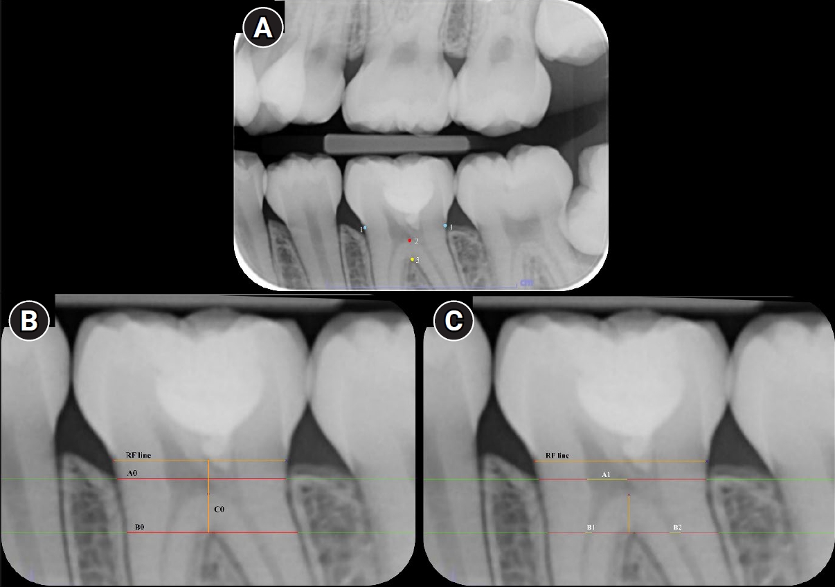
This study aimed to compare the magnitude of pulp space narrowing over time—measured as the change in pulp/tooth proportion from baseline—between mandibular molars treated with different types of vital pulp therapy (VPT) and their contralateral sound molars (controls). This study also investigated factors influencing the magnitude of pulp space narrowing in molars that have undergone VPT.
Methods
This retrospective cohort study involved the assessment of bitewing radiographs of VPT-treated molars and controls at baseline and follow-up. Using reference points and lines on the radiograph, pulp/tooth proportions were measured by examiners. The intraclass correlation coefficient (ICC) was used to report examiner reliability. The changes in pulp/tooth proportions from baselines were compared between subgroups using multilevel mixed effect linear regression and the Wald test.
Results
A total of 382 bitewing radiographs from 134 teeth were included. The follow-up period ranged from 6 to 84 months (mean, 27.12 ± 17.67 months). ICC values indicated good to excellent examiner reliability. Compared to the controls, changes in pulp/tooth proportion from baselines, indicating pulp space narrowing, were significantly greater in teeth with partial pulpotomy (at pulp chamber width) and coronal pulpotomy (at pulp canal width). Factors affecting the magnitude of pulp space narrowing included the more invasive type of VPT and the more severe preoperative diagnosis.
Conclusions
The magnitude of pulp space narrowing was greater in VPT-treated molars than in controls. The more invasive type of VPT and severe preoperative diagnosis were factors contributing to the magnitude of pulp space narrowing.
- 1,047 View
- 60 Download

- Neuropeptide Y regulation of dental pulp neurogenic inflammation provoked by tooth bleaching agents: a descriptive comparative clinical study
- Javier Caviedes-Bucheli, Néstor Ríos-Osorio, Mario Pérez-Villota, Karolina Aucú-Miño, Diana Escobar-Mafla, Hernán Darío Muñoz-Alvear, José Francisco Gomez-Sosa, Luis Diaz-Barrera, Edgar Güiza – Cristancho, Hugo Roberto Munoz
- Restor Dent Endod 2026;51(1):e10. Published online February 13, 2026
- DOI: https://doi.org/10.5395/rde.2026.51.e10
-
Abstract
PDFPubReaderePub
- Objectives
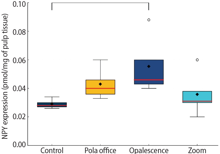
This study aimed to assess the expression of neuropeptide Y (NPY) in human dental pulp after tooth bleaching with three in-office hydrogen peroxide (H2O2)-based systems.
Methods
Forty pulps were collected from premolars scheduled for extraction and divided into four groups (n = 10): Control (no bleaching; basal NPY values); Pola Office (35% H2O2, 8 minutes); Opalescence Boost (40% H2O2, 20 minutes); and Zoom (25% H2O2 + cold blue light, 15 minutes). After extraction, pulps were fixed in 4% formaldehyde and processed. NPY levels were quantified using enzyme-linked immunosorbent assay. Data distribution was assessed with the Shapiro-Wilk test. One-way analysis of variance and Tukey post-hoc test with Bonferroni correction were applied (p < 0.05).
Results
NPY expression differed significantly among groups (p = 0.0097). The control group showed the lowest mean expression (0.026 ± 0.002 pmol/mg of pulp tissue), followed by Zoom (0.031 ± 0.005 pmol/mg), Pola Office (0.040 ± 0.004 pmol/mg), and Opalescence Boost, which exhibited the highest NPY expression (0.044 ± 0.004 pmol/mg). Post-hoc analysis revealed a statistically significant difference between the control and Opalescence Boost groups (p = 0.0122).
Conclusions
The increase in NPY expression—particularly with Opalescence Boost—indicates that in-office bleaching agents can elicit measurable neurobiological responses in pulp tissue after a single application. The significant difference between the control and Opalescence Boost groups suggests a possible H2O2 concentration- or formulation-dependent effect on pulpal neuropeptide activity, underscoring the need for further research on the biological impact of bleaching treatments.
- 1,595 View
- 90 Download
- Effect of combined application of premixed bioceramic paste and diode laser in vital pulp therapy: an immunohistochemical randomized controlled split-mouth in vivo animal experiment
- Mo’men A. Salama, Dalia M. Fayyad, Mohamed I. Rabie, Manar A. A. Selim, Mahmoud F. Ahmed
- Restor Dent Endod 2026;51(1):e4. Published online January 20, 2026
- DOI: https://doi.org/10.5395/rde.2026.51.e4
-
Abstract
PDFPubReaderePub
- Objectives
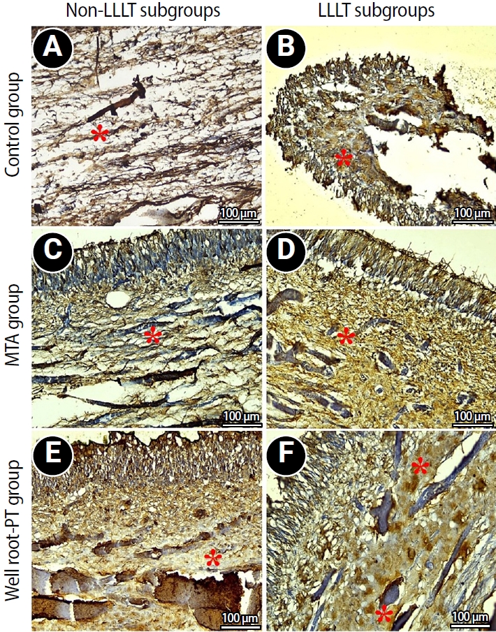
This study aimed to evaluate the effect of premixed bioceramic paste (Well-Root PT; Vericom) compared to mineral trioxide aggregate (MTA) on the expression of the mineralization-related marker dentin sialoprotein (DSP) in dental pulp following direct pulp capping, with or without prior diode laser application.
Methods
Direct pulp exposures were performed in the upper and lower incisors of eight dogs (n = 96 teeth). Cavities (Class V) were created and received pulp capping with either Well-Root PT (n = 32), MTA (n = 32), or no capping material (polytetrafluoroethylene disc only) (n = 32), with or without the application of a diode laser. Immunohistochemical analysis of DSP expression was conducted and quantified as the mean area percentage using ImageJ software at 2 and 8 weeks posttreatment.
Results
Both the Well-Root PT and MTA groups showed significantly increased DSP expression compared to the control group at both 2 and 8 weeks (p < 0.05). No significant difference in the mean area percentage of DSP expression was found between the Well-Root PT and MTA groups. The diode laser application did not produce a significant effect on DSP expression. Within-group comparison revealed a significant increase in DSP expression between the 2- and 8-week follow-up periods (p < 0.05).
Conclusions
Well-Root PT demonstrated comparable efficacy to MTA in promoting DSP expression, supporting its use as an effective direct pulp capping material. Diode laser application prior to capping had no effect on DSP expression in this experimental model.
- 2,085 View
- 143 Download
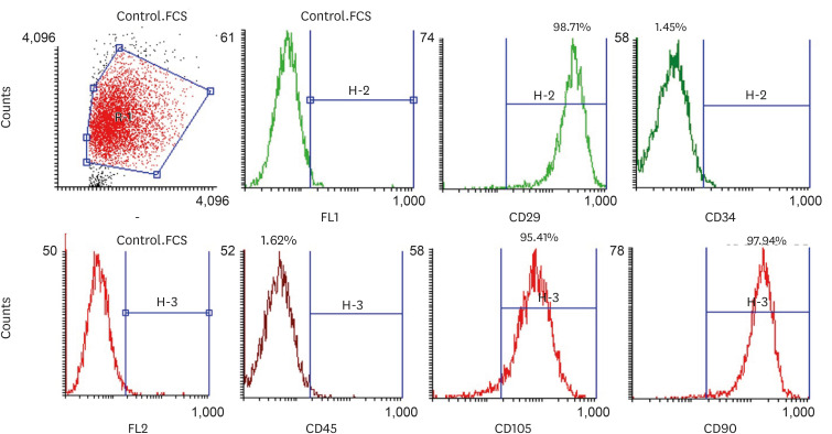
- Resolvin E1 incorporated carboxymethyl chitosan scaffold accelerates repair of dental pulp stem cells under inflammatory conditions: a laboratory investigation
- Hemalatha P Balasubramanian, Nandini Suresh, Vishnupriya Koteeswaran, Velmurugan Natanasabapathy
- Restor Dent Endod 2025;50(4):e40. Published online November 28, 2025
- DOI: https://doi.org/10.5395/rde.2025.50.e40
-
Abstract
PDFSupplementary MaterialPubReaderePub
- Objectives
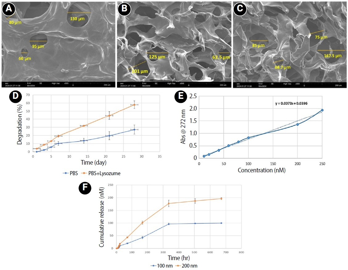
This study fabricated and characterized a resolvin E1 (RvE1)-loaded carboxymethyl chitosan (CMC) scaffold and determined its cytotoxicity and mineralization potential on inflamed human dental pulp stem cells (hDPSCs).
Methods
CMC scaffold incorporated with two concentrations of RvE1 (100 and 200 nM) was fabricated and characterized. The scaffolds’ porosity, drug release kinetics, and degradation were assessed. The impact of RvE1 on inflamed hDPSCs proliferation, proinflammatory gene expression (tumor necrosis factor alpha [TNF-α]), alkaline phosphatase activity, and alizarin red S staining was evaluated.
Results
Scanning electron microscopy analysis demonstrated a highly porous interconnected microstructure. Release kinetics showed gradual RvE1 release peaking at day 14. Cumulative degradation of the CMC scaffold at 28 days was 57.35%. Inflamed hDPSCs exposed to 200 nM RvE1-CMC scaffold exhibited significantly improved viability compared to 100 nM. Both RvE1-CMC scaffolds significantly suppressed the expression of TNF-α at 7 days. Alkaline phosphatase activity was enhanced by both RvE1 concentrations on days 7 and 14. Alizarin red staining revealed superior mineralization potential of 200 nM RvE1 on days 14 and 21.
Conclusions
This study concludes 200 nM RvE1-CMC scaffold is a promising therapy for inflamed pulp conditions, enhancing cell proliferation and biomineralization potential in inflamed hDPSCs.
- 1,489 View
- 63 Download
- Structural and morphological characterization of silver nanoparticles intruded mineral trioxide aggregate admixture as a chair-side restorative medicament: an in vitro experimental study
- H. Murali Rao, Rajkumar Krishnan, Chitra Shivalingam, Ramya Ramadoss
- Restor Dent Endod 2025;50(3):e30. Published online August 8, 2025
- DOI: https://doi.org/10.5395/rde.2025.50.e30

-
Abstract
PDFPubReaderePub
- Objectives
The aim of this study was to create a rapid admixture of mineral trioxide aggregate (MTA) and silver nanoparticles (AgNPs) for chairside use in clinical settings to remediate the challenges associated with root canal treatment and pulp capping.
Methods
Synthesized AgNPs at ratios of 10 and 25% were added to commercially available MTA to create an admixture. The admixture was subjected to structural and morphological assessment using X-ray diffraction analysis (XRD), Fourier transform infrared (FT-IR) analysis, Raman spectroscopy, and scanning electron microscopy. Antioxidant activity was measured using the hydroxyl radical scavenging assay. A significance level of 0.05 was applied to determine statistical differences.
Results
The addition of AgNPs decreased the carbonate peak intensity in XRD and FT-IR. The rod-like morphology of MTA was changed to a flake-like morphology with the addition of AgNPs. Antibacterial efficacy enhanced proportionally with the augmentation of AgNPs concentration.
Conclusions
The creation of rapid admixture of MTA and AgNPs during chairside use in clinical settings can deliver beneficial characteristics of enhanced morphological features favoring mineralization and profound antibacterial effects to overcome the challenges associated with root canal treatment and pulp capping.
- 2,820 View
- 98 Download
- 1 Web of Science
- Comparison of YouTube, TikTok, and Instagram as digital sources for obtaining information about pulp therapy in primary and permanent teeth
- Hüseyin Gürkan Güneç, Emine Kaya, Dila Nur Okumuş, Merve Gül Erence
- Restor Dent Endod 2025;50(3):e26. Published online July 24, 2025
- DOI: https://doi.org/10.5395/rde.2025.50.e26
-
Abstract
PDFPubReaderePub
- Objectives
This study aimed to compare the content, educational quality, and dependability of videos on Instagram, TikTok, and YouTube about pulp therapy (PT) in pediatric dentistry and endodontics.
Methods
Three popular video sites, Instagram (Meta Platforms, Inc.,), TikTok (ByteDance Ltd.), and YouTube (Google LLC), were searched for PT content to analyze for compliance with the American Association of Endodontists and American Academy of Pediatric Dentistry guidelines for clinical endodontists and pediatric dentists. The searched hashtags were #pulpaltherapy, #pulpaltreatment, #pulptherapy, and #pulptreatment. The classification of 158 English-language videos was based on several variables: communication quality, duration, likes and dislikes, views, source, treatment, and genre. The videos were evaluated using a usefulness score and the Global Quality Scale (GQS), Video Information and Quality Index (VIQI), Journal of the American Medical Association (JAMA) score, and modified DISCERN score to rate their quality and reliability. The majority of the videos were published by healthcare professionals, dental clinics, and universities.
Results
Significant relationships existed between video length, source of upload, usefulness score, tooth type, pulp status, and VIQI, JAMA, GQS, and DISCERN scores for all three platforms (p<0 .05). A statistically significant relationship existed of YouTube, TikTok, and Instagram with the number of views, number of months since upload, view rates, comments and likes (p< 0.05).
Conclusions
TikTok and Instagram reel videos provided high- to moderate-quality information about PT, especially in children, but YouTube may provide more reliable information than other social media tools. -
Citations
Citations to this article as recorded by
- Evaluating the reliability and educational quality of YouTube™ and TikTok™ videos on custom subperiosteal implants: a cross-sectional methodological analysis
Göksel Tımarcıoğlu, Erdal Cem Kargu, Hülya Çerçi Akçay, Başak Tımarcıoğlu
BMC Oral Health.2026;[Epub] CrossRef - New Technologies and Materials in Oral Health and Dental Care of Pediatric Dentistry
Giuseppe Minervini
Children.2025; 12(10): 1310. CrossRef
- Evaluating the reliability and educational quality of YouTube™ and TikTok™ videos on custom subperiosteal implants: a cross-sectional methodological analysis
- 3,463 View
- 88 Download
- 2 Web of Science
- 2 Crossref
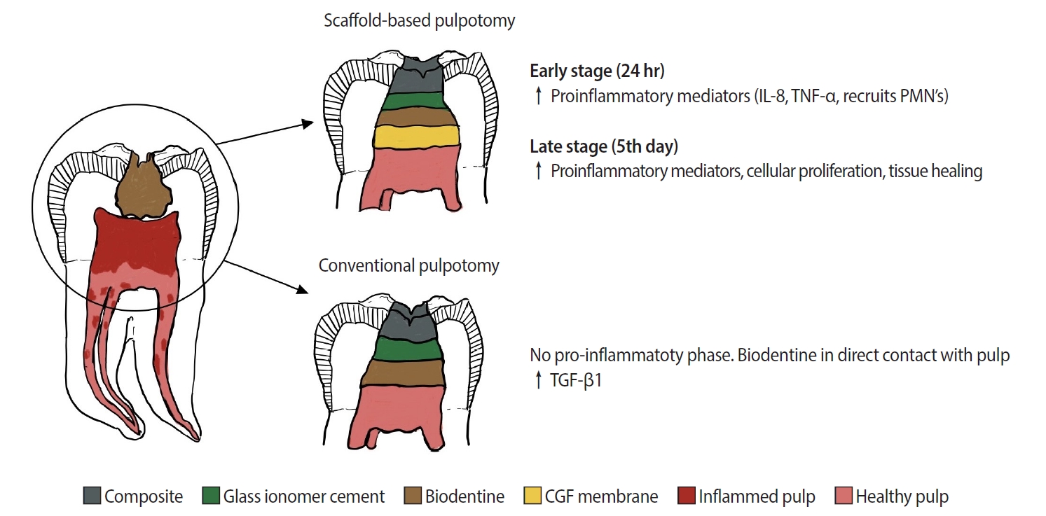
- Concentrated growth factor scaffold-based pulpotomy of permanent molars with symptomatic irreversible pulpitis
- Arthi K. Harith, Vishnupriya Koteeswaran, Dinesh Kowsky, Natanasabapathy Velmurugan, Suresh Nandini
- Restor Dent Endod 2025;50(1):e1. Published online January 17, 2025
- DOI: https://doi.org/10.5395/rde.2025.50.e1
-
Abstract
PDFPubReaderePub
- Objectives
Pulpotomy is a minimally invasive procedure that aims to retain the vitality of the radicular pulp by removing the inflamed coronal pulp tissue. This case series presents the successful management of symptomatic irreversible pulpitis by pulpotomy with concentrated growth factor (CGF) scaffolds.
Methods
Six permanent mandibular molars with a diagnosis of symptomatic irreversible pulpitis were included. Under Local anesthesia and rubber dam isolation, caries were excavated using high-speed bur under coolant. Full coronal pulpotomy was done and hemostasis was achieved. CGF membrane was prepared and placed over the radicular pulp and layered with Biodentine (Septodont). Final restoration of type IX glass ionomer cement and bulk fill composite resin was placed. Patients were assessed for various clinical and radiographic parameters at intervals of 1 week and 3, 6, and 12 months. Five patients fulfilled the success criteria at the end of 1 year.
Results
Pulpotomy is considered an alternative treatment modality for root canal treatment in symptomatic irreversible pulpitis aiming at alleviating symptoms and maintaining vitality. CGF scaffold when used as a capping material acts as a reservoir for growth factors with anti-inflammatory properties and enhances healing.
Conclusions
Scaffold-based pulpotomy can be considered a biological approach to healing inflamed pulp.
- 5,521 View
- 540 Download
-
Evaluation of mineral induction ability and cytotoxicity of carbonated hydroxyapatite for pulp tissue regeneration: an
in vitro study - S. Swathi Priyadharshini, Chinnasamy Ragavendran, Anand Sherwood, J. Ramana Ramya, Jogikalmat Krithikadatta
- Restor Dent Endod 2024;49(4):e40. Published online October 29, 2024
- DOI: https://doi.org/10.5395/rde.2024.49.e40
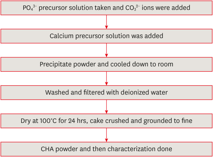
-
Abstract
PDFPubReaderePub
Objectives This study aimed to evaluate carbonated hydroxyapatite (CHA)’s ability for mineral induction and its
in vitro cytotoxicity with human dental pulp cells.Materials and Methods Precursors for the study include di-ammonium hydrogen phosphate and calcium nitrate tetrahydrate, with sodium hydrogen carbonate added to achieve different levels of carbonate substitution. The synthesized CHA samples are characterized using X-ray diffraction, Fourier transform infrared spectroscopy, and Raman spectroscopy. Scanning electron microscopy (SEM) was used to observe morphology. For 14 days at 37°C, samples were submerged in simulated body fluid to assess their mineral induction capabilities. SEM was used to confirm apatite formation on sample surfaces. The cytotoxicity assay was used to assess the vitality of the cells following their exposure to various concentrations of CHA.
Results The Joint Committee on Powder Diffraction Standards data for HA aligned well with the results from X-ray diffraction analysis of CHA across 3 different concentrations, indicating strong agreement. Fourier transform infrared spectra indicated the presence of phosphate, hydroxyl, and carbonate groups within the samples. SEM and Energy-dispersive X-ray analysis show agglomerated and flaky nanoparticles. All the samples are bioactive, but the formation of apatite differs from one another.
In vitro cytotoxicity assay showed that over 70% of cells maintain viability.Conclusions The results of this study may provide insight into the potential use of carbonated HA as a dental pulp-capping material for vital pulp therapy.
-
Citations
Citations to this article as recorded by- Smart Nanomaterials: Current State and Future Prospects in Drug Delivery and Tissue Engineering
E. Elizabeth Rani, D. Sakthi Sanjana, E. Karthikeyan, J. Nandhini
Biomedical Materials & Devices.2026; 4(2): 1455. CrossRef - Thermoresponsive Nanomaterials: Revolutionizing Cancer Theranostics
Bellarmin Michael, Mohanakrishnan Srinivasan, Karthikeyan Elumalai, Lokeshwar Ravikumar, Sivaprakash Kathiresan, Nandhini Jayaprakash
Biomedical Materials & Devices.2026; 4(3): 2697. CrossRef - Physicochemical and antibacterial evaluation of novel nano α-TCP–AgNPs biocomposites for direct pulp-capping applications
Selviana Wulansari, Hendra Dian Adhita Dharsono, Nasrul Wathoni, Rosalina Tjandrawinata, Arief Cahyanto, Moehamad Orliando Roeslan
Frontiers in Oral Health.2026;[Epub] CrossRef - Physicochemical effects of nano type-B bone substitute on pulp protective cement formulations
Njwan Fadhel SHEHAB
Dental Materials Journal.2026; 45(1): 92. CrossRef - Recycling waste for sustainability: The green synthesis of silver nanoparticles from Bougainvillea glabra green waste, and the evaluation of their antioxidant, cytotoxic, catalytic, antibacterial and in-silico molecular docking properties
Hafsa Naleem, Mathivathani Kandiah, Beneli Gunaratne, Ominda Perera
Next Research.2026; 11: 101990. CrossRef - Comparative evaluation of compressive strength and morphological interface of carbonated hydroxyapatite with other pulp capping materials: An in vitro analysis
S. Swathi Priyadharshini, Chinnasamy Ragavendran, I. Anand Sherwood, Ramanaramya Jeyapalan
Endodontology.2025; 37(1): 90. CrossRef - Bioactive Dioxo-Phosphobetaines derived from the reaction of Dichlorodinitrobenzofuroxane with various phosphines
Irina V. Galkina, Haiyan Fan, Semen R. Romanov, Dmitriy I. Bakhtiyarov, Luisa M. Usupova, Svetlana N. Egorova, Yulia V. Bakhtiyarova, Enrico Benassi
Bioorganic Chemistry.2025; 163: 108695. CrossRef - Near-infrared laser-activated PLGA-PDA core-shell nanohybrids for synergistic photothermal antibacterial therapy and sustained ion release in orthodontic white spot lesions prevention
Zezhou Feng, Yujiang Liu, Silu Sun, Minmin Si, Di Huang, Zhiyuan Feng
Journal of Dentistry.2025; 162: 106078. CrossRef - Formation and utilization of soluble microbial products in denitrifying biofilters at different carbon-to-nitrogen ratios: Microbial community characteristics
Fangyuan Jiang, Xianyang Shi
Journal of Environmental Chemical Engineering.2025; 13(6): 119554. CrossRef - Bioactivity and biocompatibility of bioceramic-based pulp capping materials in laboratory and animal models
Rafiqul Islam, Md. Refat Readul Islam, Kenta Tsuchiya, Yu Toida, Hidehiko Sano, Monica Yamauti, Hany Mohamed Aly Ahmed, Atsushi Tomokiyo
Journal of Materials Science: Materials in Medicine.2025;[Epub] CrossRef - Physical, Chemical, and Biological Properties of Graphene Nanoparticle-added Tricalcium Silicate Formulations: A Systematic Review
Soundaria Srinivasan, Deepa Gurunathan, Lakshmi Thangavelu
Journal of International Oral Health.2025; 17(6): 453. CrossRef - Advanced structural and compositional profiling of mineral trioxide aggregate incorporated with nano-carbonated hydroxyapatite: a comprehensive X-ray diffraction and energy dispersive X-ray investigation
Njwan Fadhel Shehab, Nadia Hameed Hasan, Alaa Edrees Dawood, Nawal Atiya Khalaf
Biomaterial Investigations in Dentistry.2025; 12: 216. CrossRef
- Smart Nanomaterials: Current State and Future Prospects in Drug Delivery and Tissue Engineering
- 4,775 View
- 156 Download
- 9 Web of Science
- 12 Crossref
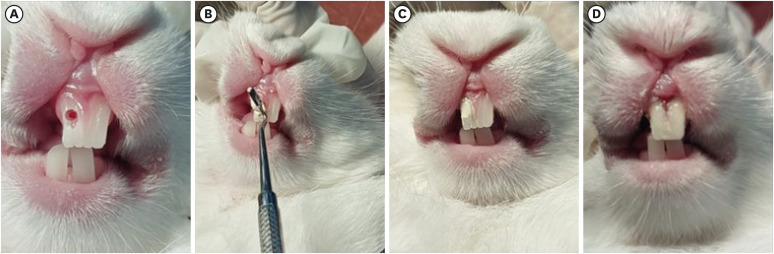
- Histological evaluation of pulp response to alendronate and Biodentine as pulp capping agents: an animal study
- Thangavel Boopathi, Sekar Manimaran, Joseline Charles Kerena, Mathew Sebeena, Kumaravadivel Karthick, Natesan Thangaraj Deepa
- Restor Dent Endod 2024;49(4):e39. Published online October 29, 2024
- DOI: https://doi.org/10.5395/rde.2024.49.e39
-
Abstract
PDFPubReaderePub
Objectives This study aimed to comparatively assess the histological response of the pulp toward alendronate and Biodentine in a direct pulp capping procedure.
Materials and Methods Twenty-four anterior teeth from 6 New Zealand rabbits were used in this study. Firstly, all rabbits were anesthetized according to their weight. Class V cavities were prepared on the buccal surfaces of anterior teeth. A pin-point exposure of the pulp was then made using a small, sterile round carbide bur and bleeding was arrested with a saline-soaked, sterile cotton pellet. The teeth under study were divided into 2 groups (
n = 12). The intentionally exposed pulp was capped with alendronate (Group 1) and Biodentine (Group 2), correspondingly. After 30 days, all rabbits were euthanized; the teeth under study were extracted and taken up for histological analysis.Results Biodentine showed an intact, very dense dentin bridge formation with a uniform odontoblast (OD) layer pattern and mild or absent inflammatory response whereas specimens capped with alendronate demonstrated a dense dentin bridge formation with non-uniform OD layer pattern and mild to moderate inflammatory response.
Conclusions Biodentine showed more biocompatibility than alendronate. However, alendronate can initiate reparative dentin formation and may be used as an alternative pulp capping agent.
-
Citations
Citations to this article as recorded by- In Vivo Evaluation of NF-κB and TGFβ-1 Modulation by Anadara granosa Shell-Derived Calcium Carbonate Bioceramic in Rat Model
Randy Nugraha Pratama, Nurhayati Natsir, Kezia Rachellea Mustakim, Juni Jekti Nugroho
European Journal of General Dentistry.2026;[Epub] CrossRef
- In Vivo Evaluation of NF-κB and TGFβ-1 Modulation by Anadara granosa Shell-Derived Calcium Carbonate Bioceramic in Rat Model
- 4,790 View
- 158 Download
- 1 Web of Science
- 1 Crossref
Review Article
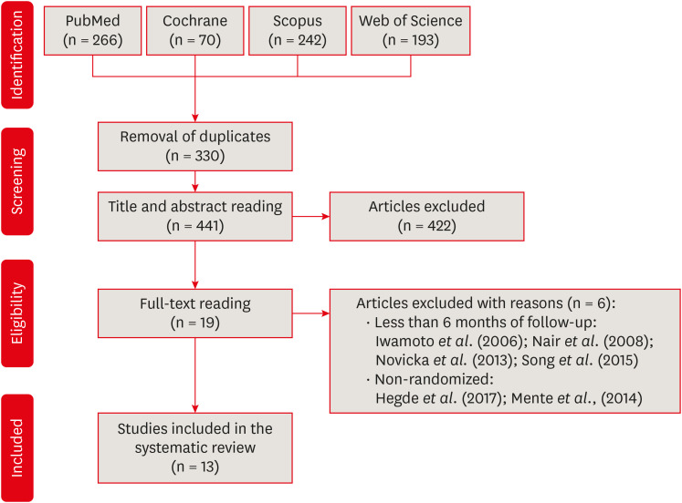
- Success rate of direct pulp capping on permanent teeth using bioactive materials: a systematic review and meta-analysis of randomized clinical trials
- Karem Paula Pinto, Gabriela Ribeiro da Silva, Cláudio Malizia Alves Ferreira, Luciana Moura Sassone, Emmanuel João Nogueira Leal da Silva
- Restor Dent Endod 2024;49(4):e34. Published online September 6, 2024
- DOI: https://doi.org/10.5395/rde.2024.49.e34
-
Abstract
PDFSupplementary MaterialPubReaderePub
This systematic review and meta-analysis aimed to evaluate the success rate of direct pulp capping (DPC) on permanent teeth, comparing the use of MTA with calcium hydroxide and calcium silicate-based cements. A systematic search was carried out in 4 databases until July 2023. The selection was based on PICOS criteria and only randomized clinical trials were included. The risk of bias was assessed using RoB-2 tool, and meta-analyses were performed using RevMan 5.3 software. The overall quality of evidence was determined using the GRADE tool. Thirteen studies were included. Meta-analyses indicated significantly higher success rate for DPC using MTA compared to calcium hydroxide, while no significant difference was observed between MTA and Biodentine, showing a success rate from 80% to 100% even after 3 years of follow-up. Five studies were classified as having high risk of bias and the GRADE assessment revealed low certainty of evidence. DPC is highly effective for permanent teeth when using MTA or Biodentine. There is a need for future well-designed randomized clinical trials to evaluate the efficacy of DPC using newer bioceramic materials.
-
Citations
Citations to this article as recorded by- Physicochemical effects of nano type-B bone substitute on pulp protective cement formulations
Njwan Fadhel SHEHAB
Dental Materials Journal.2026; 45(1): 92. CrossRef - Photobiomodulation-assisted pulp capping using nano-hydroxyapatite and mineral trioxide aggregate: Report of two cases
Priya Pal, Rhythm Bains, Promila Verma, Vivek Kumar Bains
Journal of Healthcare Research and Education.2026; 2: 2. CrossRef - Histological Tissue Response to Calcium Silicate-Based Cements Assessed in Human Tooth Culture Models: A Systematic Review
Alberto Cabrera-Fernández, Hebertt Gonzaga dos Santos Chaves, Aránzazu Díaz-Cuenca, Juan J. Segura-Egea, Jenifer Martín-González, João Peça, Diana B. Sequeira, João Miguel Marques dos Santos
Journal of Functional Biomaterials.2026; 17(2): 78. CrossRef - Translational Pathways for Smart and Bioactive Dental Biomaterials: Biocompatibility Standards, Sterilisation, Sustainability and Regulation
Katarzyna Chojnacka, Marcin Mikulewicz
MedComm – Biomaterials and Applications.2026;[Epub] CrossRef - Decision-ready evidence for vital pulp therapy: a network meta-analysis of bioactive materials in mature permanent teeth
Firas Elmsmari, Reem B. Abdelsayed, Qamar Albasoumi, Tareq Aljafarawi, Swadheena Patro, Ajinkya M. Pawar
Frontiers in Dental Medicine.2026;[Epub] CrossRef - Comparative Evaluation of Platelet-Rich Fibrin and Mineral Trioxide Aggregate in the Direct Pulp Capping of Carious Exposures: A Randomized Clinical Trial
Geeta Asthana, Rajashree Tamuli, Sadhna Manglani, Saloni Mandhare, Ragini Kulkarni, Anooja Mathirat, Parneet Kaur, P Jyothirmayee, Surabhi Landge
Cureus.2026;[Epub] CrossRef - Effects of Mauli Banana Stem Extract, Alone or Combined with Calcium Hydroxide, on DMP-1 Expression and Dentin Bridge Thickness After Direct Pulp Capping in Rat Molars
Dewi Puspitasari, Ika Kustiyah Oktaviyanti, Maharani Laillyza Apriasari, Erni Marlina, Maria Tanumihardja
European Journal of Dentistry.2026;[Epub] CrossRef - Indian Association of Conservative Dentistry and Endodontics consensus statement on deep caries management
Deepak Kumar Sharma, R. S. Mohan Kumar, Shishir Singh, Suparna Ganguly Saha, Meenal Nithin Gulve, Dipali Y. Shah, Sathish Abraham, Shruthi Nagaraja, Raksha Bhat
Journal of Conservative Dentistry and Endodontics.2025; 28(8): 714. CrossRef
- Physicochemical effects of nano type-B bone substitute on pulp protective cement formulations
- 25,078 View
- 745 Download
- 5 Web of Science
- 8 Crossref
Research Articles
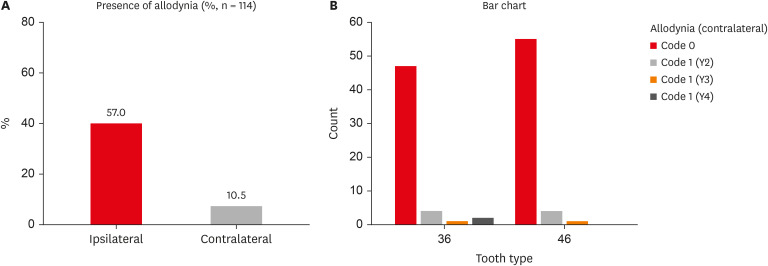
- Assessment of mechanical allodynia in healthy teeth adjacent and contralateral to endodontically diseased teeth: a clinical study
- Vaishnavi Ratnakar Patankar, Ashish K Jain, Rahul D Rao, Prajakta R Rao
- Restor Dent Endod 2024;49(3):e31. Published online July 29, 2024
- DOI: https://doi.org/10.5395/rde.2024.49.e31
-
Abstract
PDFPubReaderePub
Objectives The present study investigated the prevalence of mechanical allodynia (MA) in healthy teeth adjacent and contralateral to endodontically diseased teeth.
Materials and Methods This cross-sectional study included 114 patients with symptomatic irreversible pulpitis and apical periodontitis in permanent mandibular first molars who possessed healthy teeth adjacent and contralateral to the endodontically diseased tooth. The mechanical sensitivity of the teeth was determined by percussion testing. The presence or absence of pain on percussion in the teeth adjacent and contralateral to the endodontically diseased tooth and the tooth distal to the contralateral symmetrical tooth was recorded according to coding criteria. The prevalence of MA was computed as a percentage, and binary logistic regression analysis was done. The Fisher exact test and Mann-Whitney
U test were used for binary and ordinal data.Results Age and sex did not influence the prevalence of MA. An increased prevalence of MA was found in patients with higher levels of spontaneous pain (
p < 0.001). The prevalence of allodynia was 57% in teeth adjacent to endodontically diseased teeth and 10.5% in teeth contralateral to endodontically diseased teeth. In addition, on the ipsilateral side, there were more painful sensations distal to the diseased tooth than mesially.Conclusions Despite being disease-free, teeth adjacent and contralateral to endodontically diseased teeth exhibited pain on percussion. There was a direct association between the severity of the patient’s pain and the presence of MA.
-
Citations
Citations to this article as recorded by- Incidence and assessment of mechanical allodynia and its association with dental anxiety in endodontic pain: A clinical study
Manduwada Vishal, Sowmya Padala, E. Soujanya, Mamta Kaushik
Journal of Conservative Dentistry and Endodontics.2026; 29(5): 514. CrossRef
- Incidence and assessment of mechanical allodynia and its association with dental anxiety in endodontic pain: A clinical study
- 4,131 View
- 111 Download
- 1 Crossref
- Endodontic characteristics of mandibular premolar with dens evaginatus: a retrospective study
- Minjin Kim, Sujin Jeon, Min-Seock Seo
- Restor Dent Endod 2024;49(3):e28. Published online July 11, 2024
- DOI: https://doi.org/10.5395/rde.2024.49.e28
-
Abstract
PDFPubReaderePub
Objectives This study aimed to investigate the endodontic characteristics of mandibular premolars with dens evaginatus (DE) that require endodontic treatment.
Materials and Methods Patients who underwent endodontic treatment were enrolled. The inclusion criteria were patients who underwent root canal treatment in the lower permanent teeth with DE and were followed up for at least 1 year. Preoperative clinical and radiographic variables were obtained. The frequency distribution of the preoperative variables was compared using the χ2 or Fisher’s exact tests. The significance of the change in periapical health index (PAI) and root development stages before and after treatment was examined using the Wilcoxon signed-rank test.
Results A total of 150 teeth of 134 patients with an average age of 15.3 years were included. The percentage distribution comparison of the preoperative variables and obturation techniques revealed significant differences in pulpal and periapical diagnosis, and percussion, and especially regarding age, root development stage, and PAI. Age was the only statistically significant preoperative variable associated with root growth (
p < 0.05).Conclusions Approximately, 60% of DEs requiring endodontic treatment had immature roots. Age being the most significant predisposing factor, early treatment provides the greatest opportunity for full root development.
-
Citations
Citations to this article as recorded by- A tooth with multiple supernumerary cusps and taurodontism concurrently accompanied with other taurodont teeth: a rare case report
Zihui Tang, Hongchen Zhang, Rongrong Dang, Qiushi Zhang, Yan Huang, Yanwei Yang
Surgical and Radiologic Anatomy.2025;[Epub] CrossRef
- A tooth with multiple supernumerary cusps and taurodontism concurrently accompanied with other taurodont teeth: a rare case report
- 4,501 View
- 127 Download
- 1 Web of Science
- 1 Crossref
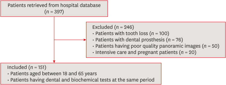
- Pulp stones: any relevance with the levels of serum calcium, parathyroid hormone, vitamin D and uric acid
- Ceyda Gürhan, Ercan Saruhan
- Restor Dent Endod 2024;49(2):e17. Published online March 26, 2024
- DOI: https://doi.org/10.5395/rde.2024.49.e17
-
Abstract
PDFPubReaderePub
Objectives This study evaluated the effect of serum calcium, parathyroid hormone (PTH), vitamin D, and uric acid levels on pulp stone formation.
Materials and Methods Patients who were admitted to the Muğla Sıtkı Koçman University, Faculty of Dentistry, Department of Oral and Maxillofacial Radiology for dental complaints were registered. Among these patients, individuals who had routine biochemical tests at the same period in the Outpatient Clinics of Muğla Sıtkı Koçman University Training and Research Hospital were included in the study. The patients with at least 1 pulp stone on panoramic radiographs recorded as the “pulp stone group” while patients without any pulp stones were the “control group”. Demographic data and serum levels of calcium, PTH, vitamin D, and uric acid were retrospectively evaluated in both groups. Student
t -test or Mann-WhitneyU test was used to evaluate the differences between the groups.Results Among 151 patients, dental pulp stone was detected in 53.6% of patients, and 82.7% of these patients were female. Female sex and pulp stone formation were significantly associated (
p = 0.001). The mean age of the pulp stone group was 43.9, while it was 39.9 in the control group, without any significant correlation between age and pulp stone (p > 0.05). Similarly, there were no significant differences in serum levels of PTH, vitamin D, uric acid and calcium between groups (p > 0.05).Conclusions According to the present study, the effect of dental factors rather than systemic factors should be considered primarily in pulp stone formation.
-
Citations
Citations to this article as recorded by- A novel deep learning-based pipeline architecture for pulp stone detection on panoramic radiographs
Ceyda Gürhan, Hasan Yiğit, Selim Yılmaz, Cihat Çetinkaya
Oral Radiology.2025; 41(2): 285. CrossRef - Vitamin D deficiency and oral health: a systematic review of literature
Saida Ziada, Aws Wishahe, Najet Mabrouk, Souad Sahtout
BMC Oral Health.2025;[Epub] CrossRef - Association between pulp stones and systemic diseases: a retrospective study using digital panoramic radiographs in a Turkish population
Buket Beytaş Alğan, Mustafa Murat Koçak, Sibel Koçak, Baran Can Sağlam
BMC Oral Health.2025;[Epub] CrossRef
- A novel deep learning-based pipeline architecture for pulp stone detection on panoramic radiographs
- 5,060 View
- 121 Download
- 4 Web of Science
- 3 Crossref
- Can different agents reduce the damage caused by bleaching gel to pulp tissue? A systematic review of basic research
- Letícia Aparecida Silva Batista, Alexandre Henrique dos Reis-Prado, Hebertt Gonzaga dos Santos Chaves, Lara Cancella de Arantes, Luís Fernando Santos Alves Morgan, Carolina Bosso André, Thaís Yumi Suzuki, Francine Benetti
- Restor Dent Endod 2023;48(4):e39. Published online November 6, 2023
- DOI: https://doi.org/10.5395/rde.2023.48.e39
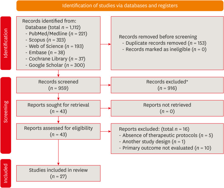
-
Abstract
PDFSupplementary MaterialPubReaderePub
Objectives This study aimed to investigate the effectiveness of different topical/systemic agents in reducing the damage caused by bleaching gel to pulp tissue or cells.
Materials and Methods Electronic searches were performed in July 2023.
In vivo andin vitro studies evaluating the effects of different topical or systemic agents on pulp inflammation or cytotoxicity after exposure to bleaching agents were included. The risk of bias was assessed.Results Out of 1,112 articles, 27 were included. Nine animal studies evaluated remineralizing/anti-inflammatories agents in rat molars subjected to bleaching with 35%–38% hydrogen peroxide (HP). Five of these studies demonstrated a significant reduction in inflammation caused by HP when combined with bioglass or MI Paste Plus (GC America), or following KF-desensitizing or Otosporin treatment (
n = 3). However, orally administered drugs did not reduce pulp inflammation (n = 4). Cytotoxicity (n = 17) was primarily assessed using the 3-(4,5-dimethylthiazol-2-yl)-2,5-diphenyltetrazolium bromide assay on human dental pulp cells and mouse dental papilla Cell-23 cells. Certain substances, including sodium ascorbate, butein, manganese chloride, and peroxidase, were found to reduce cytotoxicity, particularly when applied prior to bleaching. The risk of bias was high in animal studies and low in laboratory studies.Conclusions Few
in vivo studies have evaluated agents to reduce the damage caused by bleaching gel to pulp tissue. Within the limitations of these studies, it was found that topical agents were effective in reducing pulp inflammation in animals and cytotoxicity. Further analyses with human pulp are required to substantiate these findings.Trial Registration PROSPERO Identifier:
CRD42022337192 -
Citations
Citations to this article as recorded by- 3D-Printed and Bioprinted Scaffolds in Regenerative Endodontics: A Systematic Review
Hebertt Gonzaga dos Santos Chaves, Diana B. Sequeira, Vilton Cardozo Moreira Dias, Alberto Cabrera-Fernández, João Peça, Francine Benetti, João Miguel Marques dos Santos
Applied Sciences.2026; 16(8): 3940. CrossRef - Clinical Study on the Efficacy of 35% Hydrogen Peroxide Gel According to Exposure Time (40 min vs. 20 min) by Spectrophotometry
Trinidad Rincón, Maria Portillo Muñoz, Maria Lobato, Ana María Martín Casado, Laryssa Mylenna Madruga Barbosa, Alessandro Loguercio, Cristina Gómez‐Polo
Journal of Esthetic and Restorative Dentistry.2026;[Epub] CrossRef - Clareamento dental e TikTok: avaliação da qualidade do conteúdo em mídia social
Rafaele T Costa, Thayna Silva do Carmo Tavares, André Walsh-Monteiro
Ciência ET Praxis.2025; 21(36): 111. CrossRef - Synthesis, characterization and evaluation of novel bleaching gels containing bioactive glass and nano-hydroxyapatite on hydrogen peroxide diffusion, bleaching efficacy and enamel protection
Adrieli Burey, Byron Carpio-Salvatierra, Michael Favoretto, María Luján Méndez Bauer, Viviane Hass, Alessandra Reis, Alessandro D. Loguercio, Paulo Vitor Farago
Clinical Oral Investigations.2025;[Epub] CrossRef - Cytotoxicity of Bleaching Products: A Systematic Review
Mireia Montaner, José Luis Sanz, Carmen Llena, María Melo, Clara Puig-Herreros, James Ghilotti
Applied Sciences.2024; 14(9): 3680. CrossRef
- 3D-Printed and Bioprinted Scaffolds in Regenerative Endodontics: A Systematic Review
- 4,441 View
- 60 Download
- 4 Web of Science
- 5 Crossref
Review Article
- Stem cell-derived exosomes for dentin-pulp complex regeneration: a mini-review
- Dina A. Hammouda, Alaa M Mansour, Mahmoud A. Saeed, Ahmed R. Zaher, Mohammed E. Grawish
- Restor Dent Endod 2023;48(2):e20. Published online May 3, 2023
- DOI: https://doi.org/10.5395/rde.2023.48.e20
-
Abstract
PDFPubReaderePub
This mini-review was conducted to present an overview of the use of exosomes in regenerating the dentin-pulp complex (DPC). The PubMed and Scopus databases were searched for relevant articles published between January 1, 2013 and January 1, 2023. The findings of basic
in vitro studies indicated that exosomes enhance the proliferation and migration of mesenchymal cells, as human dental pulp stem cells, via mitogen-activated protein kinases and Wingless-Int signaling pathways. In addition, they possess proangiogenic potential and contribute to neovascularization and capillary tube formation by promoting endothelial cell proliferation and migration of human umbilical vein endothelial cells. Likewise, they regulate the migration and differentiation of Schwann cells, facilitate the conversion of M1 pro-inflammatory macrophages to M2 anti-inflammatory phenotypes, and mediate immune suppression as they promote regulatory T cell conversion. Basicin vivo studies have indicated that exosomes triggered the regeneration of dentin-pulp–like tissue, and exosomes isolated under odontogenic circumstances are particularly strong inducers of tissue regeneration and stem cell differentiation. Exosomes are a promising regenerative tool for DPC in cases of small pulp exposure or for whole-pulp tissue regeneration.-
Citations
Citations to this article as recorded by- Extracellular vesicles derived from dental mesenchymal stem cells for regenerative medicine: a scoping review
Maria Emília Mota, Márcia Martins Marques, Thaís Gimenez, Suely Kunimi Kubo Ariga, Tiago Góss dos Santos, Fábio Abreu Alves, Maria Stella Moreira
Molecular Biology Reports.2026;[Epub] CrossRef - Stem cell-derived exosomes in tissue regeneration of oral and maxillofacial region: A systematic review
Qianlei Dai, Qianfen Dai
Medicine.2026; 105(1): e46948. CrossRef - Stem Cell-Derived Exosomes in Wound Healing and Skin Regeneration: Emerging Therapeutic Strategies and Mechanisms
Nithin Vidiyala, Pavani Sunkishala, Prashanth Reddy Parupathi, Dinesh Nyavanandi
Cells.2026; 15(10): 872. CrossRef - REGENERATIVE POTENTIAL OF DENTAL STEM CELLS IN DENTISTRY: FROM BIOCHEMICAL MECHANISMS TO CLINICAL APPLICATIONS
A.Ye. Demkovych, O.Z. Yaremchuk, M.I. Kulitska, N.B. Hlyvka, V.Ya. Krupei, O.Ya. Lavrin
Ukrainian Dental Almanac.2026; (2): 5. CrossRef - Dental stem cell-derived extracellular vesicles for oral tissue regeneration: Mechanisms, engineering strategies, and clinical translation
Yang Liu, Peiyao Ge, Yuzhuo Ma, Haizhou Fu
Archives of Oral Biology.2026; 190: 106676. CrossRef - Cell Homing Strategies in Regenerative Endodontic Therapy
David Kim, Sahng G. Kim
Cells.2025; 14(3): 201. CrossRef - Impact of dental pulp cells-derived small extracellular vesicles on the properties and behavior of dental pulp cells: an in-vitro study
Dina A. Hammouda, Alaa M. Mansour, Ahmed R. Zaher, Mohammed E. Grawish
BMC Oral Health.2025;[Epub] CrossRef - Methodological Approaches for Economic Comparison of Mesenchymal Stem Cell and Exosome-based Therapies with Conventional Endodontic Treatments in Regenerative Endodontics
Madina A. Kurmanalina Kurmanalina, Nadiar M. Mussin, Aigul M. Sumanova, Violetta R. Detochkina, Maryam Mardani, Nader Tanideh, Amin Tamadon
West Kazakhstan Medical Journal.2025; 67(2): 188. CrossRef - Exosomal circ_0003057 promotes osteo/odontogenic differentiation of hDPSCs by binding with EIF4A3 through upregulated parental gene ANKH
Bingtao Wang, Yuanyuan Kong, Huixian Dong, Feng Lai, Zixin Guo, Liecong Lin, Jingyi Xu, Jingkun Zhang, Yiguo Jiang, Qianzhou Jiang
International Endodontic Journal.2025; 58(9): 1433. CrossRef - Mechanistic insights into dental stem cells‐derived exosomes in regenerative endodontics
Paras Ahmad, Nathan Estrin, Nima Farshidfar, Yufeng Zhang, Richard J. Miron
International Endodontic Journal.2025; 58(9): 1384. CrossRef - Development and characterization of an exosome-loaded biomimetic hydroxyapatite/gelatin scaffold for enhanced dental pulp regeneration
Yuen-Shan Tsai, Shih-Jung Cheng, Tsao-Li Chuang, Shu-Fang Chang, Feng-Huei Lin, Chun-Pin Lin
Journal of Dental Sciences.2025;[Epub] CrossRef - Exosomes as Promising Therapeutic Tools for Regenerative Endodontic Therapy
Qingyue Kong, Yujie Wang, Nan Jiang, Yifan Wang, Rui Wang, Xiaohan Hu, Jing Mao, Xin Shi
Biomolecules.2024; 14(3): 330. CrossRef - Role and Molecular Mechanism of miR-586 in the Differentiation of Dental Pulp Stem Cells into Odontoblast-like Cells
Gang Pan, Qianwen Zhou, Chenhua Pan, Yingxue Zhang
Cell Biochemistry and Biophysics.2024; 83(1): 507. CrossRef
- Extracellular vesicles derived from dental mesenchymal stem cells for regenerative medicine: a scoping review
- 11,393 View
- 150 Download
- 10 Web of Science
- 13 Crossref
Research Articles
- Effects of CTHRC1 on odontogenic differentiation and angiogenesis in human dental pulp stem cells
- Jong-soon Kim, Bin-Na Lee, Hoon-Sang Chang, In-Nam Hwang, Won-Mann Oh, Yun-Chan Hwang
- Restor Dent Endod 2023;48(2):e18. Published online April 28, 2023
- DOI: https://doi.org/10.5395/rde.2023.48.e18
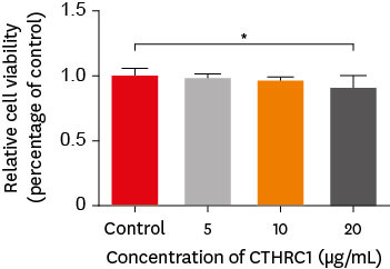
-
Abstract
PDFPubReaderePub
Objectives This study aimed to determine whether collagen triple helix repeat containing-1 (CTHRC1), which is involved in vascular remodeling and bone formation, can stimulate odontogenic differentiation and angiogenesis when administered to human dental pulp stem cells (hDPSCs).
Materials and Methods The viability of hDPSCs upon exposure to CTHRC1 was assessed with the WST-1 assay. CTHRC1 doses of 5, 10, and 20 µg/mL were administered to hDPSCs. Reverse-transcription polymerase reaction was used to detect dentin sialophosphoprotein, dentin matrix protein 1, vascular endothelial growth factor, and fibroblast growth factor 2. The formation of mineralization nodules was evaluated using Alizarin red. A scratch wound assay was conducted to evaluate the effect of CTHRC1 on cell migration. Data were analyzed using 1-way analysis of variance followed by the Tukey
post hoc test. The threshold for statistical significance was set atp < 0.05.Results CTHRC1 doses of 5, 10, and 20 µg/mL had no significant effect on the viability of hDPSCs. Mineralized nodules were formed and odontogenic markers were upregulated, indicating that CTHRC1 promoted odontogenic differentiation. Scratch wound assays demonstrated that CTHRC1 significantly enhanced the migration of hDPSCs.
Conclusions CTHRC1 promoted odontogenic differentiation and mineralization in hDPSCs.
- 2,497 View
- 40 Download
- The effects of gingival blood flow on pulpal blood flow detection using ultrasound Doppler flowmetry: animal study
- Dohyun Kim, Hyoung-Seok Ko, Soo-Yeon Park, Seung-Yeon Ryu, Sung-ho Park
- Restor Dent Endod 2023;48(1):e9. Published online January 30, 2023
- DOI: https://doi.org/10.5395/rde.2023.48.e9
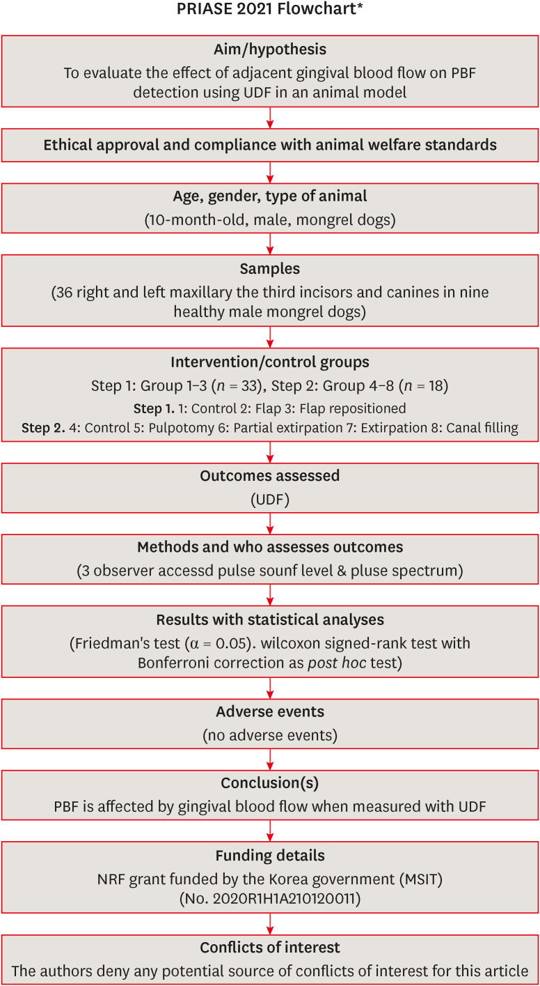
-
Abstract
PDFPubReaderePub
Objectives This study evaluated the effect of adjacent gingival blood flow on detection of pulpal blood flow (PBF) using ultrasound Doppler flowmetry (UDF) through animal study.
Materials and Methods The study included 36 right and left maxillary the third incisors and canines in 9 experimental dogs. The study included 2 main steps: In the first step, the pulse sound level (PSL) was recorded on the cervical part of each tooth without flap elevation (Group 1), with flap elevation (Group 2), and after it was repositioned in place (Group 3). In the second step, the PSL was recorded on the cervical part of each tooth (Group 4), after pulpotomy (Group 5), after partial pulp extirpation (Group 6), after complete extirpation (Group 7), and after canal filling (Group 8). In Groups 5–8, the study was performed with and without flap elevation in the left and right teeth, respectively. The PSL was graded as follows: 0, inaudible; 1, heard faintly; and 2, heard well. The difference between each group was analyzed using Friedman’s test with Wilcoxon signed-rank tests (α = 0.05).
Results In step 1, the PSL results were Group 1 > 2 and 3. In step 2, there was no significant difference between the groups when the flap was not elevated, while PSL results were Group 4 > 5 ≥ 6 and 7 ≥ 8 when the flap was elevated.
Conclusions PBF is affected by gingival blood flow when measured with UDF. UDF measurements require isolation of gingiva from the tooth.
-
Citations
Citations to this article as recorded by- Modern aspects of the use of hardware methods for diagnosing pulp vitality (Part 2. Non-traditional diagnostic methods)
K. V. Shadrina, L. Yu. Orekhova, V. D. Goncharov, V. Yu. Vashneva, E. S. Silina, E. V. Kosova, A. A. Petrov
Endodontics Today.2025; 23(3): 423. CrossRef - Exploring approaches to pulp vitality assessment: A scoping review of nontraditional methods
Farzaneh Afkhami, Patricia Paule Wright, Philip Yuan‐Ho Chien, Chun Xu, Laurence James Walsh, Ove Andreas Peters
International Endodontic Journal.2024; 57(8): 1065. CrossRef
- Modern aspects of the use of hardware methods for diagnosing pulp vitality (Part 2. Non-traditional diagnostic methods)
- 3,425 View
- 49 Download
- 1 Web of Science
- 2 Crossref
- Antimicrobial and cytotoxic properties of calcium-enriched mixture cement, Iranian propolis, and propolis with herbal extracts in primary dental pulp stem cells
- Mohammad Esmaeilzadeh, Shirin Moradkhani, Fahimeh Daneshyar, Mohammad Reza Arabestani, Sara Soleimani Asl, Soudeh Tayebi, Maryam Farhadian
- Restor Dent Endod 2023;48(1):e2. Published online December 1, 2022
- DOI: https://doi.org/10.5395/rde.2023.48.e2
-
Abstract
PDFPubReaderePub
Objectives In this study, natural substances were introduced as primary dental pulp caps for use in pulp therapy, and the antimicrobial and cytotoxic properties of these substances were investigated.
Materials and Methods In this
in vitro study, the antimicrobial properties of calcium-enriched mixture (CEM) cement, propolis, and propolis individually combined with the extracts of several medicinal plants were investigated againstEnterococcus faecalis ,Escherichia coli ,Pseudomonas aeruginosa , andStaphylococcus aureus . Then, the cytotoxicity of each substance or mixture against pulp stem cells extracted from 30 primary healthy teeth was evaluated at 4 concentrations. Data were gathered via observation, and optical density values were obtained using the 3-(4,5-dimethylthiazol-2-yl)-2,5-diphenyl-2H-tetrazolium bromide (MTT) test and recorded. SPSS software version 23 was used to analyze the data. Data were evaluated using 2-way analysis of variance and the Tukey test.Results Regarding antimicrobial properties, thyme alone and thyme + propolis had the lowest minimum inhibitory concentrations (MICs) against the growth of
S. aureus ,E. coli , andP. aeruginosa bacteria. ForE. faecalis , thyme + propolis had the lowest MIC, followed by thyme alone. At 24 and 72 hours, thyme + propolis, CEM cement, and propolis had the greatest bioviability in the primary dental pulp stem cells, and lavender + propolis had the lowest bioviability.Conclusions Of the studied materials, thyme + propolis showed the best results in the measures of practical performance as a dental pulp cap.
-
Citations
Citations to this article as recorded by- Self-Adapting Mouthguard with Nano-Hydroxyapatite and Propolis for Early Childhood Caries: Preclinical Safety and Efficacy
Mata Soslanbekovna Mustapaeva, Khadizhat Adamovna Sataeva, Elina Ilyasovna Zhabrailova, Alina Mairbekovna Sidakova, Karina Maharbekovna Mukagova, Umar Said-Asanovich Magomadov, Muslim Usmanovich Dunaev, Mehdi Usmanovich Dunaev, Khava Khuseynovna Amaeva, V
Asian Journal of Periodontics and Orthodontics.2026; 6(1): 32. CrossRef - Comprehensive review of composition, properties, clinical applications, and future perspectives of calcium-enriched mixture (CEM) cement: a systematic analysis
Saeed Asgary, Mahtab Aram, Mahta Fazlyab
BioMedical Engineering OnLine.2024;[Epub] CrossRef - Effects of aqueous and ethanolic extracts of Chinese propolis on dental pulp stem cell viability, migration and cytokine expression
Ha Bin Park, Yen Dinh, Pilar Yesares Rubi, Jennifer L. Gibbs, Benoit Michot
PeerJ.2024; 12: e18742. CrossRef
- Self-Adapting Mouthguard with Nano-Hydroxyapatite and Propolis for Early Childhood Caries: Preclinical Safety and Efficacy
- 3,483 View
- 59 Download
- 2 Web of Science
- 3 Crossref
-
The influence of sodium hypochlorite concentration on the fibrin structure of human blood clots and transforming growth factor-beta 1 release: an
ex vivo study - Anisha Mishra, Velmurugan Natanasabapathy, Nandini Suresh
- Restor Dent Endod 2022;47(4):e42. Published online October 31, 2022
- DOI: https://doi.org/10.5395/rde.2022.47.e42
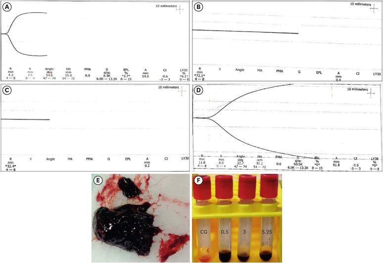
-
Abstract
PDFSupplementary MaterialPubReaderePub
Objective This study investigated the effects of various concentrations of sodium hypochlorite (NaOCl) on human whole-blood clotting kinetics, the structure of the blood clots formed, and transforming growth factor (TGF)-β1 release.
Materials and Methods Human whole blood was collected from 5 healthy volunteers and divided into 4 groups: CG (control, 0.5 mL of blood), BN0.5 (0.5 mL of blood with 0.5 mL of 0.5% NaOCl), BN3 (0.5 mL of blood with 0.5 mL of 3% NaOCl), and BN5.25 (0.5 mL of blood with 0.5 mL of 5.25% NaOCl). The effects of NaOCl on clotting kinetics, structure of fibrin and cells, and release of TGF-β1 were assessed using thromboelastography (TEG), scanning electron microscopy (SEM), and enzyme-linked immunosobent assay, respectively. Statistical analysis was conducted using the Kruskal Wallis and Mann-Whitney
U tests, followed by thepost hoc Dunn test. Ap value < 0.05 indicated statistical significance.Results The blood samples in BN0.5 and BN3 did not clot, whereas the TEG of BN5.25 showed altered clot formation. Samples from the CG and BN3 groups could only be processed with SEM, which showed that the latter lacked fibrin formation and branching of fibers, as well as clumping of red blood cells with surface roughening and distortion. TGF-β1 release was significantly highest in BN3 when all groups were compared to CG (
p < 0.05).Conclusions Each concentration of NaOCl affected the release of TGF-β1 from blood clots and altered the clotting mechanism of blood by affecting clotting kinetics and cell structure.
-
Citations
Citations to this article as recorded by- Evaluation of the cytotoxicity of a broad-spectrum antiseptic using a model of erythrocyte hemolysis in an in vitro experiment
S. P. Rubnikovich, O. E. Bekjanova, L. E. Khasanova, Sh. F. Shamsieva, S. X. Alimova, M. M. Astanakulova, N. T. Babadjanova, X. Sh. Mirzaev
Proceedings of the National Academy of Sciences of Belarus, Medical series.2026; 23(2): 95. CrossRef - Effect of Adjunctive Ozone Application Protocols on Dentin-Derived Growth Factor Release: An In Vitro Study
Sude Göbüt, Melis Oya Ateş, Ali Keleş, Fatma Avcıoğlu
Journal of Clinical Medicine.2026; 15(11): 4277. CrossRef - Cytotoxic Effects of Synthetic and Herbal Endodontic Irrigants on Human Red Blood Cells: An In Vitro Study
Panna Mangat, Bhaviya Chandel, Mampi Biswas, Sara Trivedy, Akshata Gupta, Nayan Shree, Seema Gupta
Cureus.2025;[Epub] CrossRef
- Evaluation of the cytotoxicity of a broad-spectrum antiseptic using a model of erythrocyte hemolysis in an in vitro experiment
- 3,075 View
- 39 Download
- 1 Web of Science
- 3 Crossref
- Clinical and radiographic outcomes of regenerative endodontic treatment performed by endodontic postgraduate students: a retrospective study
- Hadi Rajeh Alfahadi, Saad Al-Nazhan, Fawaz Hamad Alkazman, Nassr Al-Maflehi, Nada Al-Nazhan
- Restor Dent Endod 2022;47(2):e24. Published online May 9, 2022
- DOI: https://doi.org/10.5395/rde.2022.47.e24
-
Abstract
PDFPubReaderePub
Objectives Regenerative endodontic treatment is a clinical procedure aimed at biologically regenerating damaged root canal tissue of immature permanent teeth. This study aimed to report the outcomes of regenerative endodontic treatment performed by endodontic postgraduate students.
Materials and Methods Clinical and radiographic data of 27 patients, aged 10–22 years, who underwent regenerative treatment of immature permanent teeth from 2015 to 2019 were followed up, wherein clinical and radiographic examinations were performed for each patient. Postoperative success rate and tooth survival were analyzed, and the postoperative radiographic root area changes were quantified.
Results A total of 23 patients attended the dental appointments, showing that all teeth survived and were asymptomatic. Specifically, 7 periapical pathosis cases were completely healed, 12 were incompletely healed, and 4 cases failed. Moreover, significant differences were found between discolored and non-discolored teeth, and between the presence or absence of periapical radiolucency. Additionally, 3 anterior teeth showed complete closure of the apical foramen, while the apical foramen width was reduced in 17 teeth and failed in 3 teeth. Root length was also found to have been increased in 7 anterior and 4 posterior teeth, and the average length ranged from 4.00–0.63 mm in the anterior teeth, 2.85–1.48 mm of the mesial root, and 2.73–2.16 mm of the molar teeth distal root. Furthermore, calcified tissue deposition was observed in 7 teeth.
Conclusions A favorable outcome of regenerative endodontic treatment of immature permanent teeth with necrotic pulp was achieved with a high survival rate.
-
Citations
Citations to this article as recorded by- Regenerative Endodontics and Stem Cell-Based Therapies – A Systematic Review
Wjoud Ahmed Alshamrani, Sarah Sulaiman Alzarea, Joud Khalid Alabbas, Ayah Khalid Alabbas, Mawiyah Ibrahim Aljaddua, Osama Khattak, Rakhi Issrani
Journal of Pharmacy and Bioallied Sciences.2026; 18(Suppl 1): S29. CrossRef - Treatment outcomes and prognostic factors of regenerative endodontic procedures in immature permanent teeth
Gülsen Kiraz, Salihanur Sarı, Sümeyra Akkoç, Arzu Kaya Mumcu
BMC Oral Health.2026;[Epub] CrossRef - Pre‐Operative Factors on Prognosis of Regenerative Endodontic Procedures: A Systematic Review and Meta‐Analysis
Filipe Colombo Vitali, Alexandre Henrique dos Reis‐Prado, Pablo Silveira Santos, Ana Paula Portes Zeno, Patrícia de Andrade de Risso, Lucianne Cople Maia, Francine Benetti, Cleonice da Silveira da Teixeira
International Endodontic Journal.2025; 58(12): 1814. CrossRef - Clinical, radiographic, and biomarker perspectives of low-level laser therapy during regenerative endodontic procedures in necrotic immature young teeth: a randomized clinical study
Pragya Pandey, Neha Jasrasaria, Ramesh Bharti, Rakesh Kumar Yadav, Monika Kumari, Abinia Vaishnavi, Rahul Pandey
Lasers in Medical Science.2025;[Epub] CrossRef - Allogeneic Bone Marrow Mesenchymal Stromal Cell Transplantation Induces Dentin Pulp Complex-like Formation in Immature Teeth with Pulp Necrosis and Apical Periodontitis
Jose Francisco Gomez-Sosa, José E. Cardier, Olga Wittig, Dylana Díaz-Solano, Eloisa Lara, Kharelys Duque, Giselle Ramos-González
Journal of Endodontics.2024; 50(4): 483. CrossRef - Radiographic assessment of dental post and core placement at different educational levels in an undergraduate student clinic: a 4-year retrospective study
Turki Alshehri, Nourhan M. Aly, Raand Altayyar, Deena Alghamdi, Shahad Alotaibi, Passent Ellakany
F1000Research.2024; 12: 976. CrossRef - Evaluation of the efficacy of injectable platelet‐rich fibrin versus platelet‐rich plasma in the regeneration of traumatized necrotic immature maxillary anterior teeth: A randomized clinical trial
Maha Mohamed Abo‐Heikal, Jealan M. El‐Shafei, Samia A. Shouman, Nehal N. Roshdy
Dental Traumatology.2024; 40(1): 61. CrossRef - Radiographical assessment of post and core placement errors encountered by Saudi dental students at different educational levels
Turki Alshehri, Nourhan M. Aly, Raand Altayyar, Deena Alghamdi, Shahad Alotaibi, Passent Ellakany
F1000Research.2023; 12: 976. CrossRef
- Regenerative Endodontics and Stem Cell-Based Therapies – A Systematic Review
- 5,779 View
- 113 Download
- 8 Web of Science
- 8 Crossref
- Effects of dentin surface preparations on bonding of self-etching adhesives under simulated pulpal pressure
- Chantima Siriporananon, Pisol Senawongse, Vanthana Sattabanasuk, Natchalee Srimaneekarn, Hidehiko Sano, Pipop Saikaew
- Restor Dent Endod 2022;47(1):e4. Published online December 28, 2021
- DOI: https://doi.org/10.5395/rde.2022.47.e4
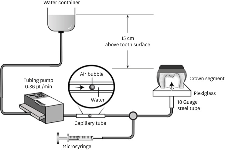
-
Abstract
PDFPubReaderePub
Objectives This study evaluated the effects of different smear layer preparations on the dentin permeability and microtensile bond strength (µTBS) of 2 self-etching adhesives (Clearfil SE Bond [CSE] and Clearfil Tri-S Bond Universal [CTS]) under dynamic pulpal pressure.
Materials and Methods Human third molars were cut into crown segments. The dentin surfaces were prepared using 4 armamentaria: 600-grit SiC paper, coarse diamond burs, superfine diamond burs, and carbide burs. The pulp chamber of each crown segment was connected to a dynamic intra-pulpal pressure simulation apparatus, and the permeability test was done under a pressure of 15 cmH2O. The relative permeability (%P) was evaluated on the smear layer-covered and bonded dentin surfaces. The teeth were bonded to either of the adhesives under pulpal pressure simulation, and cut into sticks after 24 hours water storage for the µTBS test. The resin-dentin interface and nanoleakage observations were performed using a scanning electron microscope. Statistical comparisons were done using analysis of variance and
post hoc tests.Results Only the method of surface preparation had a significant effect on permeability (
p < 0.05). The smear layers created by the carbide and superfine diamond burs yielded the lowest permeability. CSE demonstrated a higher µTBS, with these values in the superfine diamond and carbide bur groups being the highest. Microscopic evaluation of the resin-dentin interface revealed nanoleakage in the coarse diamond bur and SiC paper groups for both adhesives.Conclusions Superfine diamond and carbide burs can be recommended for dentin preparation with the use of 2-step CSE.
-
Citations
Citations to this article as recorded by- Effect of smear layer pretreatment with EDTA and sodium hypochlorite on the dentin bond durability of universal adhesives
Thanawat Ruaydee, Chantida Pawaputanon Na Mahasarakham, Vanthana Sattabanasuk, Pipop Saikaew
Frontiers in Dental Medicine.2026;[Epub] CrossRef - Determination of marginal permeability of restorations in the cervical region using a universal adhesive system: a randomized controlled open-label laboratory study
Svetlana N. Razumova, Anzhela S. Brago, Oxana R. Ruda, Artur G. Talandis, Lamara M. Khaskhanova, Ruzanna M. Bragunova, Bohdan O. Pecherskyi
Russian Journal of Dentistry.2026; 30(2): 113. CrossRef - Catechol–Phosphonate–Augmented Universal Adhesive for Hydrolysis-Resistant Dentin Bonds: A µTBS and Spectroscopic Study
Rabeia J. Khalil, Suha K. Ibrahim, Athraa H. Madhat, Ali H. Tawfieq
European Journal of Dentistry.2026;[Epub] CrossRef - The effect of different adhesive strategies and diamond burs on dentin bond strength of universal resin cements
Chavakorn Atsavathavornset, Pipop Saikaew, Choltacha Harnirattisai, Hidehiko Sano
Clinical Oral Investigations.2025;[Epub] CrossRef - Universal adhesive systems in dentistry: A narrative review
Svetlana N. Razumova, Anzhela S. Brago, Oxana R. Ruda, Zoya A. Guryeva, Elvira V. Adzhieva
Russian Journal of Dentistry.2024; 28(5): 512. CrossRef - Delayed light activation of resin composite affects the bond strength of adhesives under dynamic simulated pulpal pressure
Nattaporn Sukprasert, Choltacha Harnirattisai, Pisol Senawongse, Hidehiko Sano, Pipop Saikaew
Clinical Oral Investigations.2022; 26(11): 6743. CrossRef
- Effect of smear layer pretreatment with EDTA and sodium hypochlorite on the dentin bond durability of universal adhesives
- 4,343 View
- 65 Download
- 4 Web of Science
- 6 Crossref
- Combination of a new ultrasonic tip with rotary systems for the preparation of flattened root canals
- Karina Ines Medina Carita Tavares, Jáder Camilo Pinto, Airton Oliveira Santos-Junior, Fernanda Ferrari Esteves Torres, Juliane Maria Guerreiro-Tanomaru, Mario Tanomaru-Filho
- Restor Dent Endod 2021;46(4):e56. Published online October 27, 2021
- DOI: https://doi.org/10.5395/rde.2021.46.e56
-
Abstract
PDFPubReaderePub
Objectives This study evaluated 2 nickel-titanium rotary systems and a complementary protocol with an ultrasonic tip and a small-diameter instrument in flattened root canals.
Materials and Methods Thirty-two human maxillary second premolars with flattened canals (buccolingual diameter ≥4 times larger than the mesiodistal diameter) at 9 mm from the radiographic apex were selected. The root canals were prepared by ProDesign Logic (PDL) 30/0.01 and 30/0.05 or Hyflex EDM (HEDM) 10/0.05 and 25/0.08 (
n = 16), followed by application of the Flatsonic ultrasonic tip in the cervical and middle thirds and a PDL 25/0.03 file in the apical third (FPDL). The teeth were scanned using micro-computed tomography before and after the procedures. The percentage of volume increase, debris, and uninstrumented surface area were analyzed using the Kruskal-Wallis, Dunn, Wilcoxon, analysis of variance/Tukey, and paired and unpairedt -tests (α = 0.05).Results No significant difference was found in the volume increase and uninstrumented surface area between PDL and HEDM (
p > 0.05). PDL had a higher percentage of debris than HEDM in the middle and apical thirds (p < 0.05). The FPDL protocol resulted in less debris and uninstrumented surface area for PDL and HEDM (p < 0.05). This protocol, with HEDM, reduced debris in the middle and apical thirds and uninstrumented surface area in the apical third (p < 0.05).Conclusions High percentages of debris and uninstrumented surface area were observed after preparation of flattened root canals. The HEDM, Flatsonic tip, and 25/0.03 instrument protocol enhanced cleaning in flattened root canals.
-
Citations
Citations to this article as recorded by- Kök Kanal Tedavisi Yenilemelerinde Ultrasonik Uç Kullanımı
Ayşenur Kızıltaş Gül, Turan Mert Hisar, Seniha Miçooğulları
Selcuk Dental Journal.2025; 12(1): 157. CrossRef - Flatsonic Ultrasonic Tip Optimizes the Removal of Remaining Filling Material in Flattened Root Canals: A Micro–computed Tomographic Analysis
Airton Oliveira Santos-Junior, Karina Ines Medina Carita Tavares, Jáder Camilo Pinto, Fernanda Ferrari Esteves Torres, Juliane Maria Guerreiro-Tanomaru, Mário Tanomaru-Filho
Journal of Endodontics.2024; 50(5): 612. CrossRef - The Effect of Combined Ultrasonic Tip and Mechanized Instrumentation on the Reduction of the Percentage of Non-Instrumented Surfaces in Oval/Flat Root Canals: A Systematic Review and Meta-Analysis
Marcella Dewes Cassal, Pedro Cardoso Soares, Marcelo dos Santos
Cureus.2023;[Epub] CrossRef - Heat-treated NiTi instruments and final irrigation protocols for biomechanical preparation of flattened canals
Kleber Kildare Teodoro CARVALHO, Igor Bassi Ferreira PETEAN, Alice Corrêa SILVA-SOUSA, Rafael Verardino CAMARGO, Jardel Francisco MAZZI-CHAVES, Yara Terezinha Corrêa SILVA-SOUSA, Manoel Damião SOUSA-NETO
Brazilian Oral Research.2022;[Epub] CrossRef
- Kök Kanal Tedavisi Yenilemelerinde Ultrasonik Uç Kullanımı
- 2,678 View
- 37 Download
- 4 Web of Science
- 4 Crossref
- YouTube as a source of information about pulpotomy and pulp capping: a cross sectional reliability analysis
- Konstantinos Kodonas, Anastasia Fardi
- Restor Dent Endod 2021;46(3):e40. Published online July 6, 2021
- DOI: https://doi.org/10.5395/rde.2021.46.e40
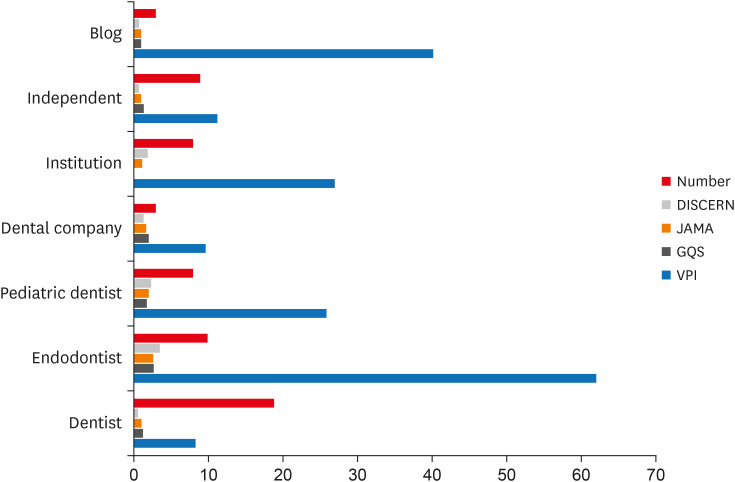
-
Abstract
PDFPubReaderePub
Objectives The purpose of this study was to critically evaluate the quality, reliability and educational content of the information of vital pulp treatment videos available on YouTube.
Materials and Methods The keywords “pulpotomy” and “pulp capping” were searched on YouTube on 5th July 2020, until 60 English language videos of each search term with a duration shorter than 15 minutes were acquired. Video characteristics were recorded and Video Power Index (VPI) was calculated. Reliability and educational quality of videos were evaluated using the Modified DISCERN score, the
Journal of American Medical Association (JAMA) benchmark criteria and Global Quality Scores (GQS). Videos were categorized by uploading source.Results Regarding pulpotomy, 31.7% of the videos were uploaded by specialists and 68.3% were directed by non-specialists. In the case of pulp capping, the corresponding percentages were 45% and 55%, respectively. Videos uploaded by specialists had significantly higher modified DISCERN, JAMA and GQS scores compared to those uploaded by non-specialists. Endodontists tended to have the highest reliability and VPI scores.
Conclusions YouTube videos on vital pulp treatment contain low educational quality or incomplete information. Low popularity of dental pulp capping and pulpotomy videos may be attributed to the specialized nature of these procedures. As YouTube represents an important source for patient information about different health topics, reliable informative videos should be uploaded by specialized dental professionals.
-
Citations
Citations to this article as recorded by- Evaluation of YouTubeTM Videos Regarding ICON as an Information Resource: A Cross-Sectional Study
Sevim Atılan Yavuz, Zeyneb Merve Ozdemır, Derya Gursel Surmelioglu
Mersin Üniversitesi Tıp Fakültesi Lokman Hekim Tıp Tarihi ve Folklorik Tıp Dergisi.2026; 16(1): 282. CrossRef - Quality and Accuracy Assessment of YouTube Videos on Vital Pulp Therapy
Divya Nangia, Vasudev Ballal, Prashant Bhasin, Esha Sukhala, Ashima Garg, Meenu G. Singla, Hemanshi Kumar
Australian Endodontic Journal.2026;[Epub] CrossRef - Assessing the Quality of YouTube® Videos on Nitrous Oxide/Oxygen Inhalation: A Multi-Dimensional Approach for Pediatric Dentists
Sanaa N. Al-Haj Ali, Nehal AlHarbi, Hessah H. Almutairi
Pesquisa Brasileira em Odontopediatria e Clínica Integrada.2025;[Epub] CrossRef - Is YouTube™ a useful resource of information about bichectomy? A cross-sectional study
H.ɪ. Durmuş, B. Ege, S. Bayazıt, M. Koparal
Annales de Chirurgie Plastique Esthétique.2025;[Epub] CrossRef - Assessing the reliability and educational value of YouTube videos on computer-controlled local anesthesia in dentistry
Hulya Cerci Akcay, Erdal Cem Kargu, Nefise Seker, Tanay Chaubal
PLOS One.2025; 20(8): e0329291. CrossRef - A content analysis of YouTube videos on interproximal enamel reduction
Weng Yan Tam, Jack Shen Tham, Smita Nimbalkar, Shilpa Gunjal, Kirti Saxena
APOS Trends in Orthodontics.2025; 0: 1. CrossRef - Comparison of YouTube, TikTok, and Instagram as digital sources for obtaining information about pulp therapy in primary and permanent teeth
Hüseyin Gürkan Güneç, Emine Kaya, Dila Nur Okumuş, Merve Gül Erence
Restorative Dentistry & Endodontics.2025; 50(3): e26. CrossRef - Evaluation of Endodontic Retreatment Videos on The Youtube Platform: Quality and Content Analysis
Birgül Özaşır, Tufan Özaşır, Derin Buğu Yüzer, Deniz İmamoğlu, Kamran Gülşahı
European Annals of Dental Sciences.2025; 52(2): 103. CrossRef - Is YouTube a reliable source for learning pre-endodontic build-up? A cross-sectional study
Merve Gökyar, İdil Özden, Hesna Sazak Öveçoğlu
Restorative Dentistry & Endodontics.2025; 50(3): e27. CrossRef - Quality of Patient-Centered eHealth Information on Erosive Tooth Wear: Systematic Search and Evaluation of Websites and YouTube Videos
Lena Holland, Amelie Friederike Kanzow, Annette Wiegand, Philipp Kanzow
Journal of Medical Internet Research.2024; 26: e49514. CrossRef - Is it safe to learn about vital pulp capping from YouTube™ videos? A content and quality analysis
Celalettin Topbaş, Tuğçe Paksoy, Ayşe Gülnihal İslamoğlu, Kemal Çağlar, Abdurrahman Kerim Kul
International Journal of Medical Informatics.2024; 185: 105409. CrossRef - Assessment of the quality of oral biopsy procedure videos shared on YouTube
A. Díaz‐Rodríguez, J. Limeres‐Posse, R. Albuquerque, V. Brailo, R. Cook, J. C. Fricain, G. Lodi, L. Monteiro, L. Silva, B. Carey, M. Diniz‐Freitas
Oral Diseases.2024; 30(5): 3081. CrossRef - İmplant üstü protezler hakkında bilgi veren internet sitelerinin okunabilirliklerinin değerlendirilmesi
Tugba TEMİZCİ
Selcuk Dental Journal.2023; 10(4): 156. CrossRef - Online Audio-Visual Information on the Treatment of OSA with Mandibular Advancement Devices: Analysis of Quality, Reliability and Contents
Serena Incerti-Parenti, Maria Lavinia Bartolucci, Elena Biondi, Andrea Fiordelli, Corrado Paganelli, Giulio Alessandri-Bonetti
Applied Sciences.2023; 13(9): 5727. CrossRef - Evaluating YouTube as a Patient Information Source for the Risks of Root Canal Treatment
Stewart McLean, Neil Cook, Alexander Rovira-Wilde, Shanon Patel, Shalini Kanagasingam
Journal of Endodontics.2023; 49(2): 155. CrossRef - Assessment of reliability and information quality of YouTube videos about root canal treatment after 2016
Myoung-jun Jung, Min-Seock Seo
BMC Oral Health.2022;[Epub] CrossRef - Is the YouTube™ a useful resource of information about orthognathic surgery?: A cross-sectional study
Seyma Bayazıt, Bilal Ege, Mahmut Koparal
Journal of Stomatology, Oral and Maxillofacial Surgery.2022; 123(6): e981. CrossRef - YoutubeTM Content Analysis as a Means of Information in Oral Medicine: A Systematic Review of the Literature
Antonio Romano, Fausto Fiori, Massimo Petruzzi, Fedora Della Vella, Rosario Serpico
International Journal of Environmental Research and Public Health.2022; 19(9): 5451. CrossRef
- Evaluation of YouTubeTM Videos Regarding ICON as an Information Resource: A Cross-Sectional Study
- 2,905 View
- 29 Download
- 15 Web of Science
- 18 Crossref
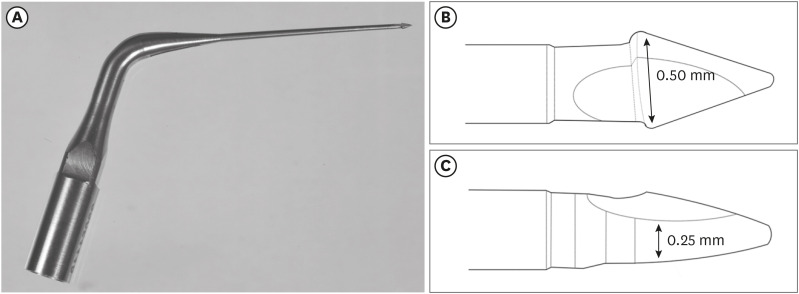
- The effectiveness of the supplementary use of the XP-endo Finisher on bacteria content reduction: a systematic review and meta-analysis
- Ludmila Smith de Jesus Oliveira, Rafaella Mariana Fontes de Bragança, Rafael Sarkis-Onofre, André Luis Faria-e-Silva
- Restor Dent Endod 2021;46(3):e37. Published online June 18, 2021
- DOI: https://doi.org/10.5395/rde.2021.46.e37
-
Abstract
PDFSupplementary MaterialPubReaderePub
Objectives This systematic review evaluated the efficacy of the supplementary use of the XP-endo Finisher on bacteria content reduction in the root canal system.
Materials and Methods In-vitro studies evaluating the use of the XP-endo Finisher on bacteria content were searched in four databases in July 2020. Two authors independently screened the studies for eligibility. Data were extracted, and risk of bias was assessed. Data were meta-analyzed by using random-effects model to compare the effect of the supplementary use (experimental) or not (control) of the XP-endo Finisher on bacteria counting reduction, and results from different endodontic protocols were combined. Four studies met the inclusion criteria while 1 study was excluded from the meta-analysis due to its high risk of bias and outlier data. The 3 studies that made it to the meta-analysis had an unclear risk of bias for at least one criterion.Results No heterogeneity was observed among the results of the studies included in the meta-analysis. The study excluded from the meta-analysis assessing the bacteria counting deep in the dentin demonstrated further bacteria reduction upon the use of the XP-endo Finisher.
Conclusions This systematic review found no evidence supporting the supplementary use of the XP-endo Finisher on further bacteria counting the reduction in the root canal.
-
Citations
Citations to this article as recorded by- Mapping risk of bias criteria in systematic reviews of in vitro endodontic studies: an umbrella review
Rafaella Rodrigues da Gama, Lucas Peixoto de Araújo, Evandro Piva, Leandro Perello Duro, Adriana Fernandes da Silva, Wellington Luiz de Oliveira da Rosa
Evidence-Based Dentistry.2025; 26(4): 179. CrossRef - Characteristics and Effectiveness of XP‐Endo Files and Systems: A Narrative Review
Sarah M. Alkahtany, Rana Alfadhel, Aseel AlOmair, Sarah Bin Durayhim, Kee Y. Kum
International Journal of Dentistry.2024;[Epub] CrossRef - Impact XP-endo finisher on the 1-year follow-up success of posterior root canal treatments: a randomized clinical trial
Ludmila Smith de Jesus Oliveira, Fabricio Eneas Diniz de Figueiredo, Janaina Araújo Dantas, Maria Amália Gonzaga Ribeiro, Carlos Estrela, Manoel Damião Sousa-Neto, André Luis Faria-e-Silva
Clinical Oral Investigations.2023; 27(12): 7595. CrossRef - Comparative analysis of the effectiveness of modern irrigants activation techniques in the process of mechanical root canal system treatment (Literature review)
Anatoliy Potapchuk, Vasyl Almashi, Arsenii Horzov, Victor Buleza
InterConf.2023; (34(159)): 200. CrossRef - Comparative analysis of the effectiveness of modern irrigants activation techniques in the protocol of chemomechanical root canal system treatment (literature review)
A. Potapchuk, V. Almashi, Y. Rak, Y. Melnyk, V. Buleza, A. Horzov
SUCHASNA STOMATOLOHIYA.2023; 114(3): 4. CrossRef - Methodological quality assessment criteria for the evaluation of laboratory‐based studies included in systematic reviews within the specialty of Endodontology: A development protocol
Venkateshbabu Nagendrababu, Paul V. Abbott, Christos Boutsioukis, Henry F. Duncan, Clovis M. Faggion, Anil Kishen, Peter E. Murray, Shaju Jacob Pulikkotil, Paul M. H. Dummer
International Endodontic Journal.2022; 55(4): 326. CrossRef
- Mapping risk of bias criteria in systematic reviews of in vitro endodontic studies: an umbrella review
- 3,490 View
- 26 Download
- 4 Web of Science
- 6 Crossref
- Evaluation of the relation between the pulp stones and direct restorations using cone beam computed tomography in a Turkish subpopulation
- Güzide Pelin Sezgin, Sema Sönmez Kaplan, Tuna Kaplan
- Restor Dent Endod 2021;46(3):e34. Published online June 8, 2021
- DOI: https://doi.org/10.5395/rde.2021.46.e34
-
Abstract
PDFPubReaderePub
Objectives This study aimed to assess the presence of pulp stones through an examination of cone beam computed tomography images and correlate their prevalence with age, sex, dental arch and side, tooth type, and restoration type and depth.
Materials and Methods Cone beam computed tomography images obtained from 673 patients and archival data on 11,494 teeth were evaluated. The associations of pulp stones with age, sex, dental arch and side, tooth type, and restoration type and depth were noted. All the measurements were subjected to a χ2 test and one sample χ2 test (
p < 0.05).Results In the study group, 163 (24.2%) patients and 379 (3.3%) teeth had at least one pulp stone. The pulp stone frequency in those aged 30–39 years was significantly greater than in those aged 18–29 and ≥ 60 years, and the frequency was higher in females than in males (
p < 0.05). The highest prevalence of pulp stones was found in maxillary dental arches and molar teeth (p < 0.05). Pulp stones were significantly more common in medium-depth restorations (p < 0.05).Conclusions Maxillary molar teeth, medium-depth restorations, individuals aged 30–39 years and females had a greater percentage of pulp stones.
-
Citations
Citations to this article as recorded by- Comparative Detection and Inter-Modality Agreement of Pulp Stones Using Digital Periapical Radiography and CBCT at Two Voxel Sizes: An Ex Vivo Study
Hassan Hamed Kaabi, Sarah Saeed Binhassan, Sultan Hamad Alrumaih, Mohammed Jamal Alotaibi, Abdullah Khalid Bakarman, Nawaf Abdulaziz Alghamdi, Hamad Abdullah Almuhaythif, Qamar Mohammadziad Hashem, Abdulfatah Samih Alazmah
Diagnostics.2026; 16(7): 961. CrossRef - Cone-Beam Computed Tomography Assessment of the Prevalence and Association of Pulp Calcification with Dental and Periodontal Pathology: A Descriptive Study
José Luis Sanz, Lucía Callado, Stefana Mantale, Jenifer Nicolás, James Ghilotti, Carmen Llena
Journal of Clinical Medicine.2025; 14(4): 1373. CrossRef - Prevalence of mineralization in the pulp chamber in patients according to CBCT data
V. A. Molokova, I. N. Antonova, V. A. Osipova
Endodontics Today.2025; 23(2): 188. CrossRef - Could carotid artery calcifications and pulp stones be an alarm sign for diabetes mellitus? A retrospective observational study
Motahare Baghestani, Mohadese Faregh, Seyed Hossein Razavi, Fatemeh Owlia
BMC Endocrine Disorders.2025;[Epub] CrossRef - Distribution and influencing factors of pulp stones based on CBCT: a retrospective observational study from southwest China
Wantong Zhang, Yao Wang, Lin Ye, Yan Zhou
BMC Oral Health.2024;[Epub] CrossRef - Prevalence and Association of Calcified Pulp Stones with Periodontitis: A Cone-Beam Computed Tomography Study in Saudi Arabian Population
Abdullah Saad Alqahtani
Journal of Pharmacy and Bioallied Sciences.2024; 16(Suppl 1): S644. CrossRef - The Prevalence And Distribution Of Pulp Stones: A Cone-Beam Computed Tomography Study İn A Group Of Turkish Patients
Mujgan Firincioglulari, Seçil Aksoy, Melis Gülbeş, Umut Aksoy, Kaan Orhan
ADO Klinik Bilimler Dergisi.2024; 13(3): 496. CrossRef - Radiographical examination of pulp stone distribution by cone beam computed tomography
Fatma Tunç, Emre Çulha, Muazzez Naz Baştürk
Journal of Health Sciences and Medicine.2024; 7(4): 472. CrossRef - Cone-Beam Computed Tomography-Based Investigation of the Prevalence and Distribution of Pulp Stones and Their Relation to Local and Systemic Factors in the Makkah Population: A Cross-Sectional Study
Laila M Kenawi, Haytham S Jaha, Mashael M Alzahrani, Jihan I Alharbi, Shahad F Alharbi, Taif A Almuqati, Rehab A Alsubhi, Wahdan M Elkwatehy
Cureus.2024;[Epub] CrossRef - Cone beam computed tomography assessment of the prevalence and association of pulp calcification with periodontitis
Lingling Xiang, Botao Wang, Yuan Zhang, Jintao Wang, Peipei Wu, Jian Zhang, Liangjun Zhong, Rui He
Odontology.2023; 111(1): 248. CrossRef - Three-dimensional analysis for detection of pulp stones in a Saudi population using cone beam computed tomography
Hassan H. Kaabi, Abdullah M. Riyahi, Nassr S. Al-Maflehi, Saleh F. Alrumayyan, Abdullah K. Bakrman, Yazeed A. Almutaw
Journal of Oral Science.2023; 65(4): 257. CrossRef
- Comparative Detection and Inter-Modality Agreement of Pulp Stones Using Digital Periapical Radiography and CBCT at Two Voxel Sizes: An Ex Vivo Study
- 3,090 View
- 36 Download
- 12 Web of Science
- 11 Crossref
- Postoperative pain after endodontic treatment of necrotic teeth with large intentional foraminal enlargement
- Ricardo Machado, Daniel Comparin, Sérgio Aparecido Ignácio, Ulisses Xavier da Silva Neto
- Restor Dent Endod 2021;46(3):e31. Published online May 31, 2021
- DOI: https://doi.org/10.5395/rde.2021.46.e31
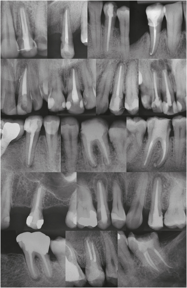
-
Abstract
PDFPubReaderePub
Objectives To evaluate postoperative pain after endodontic treatment of necrotic teeth using large intentional foraminal enlargement (LIFE).
Materials and Methods The sample included 60 asymptomatic necrotic teeth (with or without chronic apical periodontitis), and a periodontal probing depth of 3 mm, previously accessed and referred to perform endodontic treatment. After previous procedures, the position and approximate size of the apical foramen (AF) were determined by using an apex locator and K flexo-files, respectively. The chemomechanical preparation was performed with Profile 04 files 2 mm beyond the AF to achieve the LIFE, using 2.5 mL of 2.5% NaOCl at each file change. The filling was performed by Tagger's hybrid technique and EndoFill sealer. Phone calls were made to all the patients at 24, 48 and 72 hours after treatment, to classify postoperative pain. Statistical analysis was performed by different tests with a significance level of 5%.
Results Age, gender, periradicular status and tooth type did not influence postoperative pain (
p > 0.05). Only 1 patient (1.66%) reported severe pain after 72 hours. Moderate pain was reported by 7, 4 and 3 patients after 24, 48 and 72 hours, respectively (p = 0.0001). However, paired analyses showed a statistically significant difference only between 24 and 72 hours (p = 0.04). Sealer extrusion did not influence the postoperative pain (p > 0.05).Conclusions Acute or moderate postoperative pain was uncommon after endodontic treatment of necrotic teeth with LIFE.
Trial Registration The Brazilian Clinical Trials Registry Identifier:
RBR-3r967t -
Citations
Citations to this article as recorded by- Postoperative Pain After Endodontic Treatment in HIV‐Positive Patients Under HAART: A Prospective Observational Cohort Study
Marcos Felipe Iparraguirre Nuñovero, Marco Antonio Hungaro Duarte, Luciana Reis Azevedo Alanis, Bruno Cavalini Cavenago, Ulisses Xavier da Silva Neto, Everdan Carneiro
International Endodontic Journal.2026; 59(5): 788. CrossRef - Does maintaining apical patency reduce early postoperative pain after root canal treatment? A randomized controlled trial in asymptomatic vital single-rooted teeth
Ozan Arda Deger, Sehnaz Yilmaz, Kübra Gürler
Clinical Oral Investigations.2026;[Epub] CrossRef - Evaluation of Postoperative Pain Frequency in Single‐Session Endodontic Treatments With Patency and Foraminal Enlargement
Viviane Barbosa Godoy, Ana Grasiela Limoeiro, Vanessa Sandini, Vini Mehta, Wayne Martins Nascimento, Marilia Fagury Videira Marceliano‐Alves, Marcos Frozoni
Clinical and Experimental Dental Research.2026;[Epub] CrossRef - Assessment of apical extrusion in regenerative endodontics: a comparative study of different irrigation methods using three-dimensional immature tooth models
Vahide Hazal Abat, Gökçen Deniz Bayrak, Mustafa Gündoğar
Odontology.2025; 113(1): 213. CrossRef - Clinical Advances in Calcium Phosphate for Maxillomandibular Bone Regeneration: From Bench to Bedside
Seyed Ali Mostafavi Moghaddam, Hamid Mojtahedi, Amirhossein Bahador, Lotfollah Kamali Hakim, Hamid Tebyaniyan
Ceramics.2025; 8(4): 129. CrossRef - Postoperative pain after single-visit root canal treatments in necrotic teeth comparing instruments’ kinematics and apical instrumentation limits – a prospective randomized multicenter clinical trial
Ricardo Machado, Guilherme Moreira, Daniel Comparin, Arthur Pimentel Barroso, Jaqueline Nascimento, Caio Cézar Randi Ferraz, Sérgio Aparecido Ignácio, Lucas da Fonseca Roberti Garcia, Rodrigo Rodrigues Amaral, David Shadid, Ulisses Xavier da Silva Neto
BMC Oral Health.2024;[Epub] CrossRef - Assessment of mechanical allodynia in healthy teeth adjacent and contralateral to endodontically diseased teeth: a clinical study
Vaishnavi Ratnakar Patankar, Ashish K Jain, Rahul D Rao, Prajakta R Rao
Restorative Dentistry & Endodontics.2024;[Epub] CrossRef - A systematic review and meta-analysis on the effects of phototherapy on postoperative pain in conventional endodontic reintervention
Larissa Pereira Nunes, Gabriel Pereira Nunes, Túlio Morandin Ferrisse, Henrico Badaoui Strazzi-Sahyon, Eloi Dezan-Júnior, Luciano Tavares Angelo Cintra, Gustavo Sivieri-Araujo
Clinical Oral Investigations.2024;[Epub] CrossRef - The effect of intracanal cryotherapy with and without foraminal enlargement on pain prevention after endodontic treatment: a randomized clinical trial
Marcos Felipe Iparraguirre Nuñovero, Marco Antonio Hungaro Duarte, André Vinícius Kaled Segato, Ulisses Xavier da Silva Neto, Vania Portela Ditzel Westphalen, Everdan Carneiro
Scientific Reports.2024;[Epub] CrossRef - Clinical determination of anatomical diameter in different dental groups correlating them with gender, age, tooth/canal and pulpoperiradicular diagnosis: an observational clinical study
Ricardo Machado, Gabriel Filipe Pamplona, Claudemir de Souza Júnior, Jaqueline Nascimento, Eduardo Donato Eing Elgelke Back, Daniel Comparin, Sérgio Aparecido Ignácio, Stella Maria Glaci Reinke, Ana Cristina Kovalik, Ulisses Xavier da Silva Neto
Scientific Reports.2023;[Epub] CrossRef - How much to enlarge? A letter to the editor
Krishnamachari Janani, Kavalipurapu Venkata Teja, Kumar Chandan Srivatsava
Saudi Endodontic Journal.2023; 13(3): 288. CrossRef - Efficiency of diode laser in control of post-endodontic pain: a randomized controlled trial
Hend H. Ismail, Maram Obeid, Ehab Hassanien
Clinical Oral Investigations.2023; 27(6): 2797. CrossRef - Periapical Healing following Root Canal Treatment Using Different Endodontic Sealers: A Systematic Review
Akshay Khandelwal, Krishnamachari Janani, KavalipurapuVenkata Teja, Jerry Jose, Gopi Battineni, Francesco Riccitiello, Alessandra Valletta, Ajitha Palanivelu, Gianrico Spagnuolo, Vincenzo Grassia
BioMed Research International.2022;[Epub] CrossRef
- Postoperative Pain After Endodontic Treatment in HIV‐Positive Patients Under HAART: A Prospective Observational Cohort Study
- 6,501 View
- 72 Download
- 11 Web of Science
- 13 Crossref
- Cryopreservation of mesenchymal stem cells derived from dental pulp: a systematic review
- Sabrina Moreira Paes, Yasmine Mendes Pupo, Bruno Cavalini Cavenago, Thiago Fonseca-Silva, Carolina Carvalho de Oliveira Santos
- Restor Dent Endod 2021;46(2):e26. Published online April 29, 2021
- DOI: https://doi.org/10.5395/rde.2021.46.e26
-
Abstract
PDFPubReaderePub
Objectives The aim of the present systematic review was to investigate the cryopreservation process of dental pulp mesenchymal stromal cells and whether cryopreservation is effective in promoting cell viability and recovery.
Materials and Methods This systematic review was developed in accordance with the Preferred Reporting Items for Systematic Reviews and Meta-Analyses (PRISMA) statement and the research question was determined using the population, exposure, comparison, and outcomes strategy. Electronic searches were conducted in the PubMed, Cochrane Library, Science Direct, LILACS, and SciELO databases and in the gray literature (dissertations and thesis databases and Google Scholar) for relevant articles published up to March 2019. Clinical trial studies performed with dental pulp of human permanent or primary teeth, containing concrete information regarding the cryopreservation stages, and with cryopreservation performed for a period of at least 1 week were included in this study.
Results The search strategy resulted in the retrieval of 185 publications. After the application of the eligibility criteria, 21 articles were selected for a qualitative analysis.
Conclusions The cryopreservation process must be carried out in 6 stages: tooth disinfection, pulp extraction, cell isolation, cell proliferation, cryopreservation, and thawing. In addition, it can be inferred that the use of dimethyl sulfoxide, programmable freezing, and storage in liquid nitrogen are associated with a high rate of cell viability after thawing and a high rate of cell proliferation in both primary and permanent teeth.
-
Citations
Citations to this article as recorded by- Comparative study on biological characteristics of dental mesenchymal stem cells isolated from gingiva, periodontal ligament, and dental follicle and their derived conditioned medium
Xianyi He, Yichen Gao, Haiyin Wan, Xia Wang, Jie Shen, Yun He, Junliang Chen
Annals of Anatomy - Anatomischer Anzeiger.2026; 263: 152751. CrossRef - Effect of the pulp harvesting method on the viability of human dental pulp stem cells and their odontogenic differentiation potential
Justine De Visscher, Lore Vermeir, Natasja Van den Vreken, Liesbeth Temmerman, Noëmi De Roo, Jolanda van Hengel, Guy De Pauw
Cell and Tissue Banking.2025;[Epub] CrossRef - The Antimicrobial Effect of the Incorporation of Inorganic Substances into Heat-Cured Denture Base Resins—A Systematic Review
Mariana Lima, Helena Salgado, André Correia, Patrícia Fonseca
Prosthesis.2024; 6(5): 1189. CrossRef - Sphingosine-1-phosphate Treatment Improves Cryopreservation Efficiency in Human Mesenchymal Stem Cells
Seong-Ju Oh, Chan-Hee Jo, Tae-Seok Kim, Chae-Yeon Hong, Sung-Lim Lee, Young-Hoon Kang, Gyu-Jin Rho
Life.2023; 13(6): 1286. CrossRef - Time- and Concentration-Dependent Effects of the Stem Cells Derived from Human Exfoliated Deciduous Teeth on Osteosarcoma Cells
Razieh Alipour, Batool Hashemibeni, Vajihe Asgari, Hamid Bahramian
Advanced Biomedical Research.2023;[Epub] CrossRef
- Comparative study on biological characteristics of dental mesenchymal stem cells isolated from gingiva, periodontal ligament, and dental follicle and their derived conditioned medium
- 3,968 View
- 38 Download
- 4 Web of Science
- 5 Crossref
-
Hard tissue formation after direct pulp capping with osteostatin and MTA
in vivo - Ji-Hye Yoon, Sung-Hyeon Choi, Jeong-Tae Koh, Bin-Na Lee, Hoon-Sang Chang, In-Nam Hwang, Won-Mann Oh, Yun-Chan Hwang
- Restor Dent Endod 2021;46(2):e17. Published online February 25, 2021
- DOI: https://doi.org/10.5395/rde.2021.46.e17
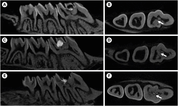
-
Abstract
PDFPubReaderePub
Objectives In recent
in vitro study, it was reported that osteostatin (OST) has an odontogenic effect and synergistic effect with mineral trioxide aggregate (MTA) in human dental pulp cells. Therefore, the aim of this study was to evaluate whether OST has a synergistic effect with MTA on hard tissue formationin vivo .Materials and Methods Thirty-two maxillary molars of Spraque-Dawley rats were used in this study. An occlusal cavity was prepared and the exposed pulps were randomly divided into 3 groups: group 1 (control; ProRoot MTA), group 2 (OST 100 μM + ProRoot MTA), group 3 (OST 10 mM + ProRoot MTA). Exposed pulps were capped with each material and cavities were restored with resin modified glass ionomer. The animals were sacrificed after 4 weeks. All harvested teeth were scanned with micro-computed tomography (CT). The samples were prepared and hard tissue formation was evaluated histologically. For immunohistochemical analysis, the specimens were sectioned and incubated with primary antibodies against dentin sialoprotein (DSP).
Results In the micro-CT analysis, it is revealed that OST with ProRoot MTA groups showed more mineralized bridge than the control (
p < 0.05). In the H&E staining, it is showed that more quantity of the mineralized dentin bridge was formed in the OST with ProRoot MTA group compared to the control (p < 0.05). In all groups, DSP was expressed in newly formed reparative dentin area.Conclusions OST can be a supplementary pulp capping material when used with MTA to make synergistic effect in hard tissue formation.
-
Citations
Citations to this article as recorded by- Trends and Future Perspectives in Additive Modifications of Calcium Silicate‐Based Cements: A Scoping Review (2015–2024)
Kenta Tsuchiya, Salvatore Sauro, Jukka P. Matinlinna, Monica Yamauti, Hidehiko Sano, Kyung‐San Min, Atsushi Tomokiyo
Australian Endodontic Journal.2026;[Epub] CrossRef - Pulpal responses to mineral trioxide aggregate with and without zinc oxide addition in mature canine teeth after full pulpotomy
Behnam Bolhari, Neda Kardouni Khouzestani, Hadi Assadian, Saeed Farzad-Mohajeri, Mohammad Mehdi Dehghan, Soheil Niavarzi, Behnam Dorost, Venkateshbabu Nagendrababu, Henry F. Duncan, Artak Heboyan, Antonio Signore, Stefano Benedicenti
Scientific Reports.2025;[Epub] CrossRef - Research Advancements in Peptides for Promoting Reparative Dentin Regeneration in Direct Pulp Capping: A Narrative Review
Jiawen Wang, Shuwei Qiao, Tianjia Huang, Junjie Lian, Song Zhu
International Journal of Peptide Research and Therapeutics.2025;[Epub] CrossRef - Biocompatibility and pro-mineralization effects of premixed calcium silicate-based materials on human dental pulp stem cells: An in vitro and in vivo study
Nyein Chan KO, Sonoko NODA, Yamato OKADA, Kento TAZAWA, Nobuyuki KAWASHIMA, Takashi OKIJI
Dental Materials Journal.2024; 43(5): 729. CrossRef - Osteostatin, a peptide for the future treatment of musculoskeletal diseases
Daniel Lozano, Arancha R. Gortazar, Sergio Portal-Núñez
Biochemical Pharmacology.2024; 223: 116177. CrossRef - Comparison of bioactive material failure rates in vital pulp treatment of permanent matured teeth – a systematic review and network meta-analysis
Péter Komora, Orsolya Vámos, Noémi Gede, Péter Hegyi, Kata Kelemen, Adél Galvács, Gábor Varga, Beáta Kerémi, János Vág
Scientific Reports.2024;[Epub] CrossRef - Hard tissue formation in pulpotomized primary teeth in dogs with nanomaterials MCM-48 and MCM-48/hydroxyapatite: an in vivo animal study
Sahar Talebi, Nosrat Nourbakhsh, Ardeshir Talebi, Amir Abbas Nourbakhsh, Abbas Haghighat, Maziar Manshayi, Hamid Reza Bakhsheshi, Razieh Karimi, Rahman Nazeri, Kenneth J.D. Mackenzie
BMC Oral Health.2024;[Epub] CrossRef - Reparative Mineralized Tissue Characterization by Different Bioactive Direct Pulp-capping Agents
Mrunal Shinde, Varsha Pandit, Sarita Singh, Aniket Jadhav, Sarah Marium, Smita Patil
Journal of the International Clinical Dental Research Organization.2024; 16(1): 8. CrossRef - Effects of mineral trioxide aggregate and methyl sulfonyl methane on pulp exposure via RUNX2 and RANKL pathways
Altar Ateş, Ayca Kurt, Tolga Mercantepe
Odontology.2024; 112(3): 895. CrossRef - Effects of barium titanate on the dielectric constant, radiopacity, and biological properties of tricalcium silicate-based bioceramics
Yoorina CHOI, Yun-Chan HWANG, Mi-Kyung YU, Kwang-Won LEE, Kyung-San MIN
Dental Materials Journal.2023; 42(1): 55. CrossRef - Bioactive potential of Bio‐C Pulpo is evidenced by presence of birefringent calcite and osteocalcin immunoexpression in the rat subcutaneous tissue
Marcela Borsatto Queiroz, Rafaela Nanami Handa Inada, Camila Soares Lopes, Juliane Maria Guerreiro‐Tanomaru, Estela Sasso‐Cerri, Mário Tanomaru‐Filho, Paulo Sérgio Cerri
Journal of Biomedical Materials Research Part B: Applied Biomaterials.2022; 110(10): 2369. CrossRef - The Influence of New Bioactive Materials on Pulp–Dentin Complex Regeneration in the Assessment of Cone Bone Computed Tomography (CBCT) and Computed Micro-Tomography (Micro-CT) from a Present and Future Perspective—A Systematic Review
Mirona Paula Palczewska-Komsa, Bartosz Gapiński, Alicja Nowicka
Journal of Clinical Medicine.2022; 11(11): 3091. CrossRef - A Breakthrough in the Era of Calcium Silicate-Based Cements: A Critical Review
Payal S Chaudhari, Manoj G Chandak, Akshay A Jaiswal, Nikhil P Mankar, Priyanka Paul
Cureus.2022;[Epub] CrossRef - Effectiveness of Direct Pulp Capping Bioactive Materials in Dentin Regeneration: A Systematic Review
Ermin Nie, Jiali Yu, Rui Jiang, Xiangzhen Liu, Xiang Li, Rafiqul Islam, Mohammad Khursheed Alam
Materials.2021; 14(22): 6811. CrossRef
- Trends and Future Perspectives in Additive Modifications of Calcium Silicate‐Based Cements: A Scoping Review (2015–2024)
- 3,972 View
- 43 Download
- 13 Web of Science
- 14 Crossref
- Efficacy of buccal piroxicam infiltration and inferior alveolar nerve block in patients with irreversible pulpitis: a prospective, double-blind, randomized clinical trial
- Saurav Paul, Sridevi Nandamuri, Aakrati Raina, Mukta Bansal
- Restor Dent Endod 2021;46(1):e9. Published online January 26, 2021
- DOI: https://doi.org/10.5395/rde.2021.46.e9
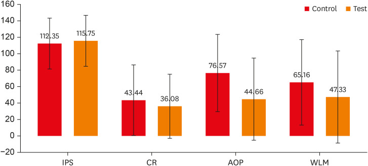
-
Abstract
PDFPubReaderePub
Objectives This randomized clinical trial aimed to assess the effectiveness of buccal infiltration with piroxicam on the anesthetic efficacy of inferior alveolar nerve block (IANB) with buccal infiltration in irreversible pulpitis, with pain assessed using the Heft-Parker visual analogue scale (HP-VAS).
Materials and Methods This study included 56 patients with irreversible pulpitis in mandibular molars, randomly distributed between 2 groups (
n = 28). After evaluating the initial pain score with the HP-VAS, each patient received IANB followed by buccal infiltration of 2% lignocaine with adrenaline (1:80,000). Five minutes later, the patients in groups 1 and 2 were given buccal infiltration with 40 mg/2 mL of piroxicam or normal saline, respectively. An access opening procedure (AOP) was performed 15 minutes post-IANB once the individual showed signs of lip numbness as well as 2 negative responses to electric pulp testing. The HP-VAS was used to grade the patient's pain during caries removal (CR), AOP, and working length measurement (WLM). Successful anesthesia was identified either by the absence of pain or slight pain through CR, AOP, and WLM, with no requirement of a further anesthetic dose. A statistical analysis was done using the Shapiro-Wilk and Mann-WhitneyU tests.Results The piroxicam group presented a significantly lower (
p < 0.05) mean pain score than the saline group during AOP.Conclusions Buccal infiltration with piroxicam enhanced the efficacy of anesthesia with IANB and buccal infiltration with lignocaine in patients with irreversible pulpitis.
-
Citations
Citations to this article as recorded by- Analyzing The Role of Fur-Friends in Mental Well-Being: Comparing Mental Well-Being of Pet Owners and Non-Owners
Jervin Dinglasan Quicho, Jesslaine Angela Sanchez, Alexandra Nicole P. Mendoza
International Journal of Multidisciplinary Applied Business and Education Research.2026; 7(3): 1053. CrossRef - Inferior alveolar nerve block success of 2% mepivacaine versus 4% articaine in patients with symptomatic irreversible pulpitis in mandibular molars: A randomized double‐blind single‐centre clinical trial
Mohammed Fawzy Omar Mohammed Habib, Sovana Tarek, Sara Mohamed Elsayed Teama, Khaled Ezzat, Randa Mohamed El Boghdadi, Abeer Marzouk, Manar Yehia Fouda, Shaimaa Ismail Gawdat, Marwa Mahmoud Bedier, Suzan Abdul Wanees Amin
International Endodontic Journal.2022; 55(11): 1177. CrossRef - Present status and future directions—Mechanisms and management of local anaesthetic failures
Masoud Parirokh, Paul V. Abbott
International Endodontic Journal.2022; 55(S4): 951. CrossRef
- Analyzing The Role of Fur-Friends in Mental Well-Being: Comparing Mental Well-Being of Pet Owners and Non-Owners
- 3,038 View
- 28 Download
- 2 Web of Science
- 3 Crossref
- Reference values for pulp oxygen saturation as a diagnostic tool in endodontics: a systematic review and meta-analysis
- Paula Lambert, Sergio Augusto Quevedo Miguens, Caroline Solda, Juliana Tomaz Sganzerla, Leandro Azambuja Reichert, Carlos Estrela, Fernando Branco Barletta
- Restor Dent Endod 2020;45(4):e48. Published online October 5, 2020
- DOI: https://doi.org/10.5395/rde.2020.45.e48
-
Abstract
PDFPubReaderePub
Objectives This systematic review aimed to identify mean oxygen saturation values (SpO2) using pulse oximetry in permanent maxillary anterior teeth.
Materials and Methods The MEDLINE, Scientific Electronic Library Online, Cochrane Central Register of Controlled Trials, EMBASE, and Literatura Latino Americana em Ciências da Saúde electronic databases were searched. Combinations and variations of “oximetry” AND “dental pulp test” were used as search terms. Studies reporting means and standard deviations of SpO2 values were included. Two reviewers independently extracted data following the Preferred Reporting Items for Systematic Reviews and Meta-Analyses checklist. Heterogeneity was assessed using the
I 2 statistic, and all analyses were performed using R software. Study quality was assessed using the Quality Assessment of Diagnostic Accuracy Studies-2 tool and the Newcastle-Ottawa scale.Results Of the 251 studies identified, 19 met the eligibility criteria and were included (total sample, 4,541 teeth). In the meta-analysis, the mean SpO2 values were 84.94% (95% confidence interval [CI], 84.85%–85.04%) for the central incisors, 89.29% (95% CI, 89.22%–89.35%) for the lateral incisors, and 89.20% (95% CI, 89.05%–89.34%) for the canines. The studies were predominantly low-quality due to the high risk of bias associated with the index test, unclear risk regarding patient selection, and concerns about outcome assessment.
Conclusions Although most studies were low-quality, the oxygen saturation levels in normal pulp could be established (minimum saturation, 77.52%). Despite the risk of bias of the included studies, the reference values reported herein are clinically relevant for assessments of changes in pulp status.
Trial Registration International Prospective Register of Systematic Reviews Identifier:
CRD42018085598 -
Citations
Citations to this article as recorded by- Lower dental pulp oxygen saturation in children with molar incisor hypomineralization: a cross-sectional study
Jade de Souza CAVALCANTE, Carlos ESTRELA, Fabrício Kitazono de CARVALHO, Francisco Wanderley Garcia de PAULA-SILVA, Manoel Damião de SOUSA-NETO, Kelly Fernanda MOLENA, Alexandra Mussolino de QUEIROZ
Brazilian Oral Research.2026;[Epub] CrossRef - Reference Values for Pulse Oximetry Testing in Permanent Teeth: A Systematic Review and Meta‐Analysis
Lilian Tietz, Theodoro Weissheimer, Cassiano Kuchenbecker Rösing, Marcus Vinicius Reis Só
International Endodontic Journal.2026;[Epub] CrossRef - Clinical Validation of Smartphone-Enabled Pulse Oximetry for Objective Pulp Vitality Assessment: A Diagnostic Accuracy Study
Celso Luiz Caldeira, Stephanie Isabel Diaz Zamalloa, Claudia Regina Guimaro Sakitani, Fernando Branco Barletta, Marinella Holzhausen
Journal of Endodontics.2025; 51(12): 1752. CrossRef - Laser Doppler Flowmetry and Continuous Tissue Oxygenation Monitoring: Best of Vitality Tests?
Herman J. J. Roeykens, Rani D’haese, Wolfgang Jacquet, Roeland J. G. De Moor, Stefan Vandeweghe
Oral.2025; 5(4): 83. CrossRef - Diagnostic accuracy of Transmitted-light plethysmography for the assessment of pulpal circulation in traumatized young permanent incisors
Satoko Kakino, Hiroaki Ohki, Kaori Kohi, Yuko Matsumura, Tsutomu Iwamoto
Scientific Reports.2025;[Epub] CrossRef - Future trends in endodontics
Foo Suanhow, Tawil Bill
Journal of Applied Biotechnology & Bioengineering.2024; 11(1): 1. CrossRef - Assessment of Pulpal Oxygen Saturation in Caries-free and Carious Maxillary Primary Central Incisors Using a Customized Dental Pulse Oximeter
Kranthi Reddy Kanumuru, Nancy Solomon, Hemalatha Ramkumar, Shankar Paulindraraj, Trophimus Gnanabagyan Jayakaran, Senthil Dakshinamoorthy
International Journal of Clinical Pediatric Dentistry.2023; 16(4): 560. CrossRef - Age-Related Variation of Pulpal Oxygen Saturation in Healthy Primary and Permanent Teeth in Children: A Clinical Study
Andreea Igna, Darian Rusu, Emilia Ogodescu, Ștefania Dinu, Marius Boariu, Adrian Voicu, Ștefan-Ioan Stratul
Journal of Clinical Medicine.2022; 12(1): 170. CrossRef - Pulp oxygen saturation measurement as a diagnostic tool for assessing pulp status in primary teeth: A systematic review and meta-analysis
Kanamarlapudi Venkata Saikiran, Deepa Gurunathan, Sainath Reddy Elicherla, Sreekanth Kumar Mallineni, Sivakumar Nuvvula
Journal of Indian Society of Pedodontics and Preventive Dentistry.2022; 40(4): 349. CrossRef - Diagnostic Value of Serum Chitinase‐3‐Like Protein 1 for Liver Fibrosis: A Meta‐analysis
Xiaoting Huang, Jialing Zhuang, Yongqiang Yang, Jiaxin Jian, Wen Ai, Chunyong Liu, Wenzhi Tang, Changyu Jiang, Yongshen He, Lesheng Huang, Se Peng, Jin Shui Pan
BioMed Research International.2022;[Epub] CrossRef - Assessment of Pulpal Status in Primary Teeth Following Direct Pulp Capping in an Experimental Canine Model
Andreea Igna, Cornel Igna, Mariana Ioana Miron, Larisa Schuszler, Roxana Dascălu, Mihaela Moldovan, Adrian Aristide Voicu, Carmen Darinca Todea, Marius Boariu, Maria-Alexandra Mârțu, Ștefan-Ioan Stratul
Diagnostics.2022; 12(8): 2022. CrossRef
- Lower dental pulp oxygen saturation in children with molar incisor hypomineralization: a cross-sectional study
- 3,465 View
- 47 Download
- 11 Crossref
- Calcium hydroxide intracanal medication effects on pain and flare-up: a systematic review and meta-analysis
- Ahmed Mohamed Ibrahim, Siza Yacoub Zakhary, Suzan Abdul Wanees Amin
- Restor Dent Endod 2020;45(3):e26. Published online May 22, 2020
- DOI: https://doi.org/10.5395/rde.2020.45.e26
-
Abstract
PDFSupplementary MaterialPubReaderePub
Objectives This study aimed to systematically review the pain and flare-up effects of calcium hydroxide (CH) as intracanal medication (ICM) in non-vital mature teeth.
Materials and Methods Electronic-databases searching for published and grey literature and manual searching were conducted. Only randomized clinical trials (RCTs) were included comparing CH to other ICMs in non-vital mature teeth. The risk of bias was assessed using the RoB 2.0 Cochrane tool. The main outcomes were pain and flare-up. Qualitative and quantitative analysis, wherever applicable, was performed. The certainty of evidence (CoE) was assessed using Grading of Recommendations Assessment, Development and Evaluation (GRADE).
Results Sixteen articles were included in 6 comparisons at different time points for different outcomes. CH reduced pain risk than no ICM within the 1–14-days interval (
p < 0.05) and than triple-antibiotic paste within the first day (p < 0.05) and was similar to corticosteroid/antibiotics combination (p > 0.05). Chlorhexidine (CHX) or CH/CHX, however, reduced pain levels than CH alone (p < 0.05). CH showed higher flare-up risk than CHX (p < 0.05). CoE, however, ranged from very low to moderate.Conclusion Most comparisons for different outcomes are based on very few studies, mostly low-powered, with an overall low CoE. Thus, the available evidence is considered insufficient to either support or refute CH effectiveness or to recommend one ICM over another. Therefore, further well-designed, larger RCTs are required.
Trial Registration PROSPERO database Identifier:
CRD42016041953 -
Citations
Citations to this article as recorded by- Interappointment pain reduction with three intracanal medicaments: A randomized clinical trial
Labdhi Maloo, Rashmi Nair, Anjali Bichpuriya, Vaishnavi Dorlikar, Sagarika Nandi
Journal of Conservative Dentistry and Endodontics.2026; 29(7): 711. CrossRef - Garre’s osteomyelitis of the mandible managed by nonsurgical re-endodontic treatment
Heegyun Kim, Jiyoung Kwon, Hyun-Jung Kim, Soram Oh, Duck-Su Kim, Ji-Hyun Jang
Restorative Dentistry & Endodontics.2024;[Epub] CrossRef - Interpretation by literature review of the use of calcium hydroxide as an intra-ductal medication
María Belén Muñoz Padilla, Verónica Alicia Vega Martínez, Camila Alejandra Villafuerte Moya
Salud, Ciencia y Tecnología.2024; 4: 924. CrossRef - ProTaper Ultimate: The Evolution of Rotary Systems and Their Clinical Applicability
Mateo-Castillo JF, Siqueira-Sandrin VDS, Moreira RA, Brandelero Junior S, Olano-Dextre TL, Claudia R Pinheiro
Journal of Operative Dentistry & Endodontics.2024; 9(1): 16. CrossRef - Comparative Evaluation of Bacterial Reduction by Laser-Activated Irrigation Technique (LAI) With Conventional Needle Irrigation (CNI) in Single-Rooted Teeth With Pulpal Necrosis: A Single-Blinded Randomized Controlled Trial
Keerthika Rajamanickam, Sandhya Raghu, J Vijayashree Priyadharsini, Delphine Priscilla Antony, Nivedhitha Malli Sureshbabu
Cureus.2023;[Epub] CrossRef - Post-Operative Endodontic Pain Management: An Overview of Systematic Reviews on Post-Operatively Administered Oral Medications and Integrated Evidence-Based Clinical Recommendations
Federica Di Spirito, Giuseppe Scelza, Roberto Fornara, Francesco Giordano, Donato Rosa, Alessandra Amato
Healthcare.2022; 10(5): 760. CrossRef - Management of External Inflammatory Root Resorption following Tooth Autotransplantation Using a Modified Combination of Triple Antibiotics
Saeed Asgary, Ardavan Parhizkar, Paulo J. Palma
Case Reports in Dentistry.2022;[Epub] CrossRef - Calcium hydroxide as an intracanal medication for postoperative pain during primary root canal therapy: A systematic review and meta-analysis with trial sequential analysis of randomised controlled trials
Muhammad Zubair Ahmad, Durre Sadaf, Khalid A Merdad, Amani Almohaimeed, Igho J. Onakpoya
Journal of Evidence-Based Dental Practice.2022; 22(1): 101680. CrossRef - Post-operative Pain and Antibacterial Efficacy of Silver Nanoparticles Formulations Intracanal Medication: A Randomized Controlled Clinical Study
Nermine Hassan, Alaa Diab, Geraldine Ahmed
Open Access Macedonian Journal of Medical Sciences.2021; 9(D): 248. CrossRef
- Interappointment pain reduction with three intracanal medicaments: A randomized clinical trial
- 6,981 View
- 103 Download
- 9 Crossref
- Bioactivity of endodontic biomaterials on dental pulp stem cells through dentin
- Bahar Javid, Narges Panahandeh, Hassan Torabzadeh, Hamid Nazarian, Ardavan Parhizkar, Saeed Asgary
- Restor Dent Endod 2020;45(1):e3. Published online November 4, 2019
- DOI: https://doi.org/10.5395/rde.2020.45.e3
-
Abstract
PDFPubReaderePub
Objectives This study investigated the indirect effect of calcium-enriched mixture (CEM) cement and mineral trioxide aggregate (MTA), as 2 calcium silicate-based hydraulic cements, on human dental pulp stem cells (hDPSCs) through different dentin thicknesses.
Materials and Methods Two-chamber setups were designed to simulate indirect pulp capping (IPC). Human molars were sectioned to obtain 0.1-, 0.3-, and 0.5-mm-thick dentin discs, which were placed between the 2 chambers to simulate an IPC procedure. Then, MTA and CEM were applied on one side of the discs, while hDPSCs were cultured on the other side. After 2 weeks of incubation, the cells were removed, and cell proliferation, morphology, and attachment to the discs were evaluated under scanning electron microscopy (SEM). Energy-dispersive X-ray (EDXA) spectroscopy was performed for elemental analysis. Alkaline phosphatase (ALP) activity was assessed quantitatively. The data were analyzed using the Kruskal-Wallis and Mann-Whitney tests.
Results SEM micrographs revealed elongated cells, collagen fibers, and calcified nucleations in all samples. EDXA verified that the calcified nucleations consisted of calcium phosphate. The largest calcifications were seen in the 0.1-mm-thick dentin subgroups. There was no significant difference in ALP activity across the CEM subgroups; however, ALP activity was significantly lower in the 0.1-mm-thick dentin subgroup than in the other MTA subgroups (
p < 0.05).Conclusions The employed capping biomaterials exerted biological activity on hDPSCs, as shown by cell proliferation, morphology, and attachment and calcific precipitations, through 0.1- to 0.5-mm-thick layers of dentin. In IPC, the bioactivity of these endodontic biomaterials is probably beneficial.
-
Citations
Citations to this article as recorded by- Dental pulp capping materials: modulators of stem cell behavior and regenerative potential
Ali Cheayto, Sara Ayoub, Sarah Ayad Al-Tameemi, Mohammad Fayyad-Kazan
Biomedical Physics & Engineering Express.2025; 11(6): 062004. CrossRef - Effect of pulp capping materials on odontogenic differentiation of human dental pulp stem cells: An in vitro study
Mahmoud M. Bakr, Mohamed Shamel, Shereen N. Raafat, Robert M. Love, Mahmoud M. Al‐Ankily
Clinical and Experimental Dental Research.2024;[Epub] CrossRef - Effects of Growth Factors on the Differentiation of Dental Stem Cells: A
Systematic Review and Meta-analysis (Part I)
Sayna Shamszadeh, Armin Shirvani, Hassan Torabzadeh, Saeed Asgary
Current Stem Cell Research & Therapy.2024; 19(4): 523. CrossRef - The Role of Growth Factor Delivery Systems on Cellular Activities of Dental
Stem Cells: A Systematic Review (Part II)
Sayna Shamszadeh, Armin Shirvani, Saeed Asgary
Current Stem Cell Research & Therapy.2024; 19(4): 587. CrossRef - Comprehensive review of composition, properties, clinical applications, and future perspectives of calcium-enriched mixture (CEM) cement: a systematic analysis
Saeed Asgary, Mahtab Aram, Mahta Fazlyab
BioMedical Engineering OnLine.2024;[Epub] CrossRef - Evaluation of dental pulp stem cells behavior after odontogenic differentiation induction by three different bioactive materials on two different scaffolds
Basma Ahmed, Mai H. Ragab, Rania A. Galhom, Hayam Y. Hassan
BMC Oral Health.2023;[Epub] CrossRef - Characterization of Dental Pulp Stem Cell Responses to Functional Biomaterials Including Mineralized Trioxide Aggregates
Sejin Bae, Bueonguk Kang, Hyungbin Lee, Harrison Luu, Eric Mullins, Karl Kingsley
Journal of Functional Biomaterials.2021; 12(1): 15. CrossRef - Incorporation of amoxicillin-loaded microspheres in mineral trioxide aggregate cement: an in vitro study
Fábio Rocha Bohns, Vicente Castelo Branco Leitune, Isadora Martini Garcia, Bruna Genari, Nélio Bairros Dornelles, Silvia Stanisçuaski Guterres, Fabrício Aulo Ogliari, Mary Anne Sampaio de Melo, Fabrício Mezzomo Collares
Restorative Dentistry & Endodontics.2020;[Epub] CrossRef
- Dental pulp capping materials: modulators of stem cell behavior and regenerative potential
- 2,472 View
- 18 Download
- 8 Crossref
Case Report
- A case report of multiple bilateral dens invaginatus in maxillary anteriors
- Shin Hye Chung, You-Jeong Hwang, Sung-Yeop You, Young-Hye Hwang, Soram Oh
- Restor Dent Endod 2019;44(4):e39. Published online October 21, 2019
- DOI: https://doi.org/10.5395/rde.2019.44.e39
-
Abstract
PDFPubReaderePub
The present report presents a case of dens invaginatus (DI) in a patient with 4 maxillary incisors. A 24-year-old female complained of swelling of the maxillary left anterior region and discoloration of the maxillary left anterior tooth. The maxillary left lateral incisor (tooth #22) showed pulp necrosis and a chronic apical abscess, and a periapical X-ray demonstrated DI on bilateral maxillary central and lateral incisors. All teeth responded to a vitality test, except tooth #22. The anatomic form of tooth #22 was similar to that of tooth #12, and both teeth had lingual pits. In addition, panoramic and periapical X-rays demonstrated root canal calcification, such as pulp stones, in the maxillary canines, first and second premolars, and the mandibular incisors, canines, and first premolars bilaterally. The patient underwent root canal treatment of tooth #22 and non-vital tooth bleaching. After a temporary filling material was removed, the invaginated mass was removed using ultrasonic tips under an operating microscope. The working length was established, and the root canal was enlarged up to #50 apical size and obturated with gutta-percha and AH 26 sealer using the continuous wave of condensation technique. Finally, non-vital bleaching was performed, and the access cavity was filled with composite resin.
-
Citations
Citations to this article as recorded by- Non-surgical endodontic management of dens invaginatus type II in an immature maxillary lateral incisor using a bioceramic apical plug: A 3-year follow-up case report
Yahya Raja Alharbi, Qayed Saad Alharbi, Shaul Hameed Kolarkodi
Saudi Endodontic Journal.2026; 16(1): 115. CrossRef - The use of three-dimensional-printed guides, static navigation, and bioactive materials to treat bilateral and double dens invaginatus
Parth Patel, Nidhi Bharti, Ankit Arora, C. Nimisha Shah
Saudi Endodontic Journal.2025; 15(2): 207. CrossRef - Endodontic Management of Dens in Dente – A Systematic Review of Case Reports and Case Series
Sanket Dilip Aras, Anamika Chetan Borkar, Sonal Kale, Sayali Maral, Prakriti Jaggi, Shailendra Sonawane
Journal of the International Clinical Dental Research Organization.2024; 16(1): 17. CrossRef - Dens invaginatus of fourteen teeth in a pediatric patient
Momoko Usuda, Tatsuya Akitomo, Mariko Kametani, Satoru Kusaka, Chieko Mitsuhata, Ryota Nomura
Pediatric Dental Journal.2023; 33(3): 240. CrossRef - The Impact of the Preferred Reporting Items for Case Reports in Endodontics (PRICE) 2020 Guidelines on the Reporting of Endodontic Case Reports
Sofian Youssef, Phillip Tomson, Amir Reza Akbari, Natalie Archer, Fayjel Shah, Jasmeet Heran, Sunmeet Kandhari, Sandeep Pai, Shivakar Mehrotra, Joanna M Batt
Cureus.2023;[Epub] CrossRef - Root Maturation of an Immature Dens Invaginatus Despite Unsuccessful Revitalization Procedure: A Case Report and Recommendations for Educational Purposes
Julia Ludwig, Marcel Reymus, Alexander Winkler, Sebastian Soliman, Ralf Krug, Gabriel Krastl
Dentistry Journal.2023; 11(2): 47. CrossRef - Conservative Management of Infraorbital Space Infection Secondary to Type III B Dens Invaginatus: A Case Report
Ashima Goyal, Aditi Kapur, Manoj A Jaiswal, Gauba Krishan, Raja Raghu, Sanjeev K Singh
Journal of Postgraduate Medicine, Education and Research.2022; 56(4): 192. CrossRef
- Non-surgical endodontic management of dens invaginatus type II in an immature maxillary lateral incisor using a bioceramic apical plug: A 3-year follow-up case report
- 3,589 View
- 35 Download
- 7 Crossref
Research Articles
- Development of a mouse model for pulp-dentin complex regeneration research: a preliminary study
- Sunil Kim, Sukjoon Lee, Han-Sung Jung, Sun-Young Kim, Euiseong Kim
- Restor Dent Endod 2019;44(2):e20. Published online May 7, 2019
- DOI: https://doi.org/10.5395/rde.2019.44.e20
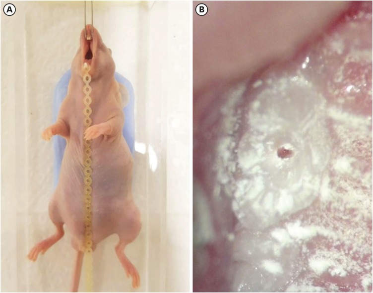
-
Abstract
PDFPubReaderePub
Objectives To achieve pulp-dentin complex regeneration with tissue engineering, treatment efficacies and safeties should be evaluated using
in vivo orthotopic transplantation in a sufficient number of animals. Mice have been a species of choice in which to study stem cell biology in mammals. However, most pulp-dentin complex regeneration studies have used large animals because the mouse tooth is too small. The purpose of this study was to demonstrate the utility of the mouse tooth as a transplantation model for pulp-dentin complex regeneration research.Materials and Methods Experiments were performed using 7-week-old male Institute of Cancer Research (ICR) mice; a total of 35 mice had their pulp exposed, and 5 mice each were sacrificed at 1, 2, 4, 7, 9, 12 and 14 days after pulp exposure. After decalcification in 5% ethylenediaminetetraacetic acid, the samples were embedded and cut with a microtome and then stained with hematoxylin and eosin. Slides were observed under a high-magnification light microscope.
Results Until 1 week postoperatively, the tissue below the pulp chamber orifice appeared normal. The remaining coronal portion of the pulp tissue was inflammatory and necrotic. After 1 week postoperatively, inflammation and necrosis were apparent in the root canals inferior to the orifices. The specimens obtained after experimental day 14 showed necrosis of all tissue in the root canals.
Conclusions This study could provide opportunities for researchers performing
in vivo orthotopic transplantation experiments with mice.-
Citations
Citations to this article as recorded by- Is dental pulp inflammation capable of causing central inflammation, behavioral, and sensory alterations? A pre-clinical study
Iago Ramirez, Igor Bassi Ferreira Petean, Francisco Wanderley Garcia de Paula-Silva, Aline Aparecida Ferraresi Tiballi, Manoel Damião Sousa-Neto, Fabiane Carneiro Lopes-Olhê, Christie Ramos Andrade Leite-Panissi, Jardel Francisco Mazzi-Chaves
Archives of Oral Biology.2025; 177: 106320. CrossRef - PRIASE 2021 guidelines for reporting animal studies in Endodontology: explanation and elaboration
V. Nagendrababu, A. Kishen, P. E. Murray, M. H. Nekoofar, J. A. P. de Figueiredo, E. Priya, J. Jayaraman, S. J. Pulikkotil, A. Jakovljevic, P. M. H. Dummer
International Endodontic Journal.2021; 54(6): 858. CrossRef
- Is dental pulp inflammation capable of causing central inflammation, behavioral, and sensory alterations? A pre-clinical study
- 2,620 View
- 13 Download
- 2 Crossref
- Effect of cooling water temperature on the temperature changes in pulp chamber and at handpiece head during high-speed tooth preparation
- Ra'fat I. Farah
- Restor Dent Endod 2019;44(1):e3. Published online December 24, 2018
- DOI: https://doi.org/10.5395/rde.2019.44.e3
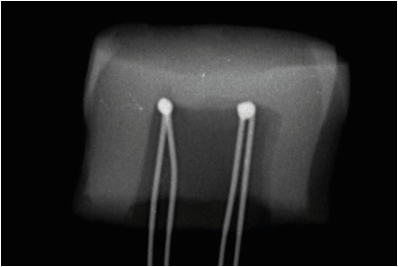
-
Abstract
PDFPubReaderePub
Objectives It was the aim of this study to evaluate the effect of cooling water temperature on the temperature changes in the pulp chamber and at the handpiece head during high-speed tooth preparation using an electric handpiece.
Materials and Methods Twenty-eight intact human molars received a standardized occlusal preparation for 60 seconds using a diamond bur in an electric handpiece, and one of four treatments were applied that varied in the temperature of cooling water applied (control, with no cooling water, 10°C, 23°C, and 35°C). The temperature changes in the pulp chamber and at the handpiece head were recorded using K-type thermocouples connected to a digital thermometer.
Results The average temperature changes within the pulp chamber and at the handpiece head during preparation increased substantially when no cooling water was applied (6.8°C and 11.0°C, respectively), but decreased significantly when cooling water was added. The most substantial drop in temperature occurred with 10°C water (−16.3°C and −10.2ºC), but reductions were also seen at 23°C (−8.6°C and −4.9°C). With 35°C cooling water, temperatures increased slightly, but still remained lower than the no cooling water group (1.6°C and 6.7ºC).
Conclusions The temperature changes in the pulp chamber and at the handpiece head were above harmful thresholds when tooth preparation was performed without cooling water. However, cooling water of all temperatures prevented harmful critical temperature changes even though water at 35°C raised temperatures slightly above baseline.
-
Citations
Citations to this article as recorded by- Efficacy Assessment of a Novel Device for Dental Aerosol Capture and Inactivation: An In Vitro Study
Yan Sun, Jia-Chun Li, Yu-Gang Li, Xiao-Yan Yu, Jie Hu, Zhe Li, Ji-Chao Cao
International Dental Journal.2026; 76(2): 109331. CrossRef - Comparison of Two Fiber Post Removal Techniques Evaluating Dentin Removal, Efficiency, and Heat Production
Matthew Fenigstein, Mazin Askar, Ahmad Maalhagh-Fard, Susan Paurazas
Dentistry Journal.2025; 13(6): 234. CrossRef - Polymethyl Methacrylate (PMMA): An Overview of Its Biological Activities, Properties, Polymerization, Modifications, and Dental and Industrial Applications
Great Iruoghene Edo, Emad Yousif, Mohammed H. Al-Mashhadani
Regenerative Engineering and Translational Medicine.2025;[Epub] CrossRef - The Effect of Water Coolant and Bur Type on Pulp Temperature When Removing Tooth Structure and Restorative Dental Materials
C Mafrici, M Kingston, R Grice, PV Abbott
Operative Dentistry.2024; 49(1): 91. CrossRef - A Finite Element Method Study on a Simulation of the Thermal Behaviour of Four Methods for the Restoration of Class II Cavities
Adela Nicoleta Staicu, Mihaela Jana Țuculină, Cristian Niky Cumpătă, Ana Maria Rîcă, Maria Cristina Beznă, Dragoș Laurențiu Popa, Alexandru Dan Popescu, Oana Andreea Diaconu
Journal of Functional Biomaterials.2024; 15(4): 86. CrossRef - In vitro comparison of guide planes for removable partial dentures prepared with CAD-CAM-assisted templates, guiding rod templates, and freehand
Ni Cheng, Hai Yu, Wenxi Shan, Jiang Wu
Journal of Dentistry.2024; 149: 105322. CrossRef - Comparison of antimicrobial efficacy of different disinfectants on the biofilm formation in dental unit water systems using dip slide and conventional methods: A pilot study
Pelin Özmen, Hilal Erdoğan, Aslıhan Güngördü, Bülent Pişkin, Funda Kont Çobankara, Serdar Sütcü, Nesrin Şahin
Microscopy Research and Technique.2024; 87(6): 1241. CrossRef - Analysis of the Pulpal Blood Flow Microdynamics during Prosthetic Tooth Preparation Using Diamond Burs with Different Degrees of Wear
Edmond Ciora, Mariana Miron, Diana Lungeanu, Andreea Igna, Anca Jivanescu
Dentistry Journal.2024; 12(6): 178. CrossRef - Bacterial contamination potential of personal protective equipment itself in dental aerosol-producing treatments
Madline Priska Gund, Jusef Naim, Stefan Rupf, Barbara Gärtner, Matthias Hannig
Odontology.2024; 112(2): 309. CrossRef - Patient satisfaction before and after occlusal adjustment using a visual analog scale
Ha-Rim Lee, Sun-Haeng Lee, Gyeong-Je Lee
Oral Biology Research.2023; 47(1): 8. CrossRef - Heat generated during dental treatments affecting intrapulpal temperature: a review
Xin Er Lau, Xiaoyun Liu, Helene Chua, Wendy Jingwen Wang, Maykon Dias, Joanne Jung Eun Choi
Clinical Oral Investigations.2023; 27(5): 2277. CrossRef - “The effect of diode laser 940 nm and 445 nm on the rise in temperature of a pulp simulating material: in vitro study”
Basant Bahaaeldin, Ola Ibrahim Fahmy, Amira Zoghaby, Rene Franzen
Lasers in Dental Science.2023; 7(3): 147. CrossRef - Evaluation of the Pulp Chamber Temperature during Tooth Veneer Preparation Using Burs with Different Degrees of Wear—A Preliminary In Vitro Study
Edmond Ciora, Mariana Miron, Daliana Bojoga, Diana Lungeanu, Anca Jivanescu
Dentistry Journal.2023; 11(8): 197. CrossRef - Effect of spray air settings of speed-increasing contra-angle handpieces on intrapulpal temperatures, drilling times, and coolant spray pattern
Edina Lempel, József Szalma
Clinical Oral Investigations.2022; 26(1): 523. CrossRef - Preparing guiding planes for removable partial dentures: an in vitro comparison between assisted CAD-CAM template procedure and freehand preparation
Hefei Bai, Hongqiang Ye, Hu Chen, Yong Wang, Yongsheng Zhou, Yuchun Sun
Journal of Dentistry.2022; 123: 104166. CrossRef - Yeni Tip Koronavirüs (COVID-19) Salgınının Diş Hekimlerinin Tedavi Kliniği Düzeni Üzerine Etkisi
Onur Altuğ SAKALLI, Sedanur SAKALLI, Aleyna Öykü AKBAŞAK, Selim ERKUT
ADO Klinik Bilimler Dergisi.2022; 11(2): 140. CrossRef - Aerosol suppression from a handpiece using viscoelastic solution in confined dental office
Yong Il Kim, Seongpil An, Jungwoo Huh, Yang-Soo Kim, Jihye Heo, In-Seok Song, Alexander L. Yarin, Sam S. Yoon
Physics of Fluids.2022;[Epub] CrossRef - Coronavirus Disease (COVID-19) Transmission through Aerosols in Restorative and Endodontic Practice
Ambar W. Raut, Priyatama V. Meshram, Radha A. Raut
Annals of African Medicine.2022; 21(1): 1. CrossRef - Redefining aerosol in dentistry during COVID-19 pandemic
Kanupriya Rathore, HarshvardhanSingh Rathore, Pranshu Singh, Pravin Kumar
Dental Research Journal.2022; 19(1): 53. CrossRef - Different grinding speeds affect induced regeneration capacity of human treated dentin matrix
Min Li, Sen Yang, Jinlin Song, Tiwei Fu, Panpan Liang, Zhi Gao, Jing Tang, Lijuan Guo
Journal of Biomedical Materials Research Part B: Applied Biomaterials.2022; 110(4): 755. CrossRef -
In Vivo Pulp Temperature Changes During Class V Cavity Preparation and Resin Composite Restoration in Premolars
DC Zarpellon, P Runnacles, C Maucoski, DJ Gross, U Coelho, FA Rueggeberg, CAG Arrais
Operative Dentistry.2021; 46(4): 374. CrossRef - In vivo evaluation of the virucidal efficacy of chlorhexidine and povidone-iodine mouthwashes against salivary SARS-CoV-2. A randomized-controlled clinical trial
Rola Elzein, Fadi Abdel-Sater, Soha Fakhreddine, Pierre Abi Hanna, Rita Feghali, Hassan Hamad, Fouad Ayoub
Journal of Evidence Based Dental Practice.2021; 21(3): 101584. CrossRef - Possible transmission of Covid-19 & precautions in a dental setting: A review
Sonali Gholap, Amit Mani, Shubhangi Mani, Shivani Sachdeva, Jasleen Kaur Sodhi, Hiral Vora
IP International Journal of Periodontology and Implantology.2021; 6(2): 98. CrossRef - An Evaluation of Two Systems for the Management of the Microbiological Quality of Water in Dental Unit Waterlines: Hygowater® and IGN Calbénium®
Damien Offner, Anne-Marie Musset
International Journal of Environmental Research and Public Health.2021; 18(10): 5477. CrossRef - Restoration of dental services after COVID-19: The fallow time determination with laser light scattering
Xiujie Li, Cheuk Ming Mak, Kuen Wai Ma, Hai Ming Wong
Sustainable Cities and Society.2021; 74: 103134. CrossRef - Possible aerosol transmission of COVID-19 and special precautions in dentistry
Zi-yu Ge, Lu-ming Yang, Jia-jia Xia, Xiao-hui Fu, Yan-zhen Zhang
Journal of Zhejiang University-SCIENCE B.2020; 21(5): 361. CrossRef - Yeni Koronavirüs Salgını ve Diş Hekimliği Tedavileri Üzerine Etkileri
Elif Ballıkaya, Gülce Esentürk, Gizem Erbaş Ünverdi, Zafer Cehreli
Hacettepe University Faculty of Health Sciences Journal.2020; 7(2): 92. CrossRef - Dentistry: its Role in the Transmission of SARS-CoV-2 Through Bioaerosols
Reinaldo Rodríguez Aguilar
Odovtos - International Journal of Dental Sciences.2020; 22(3): 103. CrossRef
- Efficacy Assessment of a Novel Device for Dental Aerosol Capture and Inactivation: An In Vitro Study
- 6,562 View
- 57 Download
- 28 Crossref
- Effects of the exposure site on histological pulpal responses after direct capping with 2 calcium-silicate based cements in a rat model
- Panruethai Trongkij, Supachai Sutimuntanakul, Puangwan Lapthanasupkul, Chitpol Chaimanakarn, Rebecca Wong, Danuchit Banomyong
- Restor Dent Endod 2018;43(4):e36. Published online August 22, 2018
- DOI: https://doi.org/10.5395/rde.2018.43.e36
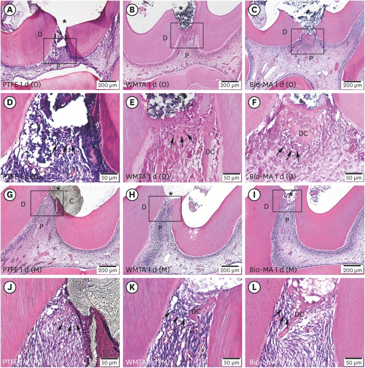
-
Abstract
PDFPubReaderePub
Objectives Direct pulp capping is a treatment for mechanically exposed pulp in which a biocompatible capping material is used to preserve pulpal vitality. Biocompatibility tests in animal studies have used a variety of experimental protocols, particularly with regard to the exposure site. In this study, pulp exposure on the occlusal and mesial surfaces of molar teeth was investigated in a rat model.
Materials and Methods A total of 58 maxillary first molars of Wistar rats were used. Forty molars were mechanically exposed and randomly assigned according to 3 factors: 1) the exposure site (occlusal or mesial), 2) the pulp-capping material (ProRoot White MTA or Bio-MA), and 3) 2 follow-up periods (1 day or 7 days) (
n = 5 each). The pulp of 6 intact molars served as negative controls. The pulp of 12 molars was exposed without a capping material (n = 3 per exposure site for each period) and served as positive controls. Inflammatory cell infiltration and reparative dentin formation were histologically evaluated at 1 and 7 days using grading scores.Results At 1 day, localized mild inflammation was detected in most teeth in all experimental groups. At 7 days, continuous/discontinuous calcified bridges were formed at exposure sites with no or few inflammatory cells. No significant differences in pulpal response according to the exposure site or calcium-silicate cement were observed.
Conclusions The location of the exposure site had no effect on rat pulpal healing. However, mesial exposures could be performed easily, with more consistent results. The pulpal responses were not significantly different between the 2 capping materials.
-
Citations
Citations to this article as recorded by- Bioactivity and biocompatibility of bioceramic-based pulp capping materials in laboratory and animal models
Rafiqul Islam, Md. Refat Readul Islam, Kenta Tsuchiya, Yu Toida, Hidehiko Sano, Monica Yamauti, Hany Mohamed Aly Ahmed, Atsushi Tomokiyo
Journal of Materials Science: Materials in Medicine.2025;[Epub] CrossRef - The road map to proper dental pulp experiments in animal models
Nuha A Elmubarak
International Dental Journal of Student's Research.2024; 11(4): 163. CrossRef - Treatment outcomes of root perforations repaired by calcium silicate-based cements with or without an accelerator: A randomized controlled trial
Kanyarat Tungputsa, Danuchit Banomyong, Sittichoke Osiri, Supachai Sutimuntanakul
Endodontology.2024; 36(4): 315. CrossRef - Biological evaluation of novel phosphorylated pullulan‐based calcium hydroxide formulations as direct pulp capping materials: An in vivo study on a rat model
Md Refat Readul Islam, Rafiqul Islam, Yunqing Liu, Yu Toida, Yasuhiro Yoshida, Hidehiko Sano, Hany Mohamed Aly Ahmed, Atsushi Tomokiyo
International Endodontic Journal.2024; 57(9): 1247. CrossRef - 3D-printed microgels supplemented with dentin matrix molecules as a novel biomaterial for direct pulp capping
Diana Cunha, Nayara Souza, Manuela Moreira, Nara Rodrigues, Paulo Silva, Cristiane Franca, Sivaporn Horsophonphong, Ashley Sercia, Ramesh Subbiah, Anthony Tahayeri, Jack Ferracane, Pamela Yelick, Vicente Saboia, Luiz Bertassoni
Clinical Oral Investigations.2022; 27(3): 1215. CrossRef - Calcium silicate and calcium aluminate cements for dentistry reviewed
Carolyn Primus, James L. Gutmann, Franklin R. Tay, Anna B. Fuks
Journal of the American Ceramic Society.2022; 105(3): 1841. CrossRef - Pulpal response to mineral trioxide aggregate containing phosphorylated pullulan-based capping material
Yu TOIDA, Shimpei KAWANO, Rafiqul ISLAM, Fu JIALE, AFM A CHOWDHURY, Shuhei HOSHIKA, Yasushi SHIMADA, Junji TAGAMI, Masahiro YOSHIYAMA, Satoshi INOUE, Ricardo M. CARVALHO, Yasuhiro YOSHIDA, Hidehiko SANO
Dental Materials Journal.2022; 41(1): 126. CrossRef - The Effect of Calcium-Silicate Cements on Reparative Dentinogenesis Following Direct Pulp Capping on Animal Models
Mihai Andrei, Raluca Paula Vacaru, Anca Coricovac, Radu Ilinca, Andreea Cristiana Didilescu, Ioana Demetrescu
Molecules.2021; 26(9): 2725. CrossRef - Histological evaluation of a novel phosphorylated pullulan‐based pulp capping material: An in vivo study on rat molars
Rafiqul Islam, Yu Toida, Fei Chen, Toru Tanaka, Satoshi Inoue, Tetsuya Kitamura, Yasuhiro Yoshida, Abu Faem Mohammad Almas Chowdhury, Hany Mohamed Aly Ahmed, Hidehiko Sano
International Endodontic Journal.2021; 54(10): 1902. CrossRef - Effectiveness of Direct Pulp Capping Bioactive Materials in Dentin Regeneration: A Systematic Review
Ermin Nie, Jiali Yu, Rui Jiang, Xiangzhen Liu, Xiang Li, Rafiqul Islam, Mohammad Khursheed Alam
Materials.2021; 14(22): 6811. CrossRef - A strontium and amorphous calcium phosphate dipped premixed injectable calcium silicate-based ceramic for dental root canal sealing
Huimin Jin, Yuzhu Li, Qingqing Wang, Menglu Dong, Mengmeng Yang, Wendy Chen, Shengrui Wang, Heng Zhang, Shunli Zheng, Chris Ying Cao, Zheng Zhou, Quan-Li Li
Ceramics International.2021; 47(23): 33738. CrossRef - Bioactive tri/dicalcium silicate cements for treatment of pulpal and periapical tissues
Carolyn M. Primus, Franklin R. Tay, Li-na Niu
Acta Biomaterialia.2019; 96: 35. CrossRef
- Bioactivity and biocompatibility of bioceramic-based pulp capping materials in laboratory and animal models
- 3,192 View
- 30 Download
- 12 Crossref
Review Article
- Triple antibiotic paste: momentous roles and applications in endodontics: a review
- Ardavan Parhizkar, Hanieh Nojehdehian, Saeed Asgary
- Restor Dent Endod 2018;43(3):e28. Published online June 20, 2018
- DOI: https://doi.org/10.5395/rde.2018.43.e28
-
Abstract
PDFPubReaderePub
This study investigated the latest findings and notions regarding ‘triple antibiotic paste’ (TAP) and its applications in dentistry, particularly endodontics. TAP is a combination of 3 antibiotics, ciprofloxacin, metronidazole, and minocycline. Despite the problems and pitfalls research pertaining to this paste has unveiled, it has been vastly used in endodontic treatments. The paste's applications vary, from vital pulp therapy to the recently introduced regeneration and revascularisation protocol. Studies have shown that the paste can eliminate the root canal microorganisms and prepare an appropriate matrix for further treatments. This combination is able to remove diverse groups of obligate and facultative gram-positive and gram-negative bacteria, providing an environment for healing. In regeneration protocol cases, this allows the development, disinfection, and possible sterilization of the root canal system, so that new tissue can infiltrate and grow into the radicular area. Moreover, TAP is capable of creating a discipline in which other wanted and needed treatments can be successfully performed. In conclusion, TAP, as an antibacterial intracanal medication, has diverse uses. Nevertheless, despite its positive effects, the paste has shown drawbacks. Further research concerning the combined paste and other intracanal medications to control microbiota is a must.
-
Citations
Citations to this article as recorded by- Endodontic Intracanal Medicaments and Agents
Anu Priya Guruswamy Pandian, Depti Bellani, Ritya Mary Jibu, Varsha Agnihotri
Dental Clinics of North America.2026; 70(1): 45. CrossRef - Regenerative Endodontic Treatment in Permanent Incisors: Two Case Reports with 6 Years of Follow-Up
María Biedma-Perea, Marcela Arenas-González, María José Barra-Soto, Carolina Caleza-Jiménez, David Ribas-Pérez
Children.2026; 13(2): 246. CrossRef - Evaluation of antimicrobial efficacy of an herbal preparation vs triple antibiotic paste against E. faecalis: an in vitro study
Chandni Dhyani, Kalpna Chaudhry, Nitin Khanduri, Leina R. Pradhan, Yoshita Gupta
International Journal of Contemporary Pediatrics.2026; 13(3): 449. CrossRef - Comparative antimicrobial efficacy of three intracanal medicaments in an in vitro Enterococcus faecalis root canal infection model
Ali Hasan, Hanan Mohammad, Maher Darwish, Mayssam Khaddam, Asmahan Zainab
BMC Oral Health.2026;[Epub] CrossRef - Comparison of the Antimicrobial Properties of Nanoemulsions Made from Phytosphingosine and Chitosan with a Triple Antibiotic Paste Against Enterococcus faecalis in Endodontics: An in-vitro Study
Ganesh Jadhav, Priya Mittal, Mohammed Abdul Kader M, Suheel Baba, Siddharth Shinde, Dhanashree Dalal, Ahmed Al Malwi, Shahabe Abullais, Siddharthan Selvaraj
International Journal of Nanomedicine.2026; Volume 21: 1. CrossRef - Chihuahuan Propolis as a Non-Antibiotic Intracanal Agent Against Enterococcus faecalis: An In Vitro Intratubular Study
Hilda Natalia Quintana-Pérez, Grissel Guadalupe Orozco-Molina, Carlos Esteban Villegas-Mercado, Sandra Aidé Santana-Delgado, Adolfo Gonzalez-Acosta, Javier Iván Martínez-Hernández, Raquel Duarte-Rico, Lydia Paulina Loya-Hernández, América Susana Mares-Gar
Microorganisms.2026; 14(4): 774. CrossRef - Intentional replantation for the management of external root resorption: A case report with clinical insights
Dipti Jitendra Khairajani, Anamika Chetan Borkar, Sanket D. Aras, Apeksha Sanjeev Gambhir, Saroj Pawar
Saudi Endodontic Journal.2026; 16(2): 287. CrossRef - Evaluation of Effect of Laser in Root Canal Disinfection in Pulp Regenerative Therapy: A Systematic Review
Kiran Kumar N, Abhishek M, Savitha B. Naik, Biji Brigit, Swetha Geervani V, M Manimozhi
Photobiomodulation, Photomedicine, and Laser Surgery.2025; 43(2): 53. CrossRef - Assessing Cell Viability: Comparative Analysis of Calcium Hydroxide, Triple Antibiotic Paste, and Their Synergistic Impact on human Dental Pulp Stem Cells
Dini Asrianti Bagio, Ibramanto Warganegara, Ike Dwi Maharti, Anggraini Margono, Citra Kusumasari, Sylva Dinie Alinda, Valeria Widita Wairooy
European Journal of General Dentistry.2025; 14(01): 073. CrossRef - Cytotoxic and Antibiofilm Properties of Antibiotic-Loaded Thermoresponsive Hydrogels for Root Canal Therapy
Cristiane Duque, Gabriela Pacheco de Almeida Braga, Juliana Machado de Carvalho, Karina Sampaio Caiaffa, Gabriel Pereira Nunes, Rafaela Laruzo Rabelo, Vanessa Rodrigues dos Santos, Geórgia Rondó Peres, Lucas da Silva Ribeiro, Emerson Rodrigues de Camargo
Processes.2025; 13(3): 661. CrossRef - Antibiofilm properties, cytotoxicity, and effect on protease activity of antibiotics and EGCG-based medications for endodontic purposes
Daniela Alvim Chrisostomo, Jesse Augusto Pereira, Polliana Mendes Candia Scaffa, Zach Gouveia, Gabriel Flores Abuna, Sergey V. Plotnikov, Anuradha Prakki, Cristiane Duque
Journal of Dentistry.2025; 156: 105660. CrossRef - The use of three-dimensional-printed guides, static navigation, and bioactive materials to treat bilateral and double dens invaginatus
Parth Patel, Nidhi Bharti, Ankit Arora, C. Nimisha Shah
Saudi Endodontic Journal.2025; 15(2): 207. CrossRef - To Assess the Cell Viability of Triple Antibiotic Paste under Confocal Microscope: An In Vitro Study
Elanthendral Saravanan, Mahesh Ramakrishnan
Journal of South Asian Association of Pediatric Dentistry.2025; 8(2): 81. CrossRef - Eficacia de la pasta triantibiótica en endodoncia: actividad antibacteriana frente a cepas resistentes de Enterococcus faecalis. Una revisión exhaustiva
Elena Patricia Cevallos Fernández, Katherine de los Ángeles Cuenca León
Anatomía Digital.2025; 8(3.1): 88. CrossRef - Efectividad de diferentes antimicóticos, junto con la pasta triantibiotica, para el tratamiento de Candida albicans en conductos radiculares
Carlos Andrés Rodríguez Tapia, Jessica María Sarmiento Ordoñez
Anatomía Digital.2025; 8(3.2): 45. CrossRef - Comparative Evaluation of Calcium Hydroxide, Triple Antibiotic Paste, and Calcium Hydroxide with 2% Chlorhexidine as Intracanal Medicaments in Reducing Interappointment Pain during Endodontic Treatment: An In Vivo Study
Itika Jain, Shyam Agrawal, Rachit Mathur, Shaista Gazal, Akshada Mungee, Babra Khan
International Journal of Clinical Pediatric Dentistry.2025; 18(6): 628. CrossRef - In Vitro Effectiveness of Endodontic Triple Antibiotic Paste Associated With Daptomycin
Sabrina S Azevedo, Gabriela C Chianca, Bruna A Thurler, Raiane C Chamon, Helvécio C Corrêa Póvoa, Leonardo S Antunes, Natalia L Pontes Póvoa Iorio
Cureus.2025;[Epub] CrossRef - Effect of Chitosan and bioactive glass nanomaterials as intracanal medicaments on TGF-β1 release from intraradicular dentin
Sarah Salah Hashem, Mohammed M. Khalefa, Mahmoud Hassan Mohamed, Hemat M. ELSheikh, Fatma Abd El-Rahman Taher
BMC Oral Health.2025;[Epub] CrossRef - Clinical and radiographic success of lesion sterilization and tissue repair in primary teeth: a systematic review and meta-analysis
Khlood Baghlaf, Rana A. Alamoudi
The Saudi Dental Journal.2025;[Epub] CrossRef - Antibacterial Effect of Cannabinoids on Bacteria Associated with Persistent Endodontic Infections
Cassandra Wieczerza, Haoyan Zhai, Mazin Askar, Zheng Zhou, Susan Paurazas
International Journal of Molecular Sciences.2025; 26(24): 11936. CrossRef - A Comparative Evaluation of Herbal Extracts and Triple Antibiotic Paste as Intracanal Medicament against Enterococcus faecalis: A Microbiological Study
Divya Singh, Rashi Singh, Nidhi Gupta, Natasha Gambhir, Saritha Golla
International Journal of Clinical Pediatric Dentistry.2024; 17(3): 285. CrossRef - Comparative Evaluation of Push-Out Bond Strength of Conventional Mineral Trioxide Aggregate, Biodentine, a Modified Mineral Trioxide Aggregate, and Two Novel Antibacterial-Enhanced Mineral Trioxide Aggregates
Arokia Rajkumar Shancy Merlin, Vignesh Ravindran, Ganesh Jeevanandan, Rajalakshmanan Eswaramoorthy, Abirami Arthanari
Cureus.2024;[Epub] CrossRef - In Vitro Evaluation of the Effect of Oleanolic Acid as a Potential Root Canal Medicament on Viability and Proliferation of Dental Pulp Stem Cells
Khalifah A. Alhaila, Manal Farouk Badawi, Mohamed G. Elbeltagy, Amany E. Badr
European Journal of General Dentistry.2024; 13(01): 051. CrossRef - Regenerative Endodontic Treatment in Dentinogenesis Imperfecta‐Induced Apical Periodontitis
Ying Liao, Ting Pan, Xianghui Xing, Sivakumar Nuvvula
Case Reports in Dentistry.2024;[Epub] CrossRef - Efecto antimicrobiano como medicación intraconducto de la pasta triantibiótica.
Paúl Sebastián Ulloa Amores, Diana Álvarez Álvarez, María Elizabeth Moscoso Abad, Magda Zulay Bastidas Calva
Revista de la Asociación Dental Mexicana.2024; 81(4): 211. CrossRef - Synthesis and Characterization of Nanosilica-Coated Antibiotics, TAP: A Comprehensive Study Utilizing XRD, EDS, FTIR, SEM, and TEM – Invitro Study
Mahaboob S. Hameed, S. Delphine P. Antony, Rajeshkumar Shanmugam, Sandhya Raghu
Journal of International Oral Health.2024; 16(5): 386. CrossRef - Association between host defence peptide IDR‐1002 and ciprofloxacin: Effects on human dental pulp cells
Danilo César Mota Martins, Maurício Gonçalves da Costa Sousa, Poliana Amanda Oliveira Silva, Lana Ribeiro Aguiar, Rosângela Vieira de Andrade, Amandda Évellin Silva‐Carvalho, Felipe Saldanha‐Araújo, Octávio Luiz Franco, Taia Maria Berto Rezende
Australian Endodontic Journal.2024; 50(3): 547. CrossRef - Comparative evaluation of antimicrobial efficacy of triple antibiotic paste and amoxicillin clavulanate paste as an intracanal medicament against Enterococcus faecalis: An in vitro study
Dhandayuthapani Sasikala, Parisa Norouzi Baghkomeh, Jamaluddin Mohammed Farzan
Dental Research Journal.2024;[Epub] CrossRef - Triple Antibiotic Paste: A Game Changer in Endodontics
Kalagi G. Panchal, Karima Virani, Vraj Patel, Aquib Ali Khan, Anam Pettiwala, Srikala S. Puranik, Srushti Joshi
Journal of Pharmacy and Bioallied Sciences.2024; 16(Suppl 3): S1913. CrossRef - Non-surgical Management of a Large Periapical Lesion: A Case Study of the Successful Application of a Modified Triple Antibacterial Paste
Srushti Awghad, Joyeeta Mahapatra, Amit Reche, Ankita Burse, Aradhana Kibe
Cureus.2024;[Epub] CrossRef - Enhancing Antimicrobial Efficacy and Synergistic Effects of Nano-Silica-Based Combinations With Doxycycline, Metronidazole, and Ciprofloxacin Against Enterococcus faecalis Biofilms
Shahul Hameed, Delphine P Antony, Rajeshkumar Shanmugam, Sandhya Raghu, Hima Sandeep Adimulapu
Cureus.2024;[Epub] CrossRef - Healing of periapical lesion by non-surgical endodontic therapy: A case series
Athira Ramesh, Rajesh Pillai, Afzal A, Anakha Santhosh, Arunima G.S, Sandeep K. V
IP Indian Journal of Conservative and Endodontics.2024; 9(2): 99. CrossRef - Effect of Calcium Hydroxide Versus Double Antibiotic Paste on Endodontic Treatment Outcomes in Teeth With Large Periapical Lesions: A Triple‐Blind Randomized Clinical Trial
Afsaneh Rahmati, Farshad Seyedein, Omid Dianat, Sara Saedi, Golriz Rostami, Alireza Akbarzadeh Baghban, Shima Sabertahan, Majid Kazem, Kee Y. Kum
International Journal of Dentistry.2024;[Epub] CrossRef - Can antimicrobial photodynamic therapy serve as an effective adjunct protocol for disinfecting the necrotic root canal system? A randomized controlled study
Remy Barazy, Hisham Alafif, Hassan Achour, Ahmad Al-Aloul, Yasser Alsayed Tolibah
BDJ Open.2024;[Epub] CrossRef - Drain and Treat: A Rubber Dam Technique for Acute Periapical Abscess
S. Grover, K. Mala, J. D’Souza
Acta Medica Bulgarica.2024; 51(s2): 143. CrossRef - Microbial Dynamics in Endodontic Pathology—From Bacterial Infection to Therapeutic Interventions—A Narrative Review
Klara Wieczorkiewicz, Anna Jarząbek, Estera Bakinowska, Kajetan Kiełbowski, Andrzej Pawlik
Pathogens.2024; 14(1): 12. CrossRef - An in vitro comparative evaluation of the effect of three intracanal medicaments – chlorhexidine gel, triple antibiotic paste, and calcium hydroxide paste on the push-out bond strength of MTA Plus, Biodentine, and calcium-enriched mixture
Gouthami Datta, Ramya Raghu, Ashish Shetty, Gautham P Manjunath, Dishant Patel, Subhashini Rajasekhara
Endodontology.2023; 35(1): 60. CrossRef - The cytotoxic effect of cysteamine and its combinations with various endodontic intracanal medications on fibroblast cells: in vitro study
Esraa Adel Mohamed Abd Elhameed ElGammal, Abeer Hashem Mahran, Salma Hassan El Ashry, Sara Hossam Fahmy
Bulletin of the National Research Centre.2023;[Epub] CrossRef - Healing of large endodontic lesions using a new combination of triple antibiotics: A case report
Saeed Asgary, Ardavan Parhizkar
Clinical Case Reports.2023;[Epub] CrossRef - Effects of Intracanal Medicaments on the Measurement Accuracy of Four Apex Locators: An In Vitro Study
Hamza Cudal, Tuğrul Aslan, Bertan Kesim
Meandros Medical and Dental Journal.2023; 24(3): 215. CrossRef - Comparative evaluation of the antibacterial efficacy of herbal agents as intracanal medicaments individually or in combination with chitosan: An in vitro RTPCR study
Gaurav Patri, Kotni Sheetal, PrasantiKumar Pradhan, Pratik Agrawal, S Lata
Journal of International Oral Health.2023; 15(1): 89. CrossRef - Recent progress in carbon dots for anti-pathogen applications in oral cavity
Yuying Jiang, Chuqiang Yin, Jianning Mo, Xiaoyu Wang, Ting Wang, Guotai Li, Qihui Zhou
Frontiers in Cellular and Infection Microbiology.2023;[Epub] CrossRef - A Comprehensive Review of Lesion Sterilization and Tissue Repair: An Alternative for Pulpectomy in Deciduous Teeth
Aparna Achanta, Amit Reche, Rishika Dakhale, Rudra R Bharate
Cureus.2023;[Epub] CrossRef - Healing of Large Endodontic Lesions Using Long‐Term Application of a New Combination of Triple Antibiotics: A Series of Cases
Saeed Asgary, Ardavan Parhizkar, Maria Beatriz Duarte Gavião
Case Reports in Dentistry.2023;[Epub] CrossRef - Non-cytotoxic Root Canal Dressing with Improved Antimicrobial Efficacy
Farzad Koosha, Jerome Cymerman, Thomas Manders, Marcia Simon, Stephen Walker, Miriam Rafailovich
Journal of Endodontics.2023; 49(2): 205. CrossRef - Triple antibiotics: A synergistic approach to combating infection
Hemant Sawhney, Anukriti Kumari, Ritik Kashwani, Geetanjali Gupta, SJ Das
IP Indian Journal of Conservative and Endodontics.2023; 8(4): 189. CrossRef - A 1-year Clinical and Radiographic Assessment of Regenerative Endodontic Therapy for Necrotic Primary Molars: A Randomized controlled Trial
Dina D Abdelmoneim, Amr M Abdelaziz, Gehan G Allam, Amira S Badran
International Journal of Clinical Pediatric Dentistry.2023; 16(2): 295. CrossRef - “BIODENTINE” THE DENTINE IN A CAPSULE AS AN APICAL BARRIER IN TRAUMATIZED MAXILLARY CENTRAL INCISOR WITH TWO YEARS FOLLOW UP.
Savita Thakur, Udai Bhanu, Gurkirat Singh Grewal
INTERNATIONAL JOURNAL OF SCIENTIFIC RESEARCH.2022; : 64. CrossRef - Long-term treatment of dentine with triple antibiotic paste promotes stem cell viability and attachment
Samiya Riaz, Ahmad Azlina, Zuliani Mahmood, Aung T. Htun
Journal of Taibah University Medical Sciences.2022; 17(4): 630. CrossRef - Non surgical management of trauma induced root resorption and large periapical
lesion using bioactive material- A case report
Tejasvini Prajapati, Sonali Kapoor, Purnil Shah, Ankit Arora, Hardik Rana
Clinical Dentistry.2022;[Epub] CrossRef - Antibacterial efficacy of antibiotic pastes versus calcium hydroxide intracanal dressing: A systematic review and meta-analysis of ex vivo studies
Mohammadreza Vatankhah, Kamyar Khosravi, Nazanin Zargar, Armin Shirvani, MohammadHossein Nekoofar, Omid Dianat
Journal of Conservative Dentistry.2022; 25(5): 463. CrossRef - Triple Antibiotic Paste: A Suitable Medicament for Intracanal Disinfection
Krutika Malu, Monika Khubchandani
Cureus.2022;[Epub] CrossRef - Present status and future directions of intracanal medicaments
Ronald Ordinola‐Zapata, W. Craig Noblett, Alejandro Perez‐Ron, Zhou Ye, Jorge Vera
International Endodontic Journal.2022; 55(S3): 613. CrossRef - The effect of four different intracanal medicaments on the push-out bond strength of root canal sealers
Shalu Maan, Vijaya Dhar Bhatt, Rohit Singh, Sayak Gupta, Syed Alay Noorain, Aashna Gill, Pradeep Kumar, Sushil Yadav, Preeti Sharma
Journal of Medicine and Life.2022; 15(4): 448. CrossRef - A Comparative Evaluation of Microhardness and Chemical Structure of Radicular Dentin with Two Combinations of TAP and MTAP: An In Vitro Study
Muthumula Daneswari, Nagireddy Venugopal Reddy, Annie P Chris, Nikhila V Reddy, Saigeeta Kondamadugu, P Niharika
International Journal of Clinical Pediatric Dentistry.2022; 15(S2): S151. CrossRef - Comparative Evaluation of Two Antibiotic Pastes for Root Canal Disinfection
Mridula Goswami, CP Baveja, Urvashi Bhushan, Sadhna Sharma
International Journal of Clinical Pediatric Dentistry.2022; 15(S1): S12. CrossRef - Management of External Inflammatory Root Resorption following Tooth Autotransplantation Using a Modified Combination of Triple Antibiotics
Saeed Asgary, Ardavan Parhizkar, Paulo J. Palma
Case Reports in Dentistry.2022;[Epub] CrossRef - Effect of triple antibiotic paste on the bond strength of epoxy and methacrylate resin-based sealers to root canal dentin
Fereshte Sobhnamayan, Alireza Adl, Mahdi Sedigh-Shams, Hossein Mirkhaghani
Journal of Conservative Dentistry.2022; 25(4): 426. CrossRef - Progress of Research on the Application of Triple Antibiotic Paste and Hydrogel Scaffold Materials in Endodontic Revascularization: A Systematic Review
Jia Zhao, Tian Jiao Wang
Evidence-Based Complementary and Alternative Medicine.2022; 2022: 1. CrossRef - The effect of different intracanal medicaments on the dislodgement resistance of mineral trioxide aggregate
Farzaneh Afkhami, Shahrzad Razavi, Sholeh Ghabraei
BMC Oral Health.2022;[Epub] CrossRef - Efficacy of Ciprofloxacin, Metronidazole and Minocycline in Ordered Mesoporous Silica against Enterococcus faecalis for Dental Pulp Revascularization: An In-Vitro Study
Cintia Micaela Chamorro-Petronacci, Beatriz Santos Torres, Rocío Guerrero-Nieves, Mario Pérez-Sayáns, Marcia Carvalho-de Abreu Fantini, Luis Carlos Cides-da-Silva, Beatriz Magariños, Berta Rivas-Mundiña
Materials.2022; 15(6): 2266. CrossRef - Antibiotic Mixtures in Noninstrumental Endodontic Treatment of Primary Teeth with Necrotic Pulps: A Systematic Review
Farah Chouchene, Fatma Masmoudi, Ahlem Baaziz, Fethi Maatouk, Hichem Ghedira, Sivakumar Nuvvula
International Journal of Dentistry.2021; 2021: 1. CrossRef - Potential apply of hydrogel-carried chlorhexidine and metronidazole in root canal disinfection
Yanhong YAN, Peng ZHOU, Haibing LU, Yun GUAN, Ming MA, Juan WANG, Guangwei SHANG, Beizhan JIANG
Dental Materials Journal.2021; 40(4): 986. CrossRef - Evaluation of the Effect of Nitrofurantoin Paste as an Intracanal Medicament on the Chemical Structure of Radicular Dentine
Mewan Abdulrahman, Bestoon Faraj, Kawa Dizaye
Sulaimani Dental Journal.2021; 8(2): 8. CrossRef - Local Drug Delivery Systems for Vital Pulp Therapy: A New Hope
Ardavan Parhizkar, Saeed Asgary, Carlo Galli
International Journal of Biomaterials.2021; 2021: 1. CrossRef - Antibiofilm and cytotoxic effect of 3,3′-dihydroxycurcumin (DHC) as photosensitizer agent in antimicrobial photodynamic therapy for endodontic purposes
Jesse Augusto Pereira, Carlos Roberto Polaquini, VanessaRodrigues dos Santos, Karina Sampaio Caiaffa, Rafaela Laruzo Rabelo, Reinaldo dos Santos Theodoro, Letícia Helena Theodoro, Luis Octavio Regasini, Cristiane Duque
Photodiagnosis and Photodynamic Therapy.2021; 36: 102534. CrossRef - An in-vitro Comparative Evaluation of Quantitative Release of Transforming Growth Factor β-1 from Dentin upon the Action of Endodontic Irrigants, Medicaments, Ultrasonic Activation, and Low-Level Laser Irradiation
Anilkumar Akhila, V. P. Prabath Singh, Kerala R. Varma, Senthil V. Vasudevan, V. Sukhithasri, Salu Sasikumar
Amrita Journal of Medicine.2021; 17(2): 34. CrossRef - Effect of triple antibiotic paste and calcium hydroxide on the rate of healing of periapical lesions: A systematic review
NKiran Kumar, Biji Brigit, BS Annapoorna, SavithaB Naik, Seema Merwade, K Rashmi
Journal of Conservative Dentistry.2021; 24(4): 307. CrossRef - Comparison of the efficacy of CanalBrush, EndoActivator, and Passive Ultrasonic Irrigation on the removal of triple antibiotic paste from root canal walls: An in vitro study
Santosh Kumar, Kavisha Desai, Aparna Palekar, Baswaraj Biradar, Ananjan Chatterjee, Khushboo Kumari
Journal of International Society of Preventive and Community Dentistry.2020; 10(4): 424. CrossRef - Apexification with Calcium Hydroxide vs. Revascularization
H. Boufdil, M. Mtalsi, S. El Arabi, B. Bousfiha, Jose López-López
Case Reports in Dentistry.2020;[Epub] CrossRef - Effect of Intracanal Medicaments and Irrigants on the Release of Transforming Growth Factor Beta 1 and Vascular Endothelial Growth Factor from Cervical Root Dentin
Lívia Nazareth Ferreira, Regina Maria Puppin-Rontani, Fernanda Miori Pascon
Journal of Endodontics.2020; 46(11): 1616. CrossRef - Incorporation of amoxicillin-loaded microspheres in mineral trioxide aggregate cement: an in vitro study
Fábio Rocha Bohns, Vicente Castelo Branco Leitune, Isadora Martini Garcia, Bruna Genari, Nélio Bairros Dornelles, Silvia Stanisçuaski Guterres, Fabrício Aulo Ogliari, Mary Anne Sampaio de Melo, Fabrício Mezzomo Collares
Restorative Dentistry & Endodontics.2020;[Epub] CrossRef - Assessment of Nitrofurantoin as an Experimental Intracanal Medicament in Endodontics
Mewan Salahalddin A. Alrahman, Bestoon Muhammed Faraj, Kawa F. Dizaye, Abdelwahab Omri
BioMed Research International.2020;[Epub] CrossRef - An in vitro assessment of effect on microhardness of dentin using vicker's hardness method
Manoj Chandak, Richa Modi, Rakesh Gogiya, Rakhi Chandak, Anuja Ikhar, Nikhil Mankar
Journal of Datta Meghe Institute of Medical Sciences University.2020; 15(2): 251. CrossRef - Inspection of the Microbiota in Endodontic Lesions
Mario Dioguardi, Giovanni Di Gioia, Gaetano Illuzzi, Claudia Arena, Vito Carlo Alberto Caponio, Giorgia Apollonia Caloro, Khrystyna Zhurakivska, Iolanda Adipietro, Giuseppe Troiano, Lorenzo Lo Muzio
Dentistry Journal.2019; 7(2): 47. CrossRef - Materials for pulpotomy in immature permanent teeth: a systematic review and meta-analysis
Yuan Chen, Xinlei Chen, Yali Zhang, Fangjie Zhou, Jiaxin Deng, Jing Zou, Yan Wang
BMC Oral Health.2019;[Epub] CrossRef - Adjunctive antimicrobial photodynamic therapy to conventional chemo-mechanical debridement of infected root canal systems: A systematic review and meta-analysis
Maryam Pourhajibagher, Abbas bahador
Photodiagnosis and Photodynamic Therapy.2019; 26: 19. CrossRef - Comparison of Antibacterial Effects of Photodynamic Therapy, Modified Triple Antibiotic Paste and Calcium Hydroxide on Root Canals Infected With Enterococcus faecalis: An In Vitro Study
Mohammad Asnaashari, Mohammad Jafar Eghbal, Amirali Sahba Yaghmayi, Mehdi Shokri, Saranaz Azari-Marhabi
Journal of Lasers in Medical Sciences.2019; 10(5): S23. CrossRef
- Endodontic Intracanal Medicaments and Agents
- 21,336 View
- 236 Download
- 78 Crossref
Research Article
-
In vivo assessment of accuracy of Propex II, Root ZX II, and radiographic measurements for location of the major foramen - Fernanda Garcia Tampelini, Marcelo Santos Coelho, Marcos de Azevêdo Rios, Carlos Eduardo Fontana, Daniel Guimarães Pedro Rocha, Sergio Luiz Pinheiro, Carlos Eduardo da Silveira Bueno
- Restor Dent Endod 2017;42(3):200-205. Published online May 16, 2017
- DOI: https://doi.org/10.5395/rde.2017.42.3.200
-
Abstract
PDFPubReaderePub
Objectives The aim of this
in vivo study was to assess the accuracy of 2 third-generation electronic apex locators (EALs), Propex II (Dentsply Maillefer) and Root ZX II (J. Morita), and radiographic technique for locating the major foramen (MF).Materials and Methods Thirty-two premolars with single canals that required extraction were included. Following anesthesia, access, and initial canal preparation with size 10 and 15 K-flex files and SX and S1 rotary ProTaper files, the canals were irrigated with 2.5% sodium hypochlorite. The length of the root canal was verified 3 times for each tooth using the 2 apex locators and once using the radiographic technique. Teeth were extracted and the actual WL was determined using size 15 K-files under a × 25 magnification. The Biostat 4.0 program (AnalystSoft Inc.) was used for comparing the direct measurements with those obtained using radiographic technique and the apex locators. Pearson's correlation analysis and analysis of variance (ANOVA) were used for statistical analyses.
Results The measurements obtained using the visual method exhibited the strongest correlation with Root ZX II (
r = 0.94), followed by Propex II (r = 0.90) and Ingle's technique (r = 0.81;p < 0.001). Descriptive statistics using ANOVA (Tukey'spost hoc test) revealed significant differences between the radiographic measurements and both EALs measurements (p < 0.05).Conclusions Both EALs presented similar accuracy that was higher than that of the radiographic measurements obtained with Ingle's technique. Our results suggest that the use of these EALs for MF location is more accurate than the use of radiographic measurements.
-
Citations
Citations to this article as recorded by- How Do Different Image Modules Impact the Accuracy of Working Length Measurements in Digital Periapical Radiography? An In Vitro Study
Vahide Hazal Abat, Rabia Figen Kaptan
Diagnostics.2025; 15(3): 305. CrossRef - Influence of maintaining apical patency in post-endodontic pain
Snigdha Shubham, Manisha Nepal, Ravish Mishra, Kishor Dutta
BMC Oral Health.2021;[Epub] CrossRef
- How Do Different Image Modules Impact the Accuracy of Working Length Measurements in Digital Periapical Radiography? An In Vitro Study
- 2,384 View
- 10 Download
- 2 Crossref
Case Report
- Mandibular bone necrosis after use of paraformaldehyde-containing paste
- Chi-hwan Lee, Yoorina Choi, Sujung Park
- Restor Dent Endod 2016;41(4):332-337. Published online November 8, 2016
- DOI: https://doi.org/10.5395/rde.2016.41.4.332
-
Abstract
PDFPubReaderePub
Paraformaldehyde has been used in the past as a pulpotomy agent. However, it has a severe cytotoxic effect and may cause alveolar bone necrosis. Depulpin, a devitalizing agent containing 49% paraformaldehyde, is no longer used frequently due to its severe side effects. In the two cases described in the present study, Depulpin was used as a devitalizing agent during root canal treatment. It caused a gradual loss of sensibility in adjacent teeth, gingival necrosis, and osteomyelitis. This case report demonstrates the serious side effects of using a paraformaldehyde-containing paste as a devitalizing agent for pulp, particularly mandibular bone necrosis.
-
Citations
Citations to this article as recorded by- Numb chin syndrome caused by paraformaldehyde-containing devitalizing agent – Case report
Jyh-Kwei Chen, Yeung-Yi Hsu, Chun-Pin Chiang, Meng-Ling Chiang
Journal of Dental Sciences.2023; 18(2): 955. CrossRef - Non-radiation and non-drug–induced maxillary osteomyelitis: Study of underlying risk factors, presentation, management and treatment outcomes
Kumar Nilesh, Pankaj Patil, Digvijay Patil, Monica Patil
Medical Journal Armed Forces India.2022; 78: S145. CrossRef - Acute toxicity potential and impact on periodontal and periapical tissue of Pulp Out: A paste contained jatropha, sidaguri, and melittin
Maria Tanumihardja, A.M. Windha, N. Musfirah, G.K. Punggawa, Andi Fatima, A.H.M. Nur Fadhila, Esfandiary, Nurhayaty Natsir, Husni Cangara, Lukman Muslimin
Toxicology Reports.2022; 9: 1788. CrossRef - Comparative evaluation of the effect of two pulpal medicaments on pain and bleeding status of mandibular molars with irreversible pulpitis post-failure of inferior alveolar nerve block: a double-blind, randomized, clinical trial
Naomi Ranjan Singh, Lora Mishra, Ajinkya M. Pawar, Nike Kurniawati, Dian Agustin Wahjuningrum
PeerJ.2022; 10: e13397. CrossRef - Dental implant restoration of mandibular bone necrosis defects caused by use of paraformaldehyde-containing paste: A case report
Won-Pyo Lee, Ho-Keel Hwang, Hyoung-Hoon Jo
Oral Biology Research.2019; 43(1): 110. CrossRef - Is Panoramic Radiography an Accurate Imaging Technique for the Detection of Endodontically Treated Asymptomatic Apical Periodontitis?
Cosimo Nardi, Linda Calistri, Giulia Grazzini, Isacco Desideri, Chiara Lorini, Mariaelena Occhipinti, Francesco Mungai, Stefano Colagrande
Journal of Endodontics.2018; 44(10): 1500. CrossRef - A case of high density abnormality in x-ray findings of mandible caused by leakage of root canal filling paste
Haruko Kashiwamura, Kyoko Oka, Yoko Tuchihashi, Hanako Yoshioka, Mayumi Kato, Atsuko Baba, Toyohiro Kagawa, Kazuhiko Okamura, Masao Ozaki
Pediatric Dental Journal.2017; 27(3): 162. CrossRef
- Numb chin syndrome caused by paraformaldehyde-containing devitalizing agent – Case report
- 4,649 View
- 25 Download
- 7 Crossref
Research Articles
-
In vitro characterization of human dental pulp stem cells isolated by three different methods - Ji-Hyun Jang, Hyeon-Woo Lee, Kyu Min Cho, Hee-Woong Shin, Mo Kwan Kang, Sang Hyuk Park, Euiseong Kim
- Restor Dent Endod 2016;41(4):283-295. Published online October 12, 2016
- DOI: https://doi.org/10.5395/rde.2016.41.4.283
-
Abstract
PDFPubReaderePub
Objectives In this study, we characterized human dental pulp cells (HDPCs) obtained by different culture methods to establish the most suitable methodology for dental tissue engineering and regenerative endodontic applications.
Materials and Methods HDPCs were isolated by the outgrowth method (HDPCs-OG), the enzymatic digestion method (collagenase/dispase/trypsin, HDPCs-ED), or the combination of both methods (HDPCs-Combined). The expression of mesenchymal stem cell markers (CD105, CD90, and CD73) was investigated.
In vitro differentiation capacities of HDPCs into adipogenic, osteogenic, and chondrogenic lineages were compared. Differentiation markers were analyzed by quantitative reverse-transcription polymerase chain reaction (RT-PCR) and western blotting.Results Our data indicated that whole HDPCs-ED, HPDCs-OG, and HDPCs-Combined could be differentiated into adipogenic, chrondrogenic, and osteogenic cell types. However, we found that the methods for isolating and culturing HDPCs influence the differentiation capacities of cells. HDPCs-OG and HDPCs-ED were preferably differentiated into adipogenic and osteogenic cells, respectively. Differentiation markers shown by RT-PCR and western blotting analysis were mostly upregulated in the treated groups compared with the control groups.
Conclusions Our findings confirmed that cell populations formed by two different culture methods and the combined culture method exhibited different properties. The results of this study could provide an insight into regenerative endodontic treatment using HDPCs.
-
Citations
Citations to this article as recorded by- Synergistic effects of collagen membrane and mineral trioxide aggregate on odontogenic differentiation and mineralization of human dental pulp stem cells: an in vitro study
Nermine Hassan, Nashwa El-Khazragy, Amir Hafez Ibrahim
BMC Oral Health.2026;[Epub] CrossRef - Effects of simulated microgravity on dental pulp stem cell stemness
Huailong Hou, Zhengjun Qiu, Jingyi Che, Yanping Li, Jingxuan Sun, Weiwei Zhang, Jinjie Ma, Shuang Zhang, Mengdi Li, Yumei Niu, Lina He
Journal of Molecular Histology.2025;[Epub] CrossRef - Validated methods for isolation and qualification of mesenchymal stromal/stem cells from different sources
Vincenzo Mattei, Francesca Santilli, Fanny Pulcini, Jessica Fabrizi, Loreto Lancia, Costantino Santacroce, Francesca Megiorni, Simona Ceccarelli, Emanuela Paldino, Roberto Gramignoli, Maria G. Roubelakis, Sadri Bahareh, Massoud Vosough, Sveva Bollini, Umb
Journal of Translational Medicine.2025;[Epub] CrossRef - ISOLATION OF HUMAN ADULT DENTAL PULP STEM CELLS USING ENZYMATIC DIGESTION
Sehrish Khan, Saima Butt, Shumaila Usman, Sana Mirza
JOURNAL OF KHYBER COLLEGE OF DENTISTRY.2024; 14(4): 9. CrossRef - Diş Hekimliğinde Oromaksillofasiyal Bölgeden Alınabilen Mezenkimal Kök Hücreler
Sefer MAHMUTOĞLU, Ayşegül MENDİ, Derviş YILMAZ
ADO Klinik Bilimler Dergisi.2022; 11(2): 184. CrossRef - Sinking Our Teeth in Getting Dental Stem Cells to Clinics for Bone Regeneration
Sarah Hani Shoushrah, Janis Lisa Transfeld, Christian Horst Tonk, Dominik Büchner, Steffen Witzleben, Martin A. Sieber, Margit Schulze, Edda Tobiasch
International Journal of Molecular Sciences.2021; 22(12): 6387. CrossRef - Isolation, Characterization, and Differentiation of Stem Cells From Various Dental Sources: An In Vitro Study
Sandeep S. Katti, Kishore Bhat, Chetana Bogar
Journal of Advanced Oral Research.2021; 12(2): 254. CrossRef - Intra-Individual Variability of Human Dental Pulp Stem Cell Features Isolated from the Same Donor
Nela Pilbauerova, Jan Schmidt, Tomas Soukup, Jan Duska, Jakub Suchanek
International Journal of Molecular Sciences.2021; 22(24): 13515. CrossRef - Comparison of Osteogenic Potentials of Dental Pulp and Bone Marrow Mesenchymal Stem Cells Using the New Cell Transplantation Platform, CellSaic, in a Rat Congenital Cleft-Jaw Model
Jinzhao Lyu, Yoshiya Hashimoto, Yoshitomo Honda, Naoyuki Matsumoto
International Journal of Molecular Sciences.2021; 22(17): 9478. CrossRef - In Vitro Characterization of Dental Pulp Stem Cells Cultured in Two Microsphere-Forming Culture Plates
Nam-Ung Bu, Hyo-Seol Lee, Bin-Na Lee, Yun-Chan Hwang, Sun-Young Kim, Seok Woo Chang, Kyoung-Kyu Choi, Duck-Su Kim, Ji-Hyun Jang
Journal of Clinical Medicine.2020; 9(1): 242. CrossRef - Micro-computed tomographic evaluation of the flow and filling ability of endodontic materials using different test models
Fernanda Ferrari Esteves Torres, Juliane Maria Guerreiro-Tanomaru, Gisselle Moraima Chavez-Andrade, Jader Camilo Pinto, Fábio Luiz Camargo Villela Berbert, Mario Tanomaru-Filho
Restorative Dentistry & Endodontics.2020;[Epub] CrossRef - Enzymatic Isolation, Amplification and Characterization of Dental Pulp Stem Cells
Nela Pilbauerova, T. Soukup, T. Suchánková Kleplová, J. Suchánek
Folia Biologica.2019; 65(3): 124. CrossRef - Metabolism as an early predictor of DPSCs aging
Dannie Macrin, Ammar Alghadeer, Yan Ting Zhao, Jason W. Miklas, Abdiasis M. Hussein, Damien Detraux, Aaron M. Robitaille, Anup Madan, Randall T. Moon, Yuliang Wang, Arikketh Devi, Julie Mathieu, Hannele Ruohola-Baker
Scientific Reports.2019;[Epub] CrossRef - The effect of platelet lysate in culture of PDLSCs: anin vitrocomparative study
Duaa A. Abuarqoub, Nazneen Aslam, Raghda B. Barham, Nidaa A. Ababneh, Diana A. Shahin, Abdallah A. Al-oweidi, Hanan D. Jafar, Mazin A. Al-Salihi, Abdalla S. Awidi
PeerJ.2019; 7: e7465. CrossRef - Progress in the use of dental pulp stem cells in regenerative medicine
Eduardo Anitua, María Troya, Mar Zalduendo
Cytotherapy.2018; 20(4): 479. CrossRef - Identification of a novel heterozygous mutation of ACAN in a Korean family with proportionate short stature
Yoo-Mi Kim, Chong Kun Cheon, Han Hyuk Lim, Han-Wook Yoo
Journal of Genetic Medicine.2018; 15(2): 102. CrossRef - Conditioned medium from relapsing-remitting multiple sclerosis patients reduces the expression and release of inflammatory cytokines induced by LPS-gingivalis in THP-1 and MO3.13 cell lines
Patrizia Ballerini, Francesca Diomede, Nicola Petragnani, Simona Cicchitti, Ilaria Merciaro, Marcos F.X.B. Cavalcanti, Oriana Trubiani
Cytokine.2017; 96: 261. CrossRef
- Synergistic effects of collagen membrane and mineral trioxide aggregate on odontogenic differentiation and mineralization of human dental pulp stem cells: an in vitro study
- 3,336 View
- 23 Download
- 17 Crossref
- Involvement of TRPA1 in the cinnamaldehyde-induced pulpal blood flow change in the feline dental pulp
- Dokyung Kim, Moon-Hwan Lee, Sung Kyo Kim
- Restor Dent Endod 2016;41(3):202-209. Published online July 29, 2016
- DOI: https://doi.org/10.5395/rde.2016.41.3.202
-
Abstract
PDFPubReaderePub
Objectives The purpose of this study was to investigate the involvement of TRPA1 in the cinnamaldehyde-induced pulpal blood flow (PBF) change in the feline dental pulp.
Materials and Methods Mandibles of eight cats were immobilized and PBF was monitored with a laser Doppler flowmetry at the mandibular canine tooth. To evaluate the effect of cinnamaldehyde on PBF, cinnamaldehyde was injected into the pulp through the lingual artery at a constant rate for 60 seconds. As a control, a mixture of 70% ethanol and 30% dimethyl sulfoxide (DMSO, vehicle) was used. To evaluate the involvement of transient receptor potential ankyrin 1 (TRPA1) in PBF change, AP18, a specific TRPA1 antagonist, was applied into the pulp through the Class V dentinal cavity followed by cinnamaldehyde-administration 3 minutes later. The paired variables of experimental data were statistically analyzed using paired
t -test. Ap value of less than 0.05 was considered as statistically significant.Results Administration of cinnamaldehyde (0.5 mg/kg, intra-arterial [i.a.]) induced significant increases in PBF (
p < 0.05). While administration of a TRPA1 antagonist, AP18 (2.5 - 3.0 mM, into the dentinal cavity [i.c.]) caused insignificant change of PBF (p > 0.05), administration of cinnamaldehyde (0.5 mg/kg, i.a.) following the application of AP18 (2.5 - 3.0 mM, i.c.) resulted in an attenuation of PBF increase from the control level (p < 0.05). As a result, a TRPA1 antagonist, AP18 effectively inhibited the vasodilative effect of cinnamaldehyde (p < 0.05).Conclusions The result of the present study provided a functional evidence that TRPA1 is involved in the mechanism of cinnamaldehyde-induced vasodilation in the feline dental pulp.
-
Citations
Citations to this article as recorded by- A simple model for the assessment of the agonistic activity of dibenzazepine derivatives by molecular moieties
Mohammad Hossein Keshavarz, Hossein Fakhraian, Norollah Saedi
Medicinal Chemistry Research.2021; 30(1): 215. CrossRef
- A simple model for the assessment of the agonistic activity of dibenzazepine derivatives by molecular moieties
- 3,013 View
- 6 Download
- 1 Crossref
- Antioxidant therapy enhances pulpal healing in bleached teeth
- Adriano Fonseca Lima, Marcelo Rocha Marques, Diana Gabriela Soares, Josimeri Hebling, Giselle Maria Marchi, Carlos Alberto de Souza Costa
- Restor Dent Endod 2016;41(1):44-54. Published online February 1, 2016
- DOI: https://doi.org/10.5395/rde.2016.41.1.44
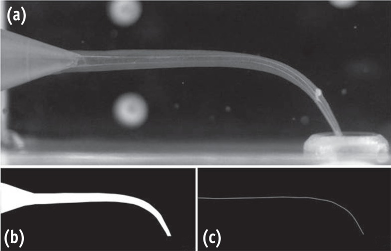
-
Abstract
PDFPubReaderePub
Objectives The purpose of this study was to evaluate the histopathological effects of an antioxidant therapy on the pulp tissue of rat teeth exposed to a bleaching gel with 35% hydrogen peroxide.
Materials and Methods Forty rats were subjected to oral ingestion by gavage of distilled water (DW) or ascorbic acid (AA) 90 min before the bleaching therapy. For the bleaching treatment, the agent was applied twice for 5 min each to buccal surfaces of the first right mandibular molars. Then, the animals were sacrificed at 6 hr, 24 hr, 3 day, or 7 day post-bleaching, and the teeth were processed for microscopic evaluation of the pulp tissue.
Results At 6 hr, the pulp tissue showed moderate inflammatory reactions in all teeth of both groups. In the DW and AA groups, 100% and 80% of teeth exhibited pulp tissue with significant necrosis and intense tissue disorganization, respectively. At 24 hr, the AA-treated group demonstrated a greater regenerative capability than the DW group, with less intense inflammatory reaction and new odontoblast layer formation in 60% of the teeth. For up to the 7 day period, the areas of pulpal necrosis were replaced by viable connective tissue, and the dentin was underlined by differentiated odontoblast-like cells in most teeth of both groups.
Conclusions A slight reduction in initial pulpal damage during post-bleaching was promoted by AA therapy. However, the pulp tissue of AA-treated animals featured faster regenerative potential over time.
-
Citations
Citations to this article as recorded by- Clinical Study on the Efficacy of 35% Hydrogen Peroxide Gel According to Exposure Time (40 min vs. 20 min) by Spectrophotometry
Trinidad Rincón, Maria Portillo Muñoz, Maria Lobato, Ana María Martín Casado, Laryssa Mylenna Madruga Barbosa, Alessandro Loguercio, Cristina Gómez‐Polo
Journal of Esthetic and Restorative Dentistry.2026;[Epub] CrossRef - Influence of dental bleaching on the pulp tissue: A systematic review of in vivo studies
Mariana Viana Donato, Alexandre Henrique dos Reis‐Prado, Lucas Guimarães Abreu, Lara Cancella de Arantes, Juliana Goto, Hebertt Gonzaga dos Santos Chaves, Luciano Tavares Angelo Cintra, André Luiz Fraga Briso, Isabella Faria da Cunha Peixoto, Francine Ben
International Endodontic Journal.2024; 57(6): 630. CrossRef - ANALYSIS OF THE INFLUENCE OF THE TEETH WHITENING PROCEDURE ON THE GUM AND CYTOKINE PROFILE OF ORAL FLUID (LITERATURE REVIEW)
S. S. Bozhik, N. V. Hasyuk, V. B. Radchuk
Bulletin of Problems Biology and Medicine.2024; 1(3): 17. CrossRef - Assessing the Viability of Laser-Activated Dental Bleaching Compared to Conventional In-Office Bleaching Methods: A Systematic Review of Clinical and In Vitro Studies
Eugenia Anagnostaki, Valina Mylona, Steven Parker, Mark Cronshaw, Martin Grootveld
Applied Sciences.2023; 13(22): 12459. CrossRef - Effects of the application of sodium ascorbate after in-office bleaching on the penetration of hydrogen peroxide, color change, and microtensile bond strength
Alexandra Mena-Serrano, María G. Granda-Albuja, Jenny Naranjo, Eliana Aldás Fierro, Michael Willian Favoreto, Alessandro D. Loguercio, Alessandra Reis
Brazilian Dental Journal.2023; 34(5): 87. CrossRef - Can different agents reduce the damage caused by bleaching gel to pulp tissue? A systematic review of basic research
Letícia Aparecida Silva Batista, Alexandre Henrique dos Reis-Prado, Hebertt Gonzaga dos Santos Chaves, Lara Cancella de Arantes, Luís Fernando Santos Alves Morgan, Carolina Bosso André, Thaís Yumi Suzuki, Francine Benetti
Restorative Dentistry & Endodontics.2023;[Epub] CrossRef - Design of a thermosensitive ibuprofen-loaded nanogel as smart material applied as anti-inflammatory in tooth bleaching: An in vivo study
Samara K.S.C.F. Moura, Milena L.V. dos Santos, Lucas A. do Nascimento, Mariana F.A. da Silva, Glória M. de França, Lucas M. da Costa, Aldo C. Medeiros, Raimundo F. Araújo-Júnior, Aurigena A. de Araújo, Cláudia N. Oliveira, André L. Dorini, Rejane A. de Ca
Journal of Drug Delivery Science and Technology.2022; 68: 103123. CrossRef - Effect of bleaching gel volume on color change and postoperative sensitivity: a randomized clinical study
Lara Maria Bueno Esteves, Paulo Henrique dos Santos, Ticiane Cestari Fagundes, Marjorie de Oliveira Gallinari, Giulia Bessa de Mello Antonaccio, Luciano Tavares Ângelo Cintra, André Luiz Fraga Briso
Clinical Oral Investigations.2022; 26(3): 2527. CrossRef - Human Dental Pulp Stem Cells Exhibit Different Biological Behaviours in Response to Commercial Bleaching Products
Carmen Llena, Mar Collado-González, Christopher Joseph Tomás-Catalá, David García-Bernal, Ricardo Elías Oñate-Sánchez, Francisco Javier Rodríguez-Lozano, Leopoldo Forner
Materials.2018; 11(7): 1098. CrossRef - Concentration‐dependent effect of bleaching agents on the immunolabelling of interleukin‐6, interleukin‐17 and CD5‐positive cells in the dental pulp
F. Benetti, J. E. Gomes‐Filho, L. L. Ferreira, G. Sivieri‐Araújo, E. Ervolino, A. L. F. Briso, L. T. A. Cintra
International Endodontic Journal.2018; 51(7): 789. CrossRef
- Clinical Study on the Efficacy of 35% Hydrogen Peroxide Gel According to Exposure Time (40 min vs. 20 min) by Spectrophotometry
- 2,453 View
- 6 Download
- 10 Crossref
-
Identification of
Enterococcus faecalis antigens specifically expressedin vivo - Seok-Woo Lee, Uttom K. Shet, Sang-Won Park, Hyun-Pil Lim, Kwi-Dug Yun, Seong Soo Kang, Se Eun Kim
- Restor Dent Endod 2015;40(4):306-311. Published online October 5, 2015
- DOI: https://doi.org/10.5395/rde.2015.40.4.306
-
Abstract
PDFPubReaderePub
Objectives Molecular mechanism of the pathogenicity of
Enterococcus faecalis (E. faecalis ), a suspected endodontic pathogen, has not yet been adequately elucidated due to limited information on its virulence factors. Here we report the identification ofin vivo expressed antigens ofE. faecalis by using a novel immunoscreening technique called change-mediated antigen technology (CMAT) and an experimental animal model of endodontic infection.Materials and Methods Among 4,500
E. coli recombinant clones screened, 19 positive clones reacted reproducibly with hyperimmune sera obtained from rabbits immunized withE. faecalis cells isolated from an experimental endodontic infection. DNA sequences from 16 of thesein vivo -induced (IVI) genes were determined.Results Identified protein antigens of
E. faecalis included enzymes involved in housekeeping functions, copper resistance protein, putative outer membrane proteins, and proteins of unknown function.Conclusions In vivo expressed antigens ofE. faecalis could be identified by using a novel immune-screening technique CMAT and an experimental animal model of endodontic infection. Detailed analysis of these IVI genes will lead to a better understanding of the molecular mechanisms involved in the endodontic infection ofE. faecalis .-
Citations
Citations to this article as recorded by-
Insights into ecology, pathogenesis, and biofilm formation of
Enterococcus faecalis
from functional genomics
Julia L. E. Willett, Gary M. Dunny, Corrella S. Detweiler
Microbiology and Molecular Biology Reviews.2025;[Epub] CrossRef - Antibody-based Screening of Porphyromonas gingivalis Proteins Specifically Produced in Patients with Chronic Periodontitis
Hye-Jung Kim, Seok-Woo Lee
International Journal of Oral Biology.2018; 43(4): 201. CrossRef - The controversial role of Enterococcus faecalis in colorectal cancer
Carolina Vieira de Almeida, Antonio Taddei, Amedeo Amedei
Therapeutic Advances in Gastroenterology.2018;[Epub] CrossRef - Model systems for the study of Enterococcal colonization and infection
H. M. Sharon Goh, M. H. Adeline Yong, Kelvin Kian Long Chong, Kimberly A. Kline
Virulence.2017; 8(8): 1525. CrossRef
-
Insights into ecology, pathogenesis, and biofilm formation of
Enterococcus faecalis
from functional genomics
- 2,327 View
- 5 Download
- 4 Crossref
- Effect of dentin treatment on proliferation and differentiation of human dental pulp stem cells
- Minjeong Park, Nan-Sim Pang, Il-Young Jung
- Restor Dent Endod 2015;40(4):290-298. Published online September 23, 2015
- DOI: https://doi.org/10.5395/rde.2015.40.4.290
-
Abstract
PDFPubReaderePub
Objectives Sodium hypochlorite (NaOCl) is an excellent bactericidal agent, but it is detrimental to stem cell survival, whereas intracanal medicaments such as calcium hydroxide (Ca[OH]2) promote the survival and proliferation of stem cells. This study evaluated the effect of sequential NaOCl and Ca[OH]2 application on the attachment and differentiation of dental pulp stem cells (DPSCs).
Materials and Methods DPSCs were obtained from human third molars. All dentin specimens were treated with 5.25% NaOCl for 30 min. DPSCs were seeded on the dentin specimens and processed with additional 1 mg/mL Ca[OH]2, 17% ethylenediaminetetraacetic acid (EDTA) treatment, file instrumentation, or a combination of these methods. After 7 day of culture, we examined DPSC morphology using scanning electron microscopy and determined the cell survival rate with 3-(4,5-dimethylthiazol-2-yl)-2,5-diphenyltetrazolium bromide assay. We measured cell adhesion gene expression levels after 4 day of culture and odontogenic differentiation gene expression levels after 4 wk using quantitative real-time polymerase chain reaction.
Results DPSCs did not attach to the dentin in the NaOCl-treated group. The gene expression levels of fibronectin-1 and secreted phosphoprotein-1 gene in both the Ca[OH]2- and the EDTA-treated groups were significantly higher than those in the other groups. All Ca[OH]2-treated groups showed higher expression levels of dentin matrix protein-1 than that of the control. The dentin sialophosphoprotein level was significantly higher in the groups treated with both Ca[OH]2 and EDTA.
Conclusions The application of Ca[OH]2 and additional treatment such as EDTA or instrumentation promoted the attachment and differentiation of DPSCs after NaOCl treatment.
-
Citations
Citations to this article as recorded by- Effect of Intracanal Medications on the Viability of Human Periodontal Ligament‐Derived Mesenchymal Stem Cells
Arian Braido, Walbert de Andrade Vieira, Bruno Cazotti Pereira, Karina Gonzales Silvério, Paulo Henrique Gabriel, Aline Cristine Gomes Matta, Emerson Alves Martins, Adriana de Jesus Soares
Australian Endodontic Journal.2026; 52(1): 35. CrossRef - Biological, antimicrobial, and apical tissue dissolution effects of ambroxol hydrochloride as an irrigant for regenerative endodontics: an in vitro study
Letycia Accioly Simoes Coelho, Ester Alves Ferreira Bordini, Fernanda Balestrero Cassiano, Mirela Cesar Barros, Renato Menezes Silva, Flaviana Bombarda de Andrade, Diana Gabriela Soares, Marco Antonio Hungaro Duarte
BMC Oral Health.2026;[Epub] CrossRef - An injectable κ-carrageenan hydrogel carrier maintains antibiofilm activity while supporting dental pulp-derived stem cell viability in a dentin-based model
Muneera A. Alajlan, Rhodanne Nicole A. Lambarte, Terrence S. Sumague, Abdurahman A. Niazy
Journal of Dentistry.2026; 171: 106713. CrossRef - Effect of Intracanal Medications on Cell Adhesion, Viability, and Differentiation During Regenerative Endodontic Procedures: A Systematic Review of In Vitro Studies
Arian Braido, Luisa Coutinho Cardim, Aline Cristine Gomes Matta, Karina Gonzales Silvério, Paulo Henrique Gabriel, Emerson Alves Martins, Adriana de Jesus Soares, Walbert A. Vieira
Journal of Endodontics.2026;[Epub] CrossRef - Effect of erbium yttrium aluminium garnet laser dentin conditioning on dental pulp stem cells viability
Aryan Jafari, Mehdi Vatanpour, Nooshin Barikrow, Pouyan Razavi, Sohrab Tour Savadkouhi
Heliyon.2024; 10(5): e26954. CrossRef - Comparison of the effect of NaOCL, curcumin, and EDTA on differentiation, proliferation, and adhesion of dental pulp stem cells
Vahid Zand, Amin Salem Milani, Carolyn Primus, Marzie Aghazade, Hadi Mokhtari, Sabete Bagheri Sabzevar, Pardis Tehranchi
Journal of Oral Biology and Craniofacial Research.2023; 13(2): 347. CrossRef - Differential Effects of Extracellular Matrix Glycoproteins Fibronectin and Laminin-5 on Dental Pulp Stem Cell Phenotypes and Responsiveness
Hyungbin Lee, Allen Bae, John Kim, Karl Kingsley
Journal of Functional Biomaterials.2023; 14(2): 91. CrossRef - Assessment of the Influence of Various Concentrations of Sodium Hypochlorite on Stem Cell Derived From Human Exfoliated Deciduous Teeth (SHED) Proliferation and Differentiation
Viral Maru, Ashwini KB, Manisha Madkaikar, R K Sarada Devi, Ashita Gada, Salil Bapat
Cureus.2022;[Epub] CrossRef - Effects of sodium hypochlorite and ethylenediaminetetraacetic acid on proliferation, osteogenic/odontogenic differentiation, and mechanosensitive gene expression of human dental pulp stem cells
Yuejun Li, Changlong Jin, Shouliang Zhao, Han Xie
Tissue and Cell.2022; 79: 101955. CrossRef - Influence of ethylenediaminetetraacetic acid on regenerative endodontics: A systematic review
Alexandre H. dos Reis‐Prado, Lucas G. Abreu, Rogéria R. Fagundes, Sabrina de C. Oliveira, Marco C. Bottino, Antônio P. Ribeiro‐Sobrinho, Francine Benetti
International Endodontic Journal.2022; 55(6): 579. CrossRef - An in-vitro Comparative Evaluation of Quantitative Release of Transforming Growth Factor β-1 from Dentin upon the Action of Endodontic Irrigants, Medicaments, Ultrasonic Activation, and Low-Level Laser Irradiation
Anilkumar Akhila, V. P. Prabath Singh, Kerala R. Varma, Senthil V. Vasudevan, V. Sukhithasri, Salu Sasikumar
Amrita Journal of Medicine.2021; 17(2): 34. CrossRef - Non-Thermal Atmospheric Pressure Plasma-Conditioned Root Dentin Promotes Attraction and Attachment of Primary Human Dental Pulp Stem Cells in Real-Time Ex Vivo
Yeon-Jee Yoo, Min-Ji Kang, Hiran Perinpanayagam, Joo-Cheol Park, Seung-Ho Baek, Kee-Yeon Kum
Applied Sciences.2021; 11(15): 6836. CrossRef - The Effects of Intracanal Irrigants and Medicaments on Dental-Derived Stem Cells Fate in Regenerative Endodontics: An update
Sara Ayoub, Ali Cheayto, Sanaa Bassam, Mehdi Najar, Antoine Berbéri, Mohammad Fayyad-Kazan
Stem Cell Reviews and Reports.2020; 16(4): 650. CrossRef - An Immunofluorescence Study to Analyze Wound Healing Outcomes of Regenerative Endodontics in an Immature Premolar with Chronic Apical Abscess
Jeen Nee Lui, Wen Yi Lim, Domenico Ricucci
Journal of Endodontics.2020; 46(5): 627. CrossRef - Dynamic Irrigation Promotes Apical Papilla Cell Attachment in an Ex Vivo Immature Root Canal Model
Sanupong Prompreecha, Thanapat Sastraruji, Phumisak Louwakul, Tanida Srisuwan
Journal of Endodontics.2018; 44(5): 744. CrossRef - Odontoblast-like differentiation and mineral formation of pulpsphere derived cells on human root canal dentin in vitro
Jörg Neunzehn, Sandra Pötschke, Christian Hannig, Hans-Peter Wiesmann, Marie-Theres Weber
Head & Face Medicine.2017;[Epub] CrossRef - Effects of Epigallocatechin Gallate, an Antibacterial Cross-linking Agent, on Proliferation and Differentiation of Human Dental Pulp Cells Cultured in Collagen Scaffolds
Young-Sun Kwon, Hee-Jin Kim, Yun-Chan Hwang, Vinicius Rosa, Mi-Kyung Yu, Kyung-San Min
Journal of Endodontics.2017; 43(2): 289. CrossRef
- Effect of Intracanal Medications on the Viability of Human Periodontal Ligament‐Derived Mesenchymal Stem Cells
- 2,664 View
- 8 Download
- 17 Crossref
- Clinical and radiographical evaluation of mineral trioxide aggregate, biodentine and propolis as pulpotomy medicaments in primary teeth
- Bharti Kusum, Kumar Rakesh, Khanna Richa
- Restor Dent Endod 2015;40(4):276-285. Published online September 9, 2015
- DOI: https://doi.org/10.5395/rde.2015.40.4.276
-
Abstract
PDFPubReaderePub
Objectives The purpose of this study was to evaluate the efficacy of mineral trioxide aggregate (MTA), Biodentine and Propolis as pulpotomy medicaments in primary dentition, both clinically and radiographically.
Materials and Methods A total of 75 healthy 3 to 10 yr old children each having at least one carious primary molar tooth were selected. Random assignment of the pulpotomy medicaments was done as follows: Group I, MTA; Group II, Biodentine; Group III, Propolis. All the pulpotomized teeth were evaluated at 3, 6, and 9 mon clinically and radiographically, based on the scoring criteria system.
Results The clinical success rates were found to be similar among the three groups at 3 and 6 mon where as a significant decrease in success rate was observed in Group III (84%) compared to both Group I (100%) and Group II (100%) at 9 mon. Radiographic success rates over a period of 9 mon in Groups I, II, and III were 92, 80, and 72%, respectively.
Conclusions Teeth treated with MTA and Biodentine showed more favorable clinical and radiographic success as compared to Propolis at 9 mon follow-up.
-
Citations
Citations to this article as recorded by- Histological Tissue Response to Calcium Silicate-Based Cements Assessed in Human Tooth Culture Models: A Systematic Review
Alberto Cabrera-Fernández, Hebertt Gonzaga dos Santos Chaves, Aránzazu Díaz-Cuenca, Juan J. Segura-Egea, Jenifer Martín-González, João Peça, Diana B. Sequeira, João Miguel Marques dos Santos
Journal of Functional Biomaterials.2026; 17(2): 78. CrossRef - The Clinical Effectiveness of Propolis on the Endodontic Treatment of Permanent Teeth: A Systematic Review of Randomized Clinical Trials and Updates
Amirah Y Aldosari, Amira M Aljared, Hanin S Alqurshy, Abdullah M Alfarran, Mohanad G Alnahdi, Sarah S Alharbi, Wed S Alharbi, Faisal T Alghamdi
Cureus.2025;[Epub] CrossRef - Natural nanoparticles versus the gold standard in direct pulp capping: a randomized clinical trial
Dalia M. Elasser, Sabah M. Sobhy, Rania Rashad Omar Taha, Nevin A. Gad, Dina M. Abdel-Ghany
The Saudi Dental Journal.2025;[Epub] CrossRef - Systematic review and meta analysis of first and second generation bioceramic materials in primary dentition pulpotomies
João Albernaz Neves, Luísa Bandeira Lopes, Marta Alves Duarte, José João Mendes, Tiago Pimentel
Scientific Reports.2025;[Epub] CrossRef - EFFICACY OF BIODENTINE VERSUS MINERAL TRIOXIDE AGGREGATE IN PULPOTOMY FOR PRIMARY TEETH: A SYSTEMATIC REVIEW AND META-ANALYSIS OF RANDOMIZED CONTROLLED TRIALS
KORINA YUN-FAN LU, JENNIFER L. GIBBS, CHENG-YU WU, MARKUS B. BLATZ, XUEHAO MA, MIN-WEN FU, KEVIN SHENG-KAI MA
Journal of Evidence-Based Dental Practice.2025; 25(4): 102191. CrossRef - Calcium Silicate-Based Cements in Restorative Dentistry: Vital Pulp Therapy Clinical, Radiographic, and Histological Outcomes on Deciduous and Permanent Dentition—A Systematic Review and Meta-Analysis
Maria Teresa Xavier, Ana Luísa Costa, João Carlos Ramos, João Caramês, Duarte Marques, Jorge N. R. Martins
Materials.2024; 17(17): 4264. CrossRef - Outcome assessment methods of bioactive and biodegradable materials as pulpotomy agents in primary and permanent teeth: a scoping review
Yasmine Elhamouly, May M. Adham, Karin M L Dowidar, Rania M. El Backly
BMC Oral Health.2024;[Epub] CrossRef - Comparative Evaluation of the Efficacy of Theracal Lc, Mineral Trioxide Aggregate, and Biodentine As Direct Pulp Capping Materials in Patients With Pulpal Exposure in Posterior Teeth: A Triple Blinded Randomized Parallel Group Clinical Trial
Joyeeta Mahapatra, Pradnya P Nikhade, Aditya Patel, Nikhil Mankar, Prachi Taori
Cureus.2024;[Epub] CrossRef - Efficacy of different endodontic irrigants in the lesion sterilization and tissue repair technique in primary molars: A randomized controlled clinical trial
Anukriti Dimri, Nikhil Srivastava, Vivek Rana, Noopur Kaushik
Journal of Indian Society of Pedodontics and Preventive Dentistry.2024; 42(4): 294. CrossRef - Clinical and Radiographic Success Rates of Pulpotomies in Primary Molars Treated with Formocresol, BiodentineTM, and Endo Repair: A Randomized Clinical Trial
Elham Farokh Gisour, Farzaneh Jalali, Fatemeh Jahanimoghadam, Tania Dehesh
Pesquisa Brasileira em Odontopediatria e Clínica Integrada.2023;[Epub] CrossRef - Evaluation and comparison of mineral trioxide aggregate and cold ceramic in primary tooth pulpotomy: Clinical and radiographic study
Bita Rasteh, Leila Basir, Shirin Taravati, Masoumeh Khataminia
Journal of Family Medicine and Primary Care.2023; 12(12): 3068. CrossRef - Comparison between the Radiographic and Clinical Rates of Success for TheraCal and MTA in Primary Tooth Pulpotomy within a 12‐Month Follow‐Up: A Split‐Mouth Clinical Trial
Sedigheh Hassanpour, Naser Asl Aminabadi, Mahdi Rahbar, Leila Erfanparast, Iole Vozza
BioMed Research International.2023;[Epub] CrossRef - Biodentine™ Pulpotomy in Stage I Primary Molars: A 12-month Follow-up
Hitaf Nasrallah, Balsam Noueiri
International Journal of Clinical Pediatric Dentistry.2023; 15(6): 660. CrossRef - Omega 3 based formulations as new possible pulpotomy agents for primary teeth
Nancy M. Metwally, Amina M. El Hosary, Gamal M. El Maghraby, Maha A. El Demellawy, Mohamed Nabil, Elsayed M. Deraz
Tanta Dental Journal.2022; 19(4): 287. CrossRef - Clinical efficacy of Er:YAG laser application in pulpotomy of primary molars: a 2-year follow-up study
Junhui Wang, Yujiang Chen, Baize Zhang, Xin Ge, Xiaojing Wang
Lasers in Medical Science.2022; 37(9): 3705. CrossRef - Comparative efficacy of medicaments or techniques for pulpotomy of primary molars: a network meta-analysis
Jiehua Guo, Na Zhang, Yuzhao Cheng
Clinical Oral Investigations.2022; 27(1): 91. CrossRef - Comparative evaluation of zinc oxide-eugenol and zinc oxide with Neem oil in root canal treatment of primary teeth
Dhvani Gordhanbhai Patel, Shoba Fernandes, Yash Bafna, Krunal Choksi, Shaila Chaudhary, Priya Mishra
AYU (An International Quarterly Journal of Research in Ayurveda).2022; 43(1): 8. CrossRef - Treatment Outcomes of Pulpotomy with Propolis in Comparison with MTA in Human Primary Molars: A 24-month Follow-up Randomized Controlled Trial
Kakarla Sri RojaRamya, Rayala Chandrasekhar, KS Uloopi, C Vinay
International Journal of Clinical Pediatric Dentistry.2022; 15(S1): S3. CrossRef - Comparison of Clinical and Radiographic Success between MTA and Biodentine in Pulpotomy of Primary Mandibular Second Molars with Irreversible Pulpitis: A Randomized Double‐Blind Clinical Trial
Alireza Eshghi, Maryam Hajiahmadi, Mohammad Hossein Nikbakht, Mona Esmaeili, Murilo Baena Lopes
International Journal of Dentistry.2022;[Epub] CrossRef - Does the use of natural products for endodontic therapy in primary teeth have sufficient evidence for clinical practice? A scoping review
Filipe Colombo Vitali, Ana Cristina Andrada, Helhen Cristina da Luz Cardoso, Gesner Francisco Xavier-Junior, Cleonice da Silveira Teixeira, Loise Pedrosa Salles, Erica Negrini Lia, Carla Massignan
Clinical Oral Investigations.2022; 26(10): 6043. CrossRef - Prevalence of pain following single-visit pulpectomy with stainless steel crown done by postgraduate students in a university sitting
Ashwin Shravan Kumar, Mahesh Ramakrishnan
Journal of Advanced Pharmaceutical Technology & Research.2022; 13(Suppl 1): S177. CrossRef - Clinical and histological response of human pulp tissue to direct pulp capping with mineral trioxide aggregate, Biodentine and propolis
Zahra Nasri, MaryamZare Jahromi, Atousa Aminzadeh
Dental Research Journal.2022; 19(1): 40. CrossRef - Long-term evaluation of primary teeth molar pulpotomies with Biodentine and MTA: a CONSORT randomized clinical trial
S. Vilella-Pastor, S. Sáez, A. Veloso, F. Guinot-Jimeno, M. Mercadé
European Archives of Paediatric Dentistry.2021; 22(4): 685. CrossRef - Tailored 70S30C Bioactive glass induces severe inflammation as pulpotomy agent in primary teeth: an interim analysis of a randomised controlled trial
Yasmine Elhamouly, Rania M. El Backly, Dalia M. Talaat, Samia S. Omar, Maha El Tantawi, Karin M. L. Dowidar
Clinical Oral Investigations.2021; 25(6): 3775. CrossRef - Efficacy of laser photobiomodulation pulpotomy in human primary teeth
Chandrashekar Murugesh Yavagal, Akshaya Lal, Viplavi Vijaysinh Chavan Patil, Puja C. Yavagal, Kiran Kumar Neelakantappa, Madhu Hariharan
Journal of Indian Society of Pedodontics and Preventive Dentistry.2021; 39(4): 436. CrossRef - SÜT DİŞİ AMPUTASYON TEDAVİSİNDE GÜNCEL YAKLAŞIMLAR
Necibe Damla ŞAHİN, Volkan ARIKAN
Atatürk Üniversitesi Diş Hekimliği Fakültesi Dergisi.2021; : 1. CrossRef - The use of propolis in dentistry, oral health, and medicine: A review
Felix Zulhendri, Rafael Felitti, James Fearnley, Munir Ravalia
Journal of Oral Biosciences.2021; 63(1): 23. CrossRef - Clinical and radiographic evaluation of pulpotomy using MTA, Biodentine and Er,Cr:YSGG laser in primary teeth. A clinical study
Pandiyan Ramanandvignesh, Kumar Gyanendra, Dhillon Jatinder Kaur Goswami Mridula
Laser Therapy.2020; 29(1): 29. CrossRef - Comparative Evaluation of Success of Biodentine and Mineral Trioxide Aggregate with Formocresol as Pulpotomy Medicaments in Primary Molars: An In Vivo Study
Ritika Malhotra, Shilpa Ahuja, Dipanshu Kumar, Kapil Gandhi, Rishabh Kapoor, Kumari Surabhi
International Journal of Clinical Pediatric Dentistry.2020; 13(2): 167. CrossRef - Comparison of Partial Pulpotomy in Permanent Molars Using Different Pulp Capping Agents and Restoration Materials
Jimi Lee, Nanyoung Lee, Sangho Lee, Myeongkwan Jih
THE JOURNAL OF THE KOREAN ACADEMY OF PEDTATRIC DENTISTRY.2020; 47(2): 148. CrossRef - A Comparative Evaluation between Propolis and Mineral Trioxide Aggregate as Pulpotomy Medicaments in Primary Molars
Kavita Madan, Sudhindra Baliga, Pranjali Deulkar, Nilima Thosar, Nilesh Rathi, Meghana Deshpande, Sphurti Bane
Journal of Evolution of Medical and Dental Sciences.2020; 9(15): 1256. CrossRef - Efficacy of Biodentine and Mineral Trioxide Aggregate in Primary Molar Pulpotomies—A Systematic Review and Meta-Analysis With Trial Sequential Analysis of Randomized Clinical Trials
Venkateshbabu Nagendrababu, Shaju Jacob Pulikkotil, Sajesh K. Veettil, Peerawat Jinatongthai, James L. Gutmann
Journal of Evidence Based Dental Practice.2019; 19(1): 17. CrossRef - Efficacy of Alternative Medicaments for Pulp Treatment in Primary Teeth in the Short Term: A Meta-analysis
Joon Soo Park, Bosky Jasani, Jilen Patel, Robert P. Anthonappa, Nigel M. King
Journal of Evidence Based Dental Practice.2019; 19(4): 101309. CrossRef - The evaluation of MTA and Biodentine as a pulpotomy materials for carious exposures in primary teeth
Burcu Nihan Çelik, Merve Safa Mutluay, Volkan Arıkan, Şaziye Sarı
Clinical Oral Investigations.2019; 23(2): 661. CrossRef - Effects of MTA and Brazilian propolis on the biological properties of dental pulp cells
Bingqing Shi, Yuming Zhao, Xiaojing Yuan
Brazilian Oral Research.2019;[Epub] CrossRef - MTA and biodentine for primary teeth pulpotomy: a systematic review and meta-analysis of clinical trials
Emyr Stringhini Junior, Manuela Gouvêa Campêlo dos Santos, Luciana Butini Oliveira, Montse Mercadé
Clinical Oral Investigations.2019; 23(4): 1967. CrossRef - Comparison of the success rate of a bioactive dentin substitute with those of other root restoration materials in pulpotomy of primary teeth
Hooman Shafaee, Mehrnoosh Alirezaie, Abdolrasoul Rangrazi, Erfan Bardideh
The Journal of the American Dental Association.2019; 150(8): 676. CrossRef - BiodentineTM versus formocresol pulpotomy technique in primary molars: a 12–month randomized controlled clinical trial
Omar Abd El Sadek El Meligy, Najlaa Mohamed Alamoudi, Sulaiman Mohamed Allazzam, Azza Abdel Mohsen El-Housseiny
BMC Oral Health.2019;[Epub] CrossRef - Randomized Controlled Trial of Pulpotomy in Primary Molars using MTA and Formocresol Compared to 3Mixtatin: A Novel Biomaterial
Zahra Jamali, Vajiheh Alavi, Ebrahim Najafpour, Naser Asl Aminabadi, Sajjad Shirazi
Journal of Clinical Pediatric Dentistry.2018; 42(5): 361. CrossRef - Recent Trends in Tricalcium Silicates for Vital Pulp Therapy
Imad About
Current Oral Health Reports.2018; 5(3): 178. CrossRef - Clinical and Radiographic Evaluations of Biodentine™ Pulpotomies in Mature Primary Molars (Stage 2)
Fouad Ayoub, Hitaf Nasrallah, Balsam El Noueiri, Charles Pilipili
International Journal of Clinical Pediatric Dentistry.2018; 11(6): 496. CrossRef - Clinical and radiographic evaluation of biodentine versus calcium hydroxide in primary teeth pulpotomies: a retrospective study
Silvia Caruso, Teresa Dinoi, Giuseppe Marzo, Vincenzo Campanella, Maria Rita Giuca, Roberto Gatto, Marco Pasini
BMC Oral Health.2018;[Epub] CrossRef - How does the pulpal response to Biodentine and ProRoot mineral trioxide aggregate compare in the laboratory and clinic?
R. Careddu, H. F. Duncan
British Dental Journal.2018; 225(8): 743. CrossRef - Mineral trioxide aggregate and other bioactive endodontic cements: an updated overview – part I: vital pulp therapy
M. Parirokh, M. Torabinejad, P. M. H. Dummer
International Endodontic Journal.2018; 51(2): 177. CrossRef - Pulp treatment for extensive decay in primary teeth
Violaine Smaïl-Faugeron, Anne-Marie Glenny, Frédéric Courson, Pierre Durieux, Michele Muller-Bolla, Helene Fron Chabouis
Cochrane Database of Systematic Reviews.2018;[Epub] CrossRef - Biodentine™ material characteristics and clinical applications: a 3 year literature review and update
S. Rajasekharan, L. C. Martens, R. G. E. C. Cauwels, R. P. Anthonappa
European Archives of Paediatric Dentistry.2018; 19(1): 1. CrossRef - Microleakage and Shear Bond Strength of Biodentine at Different Setting Time
Yong Ho Song, Nanyoung Lee, Sangho Lee, Myeongkwan Jih
THE JOURNAL OF THE KOREAN ACADEMY OF PEDTATRIC DENTISTRY.2018; 45(3): 344. CrossRef - Evaluation of Biodentine Pulpotomies in Deciduous Molars with Physiological Root Resorption (Stage 3)
Fouad Ayoub, Hitaf Nasrallah, Balsam El Noueiri, Charles Pilipili
International Journal of Clinical Pediatric Dentistry.2018; 11(5): 393. CrossRef - Clinical and radiographic comparison of biodentine, mineral trioxide aggregate and formocresol as pulpotomy agents in primary molars
P. Juneja, S. Kulkarni
European Archives of Paediatric Dentistry.2017; 18(4): 271. CrossRef - Success Rates of Pulpotomies in Primary Molars Using Calcium Silicate-Based Materials: A Randomized Control Trial
Yeliz Guven, Sermin Dicle Aksakal, Nilufer Avcu, Gulcan Unsal, Elif Bahar Tuna, Oya Aktoren
BioMed Research International.2017; 2017: 1. CrossRef - Biodentine: from biochemical and bioactive properties to clinical applications
Imad About
Giornale Italiano di Endodonzia.2016; 30(2): 81. CrossRef
- Histological Tissue Response to Calcium Silicate-Based Cements Assessed in Human Tooth Culture Models: A Systematic Review
- 3,665 View
- 36 Download
- 51 Crossref
- Changes in SIRT gene expression during odontoblastic differentiation of human dental pulp cells
- Young-Eun Jang, Su-Hee Go, Bin-Na Lee, Hoon-Sang Chang, In-Nam Hwang, Won-Mann Oh, Yun-Chan Hwang
- Restor Dent Endod 2015;40(3):223-228. Published online July 15, 2015
- DOI: https://doi.org/10.5395/rde.2015.40.3.223
-
Abstract
PDFPubReaderePub
Objectives The aim of this study was to investigate the expression of 7 different sirtuin genes (SIRT1-SIRT7) in human dental pulp cells (HDPCs), and to determine the role of SIRTs in the odontoblastic differentiation potential of HDPCs.
Materials and Methods HDPCs were isolated from freshly extracted third molar teeth of healthy patients and cultulred in odontoblastic differentiation inducing media. Osteocalcin (OCN) and dentin sialophosphoprotein (DSPP) expression was analyzed to evaluate the odontoblastic differentiation of HDPCs by reverse transcription-polymerase chain reaction (RT-PCR), while alizarin red staining was used for the mineralization assay. To investigate the expression of SIRTs during odontoblastic differentiation of HDPCs, real time PCR was also performed with RT-PCR.
Results During the culture of HDPCs in the differentiation inducing media, OCN, and DSPP mRNA expressions were increased. Mineralized nodule formation was also increased in the 14 days culture. All seven SIRT genes were expressed during the odontogenic induction period. SIRT4 expression was increased in a time-dependent manner.
Conclusions Our study identified the expression of seven different SIRT genes in HDPCs, and revealed that SIRT4 could exert an influence on the odontoblast differentiation process. Further studies are needed to determine the effects of other SIRTs on the odontogenic potential of HDPCs.
-
Citations
Citations to this article as recorded by- Biodegradable Zn‐5Dy Alloy with Enhanced Osteo/Angio‐Genic Activity and Osteointegration Effect via Regulation of SIRT4‐Dependent Mitochondrial Function
Yue Han, Xian Tong, Runqi Zhou, Yilin Wang, Yuge Chen, Liang Chen, Xinhua Hong, Linmei Wu, Zhiqiang Lin, Yichi Zhang, Xuejia Zhang, Chaoming Hu, Bin Li, Yifan Ping, Zelin Cao, Zhou Ye, Zhongchen Song, Yuncang Li, Cuie Wen, Yongsheng Zhou, Jixing Lin, Shen
Advanced Science.2024;[Epub] CrossRef - The Role of Histone Acetylation Modification in Dental Tissue-Derived Mesenchymal Stem Cells and Odontogenesis
Haoling Chen, Zijing Huang, Chuxiao Chen
Cellular Reprogramming.2023; 25(1): 11. CrossRef - Metabolic Remodeling Impacts the Epigenetic Landscape of Dental Mesenchymal Stem Cells
Haiyun Luo, Yachuan Zhou, Wenjing Liu, Jun Wang
Stem Cells International.2022; 2022: 1. CrossRef - SIRT4 regulates rat dental papilla cell differentiation by promoting mitochondrial functions
Haoling Chen, Jun Kang, Fuping Zhang, Tong Yan, Wenguo Fan, Hongwen He, Fang Huang
The International Journal of Biochemistry & Cell Biology.2021; 134: 105962. CrossRef - Sirtuins as Interesting Players in the Course of HIV Infection and Comorbidities
Karolina Jurkowska, Beata Szymańska, Brygida Knysz, Amadeusz Kuźniarski, Agnieszka Piwowar
Cells.2021; 10(10): 2739. CrossRef - Robust expression of SIRT6 inhibits pulpitis via activation of the TRPV1 channel
Jia Hu, Weiran Chen, Zailing Qiu, Hongbing Lv
Cell Biochemistry and Function.2020; 38(5): 676. CrossRef - Downregulation of microRNA‐143‐5p is required for the promotion of odontoblasts differentiation of human dental pulp stem cells through the activation of the mitogen‐activated protein kinases 14‐dependent p38 mitogen‐activated protein kinases signaling pa
Bao‐Liang Wang, Zhi Wang, Xi Nan, Qing‐Cai Zhang, Wei Liu
Journal of Cellular Physiology.2019; 234(4): 4840. CrossRef - A potential role for the silent information regulator 2 homologue 1 (SIRT1) in periapical periodontitis
H. Kudo, O. Takeichi, K. Hatori, K. Makino, K. Himi, B. Ogiso
International Endodontic Journal.2018; 51(7): 747. CrossRef - Overexpressed Sirt1 in MSCs Promotes Dentin Formation in Bmi1-Deficient Mice
H. Wang, C. Lv, Y. Gu, Q. Li, L. Xie, H. Zhang, D. Miao, W. Sun
Journal of Dental Research.2018; 97(12): 1365. CrossRef - Expression of silent information regulator 2 homolog 1 (SIRT1) in periapical granulomas
Hiroshi Kudo, Osamu Takeichi, Kosuke Makino, Keisuke Hatori, Bunnai Ogiso
Journal of Oral Science.2018; 60(3): 411. CrossRef - TET1 knockdown inhibits the odontogenic differentiation potential of human dental pulp cells
Li-Jia Rao, Bai-Cheng Yi, Qi-Meng Li, Qiong Xu
International Journal of Oral Science.2016; 8(2): 110. CrossRef
- Biodegradable Zn‐5Dy Alloy with Enhanced Osteo/Angio‐Genic Activity and Osteointegration Effect via Regulation of SIRT4‐Dependent Mitochondrial Function
- 2,481 View
- 8 Download
- 11 Crossref
Review Article
- A review of the regenerative endodontic treatment procedure
- Bin-Na Lee, Jong-Wook Moon, Hoon-Sang Chang, In-Nam Hwang, Won-Mann Oh, Yun-Chan Hwang
- Restor Dent Endod 2015;40(3):179-187. Published online March 16, 2015
- DOI: https://doi.org/10.5395/rde.2015.40.3.179
-
Abstract
PDFPubReaderePub
Traditionally, apexification has been used to treat immature permanent teeth that have lost pulp vitality. This technique promotes the formation of an apical barrier to close the open apex so that the filling materials can be confined to the root canal. Because tissue regeneration cannot be achieved with apexification, a new technique called regenerative endodontic treatment was presented recently to treat immature permanent teeth. Regenerative endodontic treatment is a treatment procedure designed to replace damaged pulp tissue with viable tissue which restores the normal function of the pulp-dentin structure. After regenerative endodontic treatment, continued root development and hard tissue deposition on the dentinal wall can occur under ideal circumstances. However, it is difficult to predict the result of regenerative endodontic treatment. Therefore, the purpose of this study was to summarize multiple factors effects on the result of regenerative endodontic treatment in order to achieve more predictable results. In this study, we investigated the features of regenerative endodontic treatment in comparison with those of other pulp treatment procedures and analyzed the factors that have an effect on regenerative endodontic treatment.
-
Citations
Citations to this article as recorded by- Efficacy of combined scaffolds and sodium hypochlorite in regenerative endodontics of immature teeth (in-vivo study)
Hanan Arb, Abeer Darrag, Neveen Shaheen, Dina Attia
Tanta Dental Journal.2026; 23(2): 240. CrossRef - Outcome evaluation of regenerative endodontic procedures using platelet derivatives: a systematic review and meta-analysis of animal models
Nafsika Alevizou, Konstantinos Kodonas, Anastasia Fardi, Christos Gogos
Odontology.2026;[Epub] CrossRef - Evaluation of the Clinical Outcomes of Regenerative Endodontic Procedures Using Autologous Platelet Concentrate: A Systematic Review and Meta-Analysis
Elnaz Mousavi, Navid Nasrabadi, Samira Jamali, Arian Haddadi
Pesquisa Brasileira em Odontopediatria e Clínica Integrada.2025;[Epub] CrossRef - Antimicrobial 3D printed gelatin scaffolds for root canal disinfection in regenerative endodontics procedures
Mateo Dallos Ortega, Jenny Aveyard, Raghda Magdy Abdelgawad, Reem El-Gendy, Alexander Ciupa, David Whetnall, Julia Behnsen, Robert J. Poole, Raechelle A. D'Sa
Biomaterials Science.2025; 13(14): 3795. CrossRef - Regenerative Endodontic Therapies: Harnessing Stem Cells, Scaffolds, and Growth Factors
Rosana Farjaminejad, Samira Farjaminejad, Franklin Garcia-Godoy
Polymers.2025; 17(11): 1475. CrossRef - Effects of combining hyaluronic acid hydrogel with injectable platelet rich fibrin on apical papilla stem cells proliferation and differentiation
Azal H. Al-Masoody, Nasrin Asadi, Hadiseh Mohammadpour, Mahshid Hodjat, Tahereh Sadat Jafarzadeh Kashi
BMC Oral Health.2025;[Epub] CrossRef - Experts consensus on management of tooth luxation and avulsion
Ruijie Huang, Chenchen Zhou, Ling Zhan, Yuan Liu, Xian Liu, Qin Du, Jun Wang, Wei Zhao, Guangtai Song, Li-an Wu, Beizhan Jiang, Yanhong Li, Hongmei Zhang, Jing Zou
International Journal of Oral Science.2024;[Epub] CrossRef - A review of tissue engineering in regenerative endodontic treatment
Eric Priyo Prasetyo, Dian Agustin Wahjuningrum, Galih Sampoerno, Wilson Sukandar, Shafy Shariz Bin Sharizal, Nurfahira Paidal, Menza Fadiyan Amriel, Nathania Elita Gunawan, Ketut Suardita, Evelyn Tjendronegoro
Conservative Dentistry Journal.2024; 14(1): 1. CrossRef - Innovative Paradigms and Established Strategies in Tooth Revitalization: A Review
Ahmad Shah Khan, Zahid Mehmood Khan, Palwasha Ishaque, Muhammad Zubair, Syeda Fatima Tu Zahra, Sana Ashfaq
Dental Update.2024; 51(8): 570. CrossRef - Explore the most recent developments and upcoming outlooks in the field of dental nanomaterials
Ali Alsuraifi, Zainab M. Sulaiman, Noor Alhuda R. Mohammed, Jassim Mohammed, Sarah Kareem Ali, Yousef Husam Abdualihamaid, Fatimah Husam, Abdullah Ayad
Beni-Suef University Journal of Basic and Applied Sciences.2024;[Epub] CrossRef - Recent Advances in Regenerative Endodontics: A Review of Current Techniques and Future Directions
Firas A Alothman, Lamia S Hakami, Ali Alnasser, Faris M AlGhamdi, Abdullah A Alamri, Basel M Almutairii
Cureus.2024;[Epub] CrossRef - Regenerative Potential of Dental Pulp Stem Cells in Response to a Bioceramic Dental Sealer and Photobiomodulation: An In Vitro Study
Hamed A Alshawkani, Mohamed Mansy, Mahmoud Al Ankily, Mohamed Shamel
The Journal of Contemporary Dental Practice.2024; 25(4): 313. CrossRef - Marginal adaptation of customized gutta percha cone with calcium silicate based sealer versus MTA and biodentine apical plugs in simulated immature permanent teeth (an in vitro study)
Mary M. Mina, Sybel M. Moussa, Mahmoud R. Aboelseoud
BMC Oral Health.2024;[Epub] CrossRef - GelMA‐based hydrogel biomaterial scaffold: A versatile platform for regenerative endodontics
Lei Huang, Xuan Chen, XiaoXia Yang, Yinchun Zhang, Xiaoling Qiu
Journal of Biomedical Materials Research Part B: Applied Biomaterials.2024;[Epub] CrossRef - Regenerative Endodontic Treatment in Dentinogenesis Imperfecta‐Induced Apical Periodontitis
Ying Liao, Ting Pan, Xianghui Xing, Sivakumar Nuvvula
Case Reports in Dentistry.2024;[Epub] CrossRef - In vitro and in vivo evaluation of iRoot BP Plus as a coronal sealing material for regenerative endodontic procedures
Ning Yang, Wenxiao Yang, Rou Shen, Shengcai Zhang, Tianchi Ma, Yao Liu
Clinical Oral Investigations.2024;[Epub] CrossRef - Comparative evaluation of pH and Ca+ ion release from MTA on interaction with platelet-rich fibrin and blood clot: an in vitro study
Sonia Khatri, Sylvia Mathew, Shruthi Nagaraja, Swaroop Hegde, Soumyadeep Ghosh, Kavimalar Ravichandran
F1000Research.2023; 12: 364. CrossRef - Photobiomodulation Therapy and Pulp-Regenerative Endodontics: A Narrative Review
Jiawen Yong, Sabine Gröger, Zuping Wu, Sabine Ruf, Yuer Ye, Xiaoyan Chen
Bioengineering.2023; 10(3): 371. CrossRef - Efficacy of disinfection procedures performed prior to regenerative endodontic therapy: An integrative review
Ketillyn da Silva Magalhães, Ana Clara Kuerten Gil, Taynara Santos Goulart, Daniela Peressoni Vieira Schuldt, Beatriz Serrato Coelho, Daniela de Rossi Figueiredo, Lucas da Fonseca Roberti Garcia, Josiane de Almeida
Australian Endodontic Journal.2023; 49(2): 418. CrossRef - Newer Prospects of Regenerative Endodontics: A Comprehensive and Updated Review of Literature
Mohammad Kamran Khan, Mahendra Kumar Jindal
Journal of the Scientific Society.2023; 50(3): 299. CrossRef - Comparative evaluation of pH and Ca+ ion release from MTA on interaction with platelet-rich fibrin and blood clot: an in vitro study
Sonia Khatri, Sylvia Mathew, Shruthi Nagaraja, Swaroop Hegde, Soumyadeep Ghosh, Kavimalar Ravichandran
F1000Research.2023; 12: 364. CrossRef - Effects of CEM cement and emdogain on proliferation and differentiation of human stem cells from the apical papilla: a comparative in vitro study
Elham Khoshbin, Leila Ghasemi, Rezvan Najafi, Hamed Karkehabadi
Biotechnology Letters.2023; 45(1): 69. CrossRef - Comparative Evaluation of the Regenerative Potential of Blood Clot and Platelet-rich Fibrin in Young Permanent Teeth Based on the Revised American Academy of Endodontics Clinical Considerations for Regenerative Procedure: 2016
Aarathi J Prakash, Saraswathi V Naik, Prabhakar Attiguppe
International Journal of Clinical Pediatric Dentistry.2023; 16(S2): S149. CrossRef - Biomechanical characterization of a fibrinogen–blood hydrogel for human dental pulp regeneration
Sofia Silvia Piglionico, Bela Varga, Orsolya Pall, Olivier Romieu, Csilla Gergely, Frédéric Cuisinier, Bernard Levallois, Ivan Vladislavov Panayotov
Biomaterials Science.2023; 11(20): 6919. CrossRef - Intracellular bacterial eradication using a novel peptide in vitro
Wing Nok Isaac Ng, Shanthini Kalimuthu, Carmen Oi Kwan Law, Angeline Hui Cheng Lee, Terrence Chi Kong Lau, Yiu Yan Leung, Gary Shun Pan Cheung, Prasanna Neelakantan
International Endodontic Journal.2023; 56(11): 1360. CrossRef - Regenerative Endodontic Treatment of Previously Treated Mature Permanent Tooth: A Case Report with 3-year Follow Up
Myung-Jin Lee
The Korean Journal of Oral and Maxillofacial Pathology.2023; 47(6): 133. CrossRef - Clinical Outcome and Comparison of Regenerative and Apexification Intervention in Young Immature Necrotic Teeth—A Systematic Review and Meta-Analysis
Pratima Panda, Lora Mishra, Shashirekha Govind, Saurav Panda, Barbara Lapinska
Journal of Clinical Medicine.2022; 11(13): 3909. CrossRef - Evaluation of Attitude and Knowledge of Endodontic, Pedodontic and SBARD Residents in Saudi Arabia toward Regenerative Endodontics—A National Survey
Ali A. Assiry, Mohmed Isaqali Karobari, Niher Tabassum Snigdha, Roshan Noor Mohamed, Syed Nahid Basheer, Mohammed Zameer
Medicina.2022; 58(4): 545. CrossRef - Effects of Intracanal Antimicrobials on Viability and Differentiation of Stem Cells From the Apical Papilla: An In Vitro Study
Gavin Raddall, Isabel Mello, Brendan M. Leung
Journal of Endodontics.2022; 48(7): 880. CrossRef - Awareness and Acceptance of Vital Pulp Therapy and Regenerative Endodontic Procedures among Dental Professionals in India: A Web-based Survey
Saloni Rathi, Priya Chauhan, Suparna Ganguly Saha, Rolly Agarwal, Simar Kaur Manocha, Mrinali Chaddha
Journal of Research and Advancement in Dentistry.2022; 14(1): 10. CrossRef - Exosomes as Biochemistry Tools for Stem Cell Differentiation: A Novel Cell-Based Treatment for Diseases
Saeed Azandeh, Darioush Bijan Nejad, Samaneh Karimi, Fereshtesadat Fakhredini
Jentashapir Journal of Cellular and Molecular Biology.2022;[Epub] CrossRef - Effect of biodentine coated with emdogain on proliferation and differentiation of human stem cells from the apical papilla
Hamed Karkehabadi, Erfan Ahmadyani, Rezvan Najafi, Elham Khoshbin
Molecular Biology Reports.2022; 49(5): 3685. CrossRef - Evaluation of the Effectiveness of Laser‐Assisted Bleaching of the Teeth Discolored due to Regenerative Endodontic Treatment
Noushin Shokouhinejad, Mehrfam Khoshkhounejad, Fatemeh Hamidzadeh, Murilo Baena Lopes
International Journal of Dentistry.2022;[Epub] CrossRef - Triple Antibiotic Paste: A Suitable Medicament for Intracanal Disinfection
Krutika Malu, Monika Khubchandani
Cureus.2022;[Epub] CrossRef - A Comparative Evaluation of Microhardness and Chemical Structure of Radicular Dentin with Two Combinations of TAP and MTAP: An In Vitro Study
Muthumula Daneswari, Nagireddy Venugopal Reddy, Annie P Chris, Nikhila V Reddy, Saigeeta Kondamadugu, P Niharika
International Journal of Clinical Pediatric Dentistry.2022; 15(S2): S151. CrossRef - Comparing Antibiotic Pastes with Electrospun Nanofibers as Modern Drug Delivery Systems for Regenerative Endodontics
Nura Brimo, Dilek Çökeliler Serdaroğlu, Busra Uysal
Current Drug Delivery.2022; 19(9): 904. CrossRef - The Advances of Blood Clots Used as Biomaterials in Regenerative Medicine
Eliza VanZweden, Rachael Tolsma, Victor Hung, Peter Awad, Robert Sawyer, Yong Li
Regenerative Medicine.2022; 17(12): 957. CrossRef - Microstructure and color stability of calcium silicate-based dental materials exposed to blood or platelet-rich fibrin
Noushin Shokouhinejad, Ibrahim Abu Tahun, Shima Saber Tahan, Fatemeh Mohandes, Mohammad H. Nekoofar, Paul M. H. Dummer
Clinical Oral Investigations.2022; 27(3): 1193. CrossRef - Results of “proroot mta” application in treatment of chronic periodontitis in teeth with incomplete root formation
N.M. Korneeva, E.A. Novikova, D.S. Popova, K.S. Rabadanova, L.Ya Rzaeva
Stomatology for All / International Dental review.2022; (2(99)): 10. CrossRef - Antimicrobial Effect of Calcium Hydroxide Combined with Electrolyzed Superoxidized Solution at Neutral pH on Enterococcus faecalis Growth
Héctor Armando Jimenez-Gonzalez, María Argelia Akemi Nakagoshi-Cepeda, Sergio Eduardo Nakagoshi-Cepeda, Víctor Hugo Urrutia-Baca, Myriam Angélica De La Garza-Ramos, Juan Manuel Solis-Soto, Ricardo Gomez-Flores, Patricia Tamez-Guerra, Yeliz Guven
BioMed Research International.2021;[Epub] CrossRef - Unpredictable Outcomes of a Regenerative Endodontic Treatment
Zahra Mohammadi, Hadi Assadian, Behnam Bolhari, Mohammadreza Sharifian, Mehrfam Khoshkhounejad, Nazanin Chitsaz, Andrea Scribante
Case Reports in Dentistry.2021;[Epub] CrossRef - Revascularization of nonvital immature incisor with asymptomatic apical periodontitis
Ema Mulyawati, Pribadi Santosa, Tunjung Nugraheni
Scientific Dental Journal.2020; 4(3): 134. CrossRef - Comparative analysis of calcium hydroxide apexification and regenerative endodontic procedure for root dentine growth stimulation in immature incisors with pulp necrosis
M.S. Rakhmanova, M.V. Korolenkova
Stomatologiya.2020; 99(6): 55. CrossRef - Antimicrobial Efficacy of a Novel Antibiotic‐Eluting Injectable Platelet‐Rich Fibrin Scaffold against a Dual‐Species Biofilm in an Infected Immature Root Canal Model
Azade Rafiee, Mahtab Memarpour, Yasaman Najibi, Bahman Khalvati, Sedigheh Kianpour, Mohammad Hossein Morowvat, Sung-Hwan Choi
BioMed Research International.2020;[Epub] CrossRef - Exosomes Derived from Stem Cells from the Apical Papilla Promote Dentine-Pulp Complex Regeneration by Inducing Specific Dentinogenesis
Xueying Zhuang, Lingli Ji, Huan Jiang, Yao Liu, Xuemei Liu, Jing Bi, Weidong Zhao, Zhenjiang Ding, Xu Chen
Stem Cells International.2020; 2020: 1. CrossRef - Injectable Biomaterials for Dental Tissue Regeneration
Håvard Jostein Haugen, Poulami Basu, Mousumi Sukul, João F Mano, Janne Elin Reseland
International Journal of Molecular Sciences.2020; 21(10): 3442. CrossRef - Viability and Stimulation of Human Stem Cells from the Apical Papilla (hSCAPs) Induced by Silicate-Based Materials for Their Potential Use in Regenerative Endodontics: A Systematic Review
José Luis Sanz, Leopoldo Forner, Alicia Almudéver, Julia Guerrero-Gironés, Carmen Llena
Materials.2020; 13(4): 974. CrossRef - An Innovative Drug Delivery System Loaded with a Modified Combination of Triple Antibiotics for Use in Endodontic Applications
Ardavan Parhizkar, Hanieh Nojehdehian, Fahimeh Tabatabaei, Saeed Asgary
International Journal of Dentistry.2020; 2020: 1. CrossRef - Defining Endodontic Residents' Clinical Experiences: A National Survey
Jonathan D. Blacher, Kamran E. Safavi, Robert H. Aseltine, Blythe M. Kaufman
Journal of Dental Education.2019; 83(5): 504. CrossRef - Coronal tooth discoloration induced by regenerative endodontic treatment using different scaffolds and intracanal coronal barriers: a 6-month ex vivo study
Noushin Shokouhinejad, Hassan Razmi, Maryam Farbod, Marzieh Alikhasi, Josette Camilleri
Restorative Dentistry & Endodontics.2019;[Epub] CrossRef - Comparative Study between Revitalization of Necrotic Immature Permanent Anterior Teeth with and without Platelet Rich Fibrin: A Randomized Controlled Trial
Rasha Adel Ragab, Amr Ezzat Abd El Lattif, Norhan Abd El Wahab El Dokky
Journal of Clinical Pediatric Dentistry.2019; 43(2): 78. CrossRef - Biomaterials and Scaffold Design Strategies for Regenerative Endodontic Therapy
Gavin Raddall, Isabel Mello, Brendan M. Leung
Frontiers in Bioengineering and Biotechnology.2019;[Epub] CrossRef - Iloprost Induces Dental Pulp Angiogenesis in a Growth Factor–free 3-Dimensional Organ Culture System
Sonntana Seang, Prasit Pavasant, Chalida N. Limjeerajarus
Journal of Endodontics.2018; 44(5): 759. CrossRef - Ratio and Rate of Induced Root Growth in Necrotic Immature Teeth
Eun Jung Sang, Ji-Soo Song, Teo Jeon Shin, Young-Jae Kim, Jung-Wook Kim, Ki-Taeg Jang, Sang-Hoon Lee, Hong-Keun Hyun
THE JOURNAL OF THE KOREAN ACADEMY OF PEDTATRIC DENTISTRY.2018; 45(2): 225. CrossRef - Triple antibiotic paste: momentous roles and applications in endodontics: a review
Ardavan Parhizkar, Hanieh Nojehdehian, Saeed Asgary
Restorative Dentistry & Endodontics.2018;[Epub] CrossRef - Traumatic avulsion and delayed replantation of maxillary incisors in an eleven-year-old child
Gokcen Deniz Bayrak
Edorium Journal of Dentistry.2018; 5(1): 1. CrossRef - Influence of Apical Diameter on the Outcome of Regenerative Endodontic Treatment in Teeth with Pulp Necrosis: A Review
Yanjun Fang, Xinhuan Wang, Jingjing Zhu, Chaonan Su, Ying Yang, Liuyan Meng
Journal of Endodontics.2018; 44(3): 414. CrossRef - Assessment of Regaining Pulp Sensibility in Mature Necrotic Teeth Using a Modified Revascularization Technique with Platelet-rich Fibrin: A Clinical Study
Mohamed Nageh, Geraldine M. Ahmed, Alaa A. El-Baz
Journal of Endodontics.2018; 44(10): 1526. CrossRef - Local drug delivery in endodontics: A literature review
Shin Hye Chung, Young-Seok Park
Journal of Drug Delivery Science and Technology.2017; 39: 334. CrossRef - Regenerative Endodontics
Kristina Feigin, Bonnie Shope
Journal of Veterinary Dentistry.2017; 34(3): 161. CrossRef - Intentional Replantation of an Avulsed Immature Permanent Incisor: A Case Report
Claudio Maniglia-Ferreira, Fabio de Almeida Gomes, Marcelo de Morais Vitoriano
Journal of Endodontics.2017; 43(8): 1383. CrossRef - Effect of acidic solutions on the microhardness of dentin and set OrthoMTA and their cytotoxicity on murine macrophage
Soram Oh, Hiran Perinpanayagam, Yoon Lee, Jae-Won Kum, Yeon-Jee Yoo, Sang-Min Lim, Seok Woo Chang, Won-Jun Shon, Woocheol Lee, Seung-Ho Baek, Kee-Yeon Kum
Restorative Dentistry & Endodontics.2016; 41(1): 12. CrossRef - Questioning the spot light on Hi-tech endodontics
Jojo Kottoor, Denzil Albuquerque
Restorative Dentistry & Endodontics.2016; 41(1): 80. CrossRef - Effects of a Bioactive Scaffold Containing a Sustained Transforming Growth Factor-β1–releasing Nanoparticle System on the Migration and Differentiation of Stem Cells from the Apical Papilla
Craig Bellamy, Suja Shrestha, Calvin Torneck, Anil Kishen
Journal of Endodontics.2016; 42(9): 1385. CrossRef - Effects of Novel 3-dimensional Antibiotic-containing Electrospun Scaffolds on Dentin Discoloration
Margaret Louise A. Porter, Eliseu A. Münchow, Maria T.P. Albuquerque, Kenneth J. Spolnik, Anderson T. Hara, Marco C. Bottino
Journal of Endodontics.2016; 42(1): 106. CrossRef
- Efficacy of combined scaffolds and sodium hypochlorite in regenerative endodontics of immature teeth (in-vivo study)
- 8,395 View
- 134 Download
- 65 Crossref
Research Article
- The success rate of bupivacaine and lidocaine as anesthetic agents in inferior alveolar nerve block in teeth with irreversible pulpitis without spontaneous pain
- Masoud Parirokh, Mohammad Hosein Yosefi, Nouzar Nakhaee, Paul V. Abbott, Hamed Manochehrifar
- Restor Dent Endod 2015;40(2):155-160. Published online March 16, 2015
- DOI: https://doi.org/10.5395/rde.2015.40.2.155
-
Abstract
PDFPubReaderePub
Objectives Achieving adequate anesthesia with inferior alveolar nerve blocks (IANB) is of great importance during dental procedures. The aim of the present study was to assess the success rate of two anesthetic agents (bupivacaine and lidocaine) for IANB when treating teeth with irreversible pulpitis.
Materials and Methods Sixty volunteer male and female patients who required root canal treatment of a mandibular molar due to caries participated in the present study. The inclusion criteria included prolonged pain to thermal stimulus but no spontaneous pain. The patients were randomly allocated to receive either 2% lidocaine with 1:80,000 epinephrine or 0.5% bupivacaine with 1:200,000 epinephrine as an IANB injection. The sensitivity of the teeth to a cold test as well as the amount of pain during access cavity preparation and root canal instrumentation were recorded. Results were statistically analyzed with the Chi-Square and Fischer's exact tests.
Results At the final step, fifty-nine patients were included in the study. The success rate for bupivacaine and lidocaine groups were 20.0% and 24.1%, respectively. There was no significant difference between the two groups at any stage of the treatment procedure.
Conclusions There was no difference in success rates of anesthesia when bupivacaine and lidocaine were used for IANB injections to treat mandibular molar teeth with irreversible pulpitis. Neither agent was able to completely anesthetize the teeth effectively. Therefore, practitioners should be prepared to administer supplemental anesthesia to overcome pain during root canal treatment.
-
Citations
Citations to this article as recorded by- A Morphometric Study of the Mandibular Foramen, Lingula, and the Incidence of Accessory Mandibular Foramina in Dry Mandibles
Yashaswi Singh, Pratibha Shakya, Noor Us Saba, Heena Singh, Navneet Kumar
Cureus.2025;[Epub] CrossRef - A Comparative Evaluation of the Effect of Photobiomodulation on the Depth of Anaesthesia During Endodontic Treatment of Teeth with Symptomatic Irreversible Pulpitis—A Prospective Clinical Trial
Akshita Balivada, R Vinay Chandra, D Krithika, B Arvind, Mrinalini Jaichander, Priyanka Girish
Journal of Pharmacy and Bioallied Sciences.2025; 17(Suppl 2): S1359. CrossRef - Is there a direct relationship between the cost and effectiveness of local anesthetics in lower molars with irreversible pulpitis? Systematic review, meta-analysis and cost-effectiveness evaluation
Luísa Figueredo de Carvalho, Adriana Poli Castilho Dugaich, Alexandra Maria de Melo Schiefler, Letícia Cristine Ramos Ribeiro, Andressa da Silva Barboza, Sheila Cristina Stolf, Rafael Guerra Lund, Juliana Silva Ribeiro de Andrade
Journal of Dentistry.2025; 163: 106175. CrossRef - Effect of topical application of amitriptyline and nortriptyline on irreversible pulpitis pain in teeth with failed pulpal anesthesia after a successful inferior alveolar nerve block: A randomized clinical trial
Armita Vali Sichani, Hossein Baharian, Navid Yaraghi, Zahra Khosravani, Asana Vali Sichani
Dental Research Journal.2024;[Epub] CrossRef - The Effect of the Anatomic Variables on the Success Rate of Anesthesia in Maxillary Molars with Irreversible Pulpitis
Masoud Parirokh, Sina Kakooei, Nouzar Nakhaee, Hamed Manochehrifar, Paul Abbott
Journal of Endodontics.2022; 48(6): 707. CrossRef - Present status and future directions—Mechanisms and management of local anaesthetic failures
Masoud Parirokh, Paul V. Abbott
International Endodontic Journal.2022; 55(S4): 951. CrossRef - Local anesthesia in oral and maxillofacial surgery: A review of current opinion
Yu-Hao Wang, Dian-Ri Wang, Ji-Yuan Liu, Jian Pan
Journal of Dental Sciences.2021; 16(4): 1055. CrossRef - ANATOMICAL STUDY OF MANDIBULAR FORAMEN IN DRY ADULT HUMAN MANDIBLES IN BIHAR STATE REGION
Vijay Kumar Singh, Md. Zahid Hussain, Subodh Kumar
GLOBAL JOURNAL FOR RESEARCH ANALYSIS.2021; : 34. CrossRef - Efficacy of buccal piroxicam infiltration and inferior alveolar nerve block in patients with irreversible pulpitis: a prospective, double-blind, randomized clinical trial
Saurav Paul, Sridevi Nandamuri, Aakrati Raina, Mukta Bansal
Restorative Dentistry & Endodontics.2021;[Epub] CrossRef - Different anesthetics on the efficacy of inferior alveolar nerve block in patients with irreversible pulpitis
Juliana Larocca de Geus, Jane Kenya Nogueira da Costa, Letícia Maíra Wambier, Bianca Medeiros Maran, Alessandro Dourado Loguercio, Alessandra Reis
The Journal of the American Dental Association.2020; 151(2): 87. CrossRef - Efficacy of local anaesthetic solutions on the success of inferior alveolar nerve block in patients with irreversible pulpitis: a systematic review and network meta‐analysis of randomized clinical trials
V. Nagendrababu, S. J. Pulikkotil, A. Suresh, S. K. Veettil, S. Bhatia, F. C. Setzer
International Endodontic Journal.2019; 52(6): 779. CrossRef - The Effect of Sphenopalatine Block on the Postoperative Pain of Endoscopic Sinus Surgery: A Meta‐analysis
Do Hyun Kim, Haram Kang, Se Hwan Hwang
Otolaryngology–Head and Neck Surgery.2019; 160(2): 223. CrossRef - Effect of four local anesthetics (tetracaine, proparacaine, lidocaine, and bupivacaine) on intraocular pressure in dogs
Ali Asghar Sarchahi, Mehdi Eskandari
International Ophthalmology.2019; 39(7): 1467. CrossRef - Strategies for managing pain during endodontic treatment
Paul V. Abbott, Masoud Parirokh
Australian Endodontic Journal.2018; 44(2): 99. CrossRef - Is mepivacaine as effective as lidocaine during inferior alveolar nerve blocks in patients with symptomatic irreversible pulpitis? A systematic review and meta‐analysis
W. A. Vieira, L. R. Paranhos, G. O. Cericato, A. Franco, M. A. G. Ribeiro
International Endodontic Journal.2018; 51(10): 1104. CrossRef - Injectable local anaesthetic agents for dental anaesthesia
Geoffrey St George, Alyn Morgan, John Meechan, David R Moles, Ian Needleman, Yuan-Ling Ng, Aviva Petrie
Cochrane Database of Systematic Reviews.2018;[Epub] CrossRef - Successful pulpal anesthesia for symptomatic irreversible pulpitis
Melissa Drum, Al Reader, John Nusstein, Sara Fowler
The Journal of the American Dental Association.2017; 148(4): 267. CrossRef - The Effect of Photobiomodulation on the Depth of Anesthesia During Endodontic Treatment of Teeth With Symptomatic Irreversible Pulpitis (Double Blind Randomized Clinical Trial)
Sholeh Ghabraei, Nasim Chiniforush, Behnam Bolhari, Mohsen Aminsobhani, Abbas Khosarvi
Journal of Lasers in Medical Sciences.2017; 9(1): 11. CrossRef - Morphometric study on mandibular foramen and incidence of accessory mandibular foramen in mandibles of south Indian population and its clinical implications in inferior alveolar nerve block
R. Shalini, C. RaviVarman, R. Manoranjitham, M. Veeramuthu
Anatomy & Cell Biology.2016; 49(4): 241. CrossRef - The Effect of Maxillary First Molar Root Length on the Success Rate of Buccal Infiltration Anesthesia
Ehsan Moradi Askari, Masoud Parirokh, Nouzar Nakhaee, Hamid Reza Hosseini, Paul V. Abbott
Journal of Endodontics.2016; 42(10): 1462. CrossRef - Efficacy of Ketorolac Buccal Infiltrations and Inferior Alveolar Nerve Blocks in Patients with Irreversible Pulpitis: A Prospective, Double-blind, Randomized Clinical Trial
Nahid Mohammadzadeh Akhlaghi, Behnoush Hormozi, Paul V. Abbott, Zohreh Khalilak
Journal of Endodontics.2016; 42(5): 691. CrossRef
- A Morphometric Study of the Mandibular Foramen, Lingula, and the Incidence of Accessory Mandibular Foramina in Dry Mandibles
- 3,947 View
- 9 Download
- 21 Crossref
Review Article
- Epigenetics: general characteristics and implications for oral health
- Ji-Yun Seo, Yoon-Jung Park, Young-Ah Yi, Ji-Yun Hwang, In-Bog Lee, Byeong-Hoon Cho, Ho-Hyun Son, Deog-Gyu Seo
- Restor Dent Endod 2015;40(1):14-22. Published online November 13, 2014
- DOI: https://doi.org/10.5395/rde.2015.40.1.14
-
Abstract
PDFPubReaderePub
Genetic information such as DNA sequences has been limited to fully explain mechanisms of gene regulation and disease process. Epigenetic mechanisms, which include DNA methylation, histone modification and non-coding RNAs, can regulate gene expression and affect progression of disease. Although studies focused on epigenetics are being actively investigated in the field of medicine and biology, epigenetics in dental research is at the early stages. However, studies on epigenetics in dentistry deserve attention because epigenetic mechanisms play important roles in gene expression during tooth development and may affect oral diseases. In addition, understanding of epigenetic alteration is important for developing new therapeutic methods. This review article aims to outline the general features of epigenetic mechanisms and describe its future implications in the field of dentistry.
-
Citations
Citations to this article as recorded by- Embracing change: Chemical modifications of nucleic acid bases as epigenetic marks
Nishu Nain, Shoaib Khan, Priyanka Phogat, Aparna Bansal, Shrikant Kukreti
Next Research.2026; 5: 101292. CrossRef - Holobiontic Intercellular Relationships Between the Oral Cavity and the Rest of the Human Organism: A Narrative Review
Vasile Burlui, Daniela Luminița Ichim, Daniela Ivona Tomița, Malina Visternicu, Alin Ciobica, Mihaela Diana Gheban
Medicina.2026; 62(7): 1365. CrossRef - Conversation between skin microbiota and the host: from early life to adulthood
Jimin Cha, Tae-Gyun Kim, Ji-Hwan Ryu
Experimental & Molecular Medicine.2025; 57(4): 703. CrossRef - Identification of two novel variants in homeodomain of
MSX1 associated with oligodontia
Ting Zeng, Xiuyou Wang, Li Xu, Xin Dong, Xili Qiu, Zhiyuan Deng, Saimin Pei, Rong Lei, Yuehong Wang, Ling Peng
Oral Science and Homeostatic Medicine.2025; 1(2): 9610029. CrossRef - DNA Methylation of COX‐2, IFN‐γ, TNF‐α, and LINE‐1 in Clinically Stable Periodontal Tissues Following Periodontal Therapy
Giulio Rasperini, Koki Yoshida, Alessandro Martinotti, Valentina Bollati, Letizia Tarantini, Farah Asa'ad
Clinical and Experimental Dental Research.2025;[Epub] CrossRef - Effect of Long Non-coding RNA and DNA Methylation on Gene Expression in Dental Fluorosis
Xiaoyan Hu, Huiru Li, Minzhi Yang, Yujiong Chen, Ailin Zeng, Jiayuan Wu, Jian Zhang, Yuan Tian, Jing Tang, Shengyan Qian, Mingsong Wu
Biological Trace Element Research.2024; 202(1): 221. CrossRef - MicroRNAs: Mighty Mite RNAs in Oral Diseases
Devapriya Appukuttan, P. S. G. Prakash
Journal of Interdisciplinary Dentistry.2024; 14(3): 145. CrossRef - Role of epigenetics in OSCC: an understanding above genetics
Priyanka P. Vatsa, Yogita Jindal, Janhavi Bhadwalkar, Ambika Chamoli, Vinal Upadhyay, Amit Mandoli
Medical Oncology.2023;[Epub] CrossRef - Downregulation of miRNA‐26 in chronic periodontitis interferes with innate immune responses and cell migration by targeting phospholipase C beta 1
Juhi R. Uttamani, Afsar R. Naqvi, Araceli Maria Valverde Estepa, Varun Kulkarni, Maria F. Brambila, Gloria Martínez, Gabriela Chapa, Christine D. Wu, Wei Li, Sona Rivas‐Tumanyan, Salvador Nares
Journal of Clinical Periodontology.2023; 50(1): 102. CrossRef - The Potential Role of Epigenetic Modifications on Different Facets in the Periodontal Pathogenesis
Samuel Laberge, Daniel Akoum, Piotr Wlodarczyk, Jean-Daniel Massé, Dominique Fournier, Abdelhabib Semlali
Genes.2023; 14(6): 1202. CrossRef - The Role of Histone Acetylation Modification in Dental Tissue-Derived Mesenchymal Stem Cells and Odontogenesis
Haoling Chen, Zijing Huang, Chuxiao Chen
Cellular Reprogramming.2023; 25(1): 11. CrossRef - Your health is in your mouth: A comprehensive view to promote general wellness
Antonia Barranca-Enríquez, Tania Romo-González
Frontiers in Oral Health.2022;[Epub] CrossRef - A Brief Landscape of Epigenetic Mechanisms in Dental Pathologies
Wojciech Tynior, Joanna Katarzyna Strzelczyk
Cytology and Genetics.2022; 56(5): 475. CrossRef - Influence of epigenetics on periodontitis and peri‐implantitis pathogenesis
Lena Larsson, Nolan M. Kavanagh, Trang V. N. Nguyen, Rogerio M. Castilho, Tord Berglundh, William V. Giannobile
Periodontology 2000.2022; 90(1): 125. CrossRef - DNA methylation alterations and their potential influence on macrophage in periodontitis
Yiyang Jiang, Jingfei Fu, Juan Du, Zhenhua Luo, Lijia Guo, Junji Xu, Yi Liu
Oral Diseases.2022; 28(2): 249. CrossRef - Stabilizing and Anti-Repressor Elements Effectively Increases Transgene Expression in Transfected CHO Cells
Qin Li, Rui-Fang Yan, Yong-Xiao Yang, Chun-liu Mi, Yan-long Jia, Tian-Yun Wang
Frontiers in Bioengineering and Biotechnology.2022;[Epub] CrossRef - Synthesis and Anticancer Potential of New Hydroxamic Acid Derivatives as Chemotherapeutic Agents
Işıl Nihan Korkmaz, Hasan Özdemir
Applied Biochemistry and Biotechnology.2022; 194(12): 6349. CrossRef - Impact of Epigenetic Alterations in the Development of Oral Diseases
Rodopi Emfietzoglou, Evangelos Pachymanolis, Christina Piperi
Current Medicinal Chemistry.2021; 28(6): 1091. CrossRef - Basics of Epigenetics and Role of Epigenetics in Diabetic Complications
Andamuthu Yamunadevi, Ramani Pratibha, Muthusamy Rajmohan, Sengottaiyan Mahendraperumal, Nalliappan Ganapathy
Journal of Pharmacy and Bioallied Sciences.2021; 13(Suppl 1): S336. CrossRef - Effects of Epigenetic Regulation on Cancer
Muhammet Mesut Nezir ENGİN, Esra ÖZEN ENGİN, Recep ERÖZ, Gorkem DULGER, Hüseyin YÜCE
Journal of Biotechnology and Strategic Health Research.2021; 5(1): 1. CrossRef - Photobiomodulation therapy improves human dental pulp stem cell viability and migration in vitro associated to upregulation of histone acetylation
Ivana M. Zaccara, Letícia B. Mestieri, Emily F. S. Pilar, Maria S. Moreira, Fabiana S. Grecca, Manoela D. Martins, Patrícia Maria Poli Kopper
Lasers in Medical Science.2020; 35(3): 741. CrossRef - The Biology of Social Adversity Applied to Oral Health
N. Gomaa, H. Tenenbaum, M. Glogauer, C. Quiñonez
Journal of Dental Research.2019; 98(13): 1442. CrossRef - The effect of DNA methylation on the miRNA expression pattern in lipopolysaccharide-induced inflammatory responses in human dental pulp cells
Zehuan Mo, Qimeng Li, Luhui Cai, Minkang Zhan, Qiong Xu
Molecular Immunology.2019; 111: 11. CrossRef - One-Carbon Metabolism Links Nutrition Intake to Embryonic Development via Epigenetic Mechanisms
Si Wu, Jun Zhang, Feifei Li, Wei Du, Xin Zhou, Mian Wan, Yi Fan, Xin Xu, Xuedong Zhou, Liwei Zheng, Yachuan Zhou
Stem Cells International.2019; 2019: 1. CrossRef - Epigenetic regulation in dental pulp inflammation
T Hui, C Wang, D Chen, L Zheng, D Huang, L Ye
Oral Diseases.2017; 23(1): 22. CrossRef - Current Concepts of Epigenetics and Its Role in Periodontitis
Lena Larsson
Current Oral Health Reports.2017; 4(4): 286. CrossRef - The periodontal war: microbes and immunity
Jeffrey L. Ebersole, Dolph Dawson, Pinar Emecen‐Huja, Radhakrishnan Nagarajan, Katherine Howard, Martha E. Grady, Katherine Thompson, Rebecca Peyyala, Ahmad Al‐Attar, Kathryn Lethbridge, Sreenatha Kirakodu, Octavio A. Gonzalez
Periodontology 2000.2017; 75(1): 52. CrossRef - Epigenetic regulatory elements: Recent advances in understanding their mode of action and use for recombinant protein production in mammalian cells
Niamh Harraghy, David Calabrese, Igor Fisch, Pierre‐Alain Girod, Valérie LeFourn, Alexandre Regamey, Nicolas Mermod
Biotechnology Journal.2015; 10(7): 967. CrossRef - Protocol for assessing maternal, environmental and epigenetic risk factors for dental caries in children
Surani Fernando, David J. Speicher, Mahmoud M. Bakr, Miles C. Benton, Rodney A. Lea, Paul A. Scuffham, Gabor Mihala, Newell W. Johnson
BMC Oral Health.2015;[Epub] CrossRef
- Embracing change: Chemical modifications of nucleic acid bases as epigenetic marks
- 3,564 View
- 32 Download
- 29 Crossref
Case Report
- Endodontic treatment of a mandibular first molar with 8 canals: a case report
- Ankit Arora, Shashi Rashmi Acharya, Padmaja Sharma
- Restor Dent Endod 2015;40(1):75-78. Published online October 13, 2014
- DOI: https://doi.org/10.5395/rde.2015.40.1.75
-
Abstract
PDFPubReaderePub
Presented here is a case where 8 canals were located in a mandibular first molar. A patient with continuing pain in mandibular left first molar even after completion of biomechanical preparation was referred by a dentist. Following basic laws of the pulp chamber floor anatomy, 8 canals were located in three steps with 4 canals in each root. In both of the roots, 4 separate canals commenced which joined into two canals and exited as two separate foramina. At 6 mon follow-up visit, the tooth was found to be asymptomatic and revealed normal radiographic periapical area. The case stresses on the fact that understanding the laws of pulp chamber anatomy and complying with them while attempting to locate additional canals can prevent missing canals.
-
Citations
Citations to this article as recorded by- Birooted Mandibular Canine: A Systematic Review and Meta-Analysis
Amelia Hoppe, Kamila Chęcińska, Maciej Chęciński, Natalia Turosz, Maciej Sikora
Journal of Clinical Medicine.2026; 15(9): 3381. CrossRef - How Do Different Image Modules Impact the Accuracy of Working Length Measurements in Digital Periapical Radiography? An In Vitro Study
Vahide Hazal Abat, Rabia Figen Kaptan
Diagnostics.2025; 15(3): 305. CrossRef - Determinants of the Number of Main Canals in a Tooth: Deciphering Potential Mechanisms
Andrea Alejandra Moreno Robalino, José Luis Álvarez Vásquez
Universitas Odontologica.2023;[Epub] CrossRef - Application Of Cone-Beam Computed Tomography In Diagnosis And Treatment Of Multiple Canals– A Case Report
Gyanendra Pratap Singh, Shruthi H Attavar, Sivaji Kavuri
Annals of Dental Specialty.2022; 10(2): 15. CrossRef - Four distal root canals in a two-rooted permanent mandibular first molar
Urvashi M. Ujariya, Foram H. Patel, Rajendra P. Bharatiya, Anjali K. Kothari
Endodontology.2022; 34(3): 212. CrossRef - Utilizing Cone-Beam Computed Tomography for Identifying and Managing Multiple Canals: A Case Report
Gyanendra Pratap Singh, Shruthi H Attavar, Sivaji Kavuri
Journal of Current Research in Oral Surgery.2022; 2(1): 37. CrossRef - Morphology and prevalence of middle canals in the mandibular molars: A systematic review
Rashmi Bansal, Sapna Hegde, Madhusudan Astekar
Journal of Oral and Maxillofacial Pathology.2018; 22(2): 216. CrossRef - Endodontic management of a permanent mandibular first molar with unusual root canal configurations: Two case reports
Mohammad Ahmad Alenezi, Mustafa Aldajani, Hind O. Al-Qathami, Seraj Al-Shommrani
Saudi Endodontic Journal.2017; 7(3): 181. CrossRef
- Birooted Mandibular Canine: A Systematic Review and Meta-Analysis
- 6,569 View
- 36 Download
- 8 Crossref
 First
First Prev
Prev

