
Search
- Page Path
- HOME > Search
Review Article
- Educational implications of a novel system for classifying root and canal anatomy in the human dentition: a narrative review
- Hany Mohamed Aly Ahmed, Paul Michael Howell Dummer
- J Korean Acad Conserv Dent ;Published online May 20, 2026
- DOI: https://doi.org/10.5395/rde.2026.51.e28 [Epub ahead of print]
-
 Abstract
 PDF
PDF PubReader
 ePub
ePub - A comprehensive understanding of both the external and internal anatomy of teeth is fundamental for the effective diagnosis and management of pulp and periapical pathoses. Recent progress in noninvasive, high-resolution imaging modalities, including cone-beam computed tomography and micro-computed tomography, has significantly enhanced the ability to examine the complex morphology of dental structures. These technological advancements have facilitated a level of anatomical detail that was previously unattainable, particularly in the assessment of crown, root, and canal systems. In response to this wealth of new anatomical data, a novel classification framework has been developed, enabling the systematic coding of root and canal configurations across all tooth types. This system offers a more nuanced and comprehensive representation of root canal anatomy compared to earlier classification models. This narrative review explores the implementation of this contemporary classification scheme in education, with a particular focus on its utility in recognizing anatomical variations and accessory canals for the benefit of undergraduate and postgraduate dental students as well as general dental practitioners.
- 1,099 View
- 45 Download

Research Articles
- In vitro assessment of geometric characteristics in canal preparation using nickel-titanium files used for minimal invasiveness: an experimental study
- EunJin Jang, Hyeon-Cheol Kim, WooCheol Lee
- Restor Dent Endod 2026;51(2):e26. Published online May 13, 2026
- DOI: https://doi.org/10.5395/rde.2026.51.e26
-
Abstract
PDFPubReaderePub
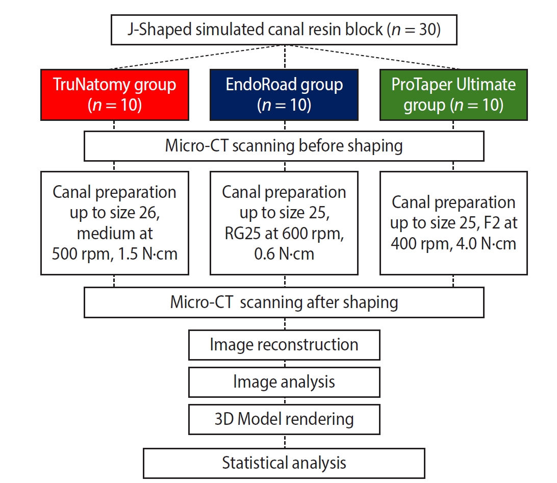
- Objectives
This study aimed to assess geometric characteristics in canal preparation using nickel-titanium (NiTi) files used for minimal invasiveness.
Methods
Thirty J-shaped simulated canals in resin blocks were instrumented with either TruNatomy (TR; Dentsply Sirona), EndoRoad (ER; Maruchi), or ProTaper Ultimate (PTU; Dentsply Sirona). The simulated canal blocks were scanned using microcomputed tomography before and after instrumentation. The scanned images were reconstructed, and the canal surface area was measured from 0.5 to 6.5 mm from the apex. Three-dimensional representative models of each group were rendered. The data were statistically analyzed using one-way analysis of variance and Kruskal-Wallis test at 95% significance level.
Results
TR showed a superior ability to maintain the canal’s center. TR demonstrated comparable apical preparation to PTU. ER showed a smaller and limited apical preparation than other systems, with a tendency for canal preparation toward the inner side of the curvature. PTU featured the largest prepared apical size among the file groups and tended to straighten the curvature by preparing the canal more towards the outward side. The surface area instrumented using each NiTi file showed statistically significant differences among the three groups at all levels except 0.5, 2.0, and 3.5 mm from the apex (p < 0.05). There was no statistically significant difference between TR and PTU at a level of 0.5 mm from the apex (p > 0.05).
Conclusions
While PTU is suitable for general canal preparation to facilitate irrigation and intracanal medication, TR and ER excel in preserving canal centering with minimal concern for canal transportation by minimally invasive preparation.
- 700 View
- 36 Download
- Determination of optimal horizontal beam angulations for canal separation in mandibular molars using cone-beam computed tomography: a retrospective image-based analysis
- Benedikt Schneider, Tamina Tepe, Daniel Rapp, Wilhelm Frank, Maria Lessani, Constantin von See, Sebastian Fitzek, Jörg Philipp Tchorz
- Restor Dent Endod 2026;51(1):e9. Published online February 26, 2026
- DOI: https://doi.org/10.5395/rde.2026.51.e9
-
Abstract
PDFPubReaderePub
- Objectives
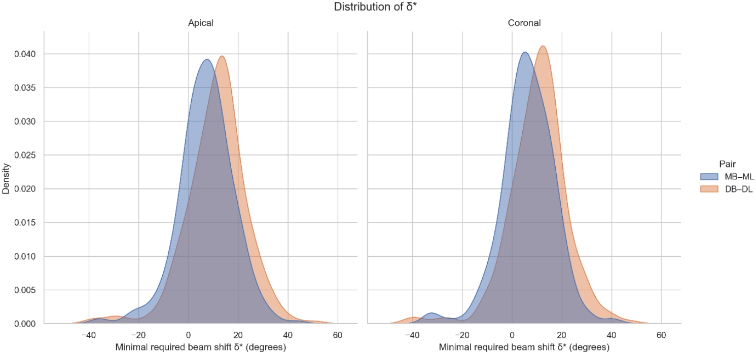
Two-dimensional intraoral radiographs often obscure canals due to superimposition, especially in mandibular molars with complex anatomy. This cone-beam computed tomography (CBCT) study identified the horizontal beam angles at which first and second molar canals overlap and derived clinically applicable angulations for enhanced canal separation.
Methods
Eighty-five CBCT datasets from 100 patients met the inclusion criteria, yielding 318 mandibular molars (160 first, 158 second). Using ImageJ, absolute horizontal overlap angles (α) were measured to determine the corresponding theoretical separation angles defined as δ* = 90° – α. Separability was modeled across horizontal beam angulation increments from −45° to +45° in five steps, and Wilson’s 95% confidence intervals were computed. Group comparisons used the Mann-Whitney U and independent t-tests (p ≤ 0.05)
Results
Minimal mesial beam angulations for effective canal separability (δ* = 90° − α) ranged from approximately 7° to 15° for mesial roots and approximately 10° to 13° for distal roots. No significant mesial differences were observed between first and second molars (p > 0.30). Distal roots of second molars exhibited significantly higher angulations (p = 0.003 coronal, p < 0.001 apical). Mesial canals achieved ≥95% separability at approximately 25° and ≥99% at approximately 35°; distal canals required approximately 30° and approximately 40°.
Conclusions
A mesial beam angulation of 30° to 35° provides probable canal differentiation in mandibular molars, separating mesial canals in ≥99% and distal canals in ≥95% of cases. This range refines previous recommendations and supports the as low as reasonably achievable (ALARA) principle.
- 1,411 View
- 36 Download
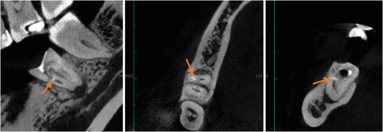
- Cone-beam computed tomography analysis of maxillary premolar canal anatomy: Ahmed’s versus Vertucci’s classifications in a Jordanian cohort
- Raidan Ba-Hattab, Muna M. Shaweesh, Nessrin A. Taha, Elham S. Abu Alhaija
- Restor Dent Endod 2026;51(1):e11. Published online February 26, 2026
- DOI: https://doi.org/10.5395/rde.2026.51.e11
-
Abstract
PDFPubReaderePub
- Objectives
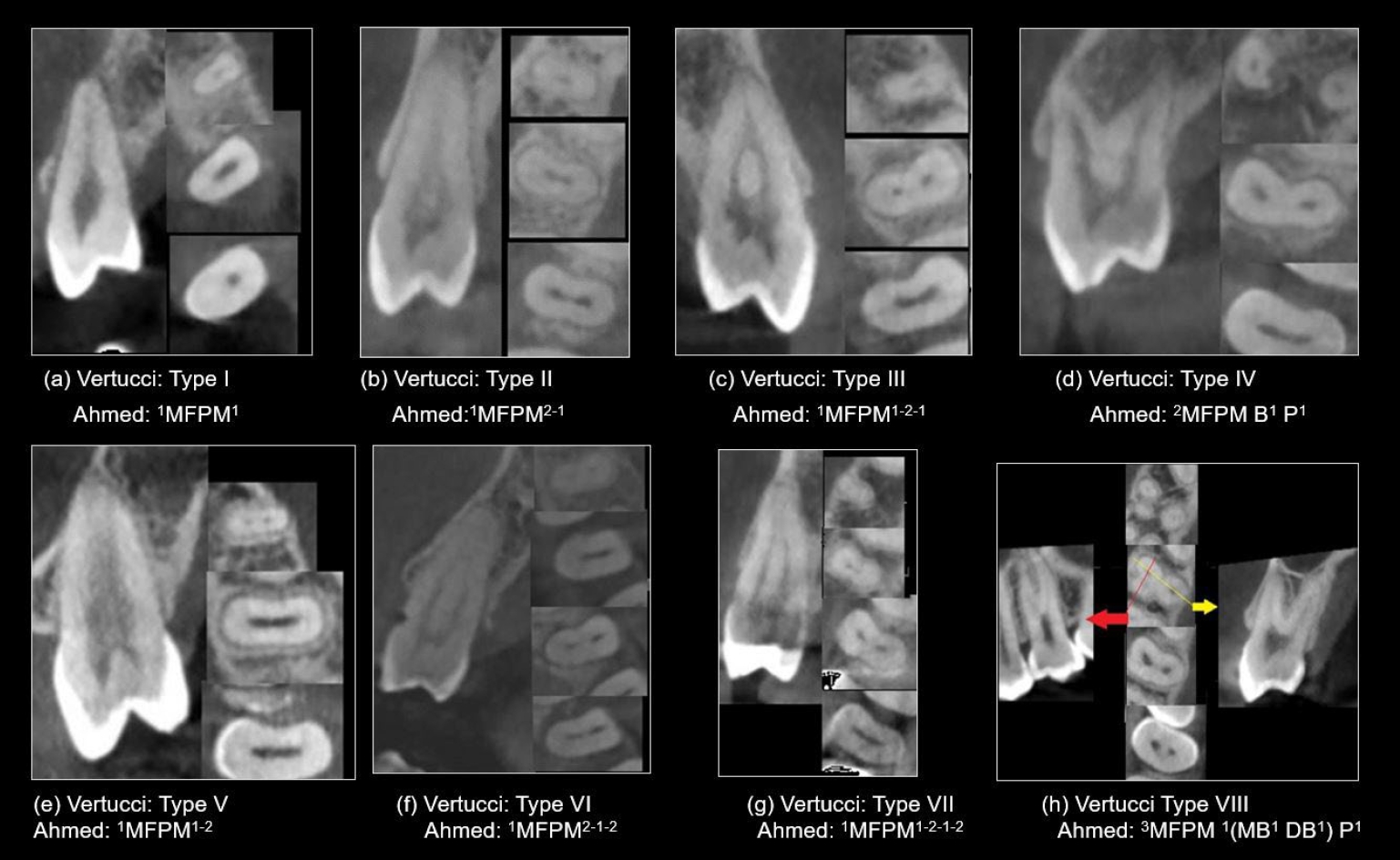
This study analyzed the root and canal configurations of maxillary premolars in a Jordanian subpopulation using cone-beam computed tomography (CBCT) and classified them based on Vertucci’s and Ahmed’s systems.
Methods
Two hundred CBCT scans of 800 maxillary premolars were retrospectively assessed for root morphology, canal configurations, and root canal divergence and merging. Data was statistically analyzed.
Results
The study included 70 males and 130 females. Most right and left maxillary first premolars (RFPM, LFPM) had two roots (59.0% and 58.5%), with a significant association between sex and root number for RFPM and LFPM (p < 0.05). In contrast, the right and left maxillary second premolars (RSPM, LSPM) mostly had a single root (87.5% and 88.5%), with no association with sex. Vertucci’s classification showed type IV as the predominant configuration in first premolars (RFPM, 65.0% and LFPM, 67.0%) and type I in second premolars (RSPM, 44.0% and LSPM, 49.0%). A significant sex association was found only with RSPM. Ahmed’s classification revealed that maxillary premolar with two separated roots and two separated canals (2MP B1 P1) was mostly found in first premolars (RFPM, 58.0% and LFPM, 56.0%), and maxillary premolar with one root and one canal (1MP1) in second premolars (RSPM, 44.0% and LSPM, 49.0%), with a significant sex association for RSPM and LSPM (p < 0.05). Age had no impact, and symmetry was observed between the right and left sides. Three-rooted premolars were identified in four cases. Almost all of Vertucci’s types and numerous codes from Ahmed’s classification were documented.
Conclusions
CBCT revealed diverse anatomical variations in the Jordanian subpopulation, with Ahmed’s classification providing more detailed canal configurations than Vertucci’s, uncovering previously overlooked variations.
- 1,074 View
- 56 Download
- Analysis of the reciprocating kinematics of the VDW Silver Reciproc, E-Connect Pro, Ecom, and Endopen endodontic motors: an in vitro experimental study
- Cristielly França, Juliana D. Bronzato, Dieimes Braambati, Adriana de-Jesus-Soares, Carla C. R. B. Félix, Michelle A. N. S. Ferreira, Marcos Frozoni
- Restor Dent Endod 2026;51(1):e5. Published online January 20, 2026
- DOI: https://doi.org/10.5395/rde.2026.51.e5
-
Abstract
PDFPubReaderePub
- Objectives
This study aimed to evaluate the actual parameters of four endodontic motors, each adjusted for reciprocating motion, and compare them to the manufacturers’ declared values.
Methods
The motors used were the VDW Silver Reciproc (VDW GmbH), E-Connect Pro (MK Life), Ecom (Woodpecker), and Endopen (Schuster Woodpecker). A custom optical target was attached to the motor contra-angle, the movements were recorded with a high-resolution camera, and the images were analyzed. Engagement, disengagement, net angles, and speed for each operation cycle, duration of clockwise (CW) and counter-clockwise (CCW) movement, duration of standstill after CW and CCW movement, and the number of cycles to complete a full rotation were analyzed. The data were statistically analyzed at a significance level of 5%. The replicability of all reciprocal parameters analyzed was statistically different from that reported by the manufacturers.
Results
There was no statistically significant difference between the VDW Silver Reciproc, Ecom, and Endopen for the engagement angle. The E-Connect Pro was the least reliable at the 150°/30° settings for both angle parameters. There was no significant difference between the set and actual cycle net angles for the VDW Silver Reciproc (p = 0.493). While the actual values for the Ecom and E-Connect Pro were significantly higher than the set (p < 0.001), the actual values for the Endopen were significantly lower than the set (p < 0.001).
Conclusions
Experiments on four commercially available reciprocating endodontic motors revealed that the actual motor values differed significantly from the set values.
- 2,212 View
- 90 Download
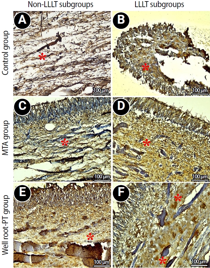
- Effect of combined application of premixed bioceramic paste and diode laser in vital pulp therapy: an immunohistochemical randomized controlled split-mouth in vivo animal experiment
- Mo’men A. Salama, Dalia M. Fayyad, Mohamed I. Rabie, Manar A. A. Selim, Mahmoud F. Ahmed
- Restor Dent Endod 2026;51(1):e4. Published online January 20, 2026
- DOI: https://doi.org/10.5395/rde.2026.51.e4
-
Abstract
PDFPubReaderePub
- Objectives
This study aimed to evaluate the effect of premixed bioceramic paste (Well-Root PT; Vericom) compared to mineral trioxide aggregate (MTA) on the expression of the mineralization-related marker dentin sialoprotein (DSP) in dental pulp following direct pulp capping, with or without prior diode laser application.
Methods
Direct pulp exposures were performed in the upper and lower incisors of eight dogs (n = 96 teeth). Cavities (Class V) were created and received pulp capping with either Well-Root PT (n = 32), MTA (n = 32), or no capping material (polytetrafluoroethylene disc only) (n = 32), with or without the application of a diode laser. Immunohistochemical analysis of DSP expression was conducted and quantified as the mean area percentage using ImageJ software at 2 and 8 weeks posttreatment.
Results
Both the Well-Root PT and MTA groups showed significantly increased DSP expression compared to the control group at both 2 and 8 weeks (p < 0.05). No significant difference in the mean area percentage of DSP expression was found between the Well-Root PT and MTA groups. The diode laser application did not produce a significant effect on DSP expression. Within-group comparison revealed a significant increase in DSP expression between the 2- and 8-week follow-up periods (p < 0.05).
Conclusions
Well-Root PT demonstrated comparable efficacy to MTA in promoting DSP expression, supporting its use as an effective direct pulp capping material. Diode laser application prior to capping had no effect on DSP expression in this experimental model.
- 2,085 View
- 143 Download
- Marginal adaptation of three root-end filling materials in cavities prepared with laser and ultrasonic tips: an in vitro comparative study
- Busra Zengin, Seda Aydemir, Nicholas Paul Chandler
- Restor Dent Endod 2025;50(4):e32. Published online September 9, 2025
- DOI: https://doi.org/10.5395/rde.2025.50.e32
-
Abstract
PDFPubReaderePub
- Objectives
This study evaluated the marginal adaptation of ProRoot MTA (Dentsply Tulsa Dental), Biodentine (Septodont), and TotalFill BC RRM (FKG) placed in root-end cavities prepared with ultrasonic or Er,Cr:YSGG laser tips, using scanning electron microscopy.
Methods
The canals of 90 extracted maxillary central incisors were prepared and obturated and their roots resected. Six groups of 15 specimens were allocated as follows: ultrasonic + ProRoot MTA, ultrasonic + Biodentine, ultrasonic + TotalFill, laser + ProRoot MTA, laser + Biodentine, and laser + TotalFill. Roots were sectioned longitudinally to expose the filling material. Apical and coronal micrographs were taken, and the greatest distance between dentin and filling material was measured. The total gap area was also calculated using further micrographs.
Results
Cavities prepared with the ultrasonic tips and filled with Biodentine showed significantly greater gap dimensions compared with TotalFill (p < 0.001) and ProRoot MTA (p = 0.007) in the apical region. The ultrasonic group showed significantly higher void values compared to the laser group for ProRoot MTA (p = 0.026), when comparing the total values of void. The Biodentine group was significantly higher than the TotalFill group in root-end cavities prepared with ultrasonic tips (p < 0.001). The Biodentine group was significantly higher than the ProRoot MTA group in root-end cavities prepared with the laser tip (p = 0.002).
Conclusions
Under the conditions of this study, it was determined that the root-end cavity preparation technique had an effect on the amount of gaps formed between the dentin and the three filling materials. -
Citations
Citations to this article as recorded by
- Marginal Adaptability of Harvard MTA and Biodentine Used as Root-End Filling Material: A Comparative SEM Study
Yaneta Kouzmanova, Ivanka Dimitrova
Materials.2025; 18(19): 4598. CrossRef
- Marginal Adaptability of Harvard MTA and Biodentine Used as Root-End Filling Material: A Comparative SEM Study
- 4,156 View
- 299 Download
- 1 Web of Science
- 1 Crossref
Case Report
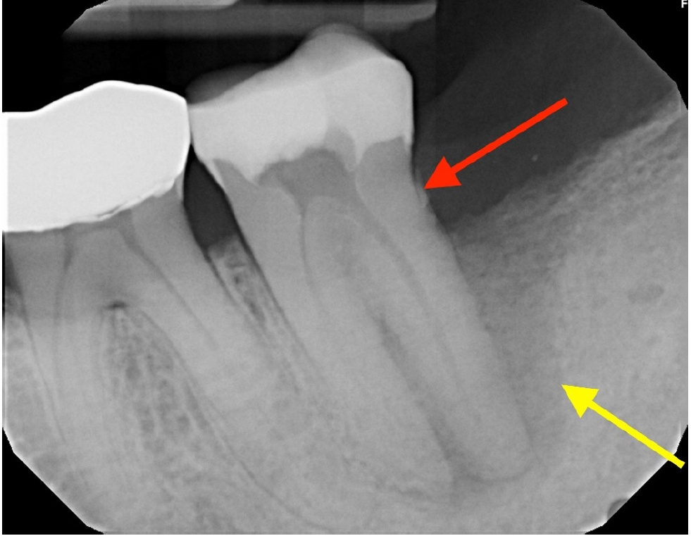
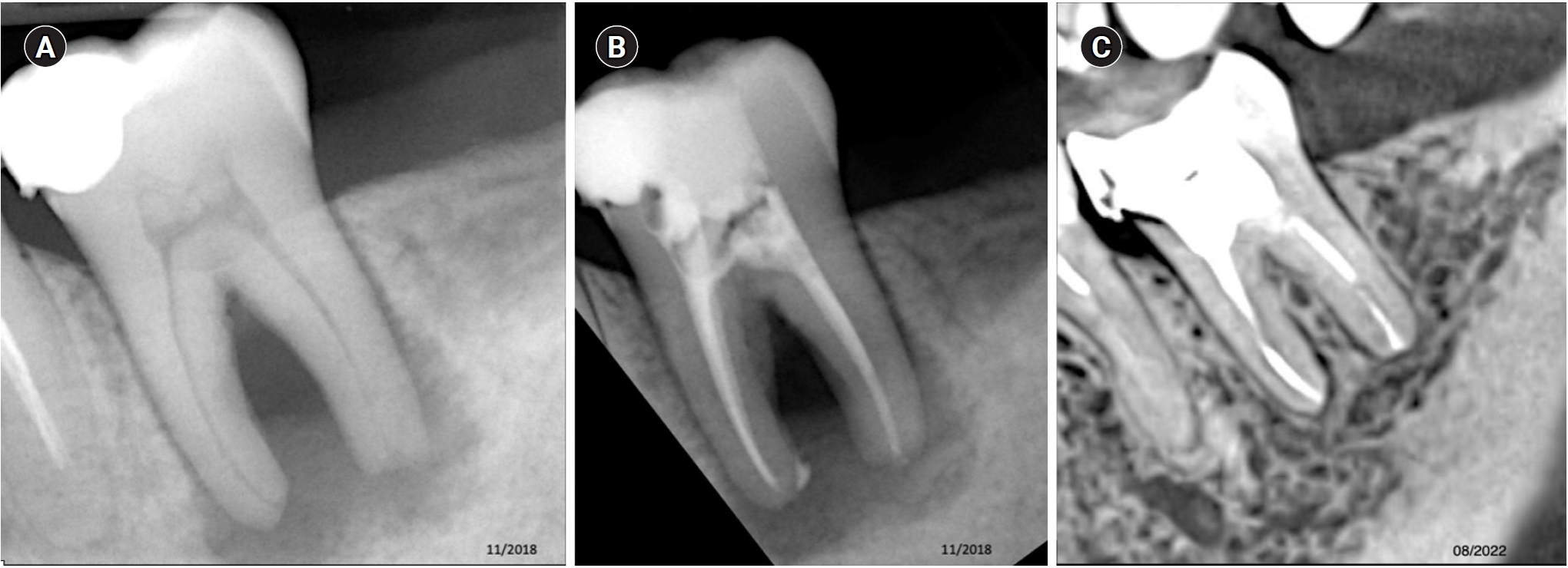
- Multidisciplinary management of an endo-perio lesion complicated by a cemental tear: a case report
- Nishanth D. Sadhak, Akshaya Pallod, Shreyas Oza
- Restor Dent Endod 2025;50(3):e31. Published online August 22, 2025
- DOI: https://doi.org/10.5395/rde.2025.50.e31
-
Abstract
PDFPubReaderePub
- Endodontic-periodontal lesions (EPLs) complicated by cemental tears present a diagnostic and therapeutic challenge. This case report describes the successful management of a 66-year-old male patient with a mandibular second molar (#18) exhibiting an EPL complicated by a cemental tear. Clinical examination revealed a draining sinus tract, deep periodontal pockets, and radiographic evidence of a “J-shaped” lesion and a radiopaque cemental fragment. The tooth had previously initiated endodontic treatment. A multidisciplinary approach involving endodontic treatment and surgical removal of the cemental tear was implemented. At 24-month follow-up, clinical and radiographic examination revealed significant improvement in periodontal health, bone regeneration, and resolution of the lesion. This case highlights the importance of considering cemental tears in the differential diagnosis of EPLs and demonstrates the efficacy of a combined endodontic-periodontal approach for achieving predictable outcomes.
- 4,673 View
- 349 Download
Research Articles
- How protocol, posts, and experience affect fracture detection in multi-rooted teeth using cone-beam computed tomography: an ex vivo experimental study
- Gleica Dal’ Ongaro Savegnago, Gabriela Marzullo de Abreu, Carolina Baumgratz Spiger, Lucas Machado Maracci, Wislem Miranda de Mello, Gabriela Salatino Liedke
- Restor Dent Endod 2025;50(3):e23. Published online July 24, 2025
- DOI: https://doi.org/10.5395/rde.2025.50.e23
-
Abstract
PDFPubReaderePub
- Objectives
This study aimed to evaluate the influence of cone-beam computed tomography (CBCT) acquisition protocol, the presence of intraradicular metal post, and examiner experience on the detection of complete root fractures in multi-rooted teeth.
Methods
Twenty human molar teeth filled with gutta-percha were placed into artificial alveoli created in bovine ribs. The sample was divided into two groups based on the presence or absence of intraradicular posts in the distal roots. CBCT scans were obtained using four acquisition protocols with varying voxel sizes (0.28, 0.2, 0.125, and 0.80 mm). Following the creation of controlled fractures using a chisel and hammer, CBCT imaging was repeated, resulting in 160 images. Five examiners assessed the images using OnDemand software (KaVo Dental GmbH). Sensitivity, specificity, and accuracy were calculated for each examiner, CBCT protocol, and post-condition. Statistical comparisons were performed using Cochran’s Q test and McNemar test, and a significance level of 5%.
Results
In teeth without metallic posts, sensitivity, specificity, and accuracy values exceeded 0.70, 0.70, and 0.80, respectively. However, the presence of metallic posts significantly reduced diagnostic performance, particularly in low-resolution protocols evaluated by less-experienced examiners.
Conclusions
CBCT acquisition protocols should be selected based on the presence of metallic posts to optimize root fracture detection in multi-rooted teeth. Examiner experience also plays a critical role in diagnostic accuracy.
- 3,205 View
- 110 Download
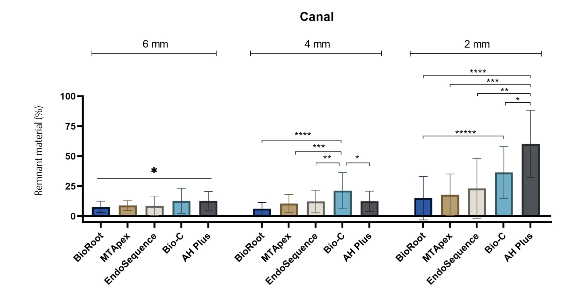
- Calcium silicate-based sealers remnants in isthmuses of mesial roots of mandibular molars: an in vitro evaluation
- David Saldanha de Brito Alencar, Ana Cristina Padilha Janini, Lauter Eston Pelepenko, Brenda Fornazaro Moraes, Francisco Haiter Neto, Marco Antonio Hungaro Duarte, Marina Angélica Marciano
- Restor Dent Endod 2025;50(3):e25. Published online July 15, 2025
- DOI: https://doi.org/10.5395/rde.2025.50.e25
-
Abstract
PDFPubReaderePub
- Objectives
Endodontic retreatment aims to address treatment failure through the removal of root canal filling materials. This in vitro study evaluated the presence of filling material remnants in the mesial root canals, specifically focusing on the isthmuses, of mandibular molars after retreatment.
Methods
One hundred extracted mandibular molar mesial roots with isthmuses were prepared with an R25 file, obturated with one of five calcium silicate-based sealers (BioRoot RCS [Septodont], MTApex [Ultradent Products Inc.], EndoSequence BC Sealer HiFlow [Brasseler USA], Bio-C Sealer [Angelus]) or an epoxy resin-based sealer (AH Plus Jet [Dentsply Maillefer]), all stained with rhodamine B, and stored at 37ºC for 30 days to allow for setting. Retreatment was subsequently performed using R40 and XP-endo Finisher R instruments (FKG Dentaire) with 2.5% sodium hypochlorite irrigation. The presence of remaining filling material was then assessed using confocal microscopy, and setting times were tested per ISO 6876:2012.
Results
AH Plus Jet showed the most remnants at 2 mm and the longest retreatment time. Calcium silicate-based sealers exhibited prolonged setting times under dry conditions, with EndoSequence BC Sealer HiFlow showing a particularly extended setting period.
Conclusions
Despite retreatment, residues remained in all canals and isthmus regions, particularly Bio-C Sealer and AH Plus Jet in apical areas, emphasizing the difficulty of complete removal and the persistence of filling material. -
Citations
Citations to this article as recorded by- Bonding effects of mechanical removal of bioceramic sealer residues using glycine or glass microparticles abrasion
Jesus Aranda, Julia de Freitas Ceccato, Eduardo Fernández Godoy, João Felipe Besegato, Joissi Ferrari Zaniboni, Regina Guenka Palma-Dibb, Milton Carlos Kuga
International Journal of Adhesion and Adhesives.2026; 148: 104289. CrossRef
- Bonding effects of mechanical removal of bioceramic sealer residues using glycine or glass microparticles abrasion
- 2,857 View
- 130 Download
- 1 Web of Science
- 1 Crossref
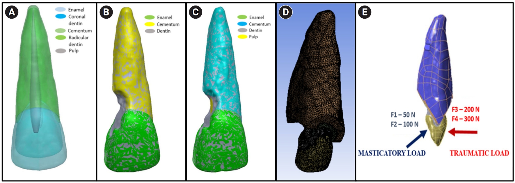
- Stress distribution of restorations in external cervical root resorption under occlusal and traumatic loads: a finite element analysis
- Padmapriya Ramanujam, Paul Kevin Abishek Karthikeyan, Vignesh Srinivasan, Selvakarthikeyan Ulaganathan, Velmurugan Natanasabapathy, Nandini Suresh
- Restor Dent Endod 2025;50(2):e21. Published online May 21, 2025
- DOI: https://doi.org/10.5395/rde.2025.50.e21
-
Abstract
PDFPubReaderePub
- Objectives
This study analyzed the stress distribution in a maxillary central incisor with external cervical resorptive defect restored with different restorative materials under normal masticatory and traumatic loading conditions using finite element analysis.
Methods
Cone-beam computed tomography of an extracted intact incisor and created resorptive models (Patel’s 3D classification-2Bd and 2Bp) in the maxillary central incisor was performed for finite element models. The 2Bd models were restored either with glass ionomer cement (GIC)/Biodentine (Septodont) or a combination of both with composite resin. 2Bp models were restored externally with a combination technique and internally with root canal treatment. The other model was external restoration with GIC and internal with fiber post. Two masticatory loads were applied at 45˚ to the palatal aspect, and two traumatic loads were applied at 90˚ to the buccal aspect. Maximum von Mises stresses were calculated, and stress distribution patterns were studied.
Results
In 2Bd models, all restorative strategies decreased stress considerably, similar to the control model under all loads. In 2Bp models, the dentin component showed maximum stress at the deepest portion of the resorptive defect, which transfers into the adjacent pulp space. In 2Bp defects, a multilayered restoration externally and root canal treatment internally provides better stress distribution compared to the placement of a fiber post.
Conclusions
Increase in load, proportionally increased von Mises stress, despite the direction or angulation of the load. Multilayered restoration is preferred for 2Bd defects, and using an internal approach of root canal treatment is suggested to restore 2Bp defects.
- 2,941 View
- 167 Download
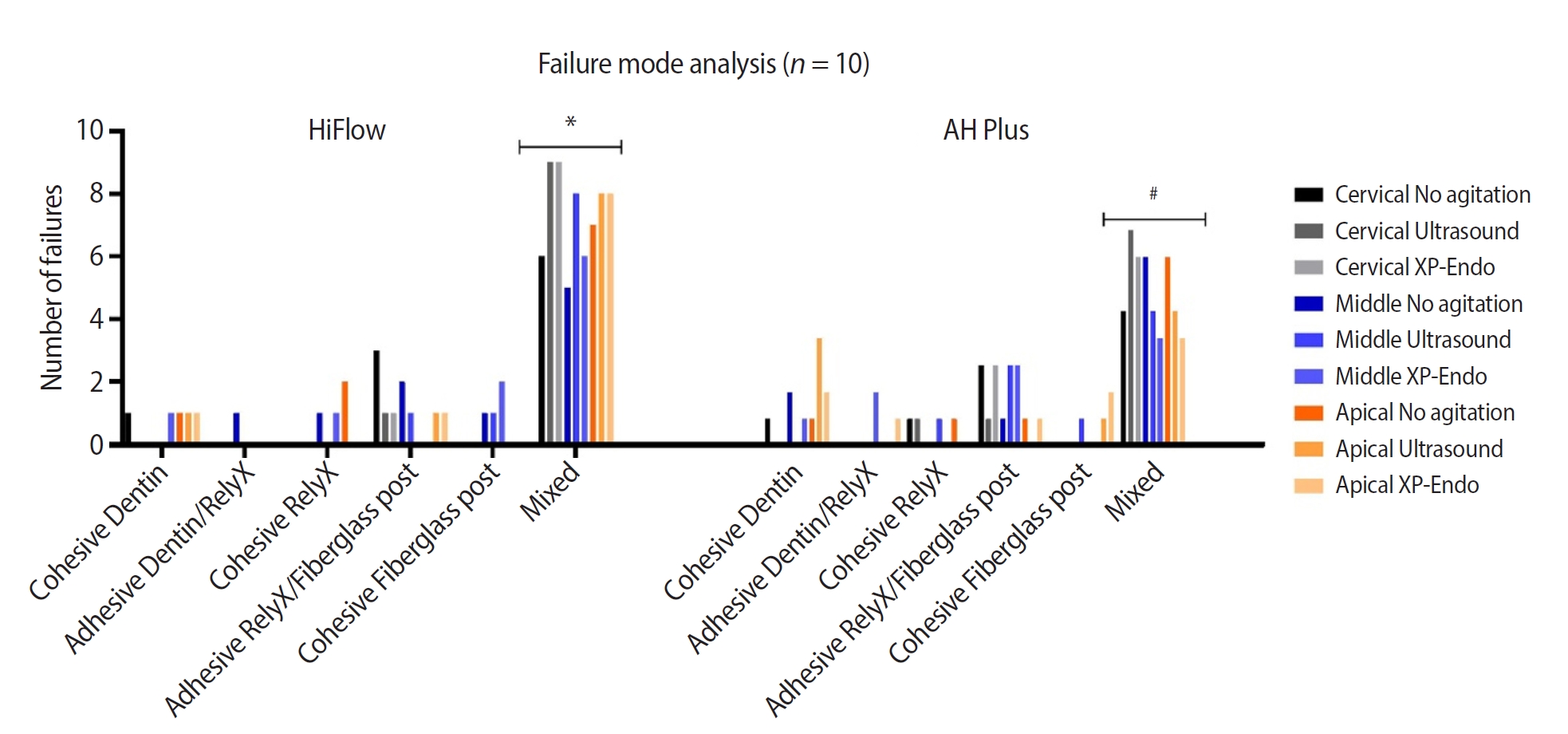
- Cleaning protocols to enhance bond strength of fiberglass posts on root canals filled with bioceramic sealer: an in vitro comparative study
- Thiago Bessa Marconato Antunes, Juliana Delatorre Bronzato, Joice Graciani, Ana Cristina Padilha Janini, Rocharles Cavalcante Fontenele, Francisco Haiter Neto, Brenda Paula Figueiredo de Almeida Gomes, Marina Angélica Marciano da Silva
- Restor Dent Endod 2025;50(2):e20. Published online May 21, 2025
- DOI: https://doi.org/10.5395/rde.2025.50.e20
-
Abstract
PDFPubReaderePub
- Objectives
This study aimed to evaluate whether the agitation protocols using ultrasonic inserts or the XP-endo Finisher R file improved the removal of two different endodontic sealer remnants and the bond strength of fiberglass posts to dentin.
Methods
Seventy-two human teeth were selected. The canals were prepared with Reciproc 50 and Easy ProDesign 30/.10 and root filled according to the endodontic sealer groups: AH Plus or EndoSequence BC Sealer HiFlow. The samples were kept at 37ºC and 95% humidity for 28 days. During the post space preparation, the obturation was removed with Largo burs, and the groups were divided according to the irrigant agitation protocols (n = 12): no agitation, agitation with R1-Clearsonic associated with E1-Irrisonic ultrasonic inserts, or agitation with XP-endo Finisher R file. The fiberglass posts were cemented with RelyX ARC. The roots were sectioned into slices and submitted to the push-out test. Micro-computed tomography analysis was used to check the effectiveness of irrigating solution agitation in the elimination of remnants.
Results
The cleaning protocols with agitation were more effective in increasing the bond strength of posts to dentin for both sealer groups compared to non-agitation (p < 0.05). There was no difference between the same cleaning protocols for the different sealers. Among the different thirds, there was no statistical difference for the same sealer in the different cleaning protocols (p > 0.05).
Conclusions
Both agitation protocols effectively clean root-filled canals sealed with resin-based and calcium silicate-based sealers during fiberglass post space preparation. These protocols result in improved bond strength compared to non-agitation methods. -
Citations
Citations to this article as recorded by- Cleaning efficacy and bond interaction of glycine-based air polishing and glass microparticles abrasion on dentin impregnated with premixed bioceramic sealer
Ândresson Aurélio Fernandes Martins, Maria Carolina Sidonio Alves, Bruno Martins Maciel, José Rodolfo Estruc Verbicário, João Felipe Besegato, Wilfredo Gustavo Escalante-Otárola, Milton Carlos Kuga
International Journal of Adhesion and Adhesives.2026; 147: 104277. CrossRef - Effect of Endodontic Sealers on the Bond Strength of Glass Fibre Posts: A Systematic Review
Thiago Bessa Marconato Antunes, Juliana D. Bronzato, Vanessa Gallego Arias Pecorari, Jennifer Santos Pereira, Talita Tartari, Adriana de Jesus Soares, Brenda P. F. A. Gomes, Marina Angélica Marciano
Australian Endodontic Journal.2026;[Epub] CrossRef - Effects of Calcium Silicate-Based Sealer Residues on Adhesive Bonding to Coronal Dentin: An in Vitro Study
Mariana Bena Gelio, Thais Piragine Leandrin, Ana Lídia Pinheiro Silva Sato, Milton Carlos Kuga, Joissi Ferrari Zaniboni
Journal of Dental Research, Dental Clinics, Dental Prospects.2026; 20(1): 25. CrossRef
- Cleaning efficacy and bond interaction of glycine-based air polishing and glass microparticles abrasion on dentin impregnated with premixed bioceramic sealer
- 5,368 View
- 249 Download
- 2 Web of Science
- 3 Crossref
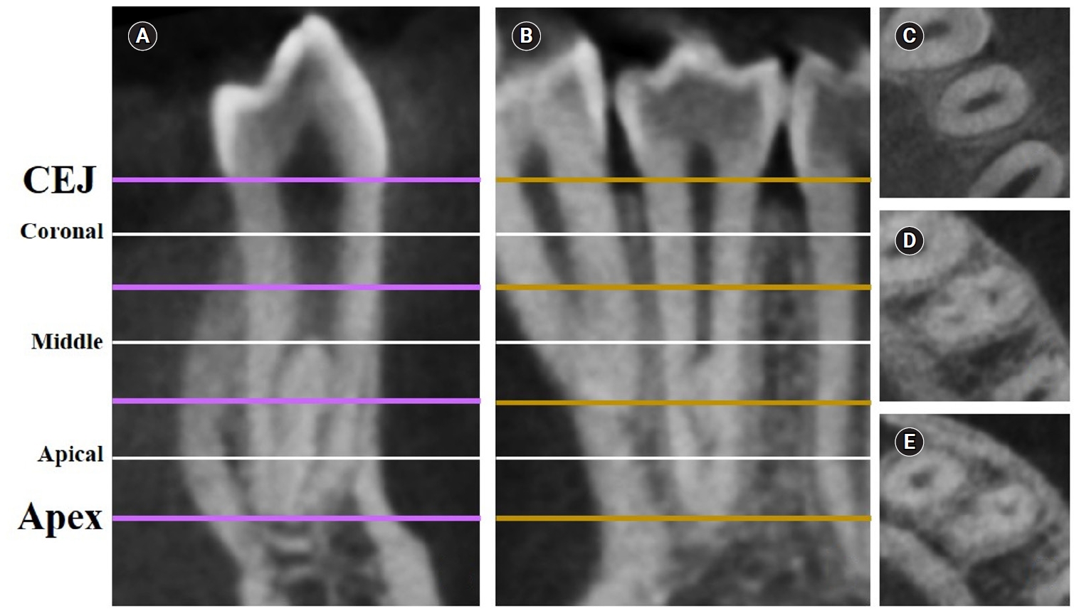
- Dentin thickness of C-shaped root canal walls in mandibular premolars based on cone-beam computed tomography: a retrospective cross-sectional study
- Elif Aslan, Ali Canberk Ulusoy, Bilge Hakan Sen, B. Guniz Baksi, Erinc Onem, Ali Mert
- Restor Dent Endod 2025;50(2):e18. Published online May 15, 2025
- DOI: https://doi.org/10.5395/rde.2025.50.e18
-
Abstract
PDFPubReaderePub
- Objectives
This study aimed to measure the dentin thickness of C-shaped canals in mandibular first and second premolars at coronal, middle, and apical root levels using cone-beam computed tomography (CBCT).
Methods
Dentin thicknesses of buccal, lingual, mesial, and distal root walls of 41 C-shaped premolars were measured at three different root levels on axial CBCT slices. The measurements were made at the midpoint of each third, along with 1 mm below and above the midpoint. C-shape configurations of the premolar root canals were also recorded. Analysis of variance, Kruskal-Wallis, and the independent samples t-tests were used for the comparisons (p = 0.05).
Results
The thickest walls for both premolars were buccal and lingual walls at all three root levels (p < 0.05). The thinnest walls for the first premolar teeth were mesial and distal walls of the lingual canal, while it was the mesial end of the buccal and lingual canals for the second premolars (p < 0.05). Dentin wall thicknesses at the mesial end of buccal and lingual canals of C1-shaped first premolars were thinner than C2-shaped first premolars at the apical level (p < 0.05).
Conclusions
Danger zones for C-shaped mandibular first and second premolars are predominantly mesial walls facing the radicular groove and distal wall of the lingual canal. CBCT imaging during endodontic treatment is recommended to avoid complications. -
Citations
Citations to this article as recorded by- Anatomical complexity in mandibular second molars: prevalence of C-shaped canals, radicular grooves, taurodontism, and radices molarum in Saudi population
Ahmed A. Madfa, Abdullah F. Alshammari, Eyad Almagadawyi, Ebtsam A. Aledaili, Afaf Al-Haddad
Scientific Reports.2025;[Epub] CrossRef
- Anatomical complexity in mandibular second molars: prevalence of C-shaped canals, radicular grooves, taurodontism, and radices molarum in Saudi population
- 4,922 View
- 157 Download
- 1 Web of Science
- 1 Crossref
- Impact of the use of high-power 810-nm diode laser as monotherapy on the clinical and tomographic success of the treatment of teeth with periapical lesions: an observational clinical study
- Fabricio Hinojosa Pedraza, Abel Victor Isidro Teves-Cordova, Murilo Priori Alcalde, Marco Antonio Hungaro Duarte
- Restor Dent Endod 2025;50(2):e15. Published online May 15, 2025
- DOI: https://doi.org/10.5395/rde.2025.50.e15
-
Abstract
PDFPubReaderePub
- Objectives
The aim of this study was to demonstrate the impact of a high-power 810-nm diode laser as monotherapy on the clinical and tomographic success of treating teeth with periapical lesions, through a series of 31 cases.
Methods
Teeth with apical lesions underwent endodontic treatment in which a high-power 810-nm diode laser with saline solution was used as monotherapy for disinfection. This type of therapy aimed to replace the traditional irrigation protocol with sodium hypochlorite. This research is the first to assess the clinical success of this alternative treatment, along with tomographic evaluations conducted over periods ranging from 2 to 7 years, analyzed using the periapical index based on cone-beam computed tomography (CBCTPAI). All cases were performed by a single clinician following the same laser protocol, which involved using 1 W of continuous power and four cycles of 20 seconds of laser activation.
Results
All teeth showed no clinical symptoms upon follow-up examination. However, the tomographic evaluation revealed that the success rates for teeth receiving primary treatment were 60% and 80% according to strict and loose criteria, respectively. For teeth requiring retreatment, the success rates were 12.5% and 37.5% using strict and loose criteria, respectively.
Conclusions
The teeth with apical lesions that underwent primary treatment did not present clinical symptoms, but they showed a moderate success rate on tomographic evaluation. However, despite lacking clinical symptoms, teeth with apical lesions that required retreatment had a very low success rate on tomographic evaluation. -
Citations
Citations to this article as recorded by- Adherence to core outcome set for endodontic treatments (COSET) international consensus: Two-year before/after bibliometric systematic review
Carolina Bender Hoppe, Pauline Mastella Lang, Lara Dotto, Mateus Silveira Martins Hartmann, Fabiana Soares Grecca
Journal of Dentistry.2026; 173: 106834. CrossRef - Diode Laser-Guided Protocol for Endo-Perio Lesions: Toward a Multi-Stage Therapeutic Strategy—A Case Series and Brief Literature Review
Ioana-Roxana Munteanu, George-Dumitru Constantin, Ruxandra-Elena Luca, Ioana Veja, Mariana-Ioana Miron
Medicina.2025; 61(12): 2157. CrossRef
- Adherence to core outcome set for endodontic treatments (COSET) international consensus: Two-year before/after bibliometric systematic review
- 5,209 View
- 232 Download
- 2 Web of Science
- 2 Crossref
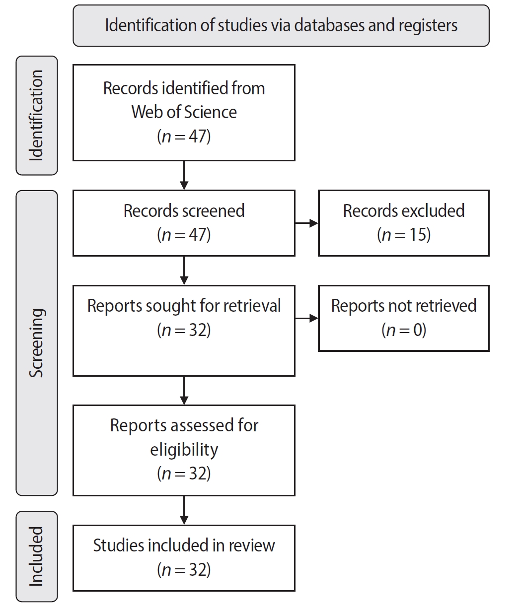
- Bibliometric analysis of the GentleWave system: trends, collaborations, and research gaps
- Raimundo Sales de Oliveira Neto, Thais de Moraes Souza, João Vitor Oliveira de Amorim, Thaine Oliveira Lima, Guilherme Ferreira da Silva, Rodrigo Ricci Vivan, Murilo Priori Alcalde, Marco Antonio Hungaro Duarte
- Restor Dent Endod 2025;50(2):e17. Published online May 12, 2025
- DOI: https://doi.org/10.5395/rde.2025.50.e17
-
Abstract
PDF
 Supplementary MaterialPubReaderePub
 - Objectives
The study aimed to conduct a bibliometric analysis of the GentleWave system (Sonendo, Inc.).
Methods
An electronic search was conducted in June 2024 using the Web of Science Collection database. Two reviewers independently screened publications, extracting data on authorship, publication details, study design, and citation metrics. Statistical analyses were performed in R to assess variable correlations, while the VOSviewer (Visualization of Similarities Viewer) software was used to map author and keyword networks.
Results
The search yielded 47 records, with 32 studies included. Publications spanned 2014 to 2024. The Journal of Endodontics published the highest number of studies (n = 15), and the International Endodontic Journal had the highest impact factor (5.4). The University of British Columbia and Sonendo, Inc. were the most frequent affiliations. Among the 32 articles, 28 were in vitro studies, primarily focusing on microbiology (n = 9). A total of 95 authors were identified, with Haapasalo and Shen being the most cited (n = 229). The articles accumulated 495 citations, demonstrating a strong positive correlation between the number of studies and citation counts (r = 0.98).
Conclusions
The analysis highlights a predominance of in vitro studies. Geographic concentration in the United States and Canada limits diversity, while the strong correlation between study numbers and citations suggests that increased publication volume enhances visibility. -
Citations
Citations to this article as recorded by- Three-year Outcomes of Conventional Versus Minimally Invasive Endodontic Treatment Protocols: A Retrospective Study
Kiavash Hossini, He Liu, Ya Shen, Jolanta Aleksejuniene, Fahda Algahtani, Ahmed Hieawy
Journal of Endodontics.2026; 52(4): 558. CrossRef
- Three-year Outcomes of Conventional Versus Minimally Invasive Endodontic Treatment Protocols: A Retrospective Study
- 4,851 View
- 104 Download
- 1 Web of Science
- 1 Crossref
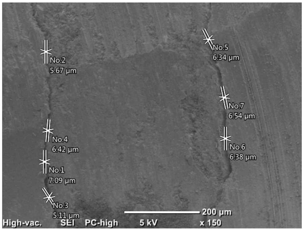
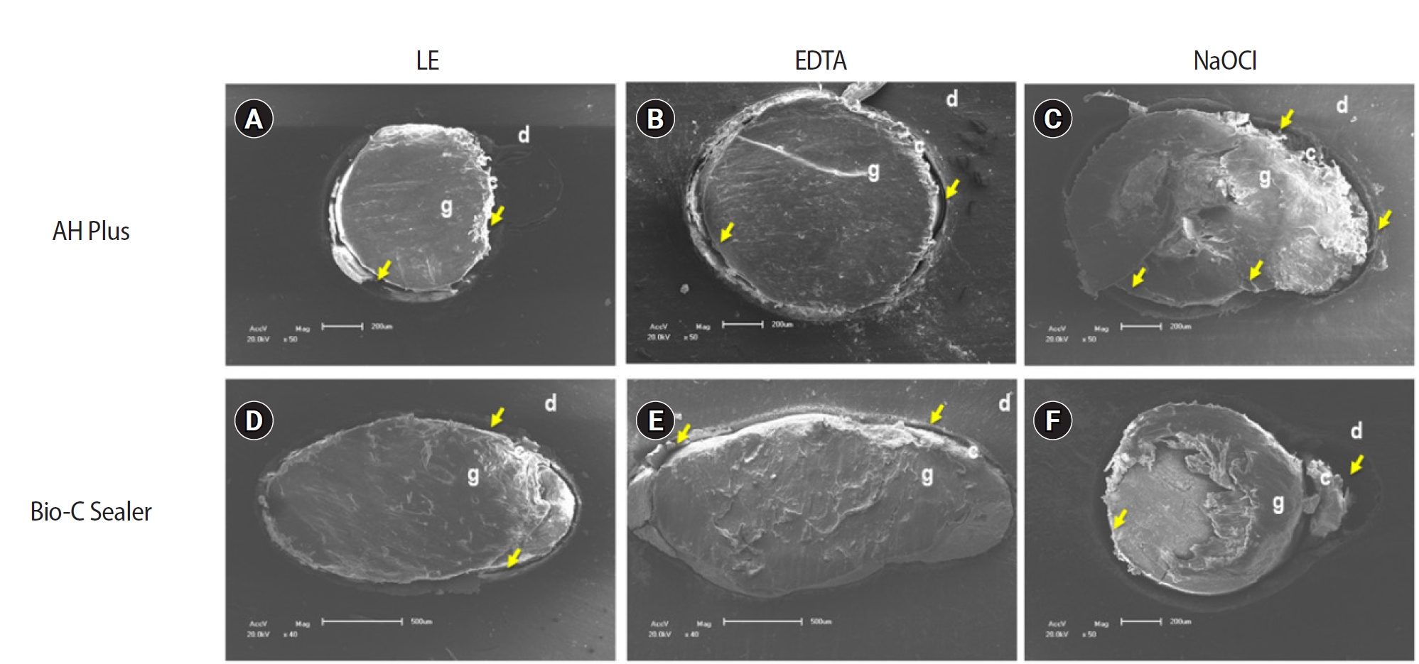
- The effect of limonene extract on the adhesion of different endodontic cements to root dentin: an in vitro experimental study
- Nayara Lima Ferraz Aguiar, Eduardo José Soares, Guilherme Nilson Alves dos Santos, Anna Luísa Araújo Pimenta, Laryssa Karla Romano, Ricardo Gariba Silva, Fernanda de Carvalho Panzeri
- Restor Dent Endod 2025;50(2):e16. Published online May 12, 2025
- DOI: https://doi.org/10.5395/rde.2025.50.e16
-
Abstract
PDFPubReaderePub
- Objectives
The study aimed to evaluate the effect of limonene extract (LE) on push-out bond strength (BS) to root dentin in endodontically treated teeth.
Methods
Single-rooted teeth were selected and instrumented using the reciprocating technique, then divided into three groups based on the final irrigating solution: 2.5% sodium hypochlorite (NaOCl), 17% ethylenediaminetetraacetic acid (EDTA), and 5% LE. The roots were further divided (n = 12) and obturated using the single-cone technique with epoxy resin-based (ERB) or bioceramic sealer (Bio-C). After 3 days, the roots were sectioned into 2-mm slices, obtaining two slices from each root third. Push-out BS testing was conducted at 0.5 mm/min, followed by failure pattern and adhesive interface analysis using scanning electron microscopy. Push-out BS data were analyzed by three-way analysis of variance and Tukey post-hoc test (p < 0.05).
Results
ERB showed higher BS when irrigated with EDTA (5.0 ± 2.3 MPa) compared to NaOCl (1.8 ± 1.1 MPa) (p = 0.0005), particularly in the cervical third. LE yielded intermediate values without significant differences from the other irrigants (3.5 ± 1.9 MPa) (p > 0.05). For Bio-C, the highest BS was observed in the apical third, especially with LE (9.4 ± 5.0 MPa), differing from other thirds and final irrigating solutions (p < 0.05). Mixed failure patterns were most prevalent, regardless of the irrigant solutions.
Conclusions
The combination of LE with Bio-C demonstrated superior BS in the apical third, suggesting its potential as a final irrigating solution in endodontic treatments.
- 3,381 View
- 242 Download
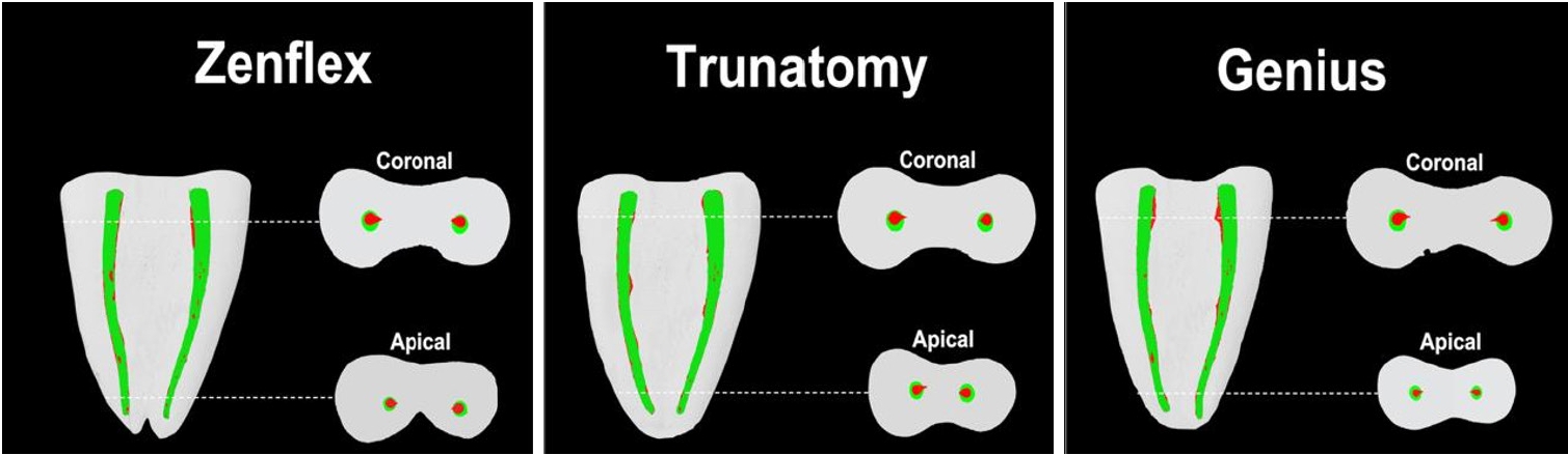
- Shaping ability and cyclic fatigue resistance between Genius ProFlex, ZenFlex, and TruNatomy rotary systems: an experimental study
- Raimundo Sales de Oliveira Neto, Murilo Priori Alcalde, Pedro Cesar Gomes Titato, Pedro Henrique Souza Calefi, Carlos Alberto Spironelli Ramos, Guilherme Ferreira da Silva, Rodrigo Ricci Vivan, Marco Antonio Hungaro Duarte
- Restor Dent Endod 2025;50(1):e9. Published online February 13, 2025
- DOI: https://doi.org/10.5395/rde.2025.50.e9
-
Abstract
PDFPubReaderePub
- Objectives
The aim of this study was to investigate the efficacy of three newly introduced rotary endodontic systems: Genius ProFlex (Medidenta), TruNatomy (Dentsply Maillefer), and ZenFlex (Kerr).
Methods
Forty-five mandibular molars with root canal curvatures <5° were utilized. Micro-computed tomography scans were performed pre- and post-preparation to assess apical transportation, centralization, percentage of dentin wear, and canal volume alterations. Eight instruments of each diameter underwent cyclic fatigue testing.
Results
The percentage of dentin wear on mesial and distal walls showed no significant differences among ZenFlex, TruNatomy, and Genius ProFlex at 1, 2, 3, and 4 mm from the apical foramen and root canal orifice (p > 0.05). Centering ability varied in the mesiolingual canal (p < 0.05). No notable differences were observed in transportation (p > 0.05). Genius ProFlex demonstrated lower volumetric changes (p < 0.05). There were significant differences in cyclic fatigue, with higher values for Genius ProFlex and lower values for TruNatomy (p < 0.05).
Conclusions
The three nickel-titanium rotary instruments are safe and efficient for root canal preparation, with Genius ProFlex exhibiting superior cyclic fatigue resistance. -
Citations
Citations to this article as recorded by- Influence of kinematic motion and instrumentation strategy on apical debris extrusion during root canal preparation: An in vitro study
Amira Alghazaly, Jumanah Aljohani, Khadijah Mohabat, Rafah Ghous
Journal of Conservative Dentistry and Endodontics.2026; 29(7): 741. CrossRef - Comparison of Shaping Ability and Apical Debris Extrusion Using 4 Different Nickel–Titanium Single‐File Systems
Siyu Li, Mengzhen Tang, Xi Wang, Jian Yang, Hyun-Do Jung
International Journal of Biomaterials.2025;[Epub] CrossRef
- Influence of kinematic motion and instrumentation strategy on apical debris extrusion during root canal preparation: An in vitro study
- 4,286 View
- 190 Download
- 1 Web of Science
- 2 Crossref
- Effect of quality of radiographs taken during root canal treatment on technical quality of root canal fillings and endodontic outcome
- Jia Min Ng, Yan Yee Lee, Prashanti Chippagiri, Elaheh Ahanin, Abhishek Parolia
- Restor Dent Endod 2025;50(1):e3. Published online January 7, 2025
- DOI: https://doi.org/10.5395/rde.2025.50.e3
-
Abstract
PDFPubReaderePub
- Objectives
This study evaluated the number and quality of working length (WL) and master cone (MC) radiographs taken during root canal treatment by dental undergraduates, and their associations with the technical quality of root canal fillings (TQRCF) and endodontic outcomes (EO).
Methods
A retrospective evaluation of radiographs from 303 root canal-treated teeth in 231 patients was conducted, with 72 patients attending recall visits to assess EO. The chi-square and one-way analysis of variance tests were performed.
Results
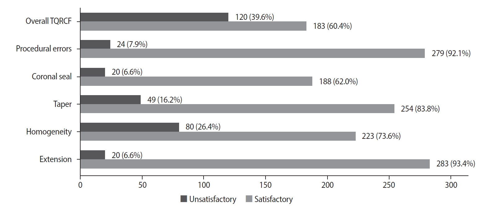
A total of 505 WL and 557 MC radiographs were reviewed, with 72.9% and 75% deemed satisfactory, respectively. Satisfactory TQRCF was achieved in 60.4% of cases. Significant associations were found between the extension of the file in WL and gutta-percha in MC radiographs and TQRCF (p = 0.000). Misinterpretation of these radiographs resulted in poor TQRCF. Furthermore, 64.2% of teeth had satisfactory EO. A significant relationship was noted between the quality of MC radiographs and both TQRCF (p = 0.043) and EO (p = 0.003).
Conclusions
Unsatisfactory MC radiographs were linked to poor TQRCF and unfavorable EO. Regular radiographic training is recommended to enhance EO. -
Citations
Citations to this article as recorded by- Radiographic evaluation of root canal fillings: can undergraduate dental students perform it?
Emine Odabaşı Tezer, Fadi Nahas, Alhabab Shbitah, İrem Dilara Kılıç, Ahmet Bölük, Meltem Öztan
BMC Medical Education.2026;[Epub] CrossRef - Assessment of radiographic errors and repetition rates in undergraduate endodontic education: a retrospective clinical study
Marwa Ameen, Abdul Rahman Saleh, Dunia Alhadi, Manal Almaslamani
The Saudi Dental Journal.2025;[Epub] CrossRef - Application of Periapical Radiography in Root Canal Treatment: A Literature Review
Jennifer Lois Violita Malau, Keizha Allysia Nabila, Widiani Harrista, Regina Amara Ginting, Tassa Kusuma Arya Putri, Jatu Rachel Keshena
Acta Odontologica Indonesia.2025; 1(2): 49. CrossRef
- Radiographic evaluation of root canal fillings: can undergraduate dental students perform it?
- 14,223 View
- 305 Download
- 2 Web of Science
- 3 Crossref
Case Report
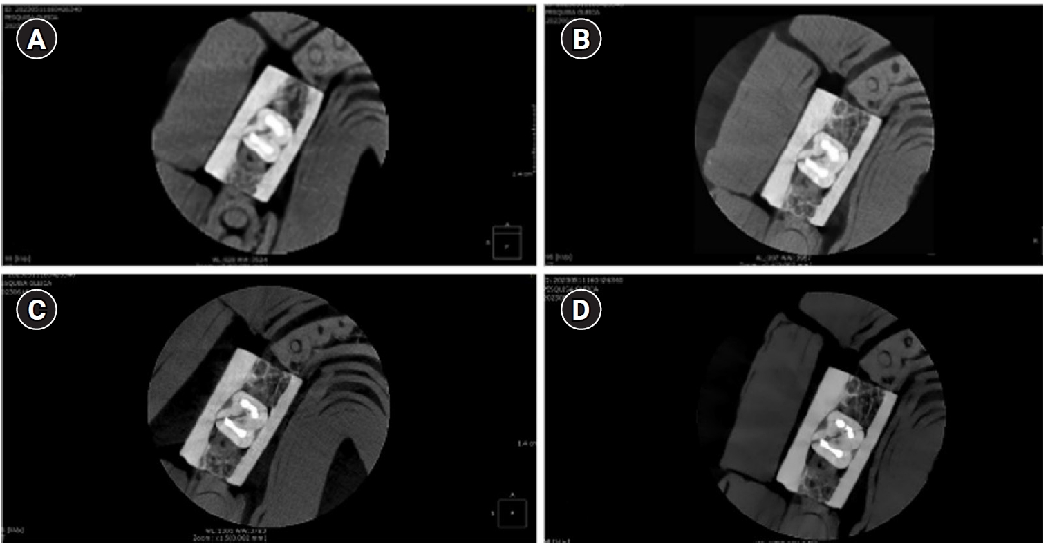
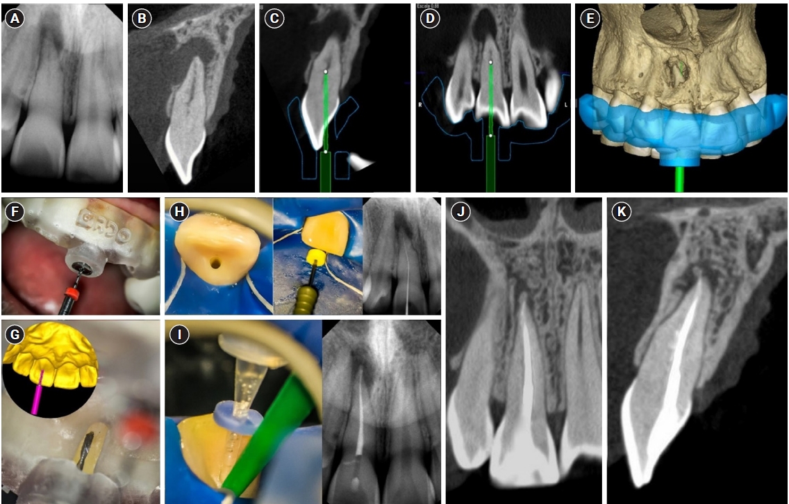
- Guided endodontics, precision and predictability: a case series of mineralized anterior teeth with follow-up cone-beam computed tomography
- Rafael Fernández-Grisales, Wilder Javier Rojas-Gutierrez, Pamela Mejía, Carolina Berruecos-Orozco, Néstor Ríos-Osorio
- Restor Dent Endod 2025;50(1):e4. Published online January 6, 2025
- DOI: https://doi.org/10.5395/rde.2025.50.e4
-
Abstract
PDFPubReaderePub
- Pulp chamber and root canal obliteration (PCO/RCO) presents a challenge for clinicians when nonsurgical endodontic treatment is indicated. Guided endodontics (GE) aims to precisely locate the root canal (RC) system while preserving as much pericervical dentin as possible. GE involves integrating cone-beam computed tomography (CBCT) of the affected tooth with a digital impression of the maxillary/mandibular arch, allowing for careful planning of the drilling path to the RC system through a three-dimensional (3D) static guide. This article reports four cases of teeth with PCO/RCO, accompanied by additional diagnoses of internal and external root resorption and horizontal tooth fracture, all successfully treated with GE. These cases highlight the clinical and radiographic success of GE treatments using CBCT, establishing this technique as a predictable approach for managing mineralized teeth.
-
Citations
Citations to this article as recorded by- Static Guided Endodontics in Primary Endodontic Treatment of Anterior Teeth: A Narrative Review
Monika Kuczmaja, Wiesława Puchalska, Agata Żółtowska
Dentistry Journal.2026; 14(4): 195. CrossRef - Effect of different rotary instrument designs (protaper ultimate and protaper gold) on postoperative pain and bacterial reduction: a randomized clinical trial
Khaled Hassan Abed, Ahmed Abdel Rahman Hashem, Reem Ahmed Lutfy, Somaia Abdellatif Eissa, Dina Ahmed Morsy
BMC Oral Health.2026;[Epub] CrossRef
- Static Guided Endodontics in Primary Endodontic Treatment of Anterior Teeth: A Narrative Review
- 5,372 View
- 384 Download
- 2 Web of Science
- 2 Crossref
Review Article
-
Influence of disinfecting solutions on the surface topography of gutta-percha cones: a systematic review of
in vitro studies - Lora Mishra, Gathani Dash, Naomi Ranjan Singh, Manoj Kumar, Saurav Panda, Franck Diemer, Monika Lukomska-Szymanska, Barbara Lapinska, Abdul Samad Khan
- Restor Dent Endod 2024;49(4):e42. Published online November 1, 2024
- DOI: https://doi.org/10.5395/rde.2024.49.e42
-
Abstract
PDFSupplementary MaterialPubReaderePub
The surface integrity of gutta-percha cones is a crucial factor in the success of endodontic procedures. Disinfecting solutions play a pivotal role in sterilizing gutta-percha cones, but their influence on gutta-percha surface topography remains a subject of concern. This systematic review aimed to present a qualitative synthesis of available laboratory studies assessing the influence of disinfecting solutions on the surface topography of gutta-percha and offers insights into the implications for clinical practice. The present review followed PRISMA 2020 guidelines. An advanced database search was performed in PubMed, Google Scholar, Embase, Scopus, LILAC, non-indexed citations and reference lists of eligible studies in May 2024. Laboratory studies, in English language, were considered for inclusion. The quality (risk of bias) of the included studies was assessed using parameters for
in vitro studies. A total of 28 studies were included in the qualitative synthesis. Based on the included in vitro studies, surface deposits and alterations in the physical properties of gutta-percha cones were observed after the disinfection protocol. A comprehensive review of the available literature indicates that the choice of disinfecting solution, its concentration, and immersion time significantly affect the surface topography of gutta-percha cones.-
Citations
Citations to this article as recorded by- In Vitro Evaluation of Disinfectants on Gutta-Percha Cones: Antimicrobial Efficacy Against Enterococcus faecalis and Candida albicans
Tringa Kelmendi, Donika Bajrami Shabani, Aida Meto, Hani Ounsi
Journal of Clinical Medicine.2025; 14(19): 6846. CrossRef
- In Vitro Evaluation of Disinfectants on Gutta-Percha Cones: Antimicrobial Efficacy Against Enterococcus faecalis and Candida albicans
- 5,277 View
- 234 Download
- 1 Web of Science
- 1 Crossref
Research Articles
-
Fracture resistance after root canal filling removal using ProTaper Next, ProTaper Universal Retreatment or hybrid instrumentation: an
ex vivo study - Hadeel Hassan Hanafy, Marwa Mahmoud Bedier, Suzan Abdul Wanees Amin
- Restor Dent Endod 2024;49(4):e38. Published online October 11, 2024
- DOI: https://doi.org/10.5395/rde.2024.49.e38
-
Abstract
PDFPubReaderePub
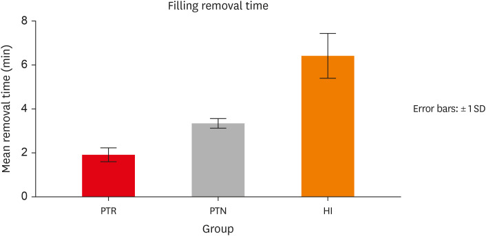
Objectives This study evaluated the effect of ProTaper Next (PTN), ProTaper Universal Retreatment (PTR) and hybrid instrumentation (HI) for canal filling removal on the fracture resistance (FR), mode of failure (MoF), and filling removal time.
Materials and Methods Ninety-six, mandibular premolars were decoronated and randomly divided into 6 groups (
n = 16), as follows: sound (S), untreated canals; prepared teeth (P), canals only prepared to ProTaper Universal finishing instrument (F4); endodontically-treated (ET), prepared and obturated canals using the single-cone technique; and groups PTN, PTR, and HI where filling was removed using PTN, PTR, or HI respectively. FR under vertical loading; MoF and time were assessed. Data were analyzed (Significance level [α] = 0.05).Results There was a significant difference in FR among all groups (
p < 0.001) (HI < P < PTN < S < ET < PTR). HI showed lower FR than S, ET and PTR, and P showed lower FR than PTR (p < 0.05). For experimental groups, there was a significant difference between every group pair (p < 0.05) No significant difference was found regarding MoF distribution (p > 0.05). HI required the highest filling removal time, while PTR required the least (p < 0.05 between every group pair).Conclusions The effect of filling removal on FR may depend on the filling removal technique/system used. PTR could be faster and protect against fracture followed by PTN; HI could adversely affect FR. FR may be associated with filling removal time.
-
Citations
Citations to this article as recorded by- Evaluation of radicular fracture resistance of maxillary premolars following non-surgical retreatment using two novel retreatment kits
Fatma M. Abu Naeem, Shaimaa Ibrahim Bakry, Ahmed S. ElSheshtawy, Ahmed Abdou, Hajer M. Abd ElHamid
BMC Oral Health.2026;[Epub] CrossRef
- Evaluation of radicular fracture resistance of maxillary premolars following non-surgical retreatment using two novel retreatment kits
- 4,004 View
- 148 Download
- 1 Web of Science
- 1 Crossref
-
Procedural errors detected by cone beam tomography in cases with indication for retreatment:
in vivo cross-sectional study - Henry Paul Valverde Haro, Carmen Rosa Garcia Rupaya, Flávio R. F. Alves
- Restor Dent Endod 2024;49(3):e26. Published online June 24, 2024
- DOI: https://doi.org/10.5395/rde.2024.49.e26
-
Abstract
PDFPubReaderePub
Objectives This study aimed to investigate the frequency and type of endodontic procedural errors in cases indicated for retreatment through cone-beam computed tomography (CBCT) analysis.
Materials and Methods The sample consisted of 96 CBCT scans, encompassing 122 permanent teeth with fully formed roots. Errors included perforation, instrument fracture, canal transportation, missed canals, and inadequate apical limit of filling. Additionally, potential risk factors were analyzed and subjected to statistical modeling.
Results The most frequent procedural error observed was the inadequate apical limit of filling, followed by canal transportation, perforation, missed canal, and instrument fracture. Statistically significant associations were identified between various procedural errors and specific factors. These include canal transportation and root canal wall, with the buccal wall being the most commonly affected; missed canal and tooth type, particularly the palatine and second mesiobuccal canal canals; inadequate apical limit of filling and root curvature, showing a higher deviation to the mesial direction in severely curved canals; inadequate apical limit of filling and the presence of calcifications, with underfilling being the most frequent; canal transportation and periapical lesion, notably with deviation to the buccal direction; and the direction of perforation and periapical lesion, most frequently occurring to buccal direction.
Conclusions CBCT emerges as a valuable tool in identifying procedural errors and associated factors, crucial for their prevention and management.
-
Citations
Citations to this article as recorded by- Regenerative endodontic treatment of a necrotic immature taurodont mandibular second molar with endodontic infection
Ali Mohammed Addokhi, Ahmed Abuhaimed
Saudi Endodontic Journal.2026; 16(2): 271. CrossRef - Repair of furcal perforations using different calcium silicate cements: An in vitro study
Ariana Esperanza Apolo Aguilar, Maria Soledad Peñaherrera Manosalvas, Henry Paul Valverde Haro
Journal of Conservative Dentistry and Endodontics.2025; 28(10): 1007. CrossRef - Impact of Downward Load and Rotational Kinematics on Root Canal Instrumentation with a Heat-Treated Nickel–Titanium Rotary Instrument
Risako Yamamoto, Keiichiro Maki, Shunsuke Kimura, Satoshi Omori, Keiko Hirano, Arata Ebihara, Yoshio Yahata, Takashi Okiji
Materials.2025; 19(1): 108. CrossRef - ANALYSIS OF THE QUALITY OF ROOT CANAL OBTURATION AND PREVALENCE OF APICAL PERIODONTITIS IN ENDODONTICALLY TREATED TEETH
Cristina Coralia Nistor, Ioana Suciu , Elena Zabrac , Ruxandra Ioana Bartok , Bogdan Dimitriu , Andreea Baluta
Romanian Journal of Oral Rehabilitation.2024; 16(4): 311. CrossRef
- Regenerative endodontic treatment of a necrotic immature taurodont mandibular second molar with endodontic infection
- 4,185 View
- 141 Download
- 2 Web of Science
- 4 Crossref
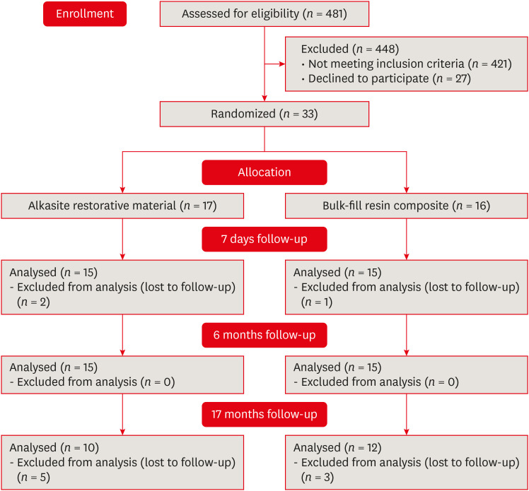
- Alkasite restorative material for endodontically treated teeth: a randomized controlled pilot study
- Davi Ariel Nobuo Bepu, Renata Siqueira Scatolin, Natalia Saud Junqueira Franco, Luiza Pejon Sanchez, Aline Evangelista Souza-Gabriel, Silmara Aparecida Milori Corona
- Restor Dent Endod 2024;49(3):e24. Published online June 11, 2024
- DOI: https://doi.org/10.5395/rde.2024.49.e24
-
Abstract
PDFPubReaderePub
Objectives This study aimed to evaluate the clinical performance of an alkasite restorative material in molars that had undergone root canal treatment.
Materials and Methods The research was registered in Brazilian Registry of Clinical Trials. The randomized clinical trial involved 33 patients, each with at least 1 mandibular molar requiring restoration after receiving endodontic treatment. Patients were randomly assigned to receive either bulk-fill resin composite (Tetric N Ceram Bulk Fill, Ivoclar Vivadent) or the alkasite restorative material (Cention N, Ivoclar Vivadent). Upon completion of the restorations, 3 calibrated professionals utilized the United States Public Health Service criteria to assess various factors, including retention, secondary caries, marginal adaptation, restoration color, marginal pigmentation, and anatomical form. Evaluations were conducted at intervals of 7 days, 6 months, and 17 months. Additionally, the assessment encompassed the presence of radiolucent lines adjacent to the restoration, material deficiencies or excess, contact points, and caries recurrence. The data underwent analysis using the Friedman and Mann-Whitney tests (α = 0.05).
Results After 17 months, the results revealed that the alkasite restorative material exhibited greater wear of anatomical shape compared to the bulk-fill resin composite (
p = 0.0189). Furthermore, the alkasite restorative material significantly differed from the natural tooth color in most cases (p = 0.0000). However, no other criteria displayed significant differences between the materials or over time (p > 0.05).Conclusions The alkasite restorative material (Cention N) emerges as a viable option for restoring endodontically treated teeth, displaying clinically acceptable alterations after a 17-month evaluation period.
Trial Registration Brazilian Registry of Clinical Trials (ReBEC) Identifier:
RBR-97kx5jv -
Citations
Citations to this article as recorded by- The Effect of Intraorifice Barrier Materials on the Fracture Resistance of Endodontically Treated Teeth: A Systematic Review and Network Meta-Analysis
Sevilay Karahan, Zeynep Buket Dağ, Emel Uzunoğlu Özyürek
Journal of Endodontics.2026; 52(5): 696. CrossRef - A Systematic Review and Meta-Analysis on the Clinical Performance and Longevity of Bioactive Composite Resin Restorations
Ahmed A. Holiel, Mounir M. Al Nakouzi, Rim Bourgi, Carlos Enrique Cuevas-Suárez, Iván Olivares Acosta, Louis Hardan, Naji Kharouf, Youssef Haikel
Journal of Composites Science.2026; 10(1): 39. CrossRef - Evaluation of Clinical Performance of Alkasite Restorative Materials: A Systematic Review and Meta-Analysis
Chloé Laporte, Rim Bourgi, Carlos Enrique Cuevas-Suárez, Naji Kharouf, Louis Hardan, Miguel Ángel Fernández-Barrera, Anh Tuan Dang, Youssef Haikel, Abigailt Flores-Ledesma
Journal of Functional Biomaterials.2026; 17(2): 93. CrossRef - 48-month clinical performance of an Alkasite restorative material versus resin composite in class II restorations: a randomized controlled trial
Ece Meral, Betül Kesim, Fatma Dilşad Öz, Sevil Gürgan
Journal of Dentistry.2026; 173: 106792. CrossRef - Alkasites in restorative dentistry: a review of their performance and properties
Alexander Bonchev, Ralitsa Bogovska-Gigova
Journal of Dentistry.2025; 160: 105916. CrossRef - Comparative Analysis of Flexural and Compressive Strengths of Bioactive Alkasite Compared to Other Ion-Releasing Restorative Materials
Hanin E. Yeslam, Fatin A. Hasanain
Biomimetics.2025; 10(11): 751. CrossRef
- The Effect of Intraorifice Barrier Materials on the Fracture Resistance of Endodontically Treated Teeth: A Systematic Review and Network Meta-Analysis
- 5,366 View
- 159 Download
- 6 Web of Science
- 6 Crossref
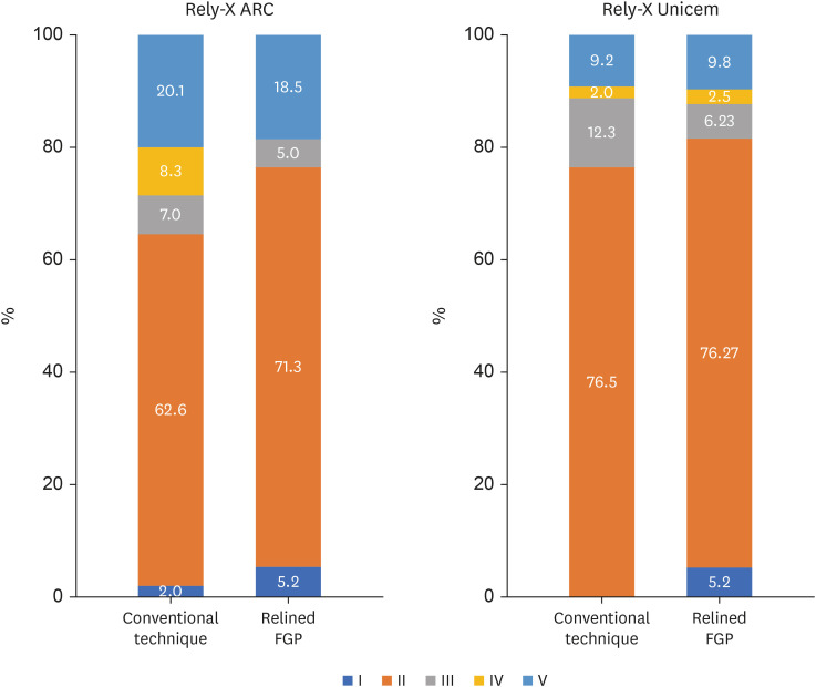
- Effects of a relined fiberglass post with conventional and self-adhesive resin cement
- Wilton Lima dos Santos Junior, Marina Rodrigues Santi, Rodrigo Barros Esteves Lins, Luís Roberto Marcondes Martins
- Restor Dent Endod 2024;49(2):e18. Published online March 27, 2024
- DOI: https://doi.org/10.5395/rde.2024.49.e18
-
Abstract
PDFPubReaderePub
Objectives This study was conducted to evaluate the mechanical properties of relined and non-relined fiberglass posts when cemented to root canal dentin using a conventional dual-cure resin cement or a self-adhesive resin cement.
Materials and Methods Two types of resin cements were utilized: conventional and self-adhesive. Additionally, 2 cementation protocols were employed, involving relined and non-relined fiberglass posts. In total, 72 bovine incisors were cemented and subjected to push-out bond strength testing (
n = 10) followed by failure mode analysis. The cross-sectional microhardness (n = 5) was assessed along the root canal, and interface analyses (n = 3) were conducted using scanning electron microscopy (SEM). Data from the push-out bond strength and cross-sectional microhardness tests were analyzed via 3-way analysis of variance and the Bonferronipost-hoc test (α = 0.05).Results For non-relined fiberglass posts, conventional resin cement exhibited higher push-out bond strength than self-adhesive cement. Relined fiberglass posts yielded comparable results between the resin cements. Type II failure was the most common failure mode for both resin cements, regardless of cementation protocol. The use of relined fiberglass posts improved the cross-sectional microhardness values for both cements. SEM images revealed voids and bubbles in the incisors with non-relined fiberglass posts.
Conclusions Mechanical properties were impacted by the cementation protocol. Relined fiberglass posts presented the highest push-out bond strength and cross-sectional microhardness values, regardless of the resin cement used (conventional dual-cure or self-adhesive). Conversely, for non-relined fiberglass posts, the conventional dual-cure resin cement yielded superior results to the self-adhesive resin cement.
-
Citations
Citations to this article as recorded by- Push-Out Bond Strength of Different Luting Cements Following Post Space Irrigation with 2% Chitosan: An In Vitro Study
Shimaa Rifaat, Ahmed Rahoma, Hind Muneer Alharbi, Sawsan Jamal Kazim, Shrouq Ali Aljuaid, Basmah Omar Alakloby, Faraz A. Farooqi, Noha Taymour
Prosthesis.2025; 7(1): 18. CrossRef
- Push-Out Bond Strength of Different Luting Cements Following Post Space Irrigation with 2% Chitosan: An In Vitro Study
- 6,397 View
- 158 Download
- 1 Web of Science
- 1 Crossref
- Prevalence of apical periodontitis and quality of root canal treatment in an adult Kuwaiti sub-population: a cross-sectional study
- Abdulrahman A. Alhailaa, Saad A Al-Nazhan, Mazen A Aldosimani
- Restor Dent Endod 2024;49(2):e16. Published online March 22, 2024
- DOI: https://doi.org/10.5395/rde.2024.49.e16
-
Abstract
PDFPubReaderePub
Objectives This cross-sectional study evaluated the prevalence of apical periodontitis (AP) and the technical quality of root canal fillings in an adult Kuwaiti subpopulation using cone-beam computed tomography (CBCT) images.
Materials and Methods Two experienced examiners analyzed 250 CBCT images obtained from Kuwaiti patients aged 15–65 years who attended government dental specialist clinics between January 2019 and September 2020. The assessment followed the radiographic scoring criteria proposed by De Moor for periapical status and the technical quality of root canal filling. Chi-square and Fisher’s exact tests were used for statistical analysis, with significance level set at
p < 0.05.Results Among the 2,762 examined teeth, 191 (6.91%) exhibited radiographic signs of AP, and 176 (6.37%) had undergone root canal filling. AP prevalence in root canal-treated teeth was 32.38%, with a significant difference between males and females. Most of the endodontically treated teeth exhibited adequate root canal filling (71.5%).
Conclusions The study demonstrated a comparable prevalence of AP and satisfactory execution of root canal treatment compared to similar studies in different countries.
-
Citations
Citations to this article as recorded by- RISK FACTORS FOR CHRONIC APICAL PERIODONTITIS ACCORDING TO THE CASE-CONTROL STUDY
N. Bagryantseva
Vrach.2026; : 43. CrossRef - A Retrospective Study of CBCT-Based Detection of Endodontic Failures and Periapical Lesions in a Romanian Cohort
Oana Andreea Diaconu, Lelia Mihaela Gheorghiță, Anca Gabriela Gheorghe, Mihaela Jana Țuculină, Maria Cristina Munteanu, Cătălina Alexandra Iacov, Virginia Maria Rădulescu, Mihaela Ionescu, Adina Andreea Mirea, Carina Alexandra Bănică
Journal of Clinical Medicine.2025; 14(18): 6364. CrossRef
- RISK FACTORS FOR CHRONIC APICAL PERIODONTITIS ACCORDING TO THE CASE-CONTROL STUDY
- 6,968 View
- 109 Download
- 1 Web of Science
- 2 Crossref
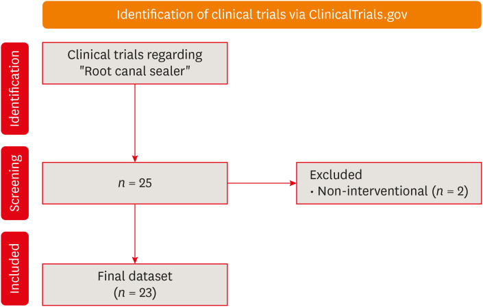
- The status of clinical trials regarding root canal sealers
- Ahmad AL Malak, Yasmina EL Masri, Mira Al Ziab, Nancy Zrara, Tarek Baroud, Pascale Salameh
- Restor Dent Endod 2024;49(1):e5. Published online January 15, 2024
- DOI: https://doi.org/10.5395/rde.2024.49.e5
-
Abstract
PDFPubReaderePub
Objectives This study aimed to present the results and analyses of clinical trials, including updates on the different functions of root canal sealers.
Materials and Methods In June 2023, we performed a comprehensive search of ClinicalTrials.gov to identify interventional clinical trials pertaining to root canal sealers. In total, 23 clinical trials conducted up to June 2023 were included in this study.
Results Approximately half of the trials (11 out of 23) were completed, while none were terminated or withdrawn. Each included trial had a minimum of 10 participants, with 11 trials having more than 100 participants. None of the assessed trials provided outcomes, and the majority (17 out of 23) lacked associated publications. In terms of geographic distribution, the USA and Canada did not contribute to any root canal sealer trials.
Conclusions This study highlights the lack of diversity in trial locations, the absence of reported results, and a scarcity of clinical trials examining the physicochemical properties of different sealers. Most published trials primarily focused on assessing the post-operative pain effect of these sealers, but no significant difference was found regarding post-operative pain control.
-
Citations
Citations to this article as recorded by- Antimicrobial additives in resin-based endodontic sealers: a scoping review of antimicrobial efficacy, physicochemical properties, and cytotoxicity
Faisal Alharamlah, Maha I. AlGhannam, Wejdan Almutairi, Faisal Alonaizan, Theeb A. Alquria, Mary Anne S. Melo, Abdulrahman A. Balhaddad
Frontiers in Dental Medicine.2026;[Epub] CrossRef
- Antimicrobial additives in resin-based endodontic sealers: a scoping review of antimicrobial efficacy, physicochemical properties, and cytotoxicity
- 5,616 View
- 77 Download
- 1 Crossref
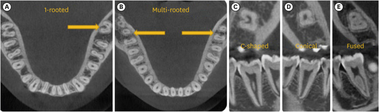
- Predictor factors of 1-rooted mandibular second molars on complicated root and canal anatomies of other mandibular teeth
- Hakan Aydın, Hatice Harorlı
- Restor Dent Endod 2024;49(1):e2. Published online January 3, 2024
- DOI: https://doi.org/10.5395/rde.2024.49.e2
-
Abstract
PDFPubReaderePub
Objectives This study aimed to determine the effects of 1-rooted mandibular second molar (MnSM) teeth on root canal anatomy complexities of the mandibular central incisor (MnCI), mandibular lateral incisor (MnLI), mandibular canine (MnCn), mandibular first premolar (MnFP), mandibular second premolar (MnSP), and mandibular first molar (MnFM) teeth.
Materials and Methods Cone-beam computed tomography images of 600 patients with full lower dentition were examined. Individuals with 1-rooted MnSMs were determined, and the complexity of root canal anatomy of other teeth was compared with individuals without 1-rooted MnSMs (Group-1; subjects with at least one 1-rooted MnSM, Group-2; subjects with more than a single root in both MnSMs). A second canal in MnCIs, MnLIs, MnCns, MnFPs, and MnSPs indicated a complicated root canal. The presence of a third root in MnFMs was recorded as complicated.
Results The prevalence of 1-rooted MnSMs was 12.2%, with the C-shaped root type being the most prevalent (9%). There were fewer complicated root canals in MnCIs (
p = 0.02), MnLIs (p < 0.001), and MnFPs (p < 0.001) in Group 1. The other teeth showed no difference between the groups (p > 0.05). According to logistic regression analysis, 1-rooted right MnSMs had a negative effect on having complex canal systems of MnLIs and MnFPs. Left MnSMs were explanatory variables on left MnLIs and both MnFPs.Conclusions In individuals with single-rooted MnSMs, a less complicated root canal system was observed in all teeth except the MnFMs.
-
Citations
Citations to this article as recorded by- Repair of furcal perforations using different calcium silicate cements: An in vitro study
Ariana Esperanza Apolo Aguilar, Maria Soledad Peñaherrera Manosalvas, Henry Paul Valverde Haro
Journal of Conservative Dentistry and Endodontics.2025; 28(10): 1007. CrossRef
- Repair of furcal perforations using different calcium silicate cements: An in vitro study
- 2,928 View
- 76 Download
- 1 Crossref
Case Report
- Ingestion and surgical retrieval of an endodontic file: a case report
- Devon Marta Ptak, Elinor Alon, Robert Bruce Amato, Julia Tassinari, Adrian Velasquez
- Restor Dent Endod 2023;48(4):e32. Published online September 2, 2023
- DOI: https://doi.org/10.5395/rde.2023.48.e32
-
Abstract
PDFPubReaderePub
Ingestions and aspirations of foreign bodies are rare, but do occasionally occur during dental treatment. Although reports exist, few include photos demonstrating the extensive surgical intervention that may be necessary to manage such events. Perhaps this lack of visualization, and associated lack of awareness, is one of the reasons some clinicians still provide non-surgical root canal therapy (NSRCT) without a rubber dam. This case report outlines the medical treatment of a 30-year-old male who initially presented to a general dentist’s office (not associated with the authors) for NSRCT of their mandibular right first molar. A rubber dam was not used for this procedure, during which the accidental ingestion of an endodontic K-file occurred. The patient was subsequently hospitalized for evaluation and treatment, consisting of numerous imaging studies, endoscopic evaluation, and surgical removal of the file from his small intestine. The ingestion of foreign bodies, and the associated complications, can be reduced through the routine use of a rubber dam, which is considered the standard of care for NSRCT. This case graphically illustrates the potential consequences associated with deviating from the standard of care and should remind clinicians that a rubber dam is necessary for all cases of NSRCT.
-
Citations
Citations to this article as recorded by- Advantages and limitations of the rubber dam. Part 2: advantages of rubber dam use
Artemy Vadimovich Karmanov, Alena Sergeevna Silkina, Natalia Alexandrovna Sokolovich
Dental Update.2026; 53(3): 198. CrossRef - Anesthesiological Management of a Sewing Needle Impacted in the Larynx in an Adult: A Case Report
Stefanie R. Senn, Felix C. Jansen, Caveh Madjdpour, Isabel Besozzi, Daniel A. Button
Clinical Case Reports.2026;[Epub] CrossRef - Dental Dam Isolation for Crown Removal, Atraumatic Tooth Extraction, Immediate Implant Placement, and Restoration Cementation: A Case Study
G Guzman-Perez, S Rojas-Rueda, F Floriani, A Unnadkat, C-C Fu, CA Jurado
Operative Dentistry.2025; 50(1): 5. CrossRef - Patient and Operator Experiences with Conventional Rubber Dam and OptiDam: A Randomized Clinical Trial
Rashed F. Binqali, Abdulwahab M. Alghamdi, Mishal S. Aloufi, Suliman A. Alharbi, Omair M. Bukhari, Reham M. Alsamman
Journal of International Society of Preventive and Community Dentistry.2025; 15(6): 554. CrossRef
- Advantages and limitations of the rubber dam. Part 2: advantages of rubber dam use
- 6,050 View
- 128 Download
- 2 Web of Science
- 4 Crossref
Research Article
- Effect of irrigation protocols on smear layer removal, bond strength and nanoleakage of fiber posts using a self-adhesive resin cement
- Rodrigo Stadler Alessi, Renata Terumi Jitumori, Bruna Fortes Bittencourt, Giovana Mongruel Gomes, João Carlos Gomes
- Restor Dent Endod 2023;48(3):e28. Published online July 27, 2023
- DOI: https://doi.org/10.5395/rde.2023.48.e28
-
Abstract
PDFPubReaderePub
Objectives This study aimed to investigate the effect of the application method of 2% chlorhexidine (CHX) and its influence on the adhesion of fiberglass posts cemented with a self-adhesive resin cement.
Materials and Methods Sixty human mandibular premolars were endodontically treated and divided into 5 groups (
n = 12), according to the canal irrigant and its application method: 2 groups with conventional syringe irrigation (CSI)—2.5% sodium hypochlorite (NaOCl) (control) and 2% CHX— and 3 groups with 2% CHX irrigation/activation—by passive ultrasonic irrigation (PUI), Easy Clean file, and XP-Endo Finisher file. Two roots per group were evaluated for smear layer (SL) removal by scanning electron microscopy. For other roots, fiber posts were luted using a self-adhesive resin cement. The roots were sectioned into 6 slices for push-out bond strength (BS) (7/group) and nanoleakage (NL) (3/group). Data from SL removal were submitted to Kruskal-Wallis and Student-Newman-Keuls tests (α = 0.05). Data from BS and NL were evaluated by 2-way analysis of variance and Tukey’s test (α = 0.05).Results For SL removal and BS, the CHX irrigation/activation promoted better values than CSI with CHX (
p < 0.05), but it was not significantly different from CSI with NaOCl (p > 0.05). For NL, the lowest values were obtained by the chlorhexidine irrigation/activation groups (p < 0.05).Conclusions Active 2% CHX irrigation can be used to improve the post space cleaning and adhesion before fiber post cementation with self-adhesive resin cements.
-
Citations
Citations to this article as recorded by- Effects of radiotherapy dose and endodontic irrigants on universal resin cement bonding to root dentin: mechanical and interfacial analyses
Lívia Ribeiro, Luíz Carlos de Lima Dias-Júnior, Paulo Henrique dos Santos, Mariana Comparotto Minamisako, Paulo Marcelo Rodrigues, Vicente Ribeiro Netto, Bruno Alexandre Pacheco de Castro Henriques, Renata Gondo Machado, Cleonice da Silveira Teixeira, Luc
International Journal of Adhesion and Adhesives.2026; 146: 104252. CrossRef - Effect of Erbium: Yttrium–Aluminum Garnet Laser, XP-Endo Finisher, and Passive Ultrasonic Irrigation in the Cleaning of Post Spaces on Push-Out Bond Strength
Dilek Hançerlioğulları, Gökhan Karadağ
Current Research in Dental Sciences.2026; 36(2): 121. CrossRef - Current trends in irrigant activation techniques among Brazilian endodontists
Mônica Pagliarini Buligon , Natália Franco Brum , Carlos Alexandre Souza Bier, Renata Domelles Morgental
Brazilian Journal of Oral Sciences.2026; 25: e269686. CrossRef - Laser‐Activated Irrigation via Photon‐Induced Photoacoustic Streaming and Shock Wave Enhanced Emission on Smear Layer Removal Efficacy, Pushout Bond Strength, and Sealer Adaptation: A SEM Assessment
Basil Almutairi, Fahad Alkhudhairy
Microscopy Research and Technique.2025; 88(6): 1806. CrossRef - The impact of passive ultrasonic irrigation on the bond strength of two different self-etch adhesives to human pulp chamber dentine: a laboratory investigation
Mohammed Turky, Jukka Matinlinna, Monika Lukomska-Szymanska, Venkateshbabu Nagendrababu, Paul M. H. Dummer, Ahmad Abdel Hamid Elheeny, Nermin Alsayed Mahmoud
BMC Oral Health.2025;[Epub] CrossRef - The effect of nanoparticles incorporation titanium dioxide and zirconium oxide within self-adhesive resin cement on the push-out bond strength of the fiber post to the radicular dentin: An in vitro study
Sawsan Hameed Al-Jubori, Maha Anwer AL-Murad
Saudi Endodontic Journal.2025; 15(2): 162. CrossRef - The Effects of Different Post Space Conditioning Procedures and Different Endodontic Sealers on the Push-Out Bond Strengths of Fiber Posts
Leyla Ayranci, Ahmet Serkan Küçükekenci, Fatih Sarı, Ahmet Çetinkaya
Clinical and Experimental Health Sciences.2025; 15(3): 620. CrossRef - Evaluation of Microleakage Using Different Luting Cements in Kedo Zirconia Crowns: An In Vitro Assessment
Guru Vishnu, Ganesh Jeevanandan
Cureus.2024;[Epub] CrossRef
- Effects of radiotherapy dose and endodontic irrigants on universal resin cement bonding to root dentin: mechanical and interfacial analyses
- 4,700 View
- 91 Download
- 7 Web of Science
- 8 Crossref
Review Article
- Does photobiomodulation on the root surface decrease the occurrence of root resorption in reimplanted teeth? A systematic review of animal studies
- Theodoro Weissheimer, Karolina Frick Bischoff, Carolina Horn Troian Michel, Bruna Barcelos Só, Manoela Domingues Martins, Matheus Albino Souza, Ricardo Abreu da Rosa, Marcus Vinícius Reis Só
- Restor Dent Endod 2023;48(3):e24. Published online June 12, 2023
- DOI: https://doi.org/10.5395/rde.2023.48.e24
-
Abstract
PDFSupplementary MaterialPubReaderePub
This review aimed to answer the following question “Does photobiomodulation treatment of the root surface decrease the occurrence of root resorption in reimplanted teeth?” Electronic searches were performed in the MEDLINE/PubMed, Cochrane Library, Scopus, Web of Science, Embase, and Grey Literature Report databases. Risk of bias was evaluated using SYRCLE Risk of Bias tool. The Grading of Recommendations, Assessment, Development, and Evaluations (GRADE) tool was used to assess the certainty of evidence. In total, 6 studies were included. Five studies reported a reduced occurrence of root resorption in teeth that received photobiomodulation treatment of the root surface prior to replantation. Only 1 study reported contradictory results. The photobiomodulation parameters varied widely among studies. GRADE assessment showed a low certainty of evidence. It can be inferred that photobiomodulation treatment of the root surface prior to replantation of teeth can reduce the occurrence of root resorption. Nonetheless, further clinical studies are needed.
Trial Registration PROSPERO Identifier: CRD42022349891
-
Citations
Citations to this article as recorded by- Liquid/Gel Mixed‐Phase Concentrated Growth Factors Enhance Periodontal and Pulpal Healing in Delayed Replantation of Immature Permanent Teeth: A Study in Beagle Dogs
Tiange Li, Xiaoxiao Yang, Yao Liu, Aochen Wang, Shu Zhu, Xu Chen, Zhenjiang Ding
International Endodontic Journal.2026; 59(7): 1430. CrossRef - Feasibility and Outcomes of Cell-based Regenerative Endodontic Therapy in Postautogenous Transplantation of a Mature Tooth: A Case Report
Noriaki Yoshihashi
Journal of Endodontics.2025; 51(1): 85. CrossRef - Evidence Mapping and Quality Assessment of Systematic Reviews in Dental Traumatology: A 54 Months Update
Nitesh Tewari, Pavithra Devi, Hemlata Nehta, Ekta Wadhwani, Rigzen Tamchos, Georgios Tsilingaridis, Vijay Prakash Mathur, Morankar Rahul
Dental Traumatology.2025; 41(6): 727. CrossRef - Photobiomodulation Literature Watch September 2023
James D. Carroll
Photobiomodulation, Photomedicine, and Laser Surgery.2024; 42(7): 498. CrossRef
- Liquid/Gel Mixed‐Phase Concentrated Growth Factors Enhance Periodontal and Pulpal Healing in Delayed Replantation of Immature Permanent Teeth: A Study in Beagle Dogs
- 4,111 View
- 58 Download
- 4 Web of Science
- 4 Crossref
Research Article
- Effects of different calcium-silicate based materials on fracture resistance of immature permanent teeth with replacement root resorption and osteoclastogenesis
- Gabriela Leite de Souza, Gabrielle Alves Nunes Freitas, Maria Tereza Hordones Ribeiro, Nelly Xiomara Alvarado Lemus, Carlos José Soares, Camilla Christian Gomes Moura
- Restor Dent Endod 2023;48(2):e21. Published online May 5, 2023
- DOI: https://doi.org/10.5395/rde.2023.48.e21
-
Abstract
PDFSupplementary MaterialPubReaderePub
Objectives This study evaluated the effects of Biodentine (BD), Bio-C Repair (BCR), and mineral trioxide aggregate (MTA) plug on the fracture resistance of simulated immature teeth with replacement root resorption (RRR) and
in vitro -induced osteoclastogenesis.Materials and Methods Sixty bovine incisors simulating immature teeth and RRR were divided into 5 groups: BD and BCR groups, with samples completely filled with the respective materials; MTA group, which utilized a 3-mm apical MTA plug; RRR group, which received no root canal filling; and normal periodontal ligament (PL) group, which had no RRR and no root canal filling. All the teeth underwent cycling loading, and compression strength testing was performed using a universal testing machine. RAW 264.7 macrophages were treated with 1:16 extracts of BD, BCR, and MTA containing receptor activator of nuclear factor-kappa B ligand (RANKL) for 5 days. RANKL-induced osteoclast differentiation was assessed by staining with tartrate-resistant acid phosphatase. The fracture load and osteoclast number were analyzed using 1-way ANOVA and Tukey’s test (α = 0.05).
Results No significant difference in fracture resistance was observed among the groups (
p > 0.05). All materials similarly inhibited osteoclastogenesis (p > 0.05), except for BCR, which led to a lower percentage of osteoclasts than did MTA (p < 0.0001).Conclusions The treatment options for non-vital immature teeth with RRR did not strengthen the teeth and promoted a similar resistance to fractures in all cases. BD, MTA, and BCR showed inhibitory effects on osteoclast differentiation, with BCR yielding improved results compared to the other materials.
-
Citations
Citations to this article as recorded by- Effect of Restoration Strategy and Cavity Location on the Fracture Resistance of Teeth with External Cervical Resorption
Saadet Elpe, Öznur Sarıyılmaz
Journal of Endodontics.2026; 52(6): 980. CrossRef - Influence of Different Post-core Restorative Modalities on Fracture Characteristics of Immature Endodontically Treated Premolars
Wafa H Alaajam, Khalid M Abdelaziz, Malaz M Mustafa, Mohammed S Al-Ak'hali, Ashraf A Khalil, Mohammed M Al Moaleem, Hoda L Abouzeid
The Journal of Contemporary Dental Practice.2026; 27(4): 399. CrossRef - In vitro comparison of fracture strength of maxillary incisors with the simulated external root resorption cavities repaired with BioMTA or Biodentine
Tufan Ozasir, Birgul Ozasir, Nagihan Aribal, Derin Bugu Yuzer, Baris Kandemir, Kamran Gulsahi
Journal of Dental Sciences.2025; 20(3): 1532. CrossRef - Comparative Analysis of Gene Expression in Periodontal Ligament Stem Cells Exposed to Biodentine and Bio-C Repair: Implications for Cementogenesis—An In Vitro Study
Mahmoud M. Bakr, Mahmoud Al Ankily, Mohammed Meer, Mohamed Shamel
Oral.2025; 5(1): 19. CrossRef - Efficacy of Mineral Trioxide Aggregate Versus Biodentine as a Direct Pulp Capping Material in Carious Human Mature Permanent Teeth: A Systematic Review
Rashmi Misra, Nikita Toprani, Sumita Bhagwat, Aashaka Vaishnav, Aastha Dureja, Omkar Bhosale
Cureus.2025;[Epub] CrossRef - Evaluation of Different Techniques and Materials for Filling in 3-dimensional Printed Teeth Replicas with Perforating Internal Resorption by Means of Micro–Computed Tomography
Angelo J.S. Torres-Carrillo, Helena C. Assis, Rodrigo E. Salazar-Gamarra, Leonardo Moreira Teodosio, Alice C. Silva-Sousa, Jardel F. Mazzi-Chaves, Priscila B. Ferreira-Soares, Manoel D. Sousa-Neto, Fabiane C. Lopes-Olhê
Journal of Endodontics.2024; 50(2): 205. CrossRef
- Effect of Restoration Strategy and Cavity Location on the Fracture Resistance of Teeth with External Cervical Resorption
- 3,695 View
- 82 Download
- 4 Web of Science
- 6 Crossref
Case Report
- Successful nonsurgical treatment of type II dens invaginatus with 5 root canals using a self-adjusting file: a case report
- George Táccio de Miranda Candeiro, Antônio Sérgio Teixeira de Menezes, Ana Carolina Saldanha de Oliveira, Flávio Rodrigues Ferreira Alves
- Restor Dent Endod 2023;48(2):e17. Published online April 27, 2023
- DOI: https://doi.org/10.5395/rde.2023.48.e17
-
Abstract
PDFPubReaderePub
The present report describes the endodontic treatment of an Oehlers type II dens invaginatus in a maxillary lateral incisor with 5 root canals, an extremely rare condition. Apical periodontitis and related symptoms were noted. Cone-beam computed tomography was used to aid the diagnosis, reveal tooth morphology, and assist in canal location. The pulp chamber was carefully accessed, and the root canals were explored under magnification. All root canals were prepared with an R25 Reciproc Blue system and sodium hypochlorite (NaOCl) irrigation. After initial preparation, a self-adjusting file (SAF) with NaOCl and ethylenediaminetetraacetic acid was used to complement the disinfection. Additionally, calcium hydroxide medication was applied. Vertical compaction was used to fill the canals with a calcium silicate-based endodontic sealer and gutta-percha. After 12 months, the patient exhibited healing of the periapical region, absence of symptoms, and normal dental function. In conclusion, this nonsurgical treatment protocol was successful in promoting the cure of apical periodontitis. Both complementary disinfection with an SAF and use of calcium hydroxide medication should be considered when choosing the best treatment approach for dens invaginatus with very complex anatomy.
-
Citations
Citations to this article as recorded by- Non-surgical endodontic management of dens invaginatus type II in an immature maxillary lateral incisor using a bioceramic apical plug: A 3-year follow-up case report
Yahya Raja Alharbi, Qayed Saad Alharbi, Shaul Hameed Kolarkodi
Saudi Endodontic Journal.2026; 16(1): 115. CrossRef
- Non-surgical endodontic management of dens invaginatus type II in an immature maxillary lateral incisor using a bioceramic apical plug: A 3-year follow-up case report
- 3,027 View
- 89 Download
- 1 Crossref
Research Articles
-
Influence of CBCT parameters on image quality and the diagnosis of vertical root fractures in teeth with metallic posts: an
ex vivo study - Larissa Pereira Lagos de Melo, Polyane Mazucatto Queiroz, Larissa Moreira-Souza, Mariana Rocha Nadaes, Gustavo Machado Santaella, Matheus Lima Oliveira, Deborah Queiroz Freitas
- Restor Dent Endod 2023;48(2):e16. Published online April 27, 2023
- DOI: https://doi.org/10.5395/rde.2023.48.e16
-
Abstract
PDFPubReaderePub
Objectives The aim of this study was to evaluate the influence of peak kilovoltage (kVp) and a metal artifact reduction (MAR) tool on image quality and the diagnosis of vertical root fracture (VRF) in cone-beam computed tomography (CBCT).
Materials and Methods Twenty single-rooted human teeth filled with an intracanal metal post were divided into 2 groups: control (
n = 10) and VRF (n = 10). Each tooth was placed into the socket of a dry mandible, and CBCT scans were acquired using a Picasso Trio varying the kVp (70, 80, 90, or 99), and the use of MAR (with or without). The examinations were assessed by 5 examiners for the diagnosis of VRF using a 5-point scale. A subjective evaluation of the expression of artifacts was done by comparing random axial images of the studied protocols. The results of the diagnoses were analyzed using 2-way analysis of variance and the Tukeypost hoc test, the subjective evaluations were compared using the Friedman test, and intra-examiner reproducibility was evaluated using the weighted kappa test (α = 5%).Results The kVp and MAR did not influence the diagnosis of VRF (
p > 0.05). According to the subjective classification, the 99 kVp protocol with MAR demonstrated the least expression of artifacts, while the 70 kVp protocol without MAR led to the most artifacts.Conclusions Protocols with higher kVp combined with MAR improved the image quality of CBCT examinations. However, those factors did not lead to an improvement in the diagnosis of VRF.
-
Citations
Citations to this article as recorded by- Digital Dentistry Society Quality Forum: Clinical recommendations on cone-beam computed tomography for the digital dentistry workflow
Hugo Gaêta-Araujo, Rocharles Cavalcante Fontenele, Reinhilde Jacobs
Digital Dentistry Journal.2026; 3(1): 100065. CrossRef - Photon‐Counting CT for Diagnosing Vertical Root Fractures in Teeth With Metal Posts: An Ex Vivo Comparative Analysis With Four CBCT Devices
Renata M. S. Leal, Fernanda B. Fagundes, Maria F. S. A. Bortoletto, Samuel C. Kluthcovsky, Walter Coudyzer, Bruno C. Cavenago, Reinhilde Jacobs, Rocharles Cavalcante Fontenele
International Endodontic Journal.2026; 59(4): 718. CrossRef - Cone‑beam computed tomography in endodontics: Historical development, technical principles, clinical applications and operative outcomes (Review)
Anshuman Shetty, Shivprasad Rai
World Academy of Sciences Journal.2026; 8(4): 1. CrossRef - Diagnostic Performance of Iterative Reconstruction of Cone-beam Computed Tomography for Detecting Vertical Root Fractures in the Presence of Metal Artifacts
Matheus Barros-Costa, Gustavo Santaella, Christiano Oliveira-Santos, Deborah Queiroz Freitas, William C. Scarfe, Francisco Carlos Groppo
Journal of Endodontics.2025; 51(6): 715. CrossRef - Radiographic and Clinical Outcomes of Laser-Enhanced Disinfection in Endodontic Therapy
Janos Kantor, Sorana Maria Bucur, Eugen Silviu Bud, Victor Nimigean, Ioana Maria Crișan, Mariana Păcurar
Journal of Clinical Medicine.2025; 14(12): 4055. CrossRef - Exploring Diagnostic Reliability of CBCT for Vertical Root Fractures: A Systematic Review and Meta‐Analytical Approach
Luiz Carlos de Lima Dias-Junior, Diego Leonardo de Souza, Adriana Pinto Bezerra, Marcio Correa, Cleonice da Silveira Teixeira, Eduardo Antunes Bortoluzzi, Lucas da Fonseca Roberti Garcia, Stefano Corbella
International Journal of Dentistry.2025;[Epub] CrossRef - Deep learning for dentomaxillofacial cone-beam computed tomography image quality enhancement: A pilot study
Ali Nazari, Seyed Mohammad Yousef Najafi, Reza Abbasi, Hossein Mohammad-Rahimi, Parisa Motie, Mina Iranparvar Alamdari, Mehdi Hosseinzadeh, Ruben Pauwels, Falk Schwendicke
Imaging Science in Dentistry.2025; 55(3): 271. CrossRef - Diagnostic Accuracy of Intraoral, Extraoral and Cone Beam Computed Tomography (CBCT)-Generated Bitewings for Detecting Approximal Caries and Periodontal Bone Loss
Jyoti Mago, Alan G Lurie, Aadarsh Gopalakrishna, Aditya Tadinada
Cureus.2025;[Epub] CrossRef - Vertical root fracture diagnosis in teeth with metallic posts: Impact of metal artifact reduction and sharpening filters
Débora Costa Ruiz, Lucas P. Lopes Rosado, Rocharles Cavalcante Fontenele, Amanda Farias-Gomes, Deborah Queiroz Freitas
Imaging Science in Dentistry.2024; 54(2): 139. CrossRef - Comparing standard- and low-dose CBCT in diagnosis and treatment decisions for impacted mandibular third molars: a non-inferiority randomised clinical study
Kuo Feng Hung, Andy Wai Kan Yeung, May Chun Mei Wong, Michael M. Bornstein, Yiu Yan Leung
Clinical Oral Investigations.2024;[Epub] CrossRef
- Digital Dentistry Society Quality Forum: Clinical recommendations on cone-beam computed tomography for the digital dentistry workflow
- 4,295 View
- 71 Download
- 7 Web of Science
- 10 Crossref
- Buckling resistance, torque, and force generation during retreatment with D-RaCe, HyFlex Remover, and Mtwo retreatment files
- Yoojin Kim, Seok Woo Chang, Soram Oh
- Restor Dent Endod 2023;48(1):e10. Published online February 6, 2023
- DOI: https://doi.org/10.5395/rde.2023.48.e10
-
Abstract
PDFPubReaderePub
Objectives This study compared the buckling resistance of 3 nickel-titanium (NiTi) retreatment file systems and the torque/force generated during retreatment.
Materials and Methods The buckling resistance was compared among the D-RaCe (DR2), HyFlex Remover, and Mtwo R25/05 retreatment systems. J-shaped canals within resin blocks were prepared with ProTaper NEXT X3 and obturated by the single-cone technique with AH Plus. After 4 weeks, 4 mm of gutta-percha in the coronal aspect was removed with Gates-Glidden drills. Retreatment was then performed using DR1 (size 30, 10% taper) followed by DR2 (size 25, 4% taper), HyFlex Remover (size 30, 7% taper), or Mtrwo R25/05 (size 25, 5% taper) (15 specimens in each group). Further apical preparation was performed with WaveOne Gold Primary. The clockwise torque and upward force generated during retreatment were recorded. After retreatment, resin blocks were examined using stereomicroscopy, and the percentage of residual filling material in the canal area was calculated. Data were analyzed using 1-way analysis of variance with the Tukey test.
Results The HyFlex Remover files exhibited the greatest buckling resistance (
p < 0.05), followed by the Mtwo R25/05. The HyFlex Remover and Mtwo R25/05 files generated the highest maximum clockwise torque and upward force, respectively (p < 0.05). The DR1 and DR2 files generated the least upward force and torque (p < 0.05). The percentage of residual filling material after retreatment was not significantly different between file systems (p > 0.05).Conclusions NiTi retreatment instruments with higher buckling resistance generated greater clockwise torque and upward force.
-
Citations
Citations to this article as recorded by- Time Required to Retreat Carrier-Based Obturation: Comparison Between Two Techniques at Two Levels of Experience
Matteo Salvadori, Elisabetta Audino, Miriam Facchinetti, Vikas Kumar, Mario Alovisi, Luca Visconti, Stefano Salgarello
Dentistry Journal.2026; 14(3): 173. CrossRef - Comparative Evaluation of Rotary and Reciprocating Systems in Root Canal Retreatment with Subsequent Apical Enlargement
Burçin Arıcan, Ayşe Tuba Özalp Koca, Taha Özyürek, Fatma Macit Ermiş
European Journal of General Dentistry.2026;[Epub] CrossRef - Comparative evaluation of the efficacy of three different retreatment files in removing root canal filling material: An In vitro confocal microscopy study
Meghna Sarah Abraham, Aravind R. Kudva, Prathap M. S. Nair, Shravan Kini, Samreena Kalander, Faseeh Muhammed Bin Farookh
Endodontology.2025; 37(2): 136. CrossRef - Efficacy of Endodontic Files in Root Canal Retreatment: A Systematic Review of In Vitro Studies
Anna Soler-Doria, José Luis Sanz, Marcello Maddalone, Leopoldo Forner
Journal of Functional Biomaterials.2025; 16(8): 293. CrossRef - Postoperative Pain Following Single‐Visit Nonsurgical Retreatment Using Minimally Invasive Rotary vs. Reciprocating Nickel‐Titanium File Systems: A Two‐Arm Parallel Randomized Clinical Trial
Hüseyin Gürkan Güneç, Büşra Pehlivan, Celalettin Topbaş, Abdurrahman Kerim Kul, Dursun Ali Şirin, Sivakumar Nuvvula
Pain Research and Management.2025;[Epub] CrossRef - Comparative Evaluation of Canal Centering Ability of Single-file Retreatment System vs Multiple-file Retreatment System, with and without Gutta-Percha Solvent: An In Vitro Study
Sangkeetha Gnanasekaran, Arasappan Rajakumaran, Rajeswari Kalaiselvam, Mathan Rajan Rajendran, Seshan Rakkesh Ramesh, Manigandan Kuzhanchinathan
The Journal of Contemporary Dental Practice.2025; 26(9): 898. CrossRef - Effect of Different Downward Loads and Rotational Speeds on the Removal of Gutta-Percha and Root Canal Sealer Using a Nickel-Titanium Rotary Gutta-Percha Removal System: An Ex Vivo Study
Koki Toyoda, Shunsuke Kimura, Keiichiro Maki, Satoshi Omori, Keiko Hirano, Arata Ebihara, Takashi Okiji
Applied Sciences.2025; 16(1): 446. CrossRef - Cone-beam computed tomographic evaluation and fracture resistance of endodontically retreated teeth using hyflex remover, Mtwo, and ProTaper retreatment file systems: An in vitro study
Isha Singh, Dakshita Joy Sinha, Pallavi Sharma, Kunal Bedi, Priyanka Rani, Swapnil Vats
Saudi Endodontic Journal.2024; 14(1): 56. CrossRef - Comparison of torsional, bending, and buckling resistances of different nickel-titanium glide path files
Feyyaz Çeliker, İrem Çetinkaya
Matéria (Rio de Janeiro).2024;[Epub] CrossRef - Assessing the impact of obturation techniques, kinematics and irrigation protocols on apical debris extrusion and time required in endodontic retreatment
Eugenio Pedullà, Francesco Iacono, Martina Pitrolo, Giovanni Barbagallo, Giusy Rita Maria La Rosa, Chiara Pirani
Australian Endodontic Journal.2023; 49(3): 623. CrossRef
- Time Required to Retreat Carrier-Based Obturation: Comparison Between Two Techniques at Two Levels of Experience
- 3,617 View
- 66 Download
- 6 Web of Science
- 10 Crossref
Review Article
- Effect of endodontic sealer on postoperative pain: a network meta-analysis
- Cynthia Maria Chaves Monteiro, Ana Cristina Rodrigues Martins, Alessandra Reis, Juliana Larocca de Geus
- Restor Dent Endod 2023;48(1):e5. Published online December 29, 2022
- DOI: https://doi.org/10.5395/rde.2023.48.e5
-
Abstract
PDFPubReaderePub
This systematic review and network meta-analysis aimed to answer the following focused research question: “Does the type of endodontic sealer affect the postoperative pain in patients who received endodontic treatment?” Different databases and grey literature were surveyed. Only one randomized controlled trial were included. The risk of bias in the studies was evaluated by using the Cochrane Collaboration’s tool. A random-effects meta-analysis was conducted to compare the risk and intensity of postoperative pain. The quality of the body of evidence was assessed using the Grading of Recommendations Assessment, Development, and Evaluation approach. Out of 11,601 studies, 15 remained for qualitative analyses and 12 for meta-analysis. Seven studies were classified at high risk of bias, and 8 studies raised some concerns. No significant differences between the endodontic materials were observed in the direct comparisons, both in risk and in intensity of postoperative pain (pairwise comparisons with 2 studies: I2 = 0%;
p > 0.05 and 8 studies: I2 = 23%;p > 0.05, respectively). The certainty of the evidence was graded as low or moderate. There was no difference in the risk and intensity of postoperative pain after filling with different endodontic sealers. Further systematic reviews should be conducted.Trial Registration PROSPERO Identifier:
CRD42020215314 -
Citations
Citations to this article as recorded by- Does the Use of a Bioceramic Sealer Reduce Postoperative Pain Compared With an Epoxy Resin‐Based Sealer After Primary Root Canal Treatment and Retreatment?—An Umbrella Review
Lokhasudhan Govindaraju, Rajeswari Kalaiselvam, Mathan Rajan Rajendran, Aleksandar Jakovljevic, Jelena Jacimovic, Henry F. Duncan, Venkateshbabu Nagendrababu
International Endodontic Journal.2026; 59(3): 341. CrossRef - Evidence synthesis of postoperative pain with bioceramic vs. epoxy resin sealers: umbrella review of randomized trials within existing systematic reviews
Mrunali Dahikar, Ashish Mandwe, Kulvinder Singh Banga, Alexander Maniangat Luke, Suraj Arora, Unmesh Khanvilkar, Ajinkya M. Pawar
Frontiers in Dental Medicine.2026;[Epub] CrossRef - Factors Influencing Apical Extrusion of 2 Types of Endodontic Sealers with Different Delivery Systems
Shumaila Iqbal, Nicholas S. Adams, Josette Camilleri
Journal of Endodontics.2026; 52(5): 806. CrossRef - Evaluation of Postoperative Pain Frequency in Single‐Session Endodontic Treatments With Patency and Foraminal Enlargement
Viviane Barbosa Godoy, Ana Grasiela Limoeiro, Vanessa Sandini, Vini Mehta, Wayne Martins Nascimento, Marilia Fagury Videira Marceliano‐Alves, Marcos Frozoni
Clinical and Experimental Dental Research.2026;[Epub] CrossRef - Silicone vs. Silicon/Silica in Intraoral Healing: A Systematic Review
David Parker, Aditi Bopardikar, Georgios E. Romanos
Materials.2026; 19(7): 1425. CrossRef - Post-Operative Pain After Endodontic Instrumentation, Irrigation and Obturation: An Umbrella Review of Systematic Reviews Published from 2016 to 2025
Fausto Zamparini, Andrea Spinelli, Gioia Quadrini, Maria Giovanna Gandolfi, Carlo Prati
Journal of Clinical Medicine.2026; 15(12): 4775. CrossRef - Comparative Evaluation of Postoperative Pain Following Nonsurgical Endodontic Therapy with Calcium Silicate-Based Sealer and Traditional Sealers: A Systematic Review and Meta-Analysis
Guha Poulomi, Solete Pradeep, Antony Delphine, Arun Nishitha, Surendar Ramamoorthi, Choudhari Sahil, Hima Sandeep Adimulapu
Pesquisa Brasileira em Odontopediatria e Clínica Integrada.2026;[Epub] CrossRef - Effect of occlusal reduction on post-operative pain of symptomatic and asymptomatic molar teeth
Aysenur Kamacı Esen, Fatma Furuncuoğlu, Fatima Betul Basturk, Muhammet Nuri Taşcıoğlu, Masoud Parirokh
Acta Odontologica Scandinavica.2025; 84: 371. CrossRef - An Observational Study on Pain Occurrence After Root Canal Treatment: Role of Operator Experience When Using a Bioceramic Sealer
Mihai Merfea, Ioana Sofia Pop-Ciutrila, Mindra Eugenia Badea, Ada Gabriela Delean, Oana Cimponeriu, Razvan Corneliu Pop, Maria Peter, Iulia Clara Badea, Sanda Ileana Cimpean
Journal of Clinical Medicine.2025; 14(13): 4558. CrossRef - Assessment of Postoperative Pain After Single‐ or Multiple‐Visit Endodontic Therapy and Its Molecular Aspects: A Randomised Controlled Study
Larissa Nunes Rosa Bedene, Denise Piotto Leonardi, Joana Santana Couto, Bruno Marques‐da‐Silva, Marilisa Carneiro Leão Gabardo, João Arnando Brancher, Flávia Sens Fagundes Tomazinho
Australian Endodontic Journal.2025; 51(3): 668. CrossRef - Clinical and Radiographic Outcomes of Root Canal Obturation with Hydraulic Condensation and Tricalcium Silicate Bioceramic Sealer: A 12-Month Observational Study on Periapical Healing
Kostadin Zhekov, Vesela Stefanova
Journal of Functional Biomaterials.2025; 16(11): 412. CrossRef - Comparative evaluation of postoperative pain and periapical healing after root canal treatment using three different endodontic sealers: A randomized controlled clinical trial
Ruchika Pandey, Nitin Kararia, Deepak Kumar Sharma, Vishal Rathod, Anand Vilas Bansod, Dhaval Desai
Journal of Conservative Dentistry and Endodontics.2024; 27(9): 962. CrossRef - Effect of bioceramic-based and resin-based sealers on postoperative discomfort following root canal therapy: a systematic review and meta-analysis
Mansi Supare, Ajinkya M. Pawar, Kashmira Sawant, Dian Agustin Wahjuningrum, Suraj Arora, Firas Elmsmari, Mohmed Isaqali Karobari, Bhagyashree Thakur
PeerJ.2024; 12: e18198. CrossRef - Comparative Evaluation of Incidences of Post Operative Pain in Patient Treated in Single Visit Root Canal Treatment by Using Different Sealers: - An in-Vivo Study
Sadashiv Daokar, Aishwarya Ranjalkar, Kalpana Pawar, Komal Potfode, Dhanashri Padwal, Sana Khan
International Journal of Innovative Science and Research Technology (IJISRT).2024; : 2743. CrossRef
- Does the Use of a Bioceramic Sealer Reduce Postoperative Pain Compared With an Epoxy Resin‐Based Sealer After Primary Root Canal Treatment and Retreatment?—An Umbrella Review
- 8,420 View
- 163 Download
- 12 Web of Science
- 14 Crossref
Research Article
- Comparison of shaping ability of the Reciproc Blue and One Curve with or without glide path in simulated S-shaped root canals
- Vincenzo Biasillo, Raffaella Castagnola, Mauro Colangeli, Claudia Panzetta, Irene Minciacchi, Gianluca Plotino, Simone Staffoli, Luca Marigo, Nicola Maria Grande
- Restor Dent Endod 2022;47(1):e3. Published online December 28, 2022
- DOI: https://doi.org/10.5395/rde.2022.47.e3
-
Abstract
PDFPubReaderePub
Objectives This study aimed to assess the impact of a glide-path on the shaping ability of 2 single-file instruments and to compare the centering ability, maintenance of original canal curvatures and area of instrumentation in simulated S-shaped root canals.
Materials and Methods Forty simulated S-shaped root canals were used and were prepared with One Curve (group OC), One G and OC (group GOC), Reciproc Blue (group RB) and R-Pilot and RB (group PRB) and scanned before and after instrumentation. The images were analyzed using AutoCAD. After superimposing the samples, 4 levels (D1, D2, D3, and D4) and 2 angles (Δ1 and Δ2) were established to evaluate the centering ability and modification of the canal curvatures. Then, the area of instrumentation (ΔA) was measured. The data were analyzed using 2-way analysis of variance and Tukey's test for multiple comparisons (
p < 0.05).Results Regarding the centering ability in the apical part (D3, D4), the use of the glide-path yielded better results than the single-file groups. Among the groups at D4, OC showed the worst results (
p < 0.05). The OC system removed less material (ΔA) than the RB system, and for Δ1, OC yielded a worse result than RB (p < 0.05).Conclusions The glide-path improved the centering ability in the apical part of the simulated S-shaped canals. The RB system showed a better centering ability in the apical part and major respect of the canal curvatures compared with OC system.
-
Citations
Citations to this article as recorded by- Evaluation of Apical Debris Extrusion and the Remaining Canal Material during Retreatment of a Bioceramic Sealer by the XP-endo Finisher File System, Followed by Various Supplementary Methods: An in Vitro Study
Paras Mull Gehlot, Parvathi Sudeep, Annapoorna B Mariswamy
World Journal of Dentistry.2025; 15(10): 837. CrossRef - Shaping Ability of Rotary NiTi Systems in S‐Shaped Root Canals of Mandibular Molars
Renata M. S. Leal, Emmanuel J. N. L. Silva, Maria C. B. P. Campos, Clarissa T. Rodrigues, Marco A. H. Duarte, Bruno C. Cavenago
Australian Endodontic Journal.2025; 51(1): 133. CrossRef - Comparison of Debris Extrusion and Preparation Time by Traverse, R‐Motion Glider C, and Other Glide Path Systems in Severely Curved Canals
Taher Al Omari, Layla Hassouneh, Khawlah Albashaireh, Alaa Dkmak, Rami Albanna, Ali Al-Mohammed, Ahmed Jamleh, Lucas da Fonseca Roberti Garcia
International Journal of Dentistry.2025;[Epub] CrossRef - Glide Path – An Ineluctable Route for Successful Endodontic Mechanics: A Literature Review
Mahima Bharat Mehta, Anupam Sharma, Aniket Jadhav, Aishwarya Handa, Abhijit Bajirao Jadhav, Ashwini A. Narayanan
Journal of the International Clinical Dental Research Organization.2024; 16(2): 101. CrossRef - Screw-in force, torque generation, and performance of glide-path files with three rotation kinetics
Jee-Yeon Woo, Ji-Hyun Jang, Seok Woo Chang, Soram Oh
Odontology.2024; 112(3): 761. CrossRef - Glide Path in Endodontics: A Literature Review of Current Knowledge
Vlad Mircea Lup, Giulia Malvicini, Carlo Gaeta, Simone Grandini, Gabriela Ciavoi
Dentistry Journal.2024; 12(8): 257. CrossRef - In Vitro Research Methods Used to Evaluate Shaping Ability of Rotary Endodontic Files—A Literature Review
Ranya F. Elemam, Ana Mano Azul, João Dias, Khaled El Sahli, Renato de Toledo Leonardo
Dentistry Journal.2024; 12(10): 334. CrossRef - Endodontic glide path - importance and performance techniques
Milica Jovanovic-Medojevic, Мiljan Stosic, Vanja Opacic-Galic, Violeta Petrovic
Srpski arhiv za celokupno lekarstvo.2023; 151(5-6): 380. CrossRef
- Evaluation of Apical Debris Extrusion and the Remaining Canal Material during Retreatment of a Bioceramic Sealer by the XP-endo Finisher File System, Followed by Various Supplementary Methods: An in Vitro Study
- 4,204 View
- 57 Download
- 8 Web of Science
- 8 Crossref
Review Article
- Influence of the root canal filling technique on the success rate of primary endodontic treatments: a systematic review
- Daniel Feijolo Marconi, Giovana Siocheta da Silva, Theodoro Weissheimer, Isadora Ames Silva, Gabriel Barcelos Só, Leonardo Thomasi Jahnke, Jovito Adiel Skupien, Marcus Vinicius Reis Só, Ricardo Abreu da Rosa
- Restor Dent Endod 2022;47(4):e40. Published online October 11, 2022
- DOI: https://doi.org/10.5395/rde.2022.47.e40
-
Abstract
PDFSupplementary MaterialPubReaderePub
Objectives This study aimed to investigate the influence of different obturation techniques compared to cold lateral compaction on the success rate of primary non-surgical endodontic treatments.
Materials and Methods Systematic searches were performed for studies published up to May 17th, 2022 in MEDLINE/PubMed, Cochrane Library, Web of Science, Scopus, EMBASE, and Grey Literature Reports. Randomized clinical trials and nonrandomized (nonrandomized clinical trials, prospective or retrospective) studies that evaluated the success rate of primary non-surgical endodontic treatments obturated with the cold lateral compaction (control) and other obturation techniques were included. The revised Cochrane risk of bias tools for randomized trials (RoB 2) and nonrandomized studies of interventions (ROBINS-I) were used to evaluate the risk of bias. The Grading of Recommendations Assessment, Development, and Evaluation (GRADE) tool was used to evaluate the certainty of evidence.
Results Eleven studies (4 randomized clinical trials (RCTs), 4 prospective, and 3 retrospectives) were included. Two RCTs were classified as having some concerns risk of bias and 2 as a low risk of bias. Two nonrandomized studies were classified as having a critical risk of bias and 5 as having a moderate risk of bias. The GRADE analysis demonstrated a very low to moderate certainty of evidence.
Conclusions This systematic review generally evidenced no differences in the success rate of primary non-surgical endodontic treatments when the cold lateral compaction technique and other obturation techniques are performed. Further well-designed studies are still necessary.
-
Citations
Citations to this article as recorded by- Assessing Sealing Ability of C-Root SP Strontium Silicate Sealer With Different Obturation Techniques: An in vitro Study
Suixin Hu, Jianshe Li, Meng Xu, Laiqing Xu, Yangming Yin, Peng Xue, Liping Dong, Lin Wang, Huixia He, Ying Liu, Qiang Luo, Fei Chen
International Dental Journal.2026; 76(1): 109283. CrossRef - Effect of root canal filling techniques and materials on endodontic treatment outcomes: a systematic review and meta-analysis
Ahmed Mushtaq, Sura Alsanafi, Firas Elmsmari, José A. González, Marc Garcia-Font, Francesc Abella Sans, Kelvin I. Afrashtehfar, Paul V. Abbott
Scientific Reports.2026;[Epub] CrossRef - Dimensional and cross-sectional compatibility of contemporary nickel–titanium files and their corresponding gutta-percha cones: A micro-computed tomography study
Cem Sarkan, Faruk Haznedaroglu
The Saudi Dental Journal.2026;[Epub] CrossRef - Comparative Analysis Of Obturation Techniques In Endodontics: Lateral Vs. Thermoplasticized. Thermoplasticized
Juan Esteban Díaz Pacheco , Rómulo Guillermo López Torres , Verónica Alejandra Salame Ortíz
Salud, Ciencia y Tecnología.2025; 5: 1626. CrossRef - Effect of ultrasonic activation of endodontic sealers on root canal filling quality during the single-cone obturation procedure: a systematic review and meta-analysis of laboratory-based studies
Shuting Feng, Weiqing Zhou, Xiaojun Chu, Shuaimei Xu, Xiongqun Zeng
Odontology.2025; 113(4): 1380. CrossRef - In Vitro and In Vivo Evaluation of a New Experimental Polydimethylsiloxane-Based Endodontic Sealer
Fabiola Cardoso Maldonado, Cesar Gaitan Fonseca, Carlos Bermudez Jimenez, Luis Alejandro Aguilera Galaviz, Margarita L. Martinez-Fierro, Lorena Troncoso Vazquez, Martha Eugenia Reyes Ortiz
Journal of Functional Biomaterials.2025; 16(11): 402. CrossRef - Evaluation of three obturation techniques in 3D-printed models of oval canals with standardized prepared morphology: a micro-CT study
Wenjun Xia, Qisheng Gu, Yingshuang Song, Yunjia Liu, Xuetao Deng, Wenhao Qian
BMC Oral Health.2025;[Epub] CrossRef - Clinical and Radiographic Failure of Nonsurgical Endodontic Treatment and Retreatment Using Single-cone Technique With Calcium Silicate-based Sealers: A Systematic Review and Meta-analysis
Mohammad A. Sabeti, Negah Karimpourtalebi, Arash Shahravan, Omid Dianat
Journal of Endodontics.2024; 50(6): 735. CrossRef - Method of microbial decontamination of endodontic absorbent paper points: a randomised experimental study
O. A. Pavlovskaya, O. A. Kachanova, V. V. Volobuev, M. N. Mitropanova, A. R. Gazarova, V. Y. Zobenko, A. G. Uvarova
Pediatric dentistry and dental prophylaxis.2024; 24(2): 157. CrossRef - The Push-Out Bond Strength, Surface Roughness, and Antimicrobial Properties of Endodontic Bioceramic Sealers Supplemented with Silver Nanoparticles
Karla Navarrete-Olvera, Nereyda Niño-Martínez, Idania De Alba-Montero, Nuria Patiño-Marín, Facundo Ruiz, Horacio Bach, Gabriel-Alejandro Martínez-Castañón
Molecules.2024; 29(18): 4422. CrossRef - Clinical outcome of non-surgical root canal treatment using different sealers and techniques of obturation in 237 patients: A retrospective study
Mateusz Radwanski, Krystyna Pietrzycka, Tan Fırat Eyüboğlu, Mutlu Özcan, Monika Lukomska-Szymanska
Clinical Oral Investigations.2024;[Epub] CrossRef
- Assessing Sealing Ability of C-Root SP Strontium Silicate Sealer With Different Obturation Techniques: An in vitro Study
- 8,971 View
- 145 Download
- 10 Web of Science
- 11 Crossref
Research Articles
- The prevalence and characteristics of external cervical resorption based on cone-beam computed tomographic imaging: a cross-sectional study
- Matheus Diniz Ferreira, Matheus Barros-Costa, Felipe Ferreira Costa, Deborah Queiroz Freitas
- Restor Dent Endod 2022;47(4):e39. Published online October 11, 2022
- DOI: https://doi.org/10.5395/rde.2022.47.e39

-
Abstract
PDFPubReaderePub
Objectives This study investigated the prevalence and characteristics of external cervical resorption (ECR) regarding sex, age, tooth, stages of progression, and portal of entry, using cone-beam computed tomography (CBCT) scans.
Materials and Methods CBCT scans of 1,313 patients from a Brazilian subpopulation comprising 883 female and 430 male patients (mean age, 55.2 years), acquired using a PreXion 3D CBCT unit, were evaluated. All permanent teeth included in the scans were evaluated for the presence of ECR according to the 3-dimensional classification and the portal of entry. The association between the presence of ECR and the factors studied was assessed using the χ2 test. Intra-observer agreement was analyzed with the kappa test (
α = 0.05).Results In total, 6,240 teeth were analyzed, of which 84 (1.35%) were affected by ECR. A significant association was found between the presence of ECR and sex, with a higher prevalence in male patients (
p = 0.002). The most frequently affected teeth were the mandibular and maxillary central incisors. The most common height was the mid-third of the root. For the portal of entry, 44% of cases were on the proximal surfaces, 40.5% on the lingual/palatal surface and 15.5% on the buccal surface. Intra-observer agreement was excellent.Conclusions The prevalence of ECR was 1.35%, with a higher prevalence in male patients and a wide age distribution. The mandibular and maxillary central incisors were the most commonly affected teeth, and cases of ECR most frequently showed a height into the mid-third of the root and proximal entry.
-
Citations
Citations to this article as recorded by- Features of external root resorption as predictors of disease progression: A CBCT cross-sectional study
Tânia Maria Soares Reis, Daniella Ribeiro Ferrari, Rafael Binato Junqueira, Priscila Dias Peyneau, Eduardo Murad Villoria, Maria Augusta Visconti, Francielle Silvestre Verner
Odontology.2026; 114(2): 720. CrossRef - External Cervical Resorption Treatment: A Single‐Center Retrospective Cohort Study of Cases Treated Over a 20‐Year Period
Terrell F. Pannkuk
Dental Traumatology.2026; 42(4): 446. CrossRef - Conservative Management of Class III Invasive Cervical Resorption Using the Heithersay Technique: A Case Report
Kalyana Ponangi
Cureus.2026;[Epub] CrossRef - Non‐Interventional Resolution of External Cervical Root Resorption: A Case Report
Farnam Pourreza-Jorshari, Georgios Patouris, Faraz Pourreza-Jorshari, Lee Feinberg, Shalini Kanagasingam, Hannah Wesley
Case Reports in Dentistry.2026;[Epub] CrossRef - Survival of single-tooth implants in sites with and without history of external cervical root resorption: a register-based cohort study
Fernando José Mota de Almeida, Jonas Norberg Cedering, Mattias Pettersson
BMC Oral Health.2026;[Epub] CrossRef - Reabsorción cervical externa. Informe de un caso // External cervical resorption: A case report
Osvaldo Zmener
Revista de la Asociación Odontológica Argentina.2026;[Epub] CrossRef - Prise en charge des lésions cervicales
C. Mocquot, L. Detzen, I. Fontanille, B. Orlik, F. Decup
EMC - Médecine buccale.2025; 18(3): 1. CrossRef - Prevalence and Characterization of External Cervical Resorption Using Cone Beam Computed Tomography
Isadora Carneiro Pereira Machado, Marilia Oliveira Morais, Adriana Lustosa Pereira Bicalho, Patricia Helena Pereira Ferrari, Juliano Martins Bueno, José Luiz Cintra Junqueira, Mariana Quirino Silveira Soares
Journal of Endodontics.2024; 50(2): 164. CrossRef - Influence of tube current and metal artifact reduction on the diagnosis of external cervical resorption in teeth adjacent to a dental implant in CBCT: an ex-vivo study
Thamiles Gonzalez-Passos, Matheus Barros-Costa, Matheus L Oliveira, Deborah Queiroz Freitas
Clinical Oral Investigations.2024;[Epub] CrossRef - Maxillary anterior teeth with extensive root resorption treated with multidisciplinary approach: A case report
Thais Machado de Carvalho Coutinho, Carollyne Souza Campello, Juliana Pires Abdelnur, Vivian Ronquete, Carlos Henrique Sardenberg Pereira, Marilia F Marceliano-Alves
International Journal of Case Reports and Images.2023; 14(1): 8. CrossRef - Clinical and radiographic features of external cervical resorption – An observational study
Shanon Patel, Francesc Abella, Kreena Patel, Paul Lambrechts, Nassr Al‐Nuaimi
International Endodontic Journal.2023; 56(12): 1475. CrossRef
- Features of external root resorption as predictors of disease progression: A CBCT cross-sectional study
- 5,196 View
- 67 Download
- 9 Web of Science
- 11 Crossref
- Apical periodontitis in mesiobuccal roots of maxillary molars: influence of anatomy and quality of root canal treatment, a CBCT study
- Samantha Jannone Carrion, Marcelo Santos Coelho, Adriana de Jesus Soares, Marcos Frozoni
- Restor Dent Endod 2022;47(4):e37. Published online September 19, 2022
- DOI: https://doi.org/10.5395/rde.2022.47.e37
-
Abstract
PDFPubReaderePub
Objectives This study aimed to evaluate the prevalence of apical periodontitis (AP) in the mesiobuccal roots of root canal-treated maxillary molars.
Materials and Methods One thousand cone-beam computed tomography images of the teeth were examined by 2 dental specialists in oral radiology and endodontics. The internal anatomy of the roots, Vertucci’s classification, quality of root canal treatment, and presence of missed canals were evaluated; additionally, the correlation between these variables and AP was ascertained.
Results A total of 1,000 roots (692 first molars and 308 second molars) encompassing 1,549 canals were assessed, and the quality of the root canal filling in the majority (56.9%) of the canals was satisfactory. AP was observed in 54.4% of the teeth. A mesiolingual canal in the mesiobuccal root (MB2 canal) was observed in 54.9% of the images, and the majority (83.5%) of these canals were not filled. Significant associations were observed between the presence of an MB2 canal and the quality of the root canal filling and the presence of AP.
Conclusions AP was detected in more than half of the images. The MB2 canals were frequently missed or poorly filled.
-
Citations
Citations to this article as recorded by- Prevalence of Unfilled MB2 Canals and Their Association with Apical Periodontitis: A CBCT-Based Cross-Sectional Study in a German Population
Maythem Al Fartousi, Christian Ralf Gernhardt
Diagnostics.2026; 16(5): 796. CrossRef - When to perform cone beam computed tomography (CBCT) in primary root canal treatment? A CBCT-based cross-sectional study on the prevalence of MB2 canal in maxillary first molars
Bledar Lilaj, Elias Salzmann, Gernot Paul Hönigl, Rinet Dauti, Anton Dobsak, Sophie Pock, Barbara Cvikl
Dentomaxillofacial Radiology.2026; 55(4): 381. CrossRef - Deep learning–based detection of the second mesiobuccal canal in maxillary first molars using cone-beam computed tomography
Jiao Lin, Jialing Liu, Yuxin Jiang, Yang Liu, Bixin Wen, Shihao Li, Chenglong Li
BMC Oral Health.2026;[Epub] CrossRef - Anatomical Configuration of the MB2 Canal Using High-Resolution Cone-Beam Computed Tomography
Luciana Magrin Blank-Gonçalves, Emmanuel João Nogueira Leal da Silva, Monikelly do Carmo Chagas Nascimento, Ana Grasiela Limoeiro, Luiz Roberto Coutinho Manhães-Jr
Journal of Endodontics.2025; 51(5): 609. CrossRef - The Effect of Age and Gender on the Distance Between the Maxillary Sinus Cortical Bone and Maxillary Molars: A Cone-Beam Tomography Analysis
Thaysa Menezes Constantino, Marília Fagury Videira Marceliano-Alves, Vivian Ronquete, Ana Grasiela da Silva Limoeiro, Pablo Andres Amoroso-Silva, Mariano Simon Pedano, Tchilalo Boukpessi, Fábio Vidal, Thais Machado de Carvalho Coutinho
Sinusitis.2025; 9(1): 9. CrossRef - Retrospective study of the morphology of third maxillary molars among the population of Lower Silesia based on analysis of cone beam computed tomography
Anna Olczyk, Barbara Malicka, Katarzyna Skośkiewicz-Malinowska, Mohmed Isaqali Karobari
PLOS ONE.2024; 19(2): e0299123. CrossRef - Relationship between apical periodontitis and missed canals in mesio-buccal roots of maxillary molars: CBCT study
Badi B. Alotaibi, Kiran I. Khan, Muhammad Q. Javed, Smita D. Dutta, Safia S. Shaikh, Nawaf M. Almutairi
Journal of Taibah University Medical Sciences.2024; 19(1): 18. CrossRef - APICAL PERIODONTITIS IN MAXILLARY MOLARS WITH MISSED SECOND MESIO-BUCCAL ROOT CANAL: A CBCT STUDY
Cristina Coralia Nistor, Ioana Suciu , Ecaterina Ionescu , Anca Dragomirescu , Elena Coculescu , Andreea Baluta
Romanian Journal of Oral Rehabilitation.2024; 16(3): 100. CrossRef - Anatomic Comparison of Contralateral Maxillary Second Molars Using High-Resolution Micro-CT
Ghassan Dandache, Umut Aksoy, Mehmet Birol Ozel, Kaan Orhan
Symmetry.2023; 15(2): 420. CrossRef
- Prevalence of Unfilled MB2 Canals and Their Association with Apical Periodontitis: A CBCT-Based Cross-Sectional Study in a German Population
- 4,551 View
- 72 Download
- 8 Web of Science
- 9 Crossref
- Effect of intracanal cryotherapy on postoperative pain after endodontic treatment: systematic review with meta-analysis
- Fernanda Garcias Hespanhol, Ludmila Silva Guimarães, Lívia Azeredo Alves Antunes, Leonardo Santos Antunes
- Restor Dent Endod 2022;47(3):e30. Published online July 4, 2022
- DOI: https://doi.org/10.5395/rde.2022.47.e30
-
Abstract
PDFPubReaderePub
Objectives This study aimed to evaluate the effectiveness of final irrigation with cold saline solution after endodontic treatment compared with saline solution at room temperature against postoperative pain following endodontic treatment.
Materials and Methods A broad search was performed in the PubMed, Web of Science, Scopus, Cochrane Library, Virtual Health Library (LILACS), and Grey Literature databases. Two independent reviewers performed data extraction, risk of bias using the Cochrane methodology, and certainty of evidence using the Grading of Recommendations, Assessment, Development and Evaluations (GRADE) approach.
Results Eight studies were included in qualitative synthesis. Intracanal cryotherapy favored the reduction of postoperative pain in the systematic review. Four studies were included in meta-analyses. The meta-analysis showed that intracanal cryotherapy reduced postoperative pain in teeth with symptomatic apical periodontitis (SAP) at 24 hours. There was no association between intracanal cryotherapy and control (room temperature) groups in teeth with normal periapical tissue with respect to postoperative pain at 24 hours and 48 hours.
Conclusions Intracanal cryotherapy was effective in reducing postoperative pain after endodontic treatment in teeth with SAP.
-
Citations
Citations to this article as recorded by- Postoperative Pain After Endodontic Treatment in HIV‐Positive Patients Under HAART: A Prospective Observational Cohort Study
Marcos Felipe Iparraguirre Nuñovero, Marco Antonio Hungaro Duarte, Luciana Reis Azevedo Alanis, Bruno Cavalini Cavenago, Ulisses Xavier da Silva Neto, Everdan Carneiro
International Endodontic Journal.2026; 59(5): 788. CrossRef - Effect of low-temperature intracanal sodium hypochlorite on root surface temperature reduction and organic tissue dissolution: an in vitro study
Marcos Felipe Iparraguirre Nuñovero, Marco Antonio Hungaro Duarte, Ulisses Xavier da Silva Neto, Vânia Portela Ditzel Westphalen, Pedro Cesar Gomes Titato, Bruno Cavalini Cavenago, Everdan Carneiro
Scientific Reports.2026;[Epub] CrossRef - Effectiveness of intracanal cryotherapy in reducing post-endodontic pain in irreversible pulpitis: a systematic review and meta-analysis
Maged Mohamed, Asmaa Abdelmajeed, Muhammad Salah-Uddin Anwar Laithy, Dina Abozaid
Scientific Reports.2026;[Epub] CrossRef - Post-Operative Pain After Endodontic Instrumentation, Irrigation and Obturation: An Umbrella Review of Systematic Reviews Published from 2016 to 2025
Fausto Zamparini, Andrea Spinelli, Gioia Quadrini, Maria Giovanna Gandolfi, Carlo Prati
Journal of Clinical Medicine.2026; 15(12): 4775. CrossRef - Impact of intracanal cryotherapy on postoperative pain in symptomatic apical periodontitis: A systematic review and meta-analysis of randomized clinical trials
Nishtha K. Patel, Prerak Doshi, Shaily R. Dalal, Pooja R. Kesharani, Shilpa S. Shah, Mohil H. Kale
Endodontology.2025; 37(2): 101. CrossRef - Evaluation of Post‐Endodontic Pain Reduction Using Intracanal Cryotherapy in Symptomatic Apical Periodontitis
Anam Fayyaz Bashir, Ussamah Waheed Jatala, Muhammad Amber Fareed, Sheryar Sheryar, Saadia Ahmad Chattha, Saima Razaq Khan, Shahzad Ahmad, Shazia Iqbal, Muhammad Sohail Zafar, Shahzad Ali
Australian Endodontic Journal.2025; 51(3): 677. CrossRef - Comparing cryotherapy and ketorolac tromethamine against room-temperature saline irrigation using interleukin-8 levels and post-operative pain within single-visit endodontic treatment of symptomatic irreversible pulpitis superimposed by apical periodontit
Yousra Khaled Ezzat, Alaa Diab, Olfat Shaker, Sarah Abouelenien
BMC Oral Health.2025;[Epub] CrossRef - Determining Efficacy of Intracanal Cryotherapy on Post Endodontic Pain in Irreversible Pulpitis
Anam Fayyaz Bashir, Ussamah Waheed Jatala, Moeen ud din Ahmad, Muhammad Talha Khan, Saima Razzaq Khan, Aisha Arshad Butt
Pakistan Journal of Health Sciences.2024; : 68. CrossRef - The effect of intracanal cryotherapy with and without foraminal enlargement on pain prevention after endodontic treatment: a randomized clinical trial
Marcos Felipe Iparraguirre Nuñovero, Marco Antonio Hungaro Duarte, André Vinícius Kaled Segato, Ulisses Xavier da Silva Neto, Vania Portela Ditzel Westphalen, Everdan Carneiro
Scientific Reports.2024;[Epub] CrossRef - Effect of cryotherapy duration on experimentally induced connective tissue inflammationin vivo
Jorge Vera, Mayra Alejandra Castro-Nuñez, María Fernanda Troncoso-Cibrian, Ana Gabriela Carrillo-Varguez, Edgar Ramiro Méndez Sánchez, Viviana Sarmiento, Lourdes Lanzagorta-Rebollo, Prasanna Neelakantan, Monica Romero, Ana Arias
Restorative Dentistry & Endodontics.2023;[Epub] CrossRef - Evaluation of knowledge and awareness of pediatric oral health among school teachers of Hazaribag before and after oral health education.
Vipin Ahuja, Annapurna Ahuja, Nilima Thosar
F1000Research.2023; 12: 1292. CrossRef
- Postoperative Pain After Endodontic Treatment in HIV‐Positive Patients Under HAART: A Prospective Observational Cohort Study
- 4,805 View
- 103 Download
- 10 Web of Science
- 11 Crossref
- Association between cigarette smoking and the prevalence of post-endodontic periapical pathology: a systematic review and meta-analysis
- Néstor Ríos-Osorio, Hernan Darío Muñoz-Alvear, Fabio Andrés Jiménez-Castellanos, Sara Quijano-Guauque, Oscar Jiménez-Peña, Herney Andrés García-Perdomo, Javier Caviedes-Bucheli
- Restor Dent Endod 2022;47(3):e27. Published online June 13, 2022
- DOI: https://doi.org/10.5395/rde.2022.47.e27
-
Abstract
PDFPubReaderePub
Objectives This systematic review and meta-analysis aimed to assess the association of cigarette smoking with the prevalence of post-endodontic apical periodontitis in humans.
Materials and Methods We searched through PubMed/Medline, Web of Science, and Scopus from inception to December 2020. Risk of bias was performed by using the Newcastle-Ottawa Scale for cross-sectional, cohort, and case-control studies. We performed the statistical analysis in Review Manager 5.3 (RevMan 5.3).
Results 6 studies met the inclusion criteria for qualitative and quantitative synthesis. Statistical analysis of these studies suggests that there were no differences in the prevalence of post endodontic apical periodontitis (AP) when comparing non-smokers
vs smoker subjects regarding patients (odds ratio [OR], 0.68; 95% confidence interval [CI], 0.31–1.49; I2 = 58%) and teeth (OR, 1.71; 95% CI, 0.99–2.93; I2 = 72%).Conclusions Our findings suggest that there was no association between cigarette smoking and post-endodontic apical periodontitis, as we did not find statistical differences in the prevalence of post-endodontic AP when comparing non-smokers
vs smoker subjects. Therefore, smoking should not be considered a risk factor associated with endodontic failure.-
Citations
Citations to this article as recorded by- The role of smoking as a risk indicator for apical periodontitis and endodontic status: a cross-sectional study of a portuguese adult sample
Isabel Silva Martins, Natália Pestana de Vasconcelos, Américo Santos Afonso, Ana Cristina Braga, Irene Pina-Vaz
Odontology.2026;[Epub] CrossRef - RISK FACTORS FOR CHRONIC APICAL PERIODONTITIS ACCORDING TO THE CASE-CONTROL STUDY
N. Bagryantseva
Vrach.2026; : 43. CrossRef
- The role of smoking as a risk indicator for apical periodontitis and endodontic status: a cross-sectional study of a portuguese adult sample
- 4,018 View
- 49 Download
- 1 Web of Science
- 2 Crossref
Case Report
- Persistent pain after successful endodontic treatment in a patient with Wegener’s granulomatosis: a case report
- Ricardo Machado, Jorge Aleixo Pereira, Filipe Colombo Vitali, Michele Bolan, Elena Riet Correa Rivero
- Restor Dent Endod 2022;47(3):e26. Published online June 9, 2022
- DOI: https://doi.org/10.5395/rde.2022.47.e26
-
Abstract
PDFPubReaderePub
Wegener’s granulomatosis (WG) is a condition with immune-mediated pathogenesis that can present oral manifestations. This report describes the case of a patient diagnosed with WG 14 years previously, who was affected by persistent pain of non-odontogenic origin after successful endodontic treatment. A 39-year-old woman with WG was diagnosed with pulp necrosis and apical periodontitis of teeth #31, #32, and #41, after evaluation through a clinical examination and cone-beam computed tomography (CBCT). At the first appointment, these teeth were subjected to conventional endodontic treatment. At 6- and 12-month follow-up visits, the patient complained of persistent pain associated with the endodontically treated teeth (mainly in tooth #31), despite complete remission of the periapical lesions shown by radiographic and CBCT exams proving the effectiveness of the endodontic treatments, thus indicating a probable diagnostic of persistent pain of non-odontogenic nature. After the surgical procedure was performed to curette the lesion and section 3 mm of the apical third of tooth #31, the histopathological analysis suggested that the painful condition was likely associated with the patient's systemic condition. Based on clinical, radiographic, and histopathological findings, this unusual case report suggests that WG may be related to non-odontogenic persistent pain after successful endodontic treatments.
-
Citations
Citations to this article as recorded by- Toothaches of Non-odontogenic Origin
Davis C. Thomas, Tanvee Somaiya, Ahana Ajayakumar, Vaishnavi Prabhakar
Dental Clinics of North America.2026; 70(1): 209. CrossRef - Prevalence of persistente pain after endodontic treatment
Edmundo Duarte Martins, Allya Francisca Marques Borges, Lidiane Oliveira Leão, Bianca Marques de Mattos de Araujo, José Stechman Neto, Tatiana Carvalho Kowaltschuk, Camila de Castro Corrêa Corrêa, Cristiano Miranda de Araújo, Karinna Veríssimo Meira Tave
Brazilian Journal of Oral Sciences.2026; 25: e269272. CrossRef
- Toothaches of Non-odontogenic Origin
- 6,837 View
- 84 Download
- 2 Crossref
Research Articles
-
Effectiveness and safety of rotary and reciprocating kinematics for retreatment of curved root canals: a systematic review of
in vitro studies - Lucas Pinho Simões, Alexandre Henrique dos Reis-Prado, Carlos Roberto Emerenciano Bueno, Ana Cecília Diniz Viana, Marco Antônio Húngaro Duarte, Luciano Tavares Angelo Cintra, Cleidiel Aparecido Araújo Lemos, Francine Benetti
- Restor Dent Endod 2022;47(2):e22. Published online April 6, 2022
- DOI: https://doi.org/10.5395/rde.2022.47.e22
-
Abstract
PDFPubReaderePub
Objectives This systematic review (register-osf.io/wg7ba) compared the efficacy and safety of rotary and reciprocating kinematics in the removal of filling material from curved root canals.
Materials and Methods Only
in vitro studies evaluating both kinematics during retreatment were included. A systematic search (PubMed/MEDLINE, Scopus, and other databases, until January 2021), data extraction, and risk of bias analysis (Joanna Briggs Institute checklist) were performed. Efficacy in filling removal was the primary outcome.Results The search resulted in 2,795 studies, of which 15 were included. Efficacy was measured in terms of the remaining filling material and the time required for this. Nine studies evaluated filling material removal, of which 7 found no significant differences between rotary and reciprocating kinematics. Regarding the time for filling removal, 5 studies showed no difference between both kinematics, 2 studies showed faster results with rotary systems, and other 2 showed the opposite. No significant differences were found in apical transportation, centering ability, instrument failure, dentin removed and extruded debris. A low risk of bias was observed.
Conclusions This review suggests that the choice of rotary or reciprocating kinematics does not influence the efficacy of filling removal from curved root canals. Further studies are needed to compare the kinematics safety in curved root canals.
-
Citations
Citations to this article as recorded by- Apical Third Cleaning Efficiency of Hand, Rotary, and Reciprocating Root Canal Instrumentation: A Quantitative In Vitro Scanning Electron Microscopic Study
B. Sai Krishna, Sita Mahalakshmi Koppu, Dilip Katakam, Sahithi Nammaniwar, Ambika Belam, Akshita Balivada, Divakar K. P.
Cureus.2026;[Epub] CrossRef - Selective Nonsurgical Endodontic Retreatment: Relevant Aspects for Clinical Application—A Scoping Review
Tomás Manuel Braz Marinho, Luiz Renato Paranhos, Anne Caroline Brito Cabral dos Santos, Djessyca Miranda‐e‐Paulo, Rui Barbosa de Brito Júnior, João Marcos da Costa Ribeiro, Felipe de Souza Matos
Australian Endodontic Journal.2026;[Epub] CrossRef - EVALUATION OF MICROLEAKAGE AFTER ENDODONTIC FILLING IN TEETH WITH APICAL WIDENING: A SYSTEMATIC REVIEW
Isabella da Costa Ferreira, Gabriela da Costa Ferreira, Isabella Figueiredo de Assis Macedo, Gustavo Oliveira Campos, Isabella Faria da Cunha Peixoto, Ana Cecília Diniz Viana, Rodrigo Rodrigues Amaral, Warley Luciano Fonseca Tavares
ARACÊ .2025; 7(10): e8792. CrossRef - Fifteen years of engine‐driven nickel–titanium reciprocating instruments, what do we know so far? An umbrella review
Felipe Immich, Lucas Peixoto de Araújo, Rafaella Rodrigues da Gama, Wellington Luiz de Oliveira da Rosa, Evandro Piva, Giampiero Rossi‐Fedele
Australian Endodontic Journal.2024; 50(2): 409. CrossRef - Efficacy of Various Heat-treated Retreatment File Systems on the Apical Deformity and Canal Centering Ability in a Single-rooted Teeth using Nano CT
Swathi S, Pradeep Solete, Ganesh Jeevanandan, Delphine Priscilla Antony S, Kavalipurapu Venkata Teja, Dona Sanju
The Open Dentistry Journal.2024;[Epub] CrossRef - Micro-CT evaluation of the removal of root fillings using rotary and reciprocating systems supplemented by XP-Endo Finisher, the Self-Adjusting File, or Er,Cr:YSGG laser
Gülsen Kiraz, Bulem Üreyen Kaya, Mert Ocak, Muhammet Bora Uzuner, Hakan Hamdi Çelik
Restorative Dentistry & Endodontics.2023;[Epub] CrossRef - Influence of sodium hypochlorite on cyclic fatigue resistance of nickel–titanium instruments: A systematic review and meta-analysis of in vitro studies
Alexandre Henrique dos Reis-Prado, Lucas Guimarães Abreu, Lara Cancella de Arantes, Kiani dos Santos de Paula, Sabrina de Castro Oliveira, Juliana Goto, Ana Cecília Diniz Viana, Francine Benetti
Clinical Oral Investigations.2023; 27(11): 6291. CrossRef - Retreatment of XP-endo Shaper and R-Endo files in curved root canals
Hayam Y. Hassan, Fahd M. Hadhoud, Ayman Mandorah
BMC Oral Health.2023;[Epub] CrossRef - Advancing Endodontics through Kinematics
Shilpa Bhandi, Dario Di Nardo, Francesco Pagnoni, Rosemary Abbagnale
World Journal of Dentistry.2023; 14(6): 479. CrossRef
- Apical Third Cleaning Efficiency of Hand, Rotary, and Reciprocating Root Canal Instrumentation: A Quantitative In Vitro Scanning Electron Microscopic Study
- 4,721 View
- 94 Download
- 7 Web of Science
- 9 Crossref
- Morphological characteristics of the mesiobuccal root in the presence of a second mesiobuccal canal: a micro-CT study
- Lucas P. Lopes Rosado, Matheus Lima Oliveira, Karla Rovaris, Deborah Queiroz Freitas, Frederico Sampaio Neves
- Restor Dent Endod 2022;47(1):e6. Published online January 18, 2022
- DOI: https://doi.org/10.5395/rde.2022.47.e6
-
Abstract
PDFPubReaderePub
Objectives This study investigated the internal morphology of mesiobuccal (MB) roots of maxillary molars with a second mesiobuccal (MB2) canal.
Materials and Methods Forty-seven maxillary first or second molars from Brazilians were scanned using micro-computed tomography. The following measurements were obtained from the MB roots: root thickness, root width, and dentin thickness of the buccal aspect of the first mesiobuccal (MB1) canal, between the MB1 and MB2 canals, and the palatal aspect of the MB2 and MB1 canals at 3 mm from the root apex and in the furcation region. For statistical analysis, the Student’s
t -test and analysis of variance with thepost-hoc Tukey test were used (α = 0.05).Results In maxillary molars with an MB2 canal, MB roots were significantly thicker (
p = 0.0014) and narrower (p = 0.0016) than in maxillary molars without an MB2 canal. The dentin thickness of the palatal aspect of the MB1 canal was also significantly greater than that of MB roots without an MB2 canal at 3 mm from the root apex (p = 0.0007) and in the furcation region (p < 0.0001). In the furcation region of maxillary molars with an MB2 canal, the dentin thickness between the MB1 and MB2 canals was significantly smaller than that in the buccal and palatal aspects (p < 0.0001).Conclusions The internal morphology of MB roots of maxillary molars with an MB2 canal revealed differences in dentin thickness, root diameter, and distance between the canals when compared with maxillary molars without an MB2 canal.
-
Citations
Citations to this article as recorded by- Association between lingual canal detection and buccolingual root width in mandibular anterior teeth: a retrospective CBCT Study
Önder Çam, Melis Oya Ateş, Ali Keleş
BMC Oral Health.2026;[Epub] CrossRef - Effectiveness and safety of three NiTi systems in endodontic retreatment of MB1 and MB2 root canals: a micro-CT and CBCT combined analysis
Airton Oliveira Santos-Junior, Rocharles Cavalcante Fontenele, Karina Ines Medina Carita Tavares, Fernanda Ferrari Esteves Torres, Jáder Camilo Pinto, Pedro Luis Busto Rosim, Andréa Gonçalves, Marco Antonio Hungaro Duarte, Juliane Maria Guerreiro-Tanomaru
Clinical Oral Investigations.2025;[Epub] CrossRef - Cone-beam computed tomography evaluation of root and canal morphology of maxillary molars in a Chinese kazakh population
Shuchun Yang, Chenye Li, Hui Shi, Ming Liu, Xu Wang
BMC Oral Health.2025;[Epub] CrossRef - Can maxillary molar dimensions predict the presence of the second mesiobuccal canal?
Lucas P. Lopes Rosado, Deborah Queiroz Freitas, Karla Rovaris, Matheus L. Oliveira, Frederico Sampaio Neves
Oral Radiology.2023; 39(3): 482. CrossRef - Can the detection of second mesiobuccal canals be enhanced based on the volume of adjacent canals?
Lucas P. Lopes Rosado, Deborah Q. Freitas, Karla Rovaris, Matheus L. Oliveira, Frederico S. Neves
Archives of Oral Biology.2023; 146: 105604. CrossRef - Assessment of the coronal root canal morphology of permanent maxillary first molars using digital 3D-reconstruction technology based on micro-computed tomography data
Mudan Wang, Yuxuan Gao, Qi Deng, Yuan Gao, Dongzhe Song, Dingming Huang
Journal of Dental Sciences.2023; 18(2): 586. CrossRef
- Association between lingual canal detection and buccolingual root width in mandibular anterior teeth: a retrospective CBCT Study
- 2,623 View
- 48 Download
- 7 Web of Science
- 6 Crossref
- Combination of a new ultrasonic tip with rotary systems for the preparation of flattened root canals
- Karina Ines Medina Carita Tavares, Jáder Camilo Pinto, Airton Oliveira Santos-Junior, Fernanda Ferrari Esteves Torres, Juliane Maria Guerreiro-Tanomaru, Mario Tanomaru-Filho
- Restor Dent Endod 2021;46(4):e56. Published online October 27, 2021
- DOI: https://doi.org/10.5395/rde.2021.46.e56
-
Abstract
PDFPubReaderePub
Objectives This study evaluated 2 nickel-titanium rotary systems and a complementary protocol with an ultrasonic tip and a small-diameter instrument in flattened root canals.
Materials and Methods Thirty-two human maxillary second premolars with flattened canals (buccolingual diameter ≥4 times larger than the mesiodistal diameter) at 9 mm from the radiographic apex were selected. The root canals were prepared by ProDesign Logic (PDL) 30/0.01 and 30/0.05 or Hyflex EDM (HEDM) 10/0.05 and 25/0.08 (
n = 16), followed by application of the Flatsonic ultrasonic tip in the cervical and middle thirds and a PDL 25/0.03 file in the apical third (FPDL). The teeth were scanned using micro-computed tomography before and after the procedures. The percentage of volume increase, debris, and uninstrumented surface area were analyzed using the Kruskal-Wallis, Dunn, Wilcoxon, analysis of variance/Tukey, and paired and unpairedt -tests (α = 0.05).Results No significant difference was found in the volume increase and uninstrumented surface area between PDL and HEDM (
p > 0.05). PDL had a higher percentage of debris than HEDM in the middle and apical thirds (p < 0.05). The FPDL protocol resulted in less debris and uninstrumented surface area for PDL and HEDM (p < 0.05). This protocol, with HEDM, reduced debris in the middle and apical thirds and uninstrumented surface area in the apical third (p < 0.05).Conclusions High percentages of debris and uninstrumented surface area were observed after preparation of flattened root canals. The HEDM, Flatsonic tip, and 25/0.03 instrument protocol enhanced cleaning in flattened root canals.
-
Citations
Citations to this article as recorded by- Kök Kanal Tedavisi Yenilemelerinde Ultrasonik Uç Kullanımı
Ayşenur Kızıltaş Gül, Turan Mert Hisar, Seniha Miçooğulları
Selcuk Dental Journal.2025; 12(1): 157. CrossRef - Flatsonic Ultrasonic Tip Optimizes the Removal of Remaining Filling Material in Flattened Root Canals: A Micro–computed Tomographic Analysis
Airton Oliveira Santos-Junior, Karina Ines Medina Carita Tavares, Jáder Camilo Pinto, Fernanda Ferrari Esteves Torres, Juliane Maria Guerreiro-Tanomaru, Mário Tanomaru-Filho
Journal of Endodontics.2024; 50(5): 612. CrossRef - The Effect of Combined Ultrasonic Tip and Mechanized Instrumentation on the Reduction of the Percentage of Non-Instrumented Surfaces in Oval/Flat Root Canals: A Systematic Review and Meta-Analysis
Marcella Dewes Cassal, Pedro Cardoso Soares, Marcelo dos Santos
Cureus.2023;[Epub] CrossRef - Heat-treated NiTi instruments and final irrigation protocols for biomechanical preparation of flattened canals
Kleber Kildare Teodoro CARVALHO, Igor Bassi Ferreira PETEAN, Alice Corrêa SILVA-SOUSA, Rafael Verardino CAMARGO, Jardel Francisco MAZZI-CHAVES, Yara Terezinha Corrêa SILVA-SOUSA, Manoel Damião SOUSA-NETO
Brazilian Oral Research.2022;[Epub] CrossRef
- Kök Kanal Tedavisi Yenilemelerinde Ultrasonik Uç Kullanımı
- 2,678 View
- 37 Download
- 4 Web of Science
- 4 Crossref
- Bone repair in defects filled with AH Plus sealer and different concentrations of MTA: a study in rat tibiae
- Jessica Emanuella Rocha Paz, Priscila Oliveira Costa, Albert Alexandre Costa Souza, Ingrid Macedo de Oliveira, Lucas Fernandes Falcão, Carlos Alberto Monteiro Falcão, Maria Ângela Area Leão Ferraz, Lucielma Salmito Soares Pinto
- Restor Dent Endod 2021;46(4):e48. Published online September 2, 2021
- DOI: https://doi.org/10.5395/rde.2021.46.e48
-
Abstract
PDFPubReaderePub
Objectives This study aimed to evaluate the effects on bone repair of different concentrations of mineral trioxide aggregate (MTA) added to AH Plus.
Materials and Methods Bone tissue reactions were evaluated in 30 rats (
Rattus norvegicus ) after 7 and 30 days. In the AH + MTA10, AH + MTA20, and AH + MTA30 groups, defects in the tibiae were filled with AH Plus with MTA in proportions of 10%, 20% and 30%, respectively; in the MTA-FILL group, MTA Fillapex was used; and in the control group, no sealer was used. The samples were histologically analyzed to assess bone union and maturation. The Kruskal-Wallis and Mann-Whitney tests were performed for multiple pairwise comparisons (p ≤ 0.05).Results At the 7-day time point, AH + MTA10 was superior to MTA-FILL with respect to bone union, and AH + MTA20 was superior to MTA-FILL with respect to bone maturity (
p < 0.05). At the 30-day time point, both the AH + MTA10 and AH + MTA20 experimental sealers were superior not only to MTA-FILL, but also to AH + MTA30 with respect to both parameters (p < 0.05). The results of the AH + MTA10 and AH + MTA20 groups were superior to those of the control group for both parameters and experimental time points (p < 0.05).Conclusions The results suggest the potential benefit of using a combination of these materials in situations requiring bone repair.
-
Citations
Citations to this article as recorded by- Analysis of the cytotoxicity and bioactivity of CeraSeal, BioRoot™ and AH Plus® sealers in pre-osteoblast lineage cells
Luciano Aparecido de Almeida-Junior, Giuliana de Campos Chaves Lamarque, Henry Herrera, Maya Fernanda Manfrin Arnez, Francine Lorencetti-Silva, Raquel Assed Bezerra Silva, Léa Assed Bezerra Silva, Francisco Wanderley Garcia Paula-Silva
BMC Oral Health.2024;[Epub] CrossRef - A Review of the research methods and progress of biocompatibility evaluation of root canal sealers
Xiliang Yang, Tianxia Zheng, Nuoya Yang, Zihan Yin, Wuliang Wang, Yuhong Bai
Australian Endodontic Journal.2023; 49(S1): 508. CrossRef - Effect of Vitapex Combined with AH-Plus Paste on Inflammation in Middle-Aged and Elderly Patients with Periodontal-Endodontic Disease
Rong Hu, Fulan Zhang, Xiangyu Guo, Youren Jing, Xiaowan Lin, Liping Tian, Min Tang
Computational and Mathematical Methods in Medicine.2022; 2022: 1. CrossRef
- Analysis of the cytotoxicity and bioactivity of CeraSeal, BioRoot™ and AH Plus® sealers in pre-osteoblast lineage cells
- 2,800 View
- 17 Download
- 4 Web of Science
- 3 Crossref
- Enhanced visualization of the root canal morphology using a chitosan-based endo-radiopaque solution
- Shashirekha Govind, Amit Jena, Satabdi Pattanaik, Mahaprasad Anarasi, Satyajit Mohapatra, Vinay Shivagange
- Restor Dent Endod 2021;46(3):e33. Published online June 4, 2021
- DOI: https://doi.org/10.5395/rde.2021.46.e33
-
Abstract
PDFPubReaderePub
Objectives This study aimed to investigate the efficacy of ionic and non-ionic-based contrast media (
in vitro study) and the combinatorial effect of chitosan-based endo-radiopaque solution (CERS) (in vivo study) for visualization of the root canal anatomy.Materials and Methods In vitr o study (120 teeth): The root canal of maxillary premolars and molars (in vitro group 1 and 2 respectively,n = 60 each) were analyzed using 4 different contrast media (subgroups: Omnipaque 350, Iopamidol, Xenetix 350, and Urografin 76;n = 15 each) in combination with 5.25% sodium hypochlorite (NaOCl). Based on the results of thein vitro study,in vivo study (80 teeth) was done to compare Xenetix 350 + 5.25% NaOCl with CERS (in vivo group 1 and 2 respectively,n = 40 each) on maxillary and mandibular premolars and molars. Two endodontists used radiovisiography to assess the depth of ingress and identify the aberrant root anatomy after access cavity preparation, and after initial cleaning and shaping of canals. Kruskal-Wallis test was used forin vitro comparison (p < 0.05), and Wilcoxon signed-rank test and Mann-WhitneyU test forin vivo analysis (p < 0.01).Results In vitro study, Xenetix 350 + 5.25% NaOCl facilitated a significant higher visualization (p < 0.05). Forin vivo study, CERS had a statistically significant depth of ingress (p < 0.01), and was efficient in identifying the aberrant root canal anatomy of premolars and molars.Conclusions CERS facilitates better visualization of the root canal anatomy of human premolars and molars.
-
Citations
Citations to this article as recorded by- Influence of irrigating solutions on the hydration of calcium silicate-based dental biomaterials: An in vitro study
Pradeep M. Divya, Amit Jena, Saumyakanta Mohanty, Govind Shashirekha, Rashmi Rekha Mallick, Priyanka Sarangi
Journal of Conservative Dentistry and Endodontics.2025; 28(8): 758. CrossRef - Improving Endodontic Radiograph Interpretation with TV-CLAHE for Enhanced Root Canal Detection
Barbara Obuchowicz, Joanna Zarzecka, Michał Strzelecki, Marzena Jakubowska, Rafał Obuchowicz, Adam Piórkowski, Elżbieta Zarzecka-Francica, Julia Lasek
Journal of Clinical Medicine.2025; 14(15): 5554. CrossRef - Efficacy of sonic and ultrasonic activation on irrigant penetration in different tapered preparations: An in vitro study
M. Rama Sowmya, Kavalipurapu Venkata Teja, Pradeep Solete, Sahil Choudhari, S Delphine Priscilla Antony, Mohammed Mustafa
Endodontology.2024; 36(4): 370. CrossRef - Analysis of the value of visualized root canal technique in the clinical treatment of endodontics
Nana SUN, Nannan WANG, Xin QIAN
Panminerva Medica.2023;[Epub] CrossRef
- Influence of irrigating solutions on the hydration of calcium silicate-based dental biomaterials: An in vitro study
- 2,584 View
- 26 Download
- 2 Web of Science
- 4 Crossref
Case Report
- The fate of overfilling in root canal treatments with long-term follow-up: a case series
- Vito Antonio Malagnino, Alfio Pappalardo, Gianluca Plotino, Teocrito Carlesi
- Restor Dent Endod 2021;46(2):e27. Published online April 29, 2021
- DOI: https://doi.org/10.5395/rde.2021.46.e27
-
Abstract
PDFPubReaderePub
This study describes 6 cases of endodontic overfilling with successful clinical outcomes during long-term (up to 35 years) radiographic follow-up. Successful endodontic treatment depends on proper shaping, disinfection, and obturation of root canals. Filling materials should completely fill the root canal space without exceeding the anatomical apex. Overfilling may occur when the filling material extrudes into the periapical tissues beyond the apex. The present case series describes 6 root canal treatments in which overfilling of root canal sealer and gutta-percha accidentally occurred. Patients’ teeth were periodically checked with periapical radiographs in order to evaluate the outcomes during long-term follow-up. All cases showed healing and progressive resorption of the extruded materials in the periapex. The present cases showed that if a 3-dimensional seal was present at the apical level, overfilling did not negatively affect the long-term outcomes of root canal treatment.
-
Citations
Citations to this article as recorded by- Cytotoxicity and Bone Biocompatibility of the C‐Root SP Experimental Root Canal Sealer
Xiliang Yang, Lingyun Xia, Yongji Chen, Lei Jiang, Tianxia Zheng, Yuhong Bai
Australian Endodontic Journal.2025; 51(1): 124. CrossRef - Comparative Analysis Of Obturation Techniques In Endodontics: Lateral Vs. Thermoplasticized. Thermoplasticized
Juan Esteban Díaz Pacheco , Rómulo Guillermo López Torres , Verónica Alejandra Salame Ortíz
Salud, Ciencia y Tecnología.2025; 5: 1626. CrossRef - Frecuencia de complicaciones endodónticas en la práctica clínica de estudiantes de pregrado. Revisión de literatura
Lourdes Katherine Poma Carchi, Priscila Alexandra León Castro
Anatomía Digital.2025; 8(3.2): 91. CrossRef - Clinical implications of apically extruded resin-based sealers: A long-term observation
Mukesh Kumar Hasija, Babita Meena, Lubna Ahmad, K. K. Wadhwani
Journal of Conservative Dentistry and Endodontics.2025; 28(10): 1062. CrossRef - Endodontic outcomes of traumatised permanent incisors in paediatric patients: a service evaluation
Nicola Storch, Jasmine Cachia Mintoff, Thayalan Kandiah, Sophie Marshall
Faculty Dental Journal.2025; 16(4): 136. CrossRef - The association of endodontic prognostic factors with periapical lesions and cortical bone destruction: a cross-sectional study
Nazanin Zargar, Negar Dehghani, Hoda Alimadadi, Yaser Safi, Seyedeh Yasaman Bathaei
British Dental Journal.2025;[Epub] CrossRef - Comparison between AH plus sealer and total fill bioceramic sealer performance in previously untreated and retreatment cases of maxillary incisors with large-sized periapical lesion: a randomized controlled trial
Eisa Wahbi, Hassan Achour, Yasser Alsayed Tolibah
BDJ Open.2024;[Epub] CrossRef - Comparison of Easydo Activator, ultrasonic and needle irrigation techniques on sealer penetration and smear layer removal in vitro
Shao-Hui Zhang, Zheng-Rong Gao, Ying-Hui Zhou, Li Tan, Yao Feng, Qin Ye, Jie Zhao, Ya-Qiong Zhao, Jing Hu, Yun Chen, Qiong Liu, Dusenge Marie Aimee, Yue Guo, Yun-Zhi Feng
BMC Oral Health.2024;[Epub] CrossRef - The effectiveness of single antibiotic paste nitrofurantoin V/S double antibiotic paste in alleviation of post-operative pain of patients suffering from symptomatic irreversible pulpitis—A randomized controlled trial
Hira Abbasi, Muhammad Saqib, Afsheen Maqsood, Rizwan Jouhar, Haroon Rashid, Naseer Ahmed, Mohmed Isaqali Karobari, Artak Heboyan
SAGE Open Medicine.2024;[Epub] CrossRef - Assessment of dental students’ knowledge and performance of master gutta-percha cone selection and fitting during root canal treatment: a pilot study
Sarah M. Alkahtany, Shaima E. Alabdulkareem, Wajd H. Alharbi, Norah F. Alrebdi, Tomather Sultan Askar, Sundus M. Bukhary, Amal A. Almohaimede, Kholod Khalil Al-Manei
BMC Medical Education.2024;[Epub] CrossRef - Evaluación de lesiones apicales en dientes con obturación endodóntica a partir de tomografías computarizadas de haz cónico (CBCT), usando el índice periapical (PAI) modificado
Didier Rodríguez-Lezama, Julián Camilo Mena-Falla, Luz Eugenia Duque-Gómez, Yolanda Grajales-Garay, Estephania Castro-Castaño
Revista Facultad de Odontología.2024;[Epub] CrossRef - A Review of the research methods and progress of biocompatibility evaluation of root canal sealers
Xiliang Yang, Tianxia Zheng, Nuoya Yang, Zihan Yin, Wuliang Wang, Yuhong Bai
Australian Endodontic Journal.2023; 49(S1): 508. CrossRef - The Prevalence Of Cases With Apical Sealer Extrusion Published In Recent Articles Of The Endodontic Literature
Ayman Abulhamael, Doo-Yong Lim, Kevin Chiang, Faisal Alghamdi, Rafael Roges
Annals of Dental Specialty.2022; 10(1): 62. CrossRef - The Assessment of Quality of the Root Canal Filling and the Number of Visits Needed for Completing Primary Root Canal Treatment by Operators with Different Experience
Krystyna Pietrzycka, Mateusz Radwanski, Louis Hardan, Rim Bourgi, Davide Mancino, Youssef Haikel, Monika Lukomska-Szymanska
Bioengineering.2022; 9(9): 468. CrossRef
- Cytotoxicity and Bone Biocompatibility of the C‐Root SP Experimental Root Canal Sealer
- 9,543 View
- 199 Download
- 10 Web of Science
- 14 Crossref
Research Articles
- Effects of radiation therapy on the dislocation resistance of root canal sealers applied to dentin and the sealer-dentin interface: a pilot study
- Pallavi Yaduka, Rubi Kataki, Debosmita Roy, Lima Das, Shachindra Goswami
- Restor Dent Endod 2021;46(2):e22. Published online March 29, 2021
- DOI: https://doi.org/10.5395/rde.2021.46.e22
-
Abstract
PDFPubReaderePub
Objectives This study evaluated and compared the effects of radiation therapy on the dislocation resistance of AH Plus and BioRoot RCS applied to dentin and the sealer-dentin interface.
Materials and Methods Thirty single-rooted teeth were randomly assigned to 2 groups (
n = 15 each): AH Plus (Dentsply DeTrey) and BioRoot RCS (Septodont). Each group was subdivided into control and experimental groups. The experimental group was subjected to a total radiation dose of 60 Gy. The root canals of all samples were cleaned, shaped, and obturated using the single-cone technique. Dentin slices (1 mm) were sectioned from each root third for the push-out test and scanning electron microscopy (SEM) was done to examine the sealer-dentin interface. The failure mode was determined using stereomicroscopy. Bond strength data were analyzed by the independentt -test, 1-way analysis of variance, and the Tukeypost hoc test (α = 0.05).Results Significantly lower bond strength was observed in irradiated teeth than non-irradiated teeth in the AH Plus group (
p < 0.05). The BioRoot RCS group showed no significant reduction in bond strength after irradiation (p > 0.05) and showed a higher post-irradiation bond strength (209.92 ± 172.26 MPa) than the AH Plus group. SEM revealed slightly larger gap-containing regions in irradiated specimens from both groups.Conclusions The dislocation resistance of BioRoot RCS was not significantly changed by irradiation and was higher than that of AH Plus. BioRoot RCS may be the sealer of choice for root canal treatment in patients undergoing radiation therapy.
-
Citations
Citations to this article as recorded by- The impact of radiotherapy on endodontic treatment: a scoping review
Guilherme Pauletto, Giovanna Isabel Mittmann Voigt, Sidnei Flores de Pellegrin, Yasmin Padoin, Carlos Alexandre Souza Bier
Odontology.2026; 114(1): 24. CrossRef - Effects of radiotherapy dose and endodontic irrigants on universal resin cement bonding to root dentin: mechanical and interfacial analyses
Lívia Ribeiro, Luíz Carlos de Lima Dias-Júnior, Paulo Henrique dos Santos, Mariana Comparotto Minamisako, Paulo Marcelo Rodrigues, Vicente Ribeiro Netto, Bruno Alexandre Pacheco de Castro Henriques, Renata Gondo Machado, Cleonice da Silveira Teixeira, Luc
International Journal of Adhesion and Adhesives.2026; 146: 104252. CrossRef - Push-out bond strength of bioceramic-based sealers following different irrigation activation techniques
Fatma Begüm Peker, Hümeyra Çapkın, Ahsen Narbay
Journal of Applied Biomaterials & Functional Materials.2026;[Epub] CrossRef - Endodontic Management Before, During and After Head and Neck Radiotherapy: Biological, Diagnostic and Clinical Considerations in Head and Neck Cancer Patients
Jing‐zhi Ma, Franklin Tay
International Endodontic Journal.2026;[Epub] CrossRef - Effect of radiation therapy on the micro push-out bond strength of tricalcium silicate-based repair cements used for furcation perforation sealing
Emanuelle Luize Meurer, Bruna Venzke Fischer, Luíz Carlos de Lima Dias-Júnior, Augusto Vanni Bodanezi, Mariana Comparotto Minamisako, Paulo Marcelo Rodrigues, Vicente Ribeiro Netto, Cleonice da Silveira Teixeira, Lucas da Fonseca Roberti Garcia
International Journal of Adhesion and Adhesives.2026; 150: 104390. CrossRef - Impact of radiation therapy regimen on the dislodgement resistance of endodontic sealers: A micro push-out test
Marcos Testa Magoga, Rafaela Lourdes de Sousa, Luiz Carlos Lima Dias-Junior, Rayssa Sabino-Silva, Mariana Comparotto Minamisako, Paulo Marcelo Rodrigues, Vicente Ribeiro Netto, Ricardo Machado, Cleonice da Silveira Teixeira, Lucas da Fonseca Roberti Garci
International Journal of Adhesion and Adhesives.2025; 136: 103894. CrossRef - Evaluation of the root dentin bond strength and intratubular biomineralization of a premixed calcium aluminate-based hydraulic bioceramic endodontic sealer
Yu-Na Lee, Min-Kyeong Kim, Hee-Jin Kim, Mi-Kyung Yu, Kwang-Won Lee, Kyung-San Min
Journal of Oral Science.2024; 66(2): 96. CrossRef - Effects of radiotherapy dose and application time on the load-to-failure values of teeth filled with different sealers
Ozgun Gulderen, Esma Saricam, Sedef Gökhan Açikgöz, Yılmaz Tezcan
BMC Oral Health.2024;[Epub] CrossRef - Ultrasonic activation of the endodontic sealer enhances its intratubular penetration and bond strength to irradiated root dentin
Luana Duart Jordani, Amanda Freitas da Rosa, Luiz Carlos de Lima Dias-Junior, Julia Menezes Savaris, Mariana Comparotto Minamisako, Luciano Roberto da Silva, Marcio Toshio Umeda Takashima, Eduardo Antunes Bortoluzzi, Cleonice da Silveira Teixeira, Lucas d
Odontology.2024; 112(3): 917. CrossRef - Effect of the timing of primary endodontic treatment and dosage of radiation therapy on the filling material removal
Bruna Venzke Fischer, Luiz Carlos de Lima Dias‐Junior, Mariana Comparotto Minamisako, Cristiane Maria Almeida, Luciano Roberto da Silva, Eduardo Antunes Bortoluzzi, Cleonice da Silveira Teixeira, Lucas da Fonseca Roberti Garcia
Australian Endodontic Journal.2024; 50(2): 321. CrossRef - Does radiation therapy affect adhesion of tricalcium silicate cements to root dentin?
Lochan KHULLAR, Nidambur Vasudev BALLAL, Tan Fırat EYÜBOĞLU, Mutlu ÖZCAN
Journal of Applied Oral Science.2023;[Epub] CrossRef - Effect of the timing of radiation therapy on the push‐out strength of resin cement to root dentine
Patrícia da Agostim Cancelier, Renata Gondo Machado, Júlia Menezes Savaris, Eduardo Antunes Bortoluzzi, Cleonice da Silveira Teixeira, Mariana Comparotto Minamisako, Paulo Marcelo Rodrigues, Vicente Ribeiro Netto, Kamile Leonardi Dutra‐Horstmann, Lucas da
Australian Endodontic Journal.2023; 49(S1): 122. CrossRef - Influence of irrigation and laser assisted root canal disinfection protocols on dislocation resistance of a bioceramic sealer
Ivona Bago, Ana Sandrić, Katarina Beljic-Ivanovic, Boris Pažin
Photodiagnosis and Photodynamic Therapy.2022; 40: 103067. CrossRef - Influence of 2% chlorhexidine on the dislodgement resistance of AH plus, bioroot RCS, and GuttaFlow 2 sealer to dentin and sealer-dentin interface
Debosmita Roy, Rubi Kataki, Lima Das, Khushboo Jain
Journal of Conservative Dentistry.2022; 25(6): 642. CrossRef
- The impact of radiotherapy on endodontic treatment: a scoping review
- 3,226 View
- 32 Download
- 14 Web of Science
- 14 Crossref
- Shaping ability and apical debris extrusion after root canal preparation with rotary or reciprocating instruments: a micro-CT study
- Emmanuel João Nogueira Leal da Silva, Sara Gomes de Moura, Carolina Oliveira de Lima, Ana Flávia Almeida Barbosa, Waleska Florentino Misael, Mariane Floriano Lopes Santos Lacerda, Luciana Moura Sassone
- Restor Dent Endod 2021;46(2):e16. Published online February 25, 2021
- DOI: https://doi.org/10.5395/rde.2021.46.e16
-
Abstract
PDFPubReaderePub
Objectives The aim of this study was to evaluate the shaping ability of the TruShape and Reciproc Blue systems and the apical extrusion of debris after root canal instrumentation. The ProTaper Universal system was used as a reference for comparison.
Materials and Methods Thirty-three mandibular premolars with a single canal were scanned using micro-computed tomography and were matched into 3 groups (
n = 11) according to the instrumentation system: TruShape, Reciproc Blue and ProTaper Universal. The teeth were accessed and mounted in an apparatus with agarose gel, which simulated apical resistance provided by the periapical tissue and enabled the collection of apically extruded debris. During root canal preparation, 2.5% sodium hypochlorite was used as an irrigant. The samples were scanned again after instrumentation. The percentage of unprepared area, removed dentin, and volume of apically extruded debris were analyzed. The data were analyzed using 1-way analysis of variance and the Tukey test for multiple comparisons at a 5% significance level.Results No significant differences in the percentage of unprepared area were observed among the systems (
p > 0.05). ProTaper Universal presented a higher percentage of dentin removal than the TruShape and Reciproc Blue systems (p < 0.05). The systems produced similar volumes of apically extruded debris (p > 0.05).Conclusions All systems caused apically extruded debris, without any significant differences among them. TruShape, Reciproc Blue, and ProTaper Universal presented similar percentages of unprepared area after root canal instrumentation; however, ProTaper Universal was associated with higher dentin removal than the other systems.
-
Citations
Citations to this article as recorded by- Endomotor integration, file kinematics impact on apical debris extrusion in severely curved canals
Anshika Saxena, Vineeta Nikhil
Endodontology.2026; 38(1): 87. CrossRef - Apical debris extrusion following photon-induced photoacoustic streaming, high-power laser, and other irrigation techniques: A systematic review
Helena Cristina de Assis, Benjamin Salmon, Guilherme Nilson Alves dos Santos, Iago Ramirez, Manoel Damião Sousa-Neto, Amanda Pelegrin Candemil, Fabiane Carneiro Lopes-Olhê
Photodiagnosis and Photodynamic Therapy.2026; 60: 105545. CrossRef - Post-Operative Pain After Endodontic Instrumentation, Irrigation and Obturation: An Umbrella Review of Systematic Reviews Published from 2016 to 2025
Fausto Zamparini, Andrea Spinelli, Gioia Quadrini, Maria Giovanna Gandolfi, Carlo Prati
Journal of Clinical Medicine.2026; 15(12): 4775. CrossRef - Comparison of post-operative pain prevalence after single visit endodontic treatment with two NiTi rotary files - a randomized clinical trial
M. E. Khallaf, Yousra Aly, Amira Ibrahim Mohamed
Scientific Reports.2025;[Epub] CrossRef - Evaluation of Silver-Ion-Coated Rotary Nickel Titanium Files - An In Vitro Study
Jhanvi H. Sadaria, Kondas V. Venkatesh, Dhanasekaran Sihivahanan
Indian Journal of Dental Research.2025; 36(3): 344. CrossRef - A quantitative comparison of apically extruded debris during root canal preparation using NiTi full-sequence rotary and single-file rotary systems: An in vitro study
Pallavi Goel, R. Vikram, R. Anithakumari, M. S. Adarsha, M. E. Sudhanva
Endodontology.2024; 36(3): 235. CrossRef - Extrusion of Sodium Hypochlorite in Oval-Shaped Canals: A Comparative Study of the Potential of Four Final Agitation Approaches Employing Agarose-Embedded Mandibular First Premolars
Aalisha Parkar, Kulvinder Singh Banga, Ajinkya M. Pawar, Alexander Maniangat Luke
Journal of Clinical Medicine.2024; 13(10): 2748. CrossRef - Shaping Efficiency of Rotary and Reciprocating Kinematics of Engine-driven Nickel-Titanium Instruments in Moderate and Severely curved Root Canals Using Microcomputed Tomography: A Systematic Review of Ex Vivo Studies
Claudiu Călin, Ana-Maria Focșăneanu, Friedrich Paulsen, Andreea C. Didilescu, Tiberiu Niță
Journal of Endodontics.2024; 50(7): 907. CrossRef - Intracanal removal and apical extrusion of filling material after retreatment using rotary or reciprocating instruments: A new approach using human cadavers
Thamyres M. Monteiro, Victor O. Cortes‐Cid, Marilia F. V. Marceliano‐Alves, Andrea F. Campello, Luan F. Bastos, Ricardo T. Lopes, José F. Siqueira, Flávio R. F. Alves
International Endodontic Journal.2024; 57(1): 100. CrossRef - Assessment of debris extrusion on using automated irrigation device with conventional needle irrigation – An ex vivo study
Sahil Choudhari, Kavalipurapu Venkata Teja, Raja Kumar, Sindhu Ramesh
Saudi Endodontic Journal.2023; 13(3): 263. CrossRef - Postoperative pain perception and associated risk factors in children after continuous rotation versus reciprocating kinematics: A randomised prospective clinical trial
Ahmad Abdel Hamid Elheeny, Dania Ibrahem Sermani, Mahmoud Ahmed Abdelmotelb
Australian Endodontic Journal.2023; 49(S1): 345. CrossRef - A critical analysis of research methods and experimental models to study apical extrusion of debris and irrigants
Jale Tanalp
International Endodontic Journal.2022; 55(S1): 153. CrossRef - Quantitative evaluation of apically extruded debris using TRUShape, TruNatomy, and WaveOne Gold in curved canals
Nehal Nabil Roshdy, Reham Hassan
BDJ Open.2022;[Epub] CrossRef - Shaping ability of new reciprocating or rotary instruments with two cross‐sectional designs: An ex vivo study
Isabela G. Guedes, Renata C. V. Rodrigues, Marília F. Marceliano‐Alves, Flávio R. F. Alves, Isabela N. Rôças, José F. Siqueira
International Endodontic Journal.2022; 55(12): 1385. CrossRef
- Endomotor integration, file kinematics impact on apical debris extrusion in severely curved canals
- 3,645 View
- 64 Download
- 10 Web of Science
- 14 Crossref
 First
First Prev
Prev

