
Previous issues
- Page Path
- HOME > Browse articles > Previous issues
- Volume 50 (3); August 2025
-


Research Articles
- Comparative study of the effectiveness of different bleaching agents on blood-colored extracted teeth and investigation of recoloring after bleaching: an in vitro experimental study
- Gülşen Arslan, Akın Aladağ, Ayşegül Demirbaş, Murat Türkün
- Restor Dent Endod 2025;50(3):e22. Published online July 9, 2025
- DOI: https://doi.org/10.5395/rde.2025.50.e22
-
 Abstract
 PDF
PDF PubReader
 ePub
ePub - Objectives
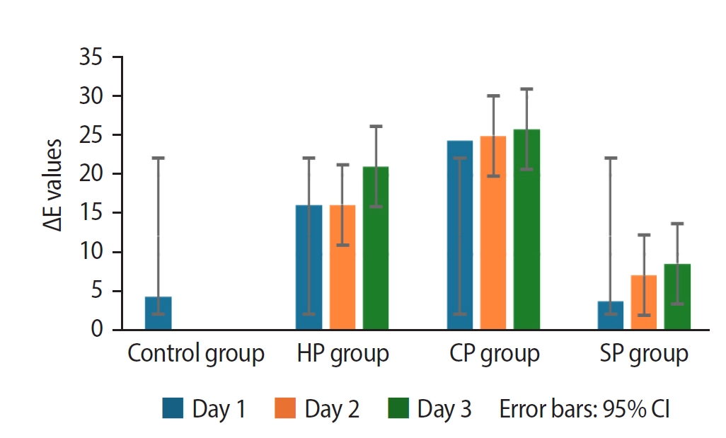
This study evaluated the efficacy of three distinct bleaching agents over time on blood-stained, devitalized teeth. Furthermore, the recoloring subsequent to bleaching will be monitored.
Methods
The study was conducted on 60 caries-free, unfilled, upper human incisors. The Freccia and Peters blood staining technique was employed, and four groups (n = 15) were identified: control, 35% hydrogen peroxide-treated, 37% carbamide peroxide-treated, and sodium perborate-treated groups. Color differences were measured using ΔE00, ΔWID, L*, a*, and b* values. To investigate tooth discoloration after bleaching, 10 unbleached teeth with three groups of 10 bleached teeth were compared by vine staining. The group of bleached teeth was restored immediately, another group waited one week, and the third group had sodium ascorbate applied and analyzed using one-way analysis of variance tests (p < 0.05).
Results
Among the groups, carbamide peroxide exhibited the most significant whitening during the 6-day bleaching process, followed by hydrogen peroxide and sodium perborate. Subsequent examination of the wine recoloring of post-bleaching samples demonstrated that bleached teeth exhibited a heightened propensity for recoloration in contrast to unbleached teeth. Notably, sodium ascorbate treatments for hydrogen peroxide neutralization and the wait-and-restore approach were not statistically significant in terms of preventing recoloration.
Conclusions
Sodium perborate is less effective and more time-consuming than hydrogen peroxide or carbamide peroxide for bleaching purposes. Carbamide peroxide is the most effective bleaching agent. The sodium ascorbate treatment and the wait-and-restore approach are ineffective in preventing recoloring. Bleached teeth have more discoloration than unbleached teeth. -
Citations
Citations to this article as recorded by
- The Effect of Adhesive Systems on Shade Matching of Composite Veneer
Fadak Al Marar, Raghad Aljarboua, Fatimah M. Alatiyyah, Shahad AlGhamdi, Faraz Ahmed Farooqi, Lama Almuhanna, Rasha AlSheikh, Abdul Samad Khan
Dentistry Journal.2026; 14(2): 85. CrossRef
- The Effect of Adhesive Systems on Shade Matching of Composite Veneer
- 4,496 View
- 282 Download
- 1 Web of Science
- 1 Crossref

- How protocol, posts, and experience affect fracture detection in multi-rooted teeth using cone-beam computed tomography: an ex vivo experimental study
- Gleica Dal’ Ongaro Savegnago, Gabriela Marzullo de Abreu, Carolina Baumgratz Spiger, Lucas Machado Maracci, Wislem Miranda de Mello, Gabriela Salatino Liedke
- Restor Dent Endod 2025;50(3):e23. Published online July 24, 2025
- DOI: https://doi.org/10.5395/rde.2025.50.e23
-
Abstract
PDFPubReaderePub
- Objectives
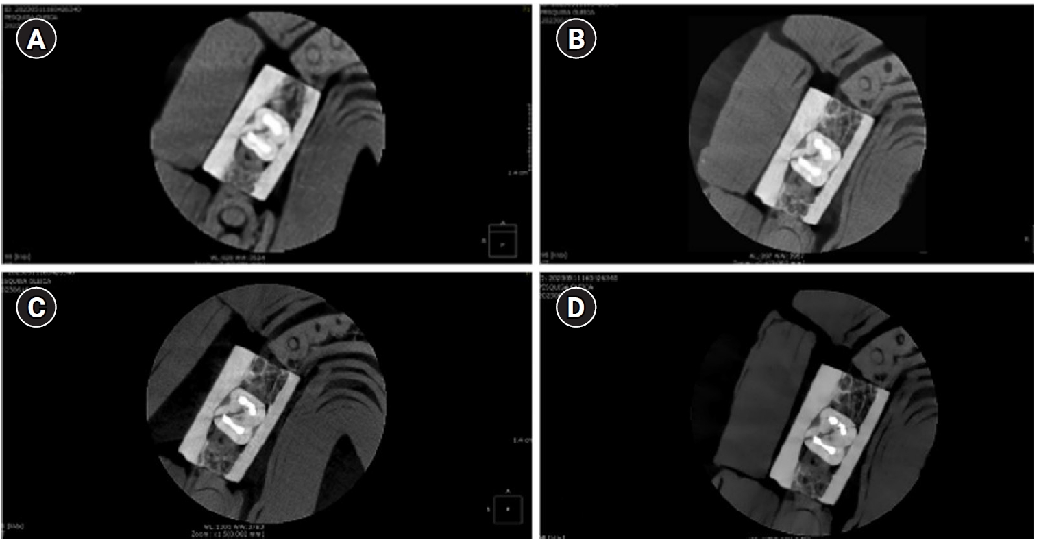
This study aimed to evaluate the influence of cone-beam computed tomography (CBCT) acquisition protocol, the presence of intraradicular metal post, and examiner experience on the detection of complete root fractures in multi-rooted teeth.
Methods
Twenty human molar teeth filled with gutta-percha were placed into artificial alveoli created in bovine ribs. The sample was divided into two groups based on the presence or absence of intraradicular posts in the distal roots. CBCT scans were obtained using four acquisition protocols with varying voxel sizes (0.28, 0.2, 0.125, and 0.80 mm). Following the creation of controlled fractures using a chisel and hammer, CBCT imaging was repeated, resulting in 160 images. Five examiners assessed the images using OnDemand software (KaVo Dental GmbH). Sensitivity, specificity, and accuracy were calculated for each examiner, CBCT protocol, and post-condition. Statistical comparisons were performed using Cochran’s Q test and McNemar test, and a significance level of 5%.
Results
In teeth without metallic posts, sensitivity, specificity, and accuracy values exceeded 0.70, 0.70, and 0.80, respectively. However, the presence of metallic posts significantly reduced diagnostic performance, particularly in low-resolution protocols evaluated by less-experienced examiners.
Conclusions
CBCT acquisition protocols should be selected based on the presence of metallic posts to optimize root fracture detection in multi-rooted teeth. Examiner experience also plays a critical role in diagnostic accuracy.
- 3,185 View
- 110 Download
- Analysis of thermal profiles on tooth structure and insert during one-piece or adapter-coupled ultrasonic insert use: an in vitro experimental study
- Gabriela Loewen Brotto, Bruno Monguilhott Crozeta, Bruno Marques-da-Silva, Alysson Nunes Diógenes, Emmanuel João Nogueira Leal da Silva, Flávia Sens Fagundes Tomazinho
- Restor Dent Endod 2025;50(3):e24. Published online July 11, 2025
- DOI: https://doi.org/10.5395/rde.2025.50.e24
-
Abstract
PDFPubReaderePub
- Objectives
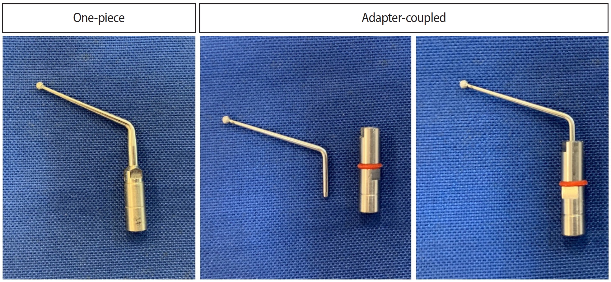
This in vitro study aimed to evaluate temperature variation on the external surface of mandibular molars and within ultrasonic inserts when using adapter-coupled versus one-piece inserts.
Methods
Twenty-four extracted human mandibular molars were divided into two groups based on the type of ultrasonic insert used: adapter-coupled and one-piece inserts. Temperature on the external surface of each tooth was measured with a thermocouple probe positioned in the furcation area, capturing data continuously. The temperature of the ultrasonic inserts was monitored in real-time using a thermal imaging camera. Measurements were taken in a controlled environment without cooling for over 120 seconds. Statistical analysis was conducted using analysis of variance (ANOVA) and two-way ANOVA with repeated measures to evaluate temperature variations between groups and over time, with significance set at 5%.
Results
In the external tooth surface temperature measurements, no significant differences were observed between the groups during the initial 15 seconds (p = 0.185) and 30 seconds (p = 0.067). However, significant differences emerged at 60 seconds (p = 0.025), 90 seconds (p = 0.024), and 120 seconds (p = 0.020), with the one-piece insert group demonstrating higher temperatures in the furcation region. Thermal imaging of the inserts revealed a significant difference at all time points (p < 0.001), with adapter-coupled inserts showing greater heating.
Conclusions
The use of ultrasonic inserts leads to a gradual rise in temperature on the external tooth surface. One-piece inserts generated higher temperatures on the tooth, while adapter-coupled inserts exhibited greater heating within the insert.
- 2,715 View
- 109 Download
- Calcium silicate-based sealers remnants in isthmuses of mesial roots of mandibular molars: an in vitro evaluation
- David Saldanha de Brito Alencar, Ana Cristina Padilha Janini, Lauter Eston Pelepenko, Brenda Fornazaro Moraes, Francisco Haiter Neto, Marco Antonio Hungaro Duarte, Marina Angélica Marciano
- Restor Dent Endod 2025;50(3):e25. Published online July 15, 2025
- DOI: https://doi.org/10.5395/rde.2025.50.e25
-
Abstract
PDFPubReaderePub
- Objectives
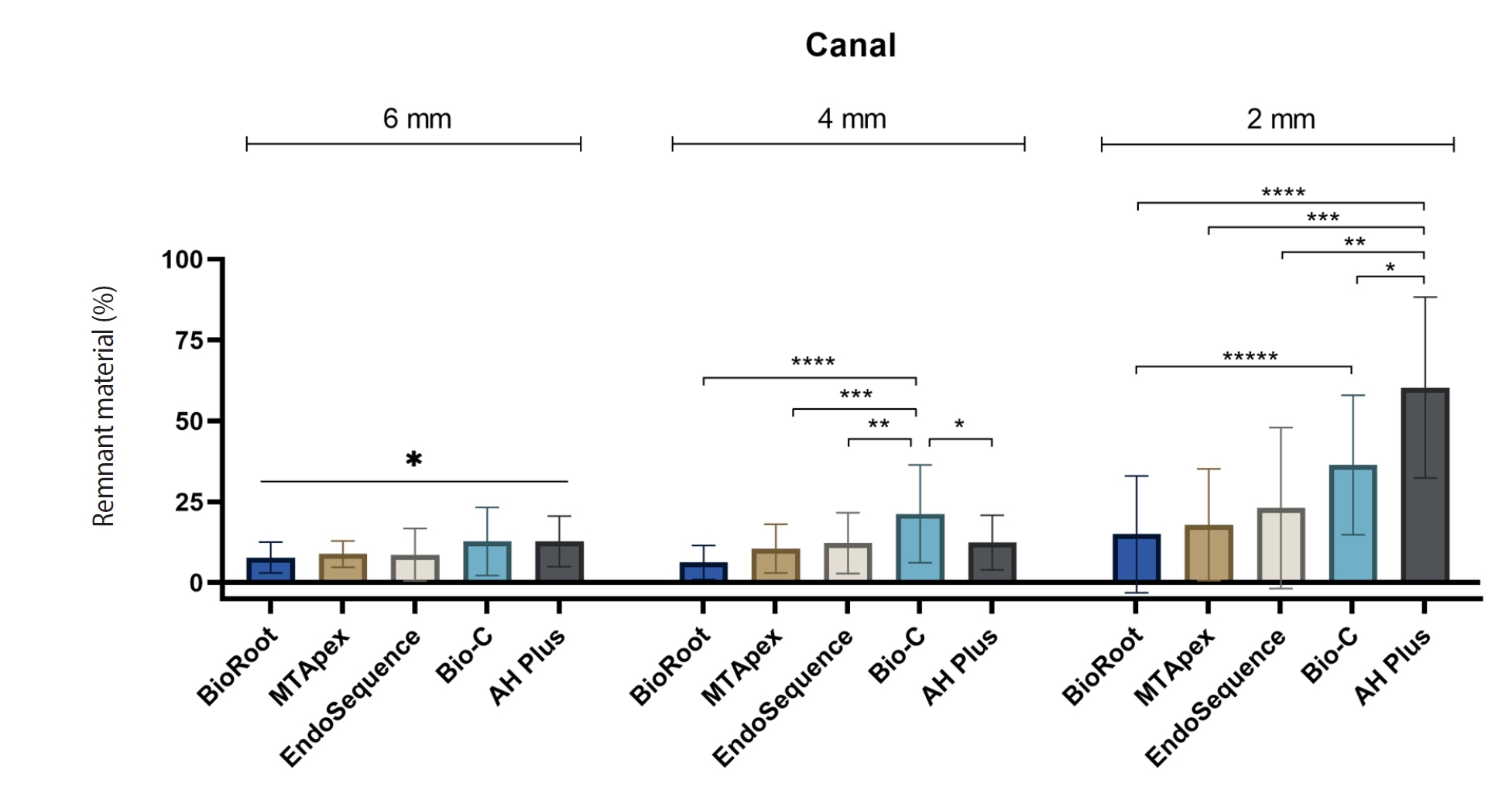
Endodontic retreatment aims to address treatment failure through the removal of root canal filling materials. This in vitro study evaluated the presence of filling material remnants in the mesial root canals, specifically focusing on the isthmuses, of mandibular molars after retreatment.
Methods
One hundred extracted mandibular molar mesial roots with isthmuses were prepared with an R25 file, obturated with one of five calcium silicate-based sealers (BioRoot RCS [Septodont], MTApex [Ultradent Products Inc.], EndoSequence BC Sealer HiFlow [Brasseler USA], Bio-C Sealer [Angelus]) or an epoxy resin-based sealer (AH Plus Jet [Dentsply Maillefer]), all stained with rhodamine B, and stored at 37ºC for 30 days to allow for setting. Retreatment was subsequently performed using R40 and XP-endo Finisher R instruments (FKG Dentaire) with 2.5% sodium hypochlorite irrigation. The presence of remaining filling material was then assessed using confocal microscopy, and setting times were tested per ISO 6876:2012.
Results
AH Plus Jet showed the most remnants at 2 mm and the longest retreatment time. Calcium silicate-based sealers exhibited prolonged setting times under dry conditions, with EndoSequence BC Sealer HiFlow showing a particularly extended setting period.
Conclusions
Despite retreatment, residues remained in all canals and isthmus regions, particularly Bio-C Sealer and AH Plus Jet in apical areas, emphasizing the difficulty of complete removal and the persistence of filling material. -
Citations
Citations to this article as recorded by- Bonding effects of mechanical removal of bioceramic sealer residues using glycine or glass microparticles abrasion
Jesus Aranda, Julia de Freitas Ceccato, Eduardo Fernández Godoy, João Felipe Besegato, Joissi Ferrari Zaniboni, Regina Guenka Palma-Dibb, Milton Carlos Kuga
International Journal of Adhesion and Adhesives.2026; 148: 104289. CrossRef
- Bonding effects of mechanical removal of bioceramic sealer residues using glycine or glass microparticles abrasion
- 2,842 View
- 130 Download
- 1 Web of Science
- 1 Crossref
- Comparison of YouTube, TikTok, and Instagram as digital sources for obtaining information about pulp therapy in primary and permanent teeth
- Hüseyin Gürkan Güneç, Emine Kaya, Dila Nur Okumuş, Merve Gül Erence
- Restor Dent Endod 2025;50(3):e26. Published online July 24, 2025
- DOI: https://doi.org/10.5395/rde.2025.50.e26
-
Abstract
PDFPubReaderePub
- Objectives
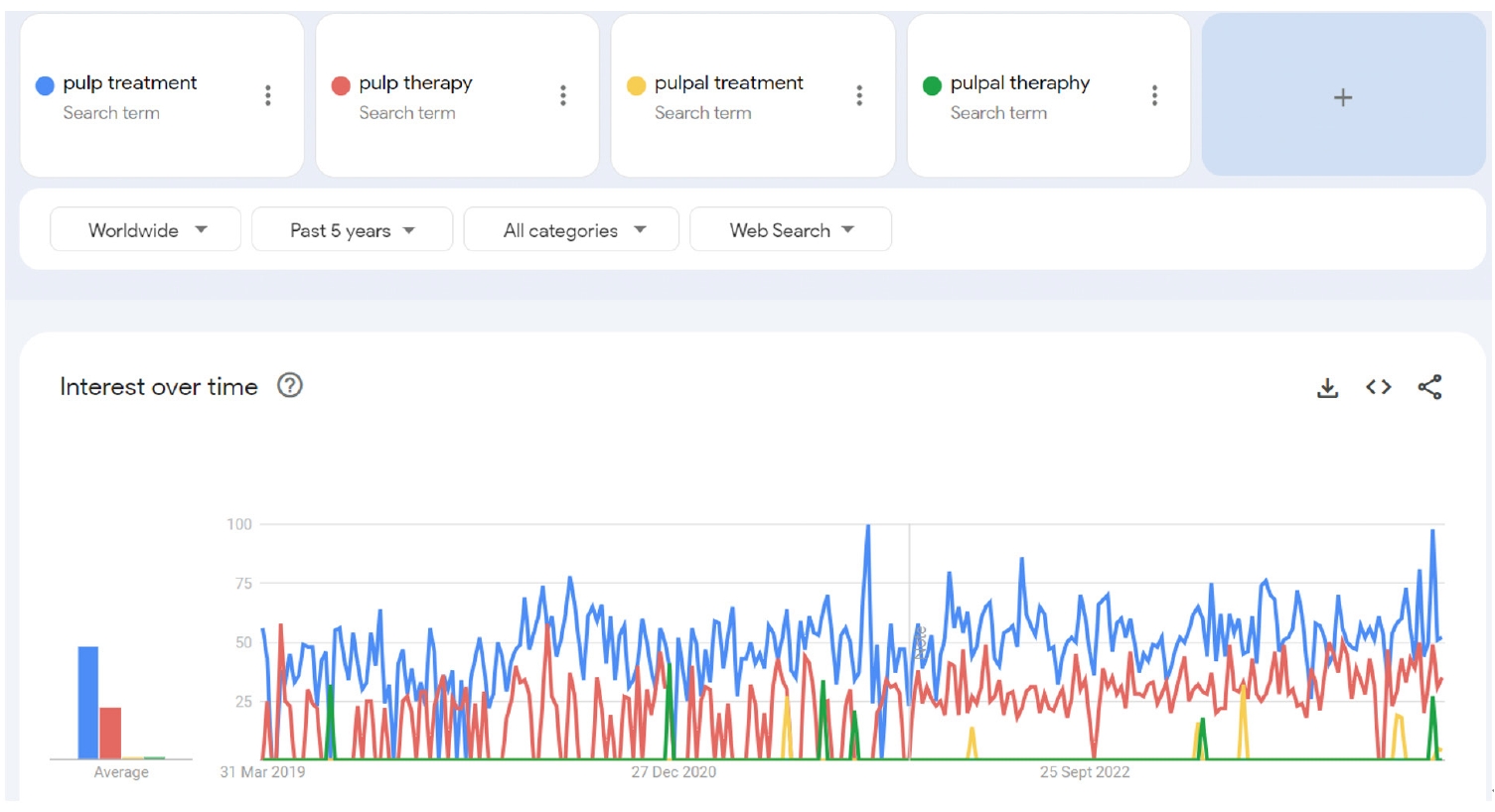
This study aimed to compare the content, educational quality, and dependability of videos on Instagram, TikTok, and YouTube about pulp therapy (PT) in pediatric dentistry and endodontics.
Methods
Three popular video sites, Instagram (Meta Platforms, Inc.,), TikTok (ByteDance Ltd.), and YouTube (Google LLC), were searched for PT content to analyze for compliance with the American Association of Endodontists and American Academy of Pediatric Dentistry guidelines for clinical endodontists and pediatric dentists. The searched hashtags were #pulpaltherapy, #pulpaltreatment, #pulptherapy, and #pulptreatment. The classification of 158 English-language videos was based on several variables: communication quality, duration, likes and dislikes, views, source, treatment, and genre. The videos were evaluated using a usefulness score and the Global Quality Scale (GQS), Video Information and Quality Index (VIQI), Journal of the American Medical Association (JAMA) score, and modified DISCERN score to rate their quality and reliability. The majority of the videos were published by healthcare professionals, dental clinics, and universities.
Results
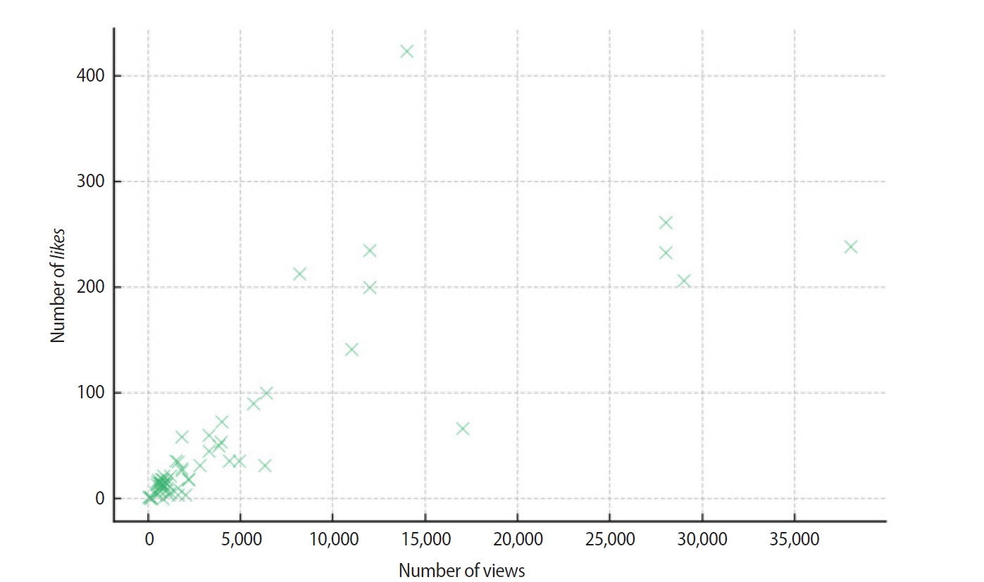
Significant relationships existed between video length, source of upload, usefulness score, tooth type, pulp status, and VIQI, JAMA, GQS, and DISCERN scores for all three platforms (p<0 .05). A statistically significant relationship existed of YouTube, TikTok, and Instagram with the number of views, number of months since upload, view rates, comments and likes (p< 0.05).
Conclusions
TikTok and Instagram reel videos provided high- to moderate-quality information about PT, especially in children, but YouTube may provide more reliable information than other social media tools. -
Citations
Citations to this article as recorded by- Evaluating the reliability and educational quality of YouTube™ and TikTok™ videos on custom subperiosteal implants: a cross-sectional methodological analysis
Göksel Tımarcıoğlu, Erdal Cem Kargu, Hülya Çerçi Akçay, Başak Tımarcıoğlu
BMC Oral Health.2026;[Epub] CrossRef - New Technologies and Materials in Oral Health and Dental Care of Pediatric Dentistry
Giuseppe Minervini
Children.2025; 12(10): 1310. CrossRef
- Evaluating the reliability and educational quality of YouTube™ and TikTok™ videos on custom subperiosteal implants: a cross-sectional methodological analysis
- 3,450 View
- 88 Download
- 2 Web of Science
- 2 Crossref
- Is YouTube a reliable source for learning pre-endodontic build-up? A cross-sectional study
- Merve Gökyar, İdil Özden, Hesna Sazak Öveçoğlu
- Restor Dent Endod 2025;50(3):e27. Published online July 24, 2025
- DOI: https://doi.org/10.5395/rde.2025.50.e27
-
Abstract
PDFPubReaderePub
- Objectives
The aim of this study is to comprehensively analyze the quality, educational value, and demographic characteristics of pre-endodontic build-up videos published on the YouTube™ platform (Google LLC).
Methods
The study was conducted on YouTube™ using the keyword “pre-endodontic build-up.” The first 100 videos retrieved from the search results were reviewed, and 61 videos meeting the inclusion criteria were analyzed. After assessing the demographic characteristics of the videos, viewing rates and interaction indices were calculated. The quality of the videos was evaluated using the DISCERN instrument and the Global Quality Scale (GQS). Statistical analyses were performed on the obtained results.
Results
A total of 61 videos were analyzed, of which 56% were uploaded by endodontists. The majority of the videos were found to be of low quality. As the DISCERN score increased, video duration, number of likes, number of comments, and view rate also increased. Additionally, a significant positive correlation was observed between the DISCERN score and the GQS value (p = 0.004). The relationship between video upload sources and various parameters was analyzed, revealing statistically significant differences (p < 0.05).
Conclusions
Considering all the evaluation methods used in this study, it is evident that the number of high-quality videos is low. This finding indicates that YouTube™ does not provide sufficient information on pre-endodontic build-up. To enhance its reliability as a source of medical information, YouTube™ should prioritize content that is not only popular but also accurate and of high quality, preferably created or endorsed by professionals. -
Citations
Citations to this article as recorded by- YouTube™ as an information source for non-surgical root canal retreatment: quality and content analysis
Betül Uslu, Arzu Kaya Mumcu
BMC Oral Health.2026;[Epub] CrossRef
- YouTube™ as an information source for non-surgical root canal retreatment: quality and content analysis
- 3,225 View
- 88 Download
- 1 Web of Science
- 1 Crossref
- Isolating design variables by assessing the impact of cross-section geometry on the mechanical performance of nickel-titanium rotary instruments: a comparative in vitro study
- Anne Rafaella Tenório Vieira, Guilherme Ferreira da Silva, Emmanuel João Nogueira Leal da Silva, Rodrigo Ricci Vivan, João Vitor Oliveira de Amorim, Thaine Oliveira Lima, Raimundo Sales de Oliveira Neto, Marco Antonio Hungaro Duarte, Murilo Priori Alcalde
- Restor Dent Endod 2025;50(3):e28. Published online July 24, 2025
- DOI: https://doi.org/10.5395/rde.2025.50.e28
-
Abstract
PDFPubReaderePub
- Objectives
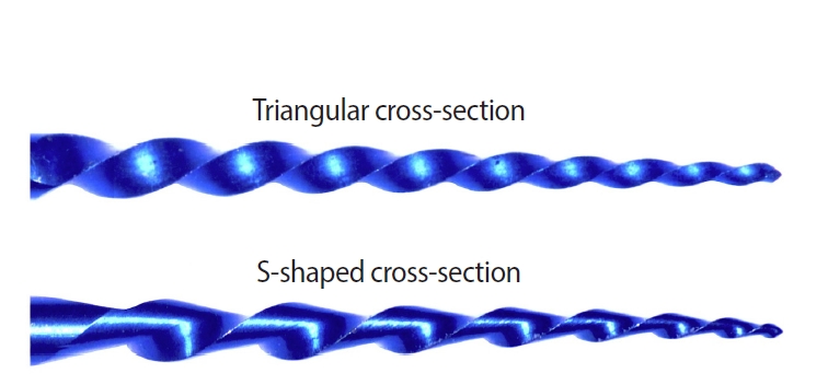
This study aimed to assess the effect of cross-section geometry on the mechanical properties of nickel-titanium (NiTi) instruments by comparing two instruments with identical tip size, taper, and thermal treatment but differing in cross-section design.
Methods
One hundred four NiTi rotary instruments, being S-shaped and triangular cross-section, manufactured with Blueish thermal treatment, were tested (n = 52 per group). Differential scanning calorimetry was employed, and the metal mass volume and cross-section area were assessed. The cyclic fatigue, torsional, and bending resistance tests were assessed. Data were analyzed using the Kolmogorov-Smirnov and Student t tests, and the level of significance was set at 5%.
Results
The instruments exhibited similar start and finish temperatures of phase transformation. The S-shaped instruments had significantly lower metal mass volume and cross-sectional area (p < 0.05). S-shaped instruments demonstrated superior cyclic fatigue resistance, greater angular deflection, and lower bending stiffness (p < 0.05).
Conclusions
Cross-section geometry significantly influences the mechanical properties of NiTi rotary instruments. -
Citations
Citations to this article as recorded by- Mechanical properties and micro-CT-based biomechanical performance of NiTi instrumentation systems in mandibular molars
Jeneffer Vieira Rodrigues, Julia Godoi-Lopes, Heitor Silva Prado, Anne Rafaella Tenório Vieira, Rafael Verardino Camargo, Graziela Bianchi Leoni, Marco Antonio Hungaro Duarte, Igor Bassi Ferreira Petean, Manoel Damião Sousa-Neto, Murilo Priori Alcalde, Fa
Odontology.2026;[Epub] CrossRef
- Mechanical properties and micro-CT-based biomechanical performance of NiTi instrumentation systems in mandibular molars
- 3,431 View
- 110 Download
- 2 Web of Science
- 1 Crossref
- Does the use of different root canal sealers and adhesive resin cements impact the bond strength of glass fiber posts?
- Ália Regina Neves de Paula Porto, Rudá França Moreira, Felipe Gonçalves Belladonna, Victor Talarico Leal Vieira, Emmanuel João Nogueira Leal da Silva
- Restor Dent Endod 2025;50(3):e29. Published online August 29, 2025
- DOI: https://doi.org/10.5395/rde.2025.50.e29
-
Abstract
PDFPubReaderePub
- Objectives
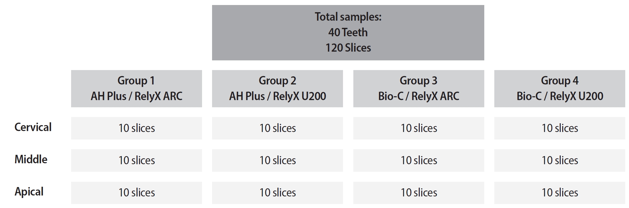
This study aimed to assess the influence of two endodontic sealers on the bond strength of glass fiber posts using conventional and self-adhesive resin cement through a push-out test. Methods: Forty central human incisors were randomly divided into four groups (n = 10) based on sealer (epoxy resin- based or calcium silicate-based) and cement (conventional and self-adhesive resin) types: AH Plus (Dentsply De- Trey)/RelyX ARC (3M ESPE), AH Plus/RelyX U200 (3M ESPE), Bio-C Sealer (Angelus)/RelyX ARC, and Bio-C Sealer/RelyX U200. After canal filling and post cementation, roots were sectioned to obtain one specimen per root third. A pushout test and failure pattern assessment were conducted, with bond strength analyzed using the one-way analysis of variance and Tukey test. Results: AH Plus/RelyX ARC showed the highest bond strength values, with a significant difference in the middle third. The most common failure was mixed (55%), while adhesive failures made up 45%, with 23.5% at the cement/post interface and 21.5% at the cement/dentin interface. Conclusions: AH Plus/RelyX ARC provided the highest bond strength values for glass fiber posts to dentin. -
Citations
Citations to this article as recorded by- Effect of Endodontic Sealers on the Bond Strength of Glass Fibre Posts: A Systematic Review
Thiago Bessa Marconato Antunes, Juliana D. Bronzato, Vanessa Gallego Arias Pecorari, Jennifer Santos Pereira, Talita Tartari, Adriana de Jesus Soares, Brenda P. F. A. Gomes, Marina Angélica Marciano
Australian Endodontic Journal.2026;[Epub] CrossRef
- Effect of Endodontic Sealers on the Bond Strength of Glass Fibre Posts: A Systematic Review
- 2,931 View
- 176 Download
- 1 Web of Science
- 1 Crossref
- Structural and morphological characterization of silver nanoparticles intruded mineral trioxide aggregate admixture as a chair-side restorative medicament: an in vitro experimental study
- H. Murali Rao, Rajkumar Krishnan, Chitra Shivalingam, Ramya Ramadoss
- Restor Dent Endod 2025;50(3):e30. Published online August 8, 2025
- DOI: https://doi.org/10.5395/rde.2025.50.e30

-
Abstract
PDFPubReaderePub
- Objectives
The aim of this study was to create a rapid admixture of mineral trioxide aggregate (MTA) and silver nanoparticles (AgNPs) for chairside use in clinical settings to remediate the challenges associated with root canal treatment and pulp capping.
Methods
Synthesized AgNPs at ratios of 10 and 25% were added to commercially available MTA to create an admixture. The admixture was subjected to structural and morphological assessment using X-ray diffraction analysis (XRD), Fourier transform infrared (FT-IR) analysis, Raman spectroscopy, and scanning electron microscopy. Antioxidant activity was measured using the hydroxyl radical scavenging assay. A significance level of 0.05 was applied to determine statistical differences.
Results
The addition of AgNPs decreased the carbonate peak intensity in XRD and FT-IR. The rod-like morphology of MTA was changed to a flake-like morphology with the addition of AgNPs. Antibacterial efficacy enhanced proportionally with the augmentation of AgNPs concentration.
Conclusions
The creation of rapid admixture of MTA and AgNPs during chairside use in clinical settings can deliver beneficial characteristics of enhanced morphological features favoring mineralization and profound antibacterial effects to overcome the challenges associated with root canal treatment and pulp capping.
- 2,801 View
- 98 Download
- 1 Web of Science
Case Report
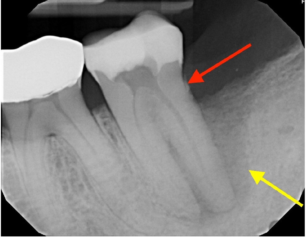
- Multidisciplinary management of an endo-perio lesion complicated by a cemental tear: a case report
- Nishanth D. Sadhak, Akshaya Pallod, Shreyas Oza
- Restor Dent Endod 2025;50(3):e31. Published online August 22, 2025
- DOI: https://doi.org/10.5395/rde.2025.50.e31
-
Abstract
PDFPubReaderePub
- Endodontic-periodontal lesions (EPLs) complicated by cemental tears present a diagnostic and therapeutic challenge. This case report describes the successful management of a 66-year-old male patient with a mandibular second molar (#18) exhibiting an EPL complicated by a cemental tear. Clinical examination revealed a draining sinus tract, deep periodontal pockets, and radiographic evidence of a “J-shaped” lesion and a radiopaque cemental fragment. The tooth had previously initiated endodontic treatment. A multidisciplinary approach involving endodontic treatment and surgical removal of the cemental tear was implemented. At 24-month follow-up, clinical and radiographic examination revealed significant improvement in periodontal health, bone regeneration, and resolution of the lesion. This case highlights the importance of considering cemental tears in the differential diagnosis of EPLs and demonstrates the efficacy of a combined endodontic-periodontal approach for achieving predictable outcomes.
- 4,652 View
- 346 Download
 First
First Prev
Prev

