
Search
- Page Path
- HOME > Search
Research Articles
- The effect of limonene extract on the adhesion of different endodontic cements to root dentin: an in vitro experimental study
- Nayara Lima Ferraz Aguiar, Eduardo José Soares, Guilherme Nilson Alves dos Santos, Anna Luísa Araújo Pimenta, Laryssa Karla Romano, Ricardo Gariba Silva, Fernanda de Carvalho Panzeri
- Restor Dent Endod 2025;50(2):e16. Published online May 12, 2025
- DOI: https://doi.org/10.5395/rde.2025.50.e16
-
 Abstract
 PDF
PDF PubReader
 ePub
ePub - Objectives
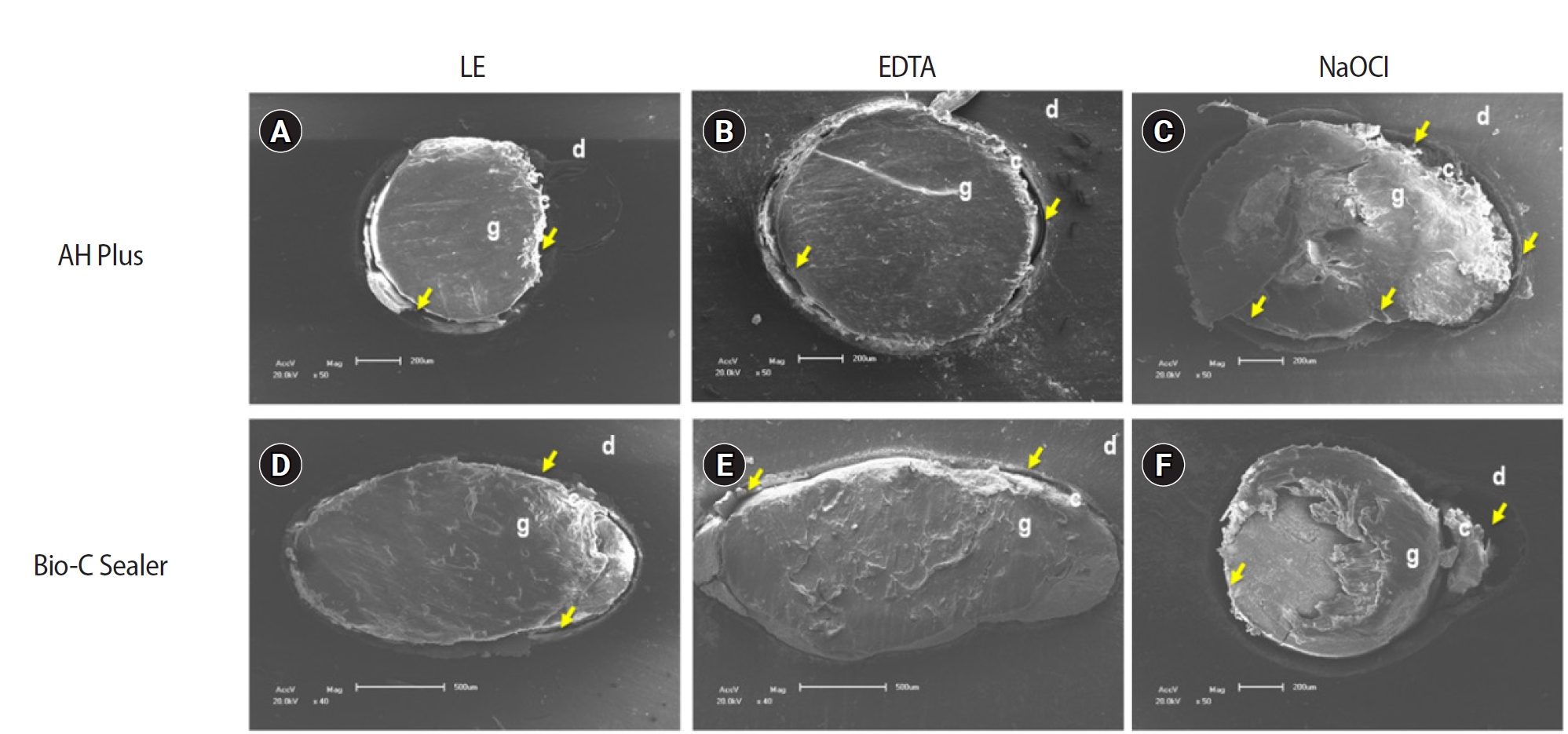
The study aimed to evaluate the effect of limonene extract (LE) on push-out bond strength (BS) to root dentin in endodontically treated teeth.
Methods
Single-rooted teeth were selected and instrumented using the reciprocating technique, then divided into three groups based on the final irrigating solution: 2.5% sodium hypochlorite (NaOCl), 17% ethylenediaminetetraacetic acid (EDTA), and 5% LE. The roots were further divided (n = 12) and obturated using the single-cone technique with epoxy resin-based (ERB) or bioceramic sealer (Bio-C). After 3 days, the roots were sectioned into 2-mm slices, obtaining two slices from each root third. Push-out BS testing was conducted at 0.5 mm/min, followed by failure pattern and adhesive interface analysis using scanning electron microscopy. Push-out BS data were analyzed by three-way analysis of variance and Tukey post-hoc test (p < 0.05).
Results
ERB showed higher BS when irrigated with EDTA (5.0 ± 2.3 MPa) compared to NaOCl (1.8 ± 1.1 MPa) (p = 0.0005), particularly in the cervical third. LE yielded intermediate values without significant differences from the other irrigants (3.5 ± 1.9 MPa) (p > 0.05). For Bio-C, the highest BS was observed in the apical third, especially with LE (9.4 ± 5.0 MPa), differing from other thirds and final irrigating solutions (p < 0.05). Mixed failure patterns were most prevalent, regardless of the irrigant solutions.
Conclusions
The combination of LE with Bio-C demonstrated superior BS in the apical third, suggesting its potential as a final irrigating solution in endodontic treatments.
- 3,095 View
- 237 Download

- Pomegranate extract on eroded dentin: antioxidant action, bond strength and morphology of the adhesive interface after aging
- Thiago Vinícius Cortez, Nathália Mancioppi Cerqueira, Julia Adornes Gallas, Wanderley Pereira Oliveira, Silmara Aparecida Milori Corona, Aline Evangelista Souza-Gabriel
- Restor Dent Endod 2024;49(1):e9. Published online January 26, 2024
- DOI: https://doi.org/10.5395/rde.2024.49.e9
-
Abstract
PDFPubReaderePub
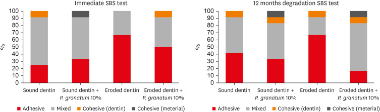
Objectives This study aimed to evaluate the effect of pomegranate solution (
Punica granatum ) on eroded dentin through antioxidant action, shear bond strength (SBS) and interface morphology.Materials and Methods The 10% pomegranate peel extract was prepared by the lyophilization method. Punicalagin polyphenol was confirmed by high-performance liquid chromatography. Antioxidant activity was evaluated by capturing the 2,2-diphenyl-1-picrylhydrazyl (DPPH) radical. For the SBS, 48 dentin fragments were divided into sound or eroded, and subdivided according to the pretreatment (
n = 12): water orP. granatum . The surfaces were restored with self-etch adhesive and a bulk-fill resin (Ecosite; DMG). The SBS was done immediately (24 hours) and after thermal cycling + water storage (12 months). For scanning electron microscopy, 48 dentin fragments (24 sound and 24 eroded) received the same treatments as for SBS (n = 6), and they were analyzed after 24 hours and 12 months.Results The
P. granatum had antioxidant action similar (p = 0.246) to the phenolic standard antioxidants. After 24 hours, eroded dentin had lower SBS than sound dentin (p < 0.001), regardless of the pretreatment. After 12 months,P. granatum maintained the SBS of sound dentin (13.46 ± 3.42 MPa) and eroded dentin (10.96 ± 1.90 MPa) statistically similar. The lowest values were found on eroded dentin treated with water (5.75 ± 1.65 MPa) (p < 0.001).P. granatum on eroded dentin caused peritubular demineralization and hybrid layer with resin tags.Conclusions The pomegranate extract had antioxidant action and preserved the adhesive interface of the eroded dentin.
-
Citations
Citations to this article as recorded by
- Spouted bed drying parameters and adjuvant effects on polyphenol retention and antioxidant activity in mango peel powders
Guilherme Henrique Alves Pinto, Fábio Bentes Freire, José Teixeira Freire, Wanderley Pereira Oliveira
The Canadian Journal of Chemical Engineering.2026;[Epub] CrossRef - Antibacterial Effects of Ethanolic Extractions of Aloe Vera, Black Tea, Pomegranate and Orange on Streptococcus mutans: An In-vitro Study
Bardia Vadiati Saberi, Soheil Taghavi Namin, Dina Maleki
Nutrition And Food In Health And Disease.2025; 12(2): 29. CrossRef - Protective effect of a novel antioxidant gel containing resveratrol and sodium fluoride on dentin erosion in the presence of acquired salivary pellicle: An in vitro study
Loraine Perez Manzoli, Luan Júlio Ruiz da Silva, George Clay dos Santos Caracas, Kalinca Furtado de Oliveira, Walessa Alana Braganca Aragão, Rafael Rodrigues Lima, Milton Carlos Kuga, Cristiane de Melo Alencar
Archives of Oral Biology.2025; 179: 106395. CrossRef - Effect of pomegranate solution alone or combined with chlorhexidine against oral multispecies biofilm
J. A. Gallas, L. L. Pelozo, S. A. M. Corona, Y. Shen, M. Haapasalo, M. D. Sousa‐Neto, A. E. Souza‐Gabriel
International Endodontic Journal.2024; 57(12): 1819. CrossRef - The effect of resveratrol application on the micro-shear bond strength of adhesive to bleached enamel
Esra Cengiz-Yanardag, Izgen Karakaya
Scientific Reports.2024;[Epub] CrossRef
- Spouted bed drying parameters and adjuvant effects on polyphenol retention and antioxidant activity in mango peel powders
- 3,307 View
- 102 Download
- 4 Web of Science
- 5 Crossref
- Is dentin biomodification with collagen cross-linking agents effective for improving dentin adhesion? A systematic review and meta-analysis
- Julianne Coelho Silva, Edson Luiz Cetira Filho, Paulo Goberlânio de Barros Silva, Fábio Wildson Gurgel Costa, Vicente de Paulo Aragão Saboia
- Restor Dent Endod 2022;47(2):e23. Published online May 6, 2022
- DOI: https://doi.org/10.5395/rde.2022.47.e23
-
Abstract
PDF
 Supplementary MaterialPubReaderePub
 Objectives The aim of this investigation was to evaluate the effectiveness of collagen cross-linking agents (CCLAs) used in combination with the adhesive technique in restorative procedures.
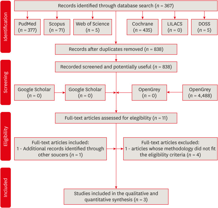
Materials and Methods In this systematic review, the authors followed the Preferred Reporting Items for Systematic Reviews and Meta-Analyses checklist. An electronic search was performed using PubMed, Scopus, Web of Science, Cochrane Library, LILACS, and DOSS, up to October 2020. The gray literature was also researched. Only randomized clinical trials were selected.
Results The selection process yielded 3 studies from the 838 retrieved. The addition of CCLAs in the retention of restorations increased the number of events. The postoperative sensitivity scores and marginal adaptation scores showed no significant difference between the CCLA and control groups, and the marginal pigmentation scores showed a significant increase in the CCLA group. There were no caries events in any group throughout the evaluation period.
Conclusions This systematic review showed that there is no clinical efficacy to justify the use of CCLAs in the protocols performed.
-
Citations
Citations to this article as recorded by- Riboflavin-ultraviolet-A collagen crosslinking treatments in improving dentin bonding and resistance to enzymatic digestion
Yung-Show Chiang, Ping-Ju Chen, Chun-Chan Ting, Yuh-Ling Chen, Shu-Fen Chuang
Journal of Dental Sciences.2025; 20(1): 109. CrossRef - Effect of dentin bio modifications and matrix metalloproteinase activity on bond strength – A systematic review and meta-analysis
D. Agarwal, S. R. Srinidhi, S. D. Aggarwal, P. Ingle, S. Tandon
Endodontics Today.2025; 23(1): 71. CrossRef - O USO DE ADESIVO AUTOCONDICIONANTE E RESINA FLOW COMO INTERFACE ADESIVA PROTETORA DA DENTINA FRENTE À IRRIGAÇÃO COM NaClO NO TRATAMENTO ENDODÔNTICO: ESTUDO IN-VITRO
Luís Daniel Ramos de Oliveira, Leandro Botelho Hanna, José Augusto Rodrigues
RECIMA21 - Revista Científica Multidisciplinar - ISSN 2675-6218.2025; 6(12): e6127063. CrossRef - Stability of dentin matrix treated with caffeic acid phenethyl ester at different concentrations
Aline Honorato Damázio, Rosanna Tarkany Basting, Enrico Coser Bridi, Fabiana Mantovani Gomes França, Flávia Lucisano Botelho do Amaral, Cecilia Pedroso Turssi, Waldemir Francisco Vieira Junior, Roberta Tarkany Basting
Brazilian Journal of Oral Sciences.2024; 23: e244006. CrossRef - Effect of Collagen Crosslinkers on Dentin Bond Strength of Adhesive Systems: A Systematic Review and Meta-Analysis
Louis Hardan, Umer Daood, Rim Bourgi, Carlos Enrique Cuevas-Suárez, Walter Devoto, Maciej Zarow, Natalia Jakubowicz, Juan Eliezer Zamarripa-Calderón, Mateusz Radwanski, Giovana Orsini, Monika Lukomska-Szymanska
Cells.2022; 11(15): 2417. CrossRef
- Riboflavin-ultraviolet-A collagen crosslinking treatments in improving dentin bonding and resistance to enzymatic digestion
- 2,912 View
- 75 Download
- 3 Web of Science
- 5 Crossref
Review Article
- Deep proximal margin rebuilding with direct esthetic restorations: a systematic review of marginal adaptation and bond strength
- Hoda S. Ismail, Ashraf I. Ali, Rabab El. Mehesen, Jelena Juloski, Franklin Garcia-Godoy, Salah H. Mahmoud
- Restor Dent Endod 2022;47(2):e15. Published online March 4, 2022
- DOI: https://doi.org/10.5395/rde.2022.47.e15
-
Abstract
PDFPubReaderePub
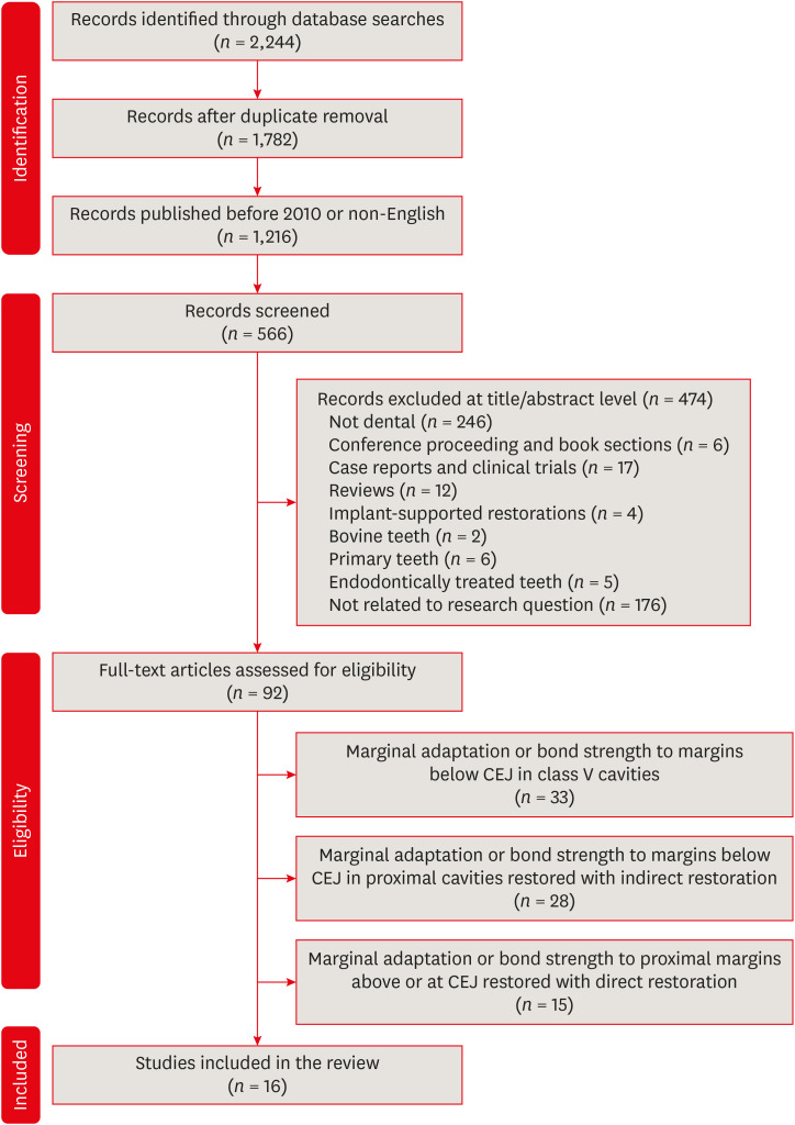
This review aimed to characterize the effect of direct restorative material types and adhesive protocols on marginal adaptation and the bond strength of the interface between the material and the proximal dentin/cementum. An electronic search of 3 databases (the National Library of Medicine [MEDLINE/PubMed], Scopus, and ScienceDirect) was conducted. Studies were included if they evaluated marginal adaptation or bond strength tests for proximal restorations under the cementoenamel junction. Only 16 studies met the inclusion criteria and were included in this review. These studies presented a high degree of heterogeneity in terms of the materials used and the methodologies and evaluation criteria of each test; therefore, only a descriptive analysis could be conducted. The included studies were individually evaluated for the risk of bias following predetermined criteria. To summarize the results of the included studies, the type of restorative material affected the test results, whereas the use of different adhesive protocols had an insignificant effect on the results. It could be concluded that various categories of resin-based composites could be a suitable choice for clinicians to elevate proximal dentin/cementum margins, rather than the open sandwich technique with resin-modified glass ionomers. Despite challenges in bonding to proximal dentin/cementum margins, different adhesive protocols provided comparable outcomes.
-
Citations
Citations to this article as recorded by- Influence of thickness and base material in class II restorations with nanofilled composites: finite element study
Fredy Hugo Cruzado-Oliva, Alexander Vega-Anticona, David Arturo Ortiz-Diaz, Heber Isac Arbildo-Vega, Franz Tito Coronel-Zubiate
Frontiers in Dental Medicine.2026;[Epub] CrossRef - An In Vitro Evaluation of Novel Bioactive Liner's Effect on Marginal Adaptation of Class II Composite Restorations: A Scanning Electron Microscope Analysis
Girija S Sajjan, Naveena Ponnada, Praveen Dalavai, Madhu Varma Kanumuri, Venkata Karteek Varma Penmatsa, B V Sindhuja
World Journal of Dentistry.2025; 15(9): 749. CrossRef - Effect of Cervical Margin Relocation With Different Injectable Restorative Materials on Fracture Resistance of Molars Received MOD CAD/CAM Onlay Restorations
Basema N. Roshdy, Radwa I. Eltoukhy, Ashraf I. Ali, Salah Hasab Mahmoud
Journal of Esthetic and Restorative Dentistry.2025; 37(6): 1522. CrossRef - Short dentin etching with universal adhesives: effect on bond strength and gingival margin adaptation
Hoda Saleh Ismail, Hanan Ahmed Nabil Soliman
BMC Oral Health.2025;[Epub] CrossRef - Awareness and Practice of Deep Margin Elevation among Dental Practitioners in India: A Cross-Sectional Survey
Mythri Padaru, Preethesh Shetty, Namith Rai, Raksha Bhat
Pesquisa Brasileira em Odontopediatria e Clínica Integrada.2025;[Epub] CrossRef - Effect of surface treatment on glass ionomers in sandwich restorations: a systematic review and meta-analysis of laboratory studies
Hoda S. Ismail, Ashraf Ibrahim Ali, Franklin Garcia-Godoy
Restorative Dentistry & Endodontics.2025; 50(2): e13. CrossRef - Do irrigation solutions effect bond strength of composite resin to deep margin elevation material? An in-vitro study
Şeref Nur Mutlu, Yasemin Derya Fidancıoğlu, Hatice Büyüközer Özkan, Hayriye Esra Ülker
BMC Oral Health.2025;[Epub] CrossRef - Two-year evaluation of periodontal parameters following deep-margin-elevation and CAD/CAM partial lithium disilicate restorations – a prospective controlled clinical trial
Tim Hausdörfer, Philipp Kanzow, Tina Rödig, Annette Wiegand, Clemens Lechte
Journal of Dentistry.2025; 160: 105901. CrossRef - Deep Margin Elevation: Current Evidence and a Critical Approach to Clinical Protocols—A Narrative Review
Athanasios Karageorgiou, Maria Fostiropoulou, Maria Antoniadou, Eftychia Pappa
Adhesives.2025; 1(3): 10. CrossRef - Comparative Micro-CT Analysis of Internal Adaptation and Closed Porosity of Conventional Layered and Thermoviscous Bulk-Fill Resin Composites Using Total-Etch or Universal Adhesives
Dóra Jordáki, Virág Veress, Tamás Kiss, József Szalma, Márk Fráter, Edina Lempel
Polymers.2025; 17(15): 2049. CrossRef - Effect of different restorative systems and aging on marginal adaptation of resin composites to deep proximal margins
Hoda S. Ismail, Ashraf I. Ali
Journal of Esthetic and Restorative Dentistry.2024; 36(2): 346. CrossRef - Management of subgingival proximal defects
Jagruti Mutalikdesai, K. C. Dhaniba, Supriya Choudhary, Promila Verma, Rhythm Bains
Asian Journal of Oral Health and Allied Sciences.2024; 14: 15. CrossRef - Effect of Deep Margin Elevation on the Pulpal and Periodontal Health of Teeth: A Systematic Review
S Srirama, S Jain, B Arul, K Prabakar, V Natanasabapathy
Operative Dentistry.2024; 49(4): 388. CrossRef - Alternative Direct Restorative Materials for Dental Amalgam: A Concise Review Based on an FDI Policy Statement
Gottfried Schmalz, Falk Schwendicke, Reinhard Hickel, Jeffrey A. Platt
International Dental Journal.2024; 74(4): 661. CrossRef - Comparison of the stress distribution in base materials and thicknesses in composite resin restorations
Min-Kwan Jung, Mi-Jeong Jeon, Jae-Hoon Kim, Sung-Ae Son, Jeong-Kil Park, Deog-Gyu Seo
Heliyon.2024; 10(3): e25040. CrossRef - Influence of curing mode and aging on the bonding performance of universal adhesives in coronal and root dentin
Hoda Saleh Ismail, Ashraf Ibrahim Ali, Mohamed Elshirbeny Elawsya
BMC Oral Health.2024;[Epub] CrossRef - CLINICAL ASSESSMENT OF THE EFFECTIVENESS OF ESTHETIC RESTORATION OF ANTERIOR TEETH
Lyudmila Tatintsyan, Minas Poghosyan, Armen Shaginyan, Hovhannes Gevorgyan, Biayna Hoveyan, Tatevik Margaryan, Arsen Kupelyan
BULLETIN OF STOMATOLOGY AND MAXILLOFACIAL SURGERY.2023; : 16. CrossRef - Deep margin elevation—Present status and future directions
Florin Eggmann, Jose M. Ayub, Julián Conejo, Markus B. Blatz
Journal of Esthetic and Restorative Dentistry.2023; 35(1): 26. CrossRef
- Influence of thickness and base material in class II restorations with nanofilled composites: finite element study
- 5,950 View
- 134 Download
- 17 Web of Science
- 18 Crossref
Research Articles
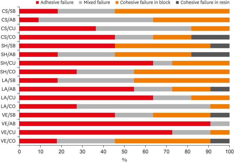
- Influence of different universal adhesives on the repair performance of hybrid CAD-CAM materials
- Gülbike Demirel, İsmail Hakkı Baltacıoğlu
- Restor Dent Endod 2019;44(3):e23. Published online May 20, 2019
- DOI: https://doi.org/10.5395/rde.2019.44.e23
-
Abstract
PDFPubReaderePub
Objectives The aim of this study was to investigate the microshear bond strength (μSBS) of different universal adhesive systems applied to hybrid computer-aided design/computer-aided manufacturing (CAD-CAM) restorative materials repaired with a composite resin.
Materials and Methods Four types of CAD-CAM hybrid block materials—Lava Ultimate (LA), Vita Enamic (VE), CeraSmart (CS), and Shofu Block HC (SH)—were used in this study, in combination with the following four adhesive protocols: 1) control: porcelain primer + total etch adhesive (CO), 2) Single Bond Universal (SB), 3) All Bond Universal (AB), and 4) Clearfil Universal Bond (CU). The μSBS of the composite resin (Clearfil Majesty Esthetic) was measured and the data were analyzed using two-way analysis of variance and the Tukey test, with the level of significance set at
p < 0.05.Results The CAD-CAM block type and block-adhesive combination had significant effects on the bond strength values (
p < 0.05). Significant differences were found between the following pairs of groups: VE/CO and VE/AB, CS/CO and CS/AB, VE/CU and CS/CU, and VE/AB and CS/AB (p < 0.05).Conclusions The μSBS values were affected by hybrid block type. All tested universal adhesive treatments can be used as an alternative to the control treatment for repair, except the AB system on VE blocks (the VE/AB group). The μSBS values showed variation across different adhesive treatments on different hybrid CAD-CAM block types.
-
Citations
Citations to this article as recorded by- Micro-shear bond strength of repaired additively manufactured resin composites after artificial aging: effect of surface treatment and universal adhesives
Neşe Ciziroğlu, Didar Dilan Hartavi, Rafat Sasany, Mutlu Özcan
Journal of Adhesion Science and Technology.2026; : 1. CrossRef - Effect of surface treatments on the bond strength of resin-repaired resin matrix CAD-CAM ceramic: A scoping review
Ana Beatriz de Souza Albergardi, João Pedro Justino de Oliveira Limírio, Jéssica Marcela de Luna Gomes, Aldiéris Alves Pesqueira, Eduardo Piza Pellizzer
Journal of Dentistry.2025; 154: 105594. CrossRef - Bond strength to aged CAD/CAM composites and polymer-infiltrated ceramic network using a universal adhesive with or without previous application of a universal primer
Clemens Lechte, Lisa Sophia Faesser, Jana Biermann, Alexandra Schmidt, Philipp Kanzow, Annette Wiegand
International Journal of Adhesion and Adhesives.2025; 140: 104017. CrossRef - The Influence of Thermocycling and Ultraviolet Aging on Surface Characteristics and the Repair Bond Strength of CAD/CAM Resin Nanoceramics
Beyza Unalan Degirmenci, Alperen Degirmenci, Zelal Seyfioglu Polat
Journal of Functional Biomaterials.2025; 16(5): 156. CrossRef - Impact of in vitro findings on clinical protocols for the adhesion of CAD-CAM blocks: A systematic integrative review and meta-analysis
Maria João Calheiros-Lobo, Ricardo Carbas, Lucas F.M. da Silva, Teresa Pinho
The Journal of Prosthetic Dentistry.2024; 131(6): 1051. CrossRef - Repair protocols for indirect monolithic restorations: a literature review
Lucas Saldanha da Rosa, Rafaela Oliveira Pilecco, Pablo Machado Soares, Marília Pivetta Rippe, Gabriel Kalil Rocha Pereira, Luiz Felipe Valandro, Cornelis Johannes Kleverlaan, Albert J. Feilzer, João Paulo Mendes Tribst
PeerJ.2024; 12: e16942. CrossRef - Bonding performance of universal adhesives with concomitant use of silanes to CAD/CAM blocks
Marina AMARAL, Jaqueline Maria Brandão RIZZATO, Victoria Caroline Souza de ALMEIDA, Priscila Christiane Suzy LIPORONI, Rayssa Ferreira ZANATTA
RGO - Revista Gaúcha de Odontologia.2023;[Epub] CrossRef - Resistencia a la fractura de una nanocerámica CAD/CAM reparada con dos tratamientos de superficie: estudio in vitro
Marcelo Geovanny Cascante-Calderón, Kevin Alejandro Reascos Flores, Inés María Villacís-Altamirano, Anggely Maite Bayas Salinas, Jessica Elizabeth Taraguay Galindo
Universitas Odontologica.2023;[Epub] CrossRef - Influence of surface treatments and adhesive protocols on repair bond strength of glass‐matrix and resin‐matrix CAD/CAM ceramics
Rana Turunç‐Oğuzman, Soner Şişmanoğlu
Journal of Esthetic and Restorative Dentistry.2023; 35(8): 1322. CrossRef - Effect of Anti-COVID-19 Mouthwashes on Shear Bond Strength of Resin-Matrix Ceramics Repaired with Resin Composite Using Universal Adhesive: An In Vitro Study
Wichuda Limsiriwong, Awiruth Klaisiri, Nantawan Krajangta
Journal of Functional Biomaterials.2023; 14(3): 158. CrossRef - Effect of ceramic primers with different chemical contents on the shear bond strength of CAD/CAM ceramics with resin cement after thermal ageing
Mehmet Uğur, İdris Kavut, Özgür Ozan Tanrıkut, Önder Cengiz
BMC Oral Health.2023;[Epub] CrossRef - Effect of microstructure of reinforced CAD/CAM hybrid composite resin block on shear bond strength of composite resin
Sung-Ho Um, Minjeong Shin, Shin-hye Chung, Young-Seok Park, Bum-Soon Lim
Korean Journal of Dental Materials.2023; 50(1): 29. CrossRef - Dentin contamination during repair procedures: A threat to universal adhesives?
Anne‐Katrin Lührs, Cosima Brachmann, Silke Jacker‐Guhr
Clinical and Experimental Dental Research.2022; 8(3): 771. CrossRef - Influence of mechanical and chemical pre-treatments on the repair of a hybrid ceramic
Sascha Niklas Jung, Stefan Rüttermann
Dental Materials.2022; 38(7): 1140. CrossRef - Influence of different repair protocols and artificial aging on bond strength of composite to a CAD/CAM polymer-infiltrated ceramic
Ece İrem OĞUZ, Gökhan ÇİÇEKCİ
Cumhuriyet Dental Journal.2021; 24(1): 37. CrossRef - REZİN MATRİKS SERAMİKLER-DERLEME
Elif Melike AKARCA, Dilara ŞAHİN, Ragibe Şenay CANAY
Atatürk Üniversitesi Diş Hekimliği Fakültesi Dergisi.2021; : 1. CrossRef - REZİN MATRİKS SERAMİKLER-DERLEME
Elif Melike AKARCA, Dilara ŞAHİN, Ragibe Şenay CANAY
Atatürk Üniversitesi Diş Hekimliği Fakültesi Dergisi.2021; : 1. CrossRef - Microshear bond strength of contemporary self-adhesive resin cements to CAD/CAM restorative materials: effect of surface treatment and aging
Soner Şişmanoğlu, Rana Turunç-Oğuzman
Journal of Adhesion Science and Technology.2020; 34(22): 2484. CrossRef - Influence of different surface treatments and universal adhesives on the repair of CAD-CAM composite resins: An in vitro study
Soner Sismanoglu, Zuhal Yildirim-Bilmez, Aysegul Erten-Taysi, Pınar Ercal
The Journal of Prosthetic Dentistry.2020; 124(2): 238.e1. CrossRef
- Micro-shear bond strength of repaired additively manufactured resin composites after artificial aging: effect of surface treatment and universal adhesives
- 4,320 View
- 19 Download
- 19 Crossref
- Influence of silver nanoparticles on resin-dentin bond strength durability in a self-etch and an etch-and-rinse adhesive system
- Zahra Jowkar, Fereshteh Shafiei, Elham Asadmanesh, Fatemeh Koohpeima
- Restor Dent Endod 2019;44(2):e13. Published online March 29, 2019
- DOI: https://doi.org/10.5395/rde.2019.44.e13
-
Abstract
PDFPubReaderePub
Objectives This study evaluated the effect of dentin pretreatment with silver nanoparticles (SNPs) and chlorhexidine (CHX) on the microshear bond strength (µSBS) durability of different adhesives to dentin.
Materials and Methods Occlusal surfaces of 120 human molars were ground to expose flat dentin surfaces. The specimens were randomly assigned to six groups (
n = 20). Three groups (A, B, and C) were bonded with Adper Single Bond 2 (SB) and the other groups (D, E, and F) were bonded with Clearfil SE Bond (SEB). Dentin was pretreated with CHX in groups B and E, and with SNPs in groups C and F. The specimens were restored with Z250 composite. Half of the bonded surfaces in each group underwent µSBS testing after 24 hours and the other half was tested after 6 months of water storage.Results SNP application was associated with a higher µSBS than was observed in the CHX and control groups for SEB after 24 hours (
p < 0.05). A significantly lower µSBS was observed when no dentin pretreatment was applied compared to dentin pretreatment with CHX and SNPs for SB after 24 hours (p < 0.05). The µSBS values of the 6-month specimens were significantly lower than those obtained from the 24-hour specimens for all groups (p < 0.05). This decrease was much more pronounced when both adhesives were used without any dentin pretreatment (p < 0.05).Conclusions SNPs and CHX reduced the degradation of resin-dentin bonds over a 6-month period for both adhesive systems.
-
Citations
Citations to this article as recorded by- Effect of nano silver fluoride pre-treatment on the microtensile bond strength of a universal adhesive to dentin: an in vitro study
Larissa Alexsandra dos Santos Silva, Anna Caroline Lima Dos Santos, Luis Felipe Espíndola-Castro, Ana Luísa Cassiano Alves, Márcia de Almeida Durão, Maria Clara Müller De Andrade, André Galembeck, Gabriela Queiroz de Melo Monteiro
Biomaterial Investigations in Dentistry.2026; 13: 198. CrossRef - An in vitro comparative evaluation of silver and chitosan nanoparticles on shear bond strength of nanohybrid composite using different adhesion protocols
Roopadevi Garlapati, Nagesh Bolla, Mayana Aameena Banu, Anila Bandlapally Sreenivasa Guptha, Niharika Halder, Ram Chowdary Basam
Journal of Conservative Dentistry and Endodontics.2025; 28(6): 522. CrossRef - Nanoparticle-enhanced dental adhesives: improving dentin bond strength through multifunctional nanotechnology
Suleiman Ibrahim Mohammad, Asokan Vasudevan, Lashin Saad Ali, Wenchang Chen
The Journal of Adhesion.2025; : 1. CrossRef - The Effect of Silver Nanoparticles on Bond Strength of Calcium Silicate-Based Sealer: An In Vitro Study
Sundus Bukhary, Sarah Alkahtany, Dalal AlDabeeb
Applied Sciences.2024; 14(21): 9817. CrossRef - Performance of self-etching adhesives on caries-affected primary dentin treated with glutaraldehyde or silver diamine fluoride
Marcelly Tupan Christoffoli Wolowski, Andressa Mioto Stabile Grenier, Victória Alícia de Oliveira, Caroline Anselmi, Mariana Sversut Gibin, Lidiane Vizioli de Castro-Hoshino, Francielle Sato, Cristina Perez, Régis Henke Scheffel, Josimeri Hebling, Mauro L
Journal of the Mechanical Behavior of Biomedical Materials.2024; 150: 106293. CrossRef - The Impact of Silver Nanoparticles on Dentinal Tubule Penetration of Endodontic Bioceramic Sealer
Sundus Bukhary, Sarah Alkahtany, Amal Almohaimede, Nourah Alkhayatt, Shahad Alsulaiman, Salma Alohali
Applied Sciences.2024; 14(24): 11639. CrossRef - Effect of silver diamine fluoride on the longevity of the bonding properties to caries-affected dentine
LP Muniz, M Wendlinger, GD Cochinski, PHA Moreira, AFM Cardenas, TS Carvalho, AD Loguercio, A Reis, FSF Siqueira
Journal of Dentistry.2024; 143: 104897. CrossRef - Evaluation of Chitosan-Oleuropein Nanoparticles on the Durability of Dentin Bonding
Shuya Zhao, Yunyang Zhang, Yun Chen, Xianghui Xing, Yu Wang, Guofeng Wu
Drug Design, Development and Therapy.2023; Volume 17: 167. CrossRef - Influence of silver nanoparticles on the resin-dentin bond strength and antibacterial activity of a self-etch adhesive system
Jia Wang, Wei Jiang, Jingping Liang, Shujun Ran
The Journal of Prosthetic Dentistry.2022; 128(6): 1363.e1. CrossRef - Marginal Integrity of Composite Restoration with and without Surface Pretreatment by Gold and Silver Nanoparticles vs Chlorhexidine: A Randomized Controlled Trial
Aya AEM Nemt-Allah, Shereen H Ibrahim, Amira F El-Zoghby
The Journal of Contemporary Dental Practice.2022; 22(10): 1087. CrossRef - Effect of Cavity Disinfectants on Dentin Bond Strength and Clinical Success of Composite Restorations—A Systematic Review of In Vitro, In Situ and Clinical Studies
Ana Coelho, Inês Amaro, Beatriz Rascão, Inês Marcelino, Anabela Paula, José Saraiva, Gianrico Spagnuolo, Manuel Marques Ferreira, Carlos Miguel Marto, Eunice Carrilho
International Journal of Molecular Sciences.2020; 22(1): 353. CrossRef
- Effect of nano silver fluoride pre-treatment on the microtensile bond strength of a universal adhesive to dentin: an in vitro study
- 2,072 View
- 18 Download
- 11 Crossref
- Do universal adhesives promote bonding to dentin? A systematic review and meta-analysis
- Ali A. Elkaffas, Hamdi H. H. Hamama, Salah H. Mahmoud
- Restor Dent Endod 2018;43(3):e29. Published online June 18, 2018
- DOI: https://doi.org/10.5395/rde.2018.43.e29
-
Abstract
PDFPubReaderePub
Objectives The aims of this study were to conduct a systematic review of the microtensile bond strength (µTBS) of multi-mode adhesives to dentin and to perform a meta-analysis to assess the significance of differences in the µTBS of one of the most commonly used universal adhesives (Scotchbond Universal, 3M ESPE) depending on whether the etch-and-rinse or self-etch mode was used.
Materials and Methods An electronic search was performed of MEDLINE/PubMed, ScienceDirect, and EBSCOhost. Laboratory studies that evaluated the µTBS of multi-mode adhesives to dentin using either the etch-and-rinse or self-etch mode were selected. A meta-analysis was conducted of the reviewed studies to quantify the differences in the µTBS of Scotchbond Universal adhesive.
Results Only 10 studies fulfilled the inclusion criteria for the systematic review. Extensive variation was found in the restorative materials, testing methodologies, and failure mode in the reviewed articles. Furthermore, variation was also observed in the dimensions of the microtensile testing beams. The meta-analysis showed no statistically significant difference between the etch-and-rinse and self-etch modes for Scotchbond Universal adhesive (
p > 0.05).Conclusions Multi-mode ‘universal’ adhesives can achieve substantial bonding to dentin, regardless of the used modes (either etch-and-rinse or self-etch).
-
Citations
Citations to this article as recorded by- The potential cytotoxic effect of recent universal adhesives with modified monomeric compositions on human gingival epithelial cells
Omar Abd El-Maksoud, Nessma Sultan, Hoda Saleh Ismail, Ramy Ahmed Wafaie, Hamdi H. Hamama, Salah Hasab Mahmoud
Scientific Reports.2026;[Epub] CrossRef - Effect of different dentin etching protocols on the immediate microtensile bond strength of two bulk-fill restorative systems in class I cavities
Eman H. Albelasy, Ahmed Gamal Raghip, Hoda Saleh Ismail
BMC Oral Health.2026;[Epub] CrossRef - Influence of Proximal-Cervical Undermined Enamel Areas on Marginal Quality and Enamel Integrity of Laboratory and CAD/CAM Ceramic Inlays and Partial Crowns
Roland Frankenberger, Katharina Friedrich, Marie-Christine Dudek, Julia Winter, Norbert Krämer, Matthias J. Roggendorf
Journal of Functional Biomaterials.2025; 16(3): 82. CrossRef - Improving Bonding Protocols: The Effect of Selective Dentin Etching with Two Different Universal Adhesives—An In Vitro Study
Sandro Ferreira, Tiago Rodrigues, Mariana Nunes, Ana Mano Azul, José João Mendes, Ana Filipa Chasqueira, Joana Costa
Polymers.2025; 17(9): 1215. CrossRef - Effect of surface treatment on glass ionomers in sandwich restorations: a systematic review and meta-analysis of laboratory studies
Hoda S. Ismail, Ashraf Ibrahim Ali, Franklin Garcia-Godoy
Restorative Dentistry & Endodontics.2025; 50(2): e13. CrossRef - Wet vs. Dry Dentin Bonding: A Systematic Review and Meta-Analysis of Adhesive Performance and Hybrid Layer Integrity
Mircea Popescu, Mădălina Malița, Andrei Vorovenci, Andreea Angela Ștețiu, Viorel Ștefan Perieanu, Radu Cătălin Costea, Mihai David, Raluca Mariana Costea, Maria Antonia Ștețiu, Andi Ciprian Drăguș, Cristina Maria Șerbănescu, Andrei Burlibașa, Oana Eftene,
Oral.2025; 5(3): 63. CrossRef - The Effect of Different Multimode Adhesives On Microleakage of Class V Composite Restorations in Three Etching Modes
Fatma Yılmaz, Sevgi Kurşun, Zeliha Öztürk
ADO Klinik Bilimler Dergisi.2025; 14(3): 177. CrossRef - Controversies about refrigeration of dental adhesives: a review
Omar Abd El-Maksoud, Hamdi Hosni Hamdan Hamama, Ramy Ahmed Wafaie, Salah Hasab Mahmoud
BDJ Open.2025;[Epub] CrossRef - Tooth-composite bond failure with a universal and an etch-and-rinse adhesive depending on mode and frequency of application
Ellen Schulz-Kornas, Mathilde Tittel, Hartmut Schneider, Maximilian Bemmann, Marco Pellino, Tobias Meissner, Florian Fuchs, Christian Hannig, Florian Tetschke, Kyung-Jin Park, Michaela Strumpski, Rainer Haak
Dental Materials.2024; 40(2): 359. CrossRef - Comparison of postoperative hypersensitivity between Total-etch and Universal adhesive system: a randomized clinical trial
Kiran Javed, Nouman Noor, Muhammad Zubair Nasir, Manzoor Ahmed Manzoor
Scientific Reports.2024;[Epub] CrossRef - Adhesion and sealing of different universal adhesive systems associated with bulk‐fill resins after using endodontic irrigation solutions: An in vitro study
Érika Mayumi Omoto, Anderson Catelan, Paulo Henrique dos Santos, Luciano Tavares Angelo Cintra, Fernanda de Souza e Silva Ramos, Caio César Pavani, André Luiz Fraga Briso, Ticiane Cestari Fagundes
Australian Endodontic Journal.2024; 50(2): 309. CrossRef - Evaluation of the effects of combined application of dimethylaminohexadecyl methacrylate and MDP on dentin bonding and antimicrobial properties
Jiadi Shen, Ming Ma, Yun Huang, Haochen Miao, Xin Wei
Journal of Materials Science.2023; 58(31): 12685. CrossRef - Efficacy of adhesive strategies for restorative dentistry: A systematic review and network meta-analysis of double-blind randomized controlled trials over 12 months of follow-up
Kevin Sheng-Kai Ma, Li-Tzu Wang, Markus B. Blatz
Journal of Prosthodontic Research.2023; 67(1): 35. CrossRef - Impact of Preceded Tumor Therapeutic Irradiation on the Microtensile Bond Strength of Universal Adhesives Applied in Self-Etch Mode to Human Dentin In Vitro
Sina Broscheit, Dirk Vordermark, Reinhard Gerlach, Christian Ralf Gernhardt
Applied Sciences.2023; 13(13): 7873. CrossRef - Effect of the Adhesive Strategy on Clinical Performance and Marginal Integrity of a Universal Adhesive in Non-Carious Cervical Lesions in a Randomized 36-Month Study
Rainer Haak, Gesa Stache, Hartmut Schneider, Matthias Häfer, Gerhard Schmalz, Ellen Schulz-Kornas
Journal of Clinical Medicine.2023; 12(18): 5776. CrossRef - Universal Adhesives in Clinical Dentistry
Fusun Ozer, Shilpa Patnaikuni
Science, Art and Religion.2023; 2(1--2): 6. CrossRef - Deep proximal margin rebuilding with direct esthetic restorations: a systematic review of marginal adaptation and bond strength
Hoda S. Ismail, Ashraf I. Ali, Rabab El. Mehesen, Jelena Juloski, Franklin Garcia-Godoy, Salah H. Mahmoud
Restorative Dentistry & Endodontics.2022;[Epub] CrossRef - Improving Properties of an Experimental Universal Adhesive by Adding a Multifunctional Dendrimer (G-IEMA): Bond Strength and Nanoleakage Evaluation
Joana Vasconcelos e Cruz, António H. S. Delgado, Samuel Félix, José Brito, Luísa Gonçalves, Mário Polido
Polymers.2022; 14(7): 1462. CrossRef - Scoping review of trials evaluating adhesive strategies in pediatric dentistry: where do simplified strategies lie?
António H. S. Delgado, Hasan Jamal, Anne Young, Paul Ashley
BMC Oral Health.2021;[Epub] CrossRef - Does acid etching prior to applying universal adhesives affect the bond strength of glass fiber post to root dentin?
Helder Callegaro Velho, Eduardo Trindade Dalence, Pablo Soares Machado, Marília Pivetta Rippe, Jovito Adiel Skupien, Vinícius Felipe Wandscher
International Journal of Adhesion and Adhesives.2021; 105: 102795. CrossRef - Does Adhesive Layer Thickness and Tag Length Influence Short/Long-Term Bond Strength of Universal Adhesive Systems? An In-Vitro Study
Naji Kharouf, Tarek Ashi, Ammar Eid, Levi Maguina, Jihed Zghal, Nairy Sekayan, Rim Bourgi, Louis Hardan, Salvatore Sauro, Youssef Haikel, Davide Mancino
Applied Sciences.2021; 11(6): 2635. CrossRef - Chronological history and current advancements of dental adhesive systems development: a narrative review
Maicon Sebold, Carolina Bosso André, Beatriz Ometto Sahadi, Lorenzo Breschi, Marcelo Giannini
Journal of Adhesion Science and Technology.2021; 35(18): 1941. CrossRef - Laboratory methods for measuring adhesive bond strength between restoration materials and hard tooth tissues
I.Ya. Poyurovskaya, A.P. Polikarpova, F.S. Rusanov
Stomatologiya.2021; 100(5): 88. CrossRef - Effect of Curcumin Suspension and Vitamin C on Dentin Shear Bond Strength and Durability. A Pilot Study
Dalia A. Abuelenain, Ensanya A. Abou Neel, Tariq S. Abuhaimed, Amal M. Alamri, Hanan S. Ammar, Sahar M. N. Bukhary
The Open Dentistry Journal.2021; 15(1): 540. CrossRef - Effect of 9.3 μm CO2 and 2.94 μm Er:YAG Laser vs. Bur Preparations on Marginal Adaptation in Enamel and Dentin of Mixed Class V Cavities Restored With Different Restorative Systems
Clara Isabel Anton y Otero, Enrico Di Bella, Ivo Krejci, Tissiana Bortolotto
Frontiers in Dental Medicine.2021;[Epub] CrossRef - Adhesion strategy and curing mode of a universal adhesive influence the bonding of dual-cured core build-up resin composite to dentin
Ahmed Eid Elsayed, Mohamed Amr Kamel, Farid Sabry El-Askary
Journal of Adhesion Science and Technology.2021; 35(1): 52. CrossRef - Influence of etching mode and composite resin type on bond strength to dentin using universal adhesive system
Stefan Dačić, Milan Miljković, Aleksandar Mitić, Goran Radenković, Marija Anđelković‐Apostolović, Milica Jovanović
Microscopy Research and Technique.2021; 84(6): 1212. CrossRef - Universal adhesives - a new direction in the development of adhesive systems
A. Tichý, K. Hosaka, J. Tagami
Česká stomatologie a praktické zubní lékařství.2020; 120(1): 4. CrossRef - Effect of Over-Etching and Prolonged Application Time of a Universal Adhesive on Dentin Bond Strength
Phoebe Burrer, Hoang Dang, Matej Par, Thomas Attin, Tobias T. Tauböck
Polymers.2020; 12(12): 2902. CrossRef - Profile of a 10-MDP-based universal adhesive system associated with chlorhexidine: Dentin bond strength and in situ zymography performance
Marina Ciccone Giacomini, Polliana Mendes Candia Scaffa, Rafael Simões Gonçalves, Giovanna Speranza Zabeu, Cristina de Mattos Pimenta Vidal, Marcela Rocha de Oliveira Carrilho, Heitor Marques Honório, Linda Wang
Journal of the Mechanical Behavior of Biomedical Materials.2020; 110: 103925. CrossRef - Universal dental adhesives: Current status, laboratory testing, and clinical performance
Sanket Nagarkar, Nicole Theis‐Mahon, Jorge Perdigão
Journal of Biomedical Materials Research Part B: Applied Biomaterials.2019; 107(6): 2121. CrossRef - Modifying Adhesive Materials to Improve the Longevity of Resinous Restorations
Wen Zhou, Shiyu Liu, Xuedong Zhou, Matthias Hannig, Stefan Rupf, Jin Feng, Xian Peng, Lei Cheng
International Journal of Molecular Sciences.2019; 20(3): 723. CrossRef
- The potential cytotoxic effect of recent universal adhesives with modified monomeric compositions on human gingival epithelial cells
- 6,297 View
- 63 Download
- 32 Crossref
Case Report
- Management of dental erosion induced by gastro-esophageal reflux disorder with direct composite veneering aided by a flexible splint matrix
- Sherin Jose Chockattu, Byathnal Suryakant Deepak, Anubhav Sood, Nandini T. Niranjan, Arun Jayasheel, Mallikarjun K. Goud
- Restor Dent Endod 2018;43(1):e13. Published online February 6, 2018
- DOI: https://doi.org/10.5395/rde.2018.43.e13
-
Abstract
PDFPubReaderePub
Dental erosion is frequently overlooked in clinical practice. The management of erosion-induced damage to the dentition is often delayed, such that extensive occlusal rehabilitation is required. These cases can be diagnosed by a careful clinical examination and a thorough review of the patient's medical history and/or lifestyle habits. This case report presents the diagnosis, categorization, and management of a case of gastro-esophageal reflux disease-induced palatal erosion of the maxillary teeth. The early management of such cases is of utmost importance to delay or prevent the progression of damage both to the dentition and to occlusal stability. Non-invasive adhesively bonded restorations aid in achieving this goal.
-
Citations
Citations to this article as recorded by- Effect of Acidic Media on Surface Topography and Color Stability of Two Different Glass Ceramics
Fatma Makkeyah, Nesrine A. Elsahn, Mahmoud M. Bakr, Mahmoud Al Ankily
European Journal of Dentistry.2025; 19(01): 173. CrossRef - Mechanical Performance and Surface Roughness of Lithium Disilicate and Zirconia-Reinforced Lithium Silicate Ceramics Before and After Exposure to Acidic Challenge
Ahmed Elsherbini, Salma M. Fathy, Walid Al-Zordk, Mutlu Özcan, Amal A. Sakrana
Dentistry Journal.2025; 13(3): 117. CrossRef - Biomechanical reinforcement by CAD-CAM materials affects stress distributions of posterior composite bridges: 3D finite element analysis.
Alaaeldin Elraggal, Islam M. Abdelraheem, David C. Watts, Sandipan Roy, Vamsi Krishna Dommeti, Abdulrahman Alshabib, Khaled Abid Althaqafi, Rania R. Afifi
Dental Materials.2024; 40(5): 869. CrossRef - Surface Properties and Wear Resistance of Injectable and Computer-Aided Design/Computer Aided Manufacturing–Milled Resin Composite Thin Occlusal Veneers
Nesrine A. Elsahn, Hatem M. El-Damanhoury, Zainab Shirazi, Abdul Rahman M. Saleh
European Journal of Dentistry.2023; 17(03): 663. CrossRef - Effect of acidic media on flexural strength and fatigue of CAD-CAM dental materials
Alaaeldin Elraggal, Rania. R Afifi, Rasha A. Alamoush, Islam Abdel Raheem, David C. Watts
Dental Materials.2023; 39(1): 57. CrossRef - Three-year Follow-up of Conservative Direct Composite Veneers on Eroded Teeth
RQ Ramos, NF Coelho, GC Lopes
Operative Dentistry.2022; 47(2): 131. CrossRef - The effects of intrinsic and extrinsic acids on nanofilled and bulk fill resin composites: Roughness, surface hardness, and scanning electron microscopy analysis
Milena F. Alencar, Mirella T. Pereira, Maria D. R. De‐Moraes, Sérgio L. Santiago, Vanara F. Passos
Microscopy Research and Technique.2020; 83(2): 202. CrossRef
- Effect of Acidic Media on Surface Topography and Color Stability of Two Different Glass Ceramics
- 3,005 View
- 27 Download
- 7 Crossref
Research Articles
- The effects of non-thermal plasma and conventional treatments on the bond strength of fiber posts to resin cement
- Maíra do Prado, Eduardo Moreira da Silva, Juliana das Neves Marques, Caroline Brum Gonzalez, Renata Antoun Simão
- Restor Dent Endod 2017;42(2):125-133. Published online April 11, 2017
- DOI: https://doi.org/10.5395/rde.2017.42.2.125
-
Abstract
PDFPubReaderePub
Objectives This study compared the effect of hexamethyldisiloxane (HMDSO) and ammonia (NH3) plasmas on the bond strength of resin cement to fiber posts with conventional treatments.
Materials and Methods Sixty-five fiber posts were divided into 5 groups: Control (no surface treatment); H2O2 (24% hydrogen peroxide for 1 min); Blasting (blasting with aluminum oxide for 30 sec); NH3 (NH3 plasma treatment for 3 min); HMDSO (HMDSO plasma treatment for 15 min). After the treatments, the Ambar adhesive (FGM Dental Products) was applied to the post surface (
n = 10). The fiber post was inserted into a silicon matrix that was filled with the conventional resin cement Allcem Core (FGM). Afterwards, the post/cement specimens were cut into discs and subjected to a push-out bond strength (POBS) test. Additionally, 3 posts in each group were evaluated using scanning electron microscopy. The POBS data were analyzed by one-way analysis of variance and the Tukey's honest significant differencepost hoc test (α = 0.05).Results The Blasting and NH3 groups showed the highest POBS values. The HMDSO group showed intermediate POBS values, whereas the Control and H2O2 groups showed the lowest POBS values.
Conclusion Blasting and NH3 plasma treatments were associated with stronger bonding of the conventional resin cement Allcem to fiber posts, in a procedure in which the Ambar adhesive was used.
-
Citations
Citations to this article as recorded by- Optimization of Bond Strength Between Heat-Polymerized PMMA and Contemporary CAD/CAM Framework Materials: A Comparative In Vitro Study
Başak Topdağı
Polymers.2025; 17(11): 1488. CrossRef - Enhancement of Composite Resin Bonding on Dental Tissues Using Cold Atmospheric Plasma Discharge: A Systematic Literature Review and Proposal for a Redaction Grid
Thibault Canceill, Cristina Canal, Alison Prosper, Djakaou Iya‐Sou, Antoine Dubuc, Nofel Merbahi, Sarah Cousty
Plasma Processes and Polymers.2025;[Epub] CrossRef - In Vitro Enhanced Bonding of Silane‐Modified Adhesive Systems in Fiber Post Cementation
Thais Pantoja de França, João Victor Frazão Câmara, Leonardo Queiroz Athias, Renata Antoun Simão, Maíra Prado, Murilo Baena Lopes
International Journal of Dentistry.2025;[Epub] CrossRef - Comparative analysis of the push-out bond strength of fiber posts: Immediate vs. delayed post-space preparation with two obturation techniques
Weilin Long, Xiongjun Xu, Li Tang, Hongwei Jiang, Yihua Huang, Miriam Fatima Zaccaro Scelza
PLOS One.2025; 20(10): e0333880. CrossRef -
Effect of Cold Atmospheric Plasma Treatment on the Bond Strength of Glass Fiber Posts
Elif Şeyma Kaban, Gizem Dilara Özdemir, Ilgın İlgenli, Utku Kürşat Ercan
Plasma Medicine.2024; 14(1): 17. CrossRef - Effect of non-thermal argon plasma on the shear strength of adhesive systems
Isabella de Almeida Guimarães Passos, Juliana das Neves Marques, João Victor Frazão Câmara, Renata Antoun Simão, Maíra do Prado, Gisele Damiana da Silveira Pereira
Polímeros.2022;[Epub] CrossRef - The Oleofobization of Paper via Plasma Treatment
Matic Resnik, Eva Levičnik, Žiga Gosar, Rok Zaplotnik, Janez Kovač, Jernej Ekar, Miran Mozetič, Ita Junkar
Polymers.2021; 13(13): 2148. CrossRef - Analysis of physical properties of facial silicones with different pigmentations submitted to nonthermal plasma treatment and accelerated aging
Marcela Borghi Paulini, Daniela Micheline dos Santos, Clóvis Lamartine de Moraes Melo Neto, Sandro Basso Bitencourt, Emily Vivianne Freitas da Silva, Fernanda Pereira de Caxias, Rafael Parra Ribeiro, Elidiane Cipriano Rangel, Mariana Vilela Sônego, Marcel
The Journal of Prosthetic Dentistry.2020; 124(6): 815.e1. CrossRef - Effect of different surface treatments on the shear bond strength of luting cements used with implant-supported prosthesis: Anin vitrostudy
Kubra Degirmenci, Serkan Saridag
The Journal of Advanced Prosthodontics.2020; 12(2): 75. CrossRef - Non-thermal plasma treatment to enhance the adhesion between enamel surface and orthodontic bracket
Salem Almoammar, Ibrahim AlShahrani, Moshabab A. Asiry, Simone Duarte, Malvin Janal, Edmund Khoo
Bio-Medical Materials and Engineering.2019; 30(4): 439. CrossRef
- Optimization of Bond Strength Between Heat-Polymerized PMMA and Contemporary CAD/CAM Framework Materials: A Comparative In Vitro Study
- 2,111 View
- 7 Download
- 10 Crossref
- Orthodontic bracket bonding to glazed full-contour zirconia
- Ji-Young Kwak, Hyo-Kyung Jung, Il-Kyung Choi, Tae-Yub Kwon
- Restor Dent Endod 2016;41(2):106-113. Published online April 14, 2016
- DOI: https://doi.org/10.5395/rde.2016.41.2.106
-
Abstract
PDFPubReaderePub
Objectives This study evaluated the effects of different surface conditioning methods on the bond strength of orthodontic brackets to glazed full-zirconia surfaces.
Materials and Methods Glazed zirconia (except for the control, Zirkonzahn Prettau) disc surfaces were pre-treated: PO (control), polishing; BR, bur roughening; PP, cleaning with a prophy cup and pumice; HF, hydrofluoric acid etching; AA, air abrasion with aluminum oxide; CJ, CoJet-Sand. The surfaces were examined using profilometry, scanning electron microscopy, and electron dispersive spectroscopy. A zirconia primer (Z-Prime Plus, Z) or a silane primer (Monobond-S, S) was then applied to the surfaces, yielding 7 groups (PO-Z, BR-Z, PP-S, HF-S, AA-S, AA-Z, and CJ-S). Metal bracket-bonded specimens were stored in water for 24 hr at 37℃, and thermocycled for 1,000 cycles. Their bond strengths were measured using the wire loop method (
n = 10).Results Except for BR, the surface pre-treatments failed to expose the zirconia substructure. A significant difference in bond strengths was found between AA-Z (4.60 ± 1.08 MPa) and all other groups (13.38 ± 2.57 - 15.78 ± 2.39 MPa,
p < 0.05). For AA-Z, most of the adhesive remained on the bracket.Conclusions For bracket bonding to glazed zirconia, a simple application of silane to the cleaned surface is recommended. A zirconia primer should be used only when the zirconia substructure is definitely exposed.
-
Citations
Citations to this article as recorded by- Evaluation of Different Surface Roughening Techniques on Clear Aligner Attachments Bonded to Monolithic Zirconia: In Vitro Study
Nehal F Albelasy, Ahmad M Hafez, Abdullah S Alhunayni
The Journal of Contemporary Dental Practice.2025; 25(12): 1104. CrossRef - An Innovative Method of Permanent Retention on Veneered Crowns
Yugandhar Garlapati, Sampath Krishna Veni, Jashva Vamsi Kogila, Polisetty Siva Krishna, K. N. Anand Kumar
Journal of Indian Orthodontic Society.2025; 59(3): 279. CrossRef - Effect of Different Primers on the Shear Bond Strength of Orthodontic Brackets Bonded to Reinforced Polyetheretherketone (PEEK) Substrate
Ahmed Akram EL-Awady, Khaled Samy ElHabbak, Hussein Ramadan Mohamed, Ahmed Elsayed Elwan, Karim Sherif Adly, Moamen Ahmed Abdalla, Ehab Mohamed Kamal, Ahmed Leithy Alameldin
Dentistry Journal.2024; 12(6): 188. CrossRef - The Effect of Various Lasers on the Bond Strength Between Orthodontic Brackets and Dental Ceramics: A Systematic Review and Meta-Analysis
Seyed Ali Mosaddad, Jaafar Abduo, Mehrnaz Zakizade, Hamid Tebyaniyan, Ahmed Hussain
Photobiomodulation, Photomedicine, and Laser Surgery.2024; 42(1): 20. CrossRef - Shear Bond Strength of Clear Aligner Attachment Using 4-META/MMA-TBB Resin Cement on Glazed Monolithic Zirconia
Kasidit Nitasnoraset, Apiwat Riddhabhaya, Chidchanok Sessirisombat, Hitoshi Hotokezaka, Noriaki Yoshida, Irin Sirisoontorn
Polymers.2024; 16(14): 1988. CrossRef - Orthodontic bonding in special circumstances
Angus Burns, Annie Hughes, Michael O’Sullivan
British Dental Journal.2024; 237(5): 400. CrossRef - Bonding Effectiveness of Saliva-Contaminated Monolithic Zirconia Ceramics Using Different Decontamination Protocols
Necla Demir, Ozge Genc, Ipek Balevi Akkese, Meral Arslan Malkoc, Mutlu Ozcan, Konstantinos Michalakis
BioMed Research International.2024; 2024: 1. CrossRef - Comparison of shear bond strength of metallic orthodontic brackets bonded to zirconia models underwent different surface conditioning methods and different primer systems
Amena Raafat Khaled, Enas Talb Al-Jwary
APOS Trends in Orthodontics.2024; 15: 251. CrossRef - Shear bond strength and ARI scores of metal brackets to glazed glass ceramics and zirconia: an in vitro study investigating surface treatment protocols
Claire Pédemay, Philippe François, Vincent Fouquet, Sarah Abdel-Gawad, Jean-Pierre Attal, Claire-Adeline Dantagnan
BMC Oral Health.2024;[Epub] CrossRef - Enhanced Bracket Retention on Reinforced Polyetheretherketone: Role of Specialized Primers in Shear Bond Strength
Mehmet Yılmaz, Ayşe Demir
International Journal of Dental Research and Allied Sciences.2024; 4(2): 64. CrossRef - Mechanical and chemical surface treatment enhances bond strength between zirconia and orthodontic brackets: an in vitro study
Nareudee Limpuangthip, Atikom Surintanasarn, Ploylada Vitavaspan
BDJ Open.2023;[Epub] CrossRef - Shear bond strength of orthodontic brackets bonded to a new version of zirconium all ceramic restoration: An in vitro comparative study
Assem Abd EL-wahab, Marwa Shamaa, Ahmed Hafez, Noha El-Wassefy, Shaza Hammad
Heliyon.2023; 9(5): e16249. CrossRef - Evaluation of the effects of different composite materials and surface roughening techniques in bonding attachments of clear aligner on monolithic zirconia
Semiha Arslan, Hamiyet Kilinc
Orthodontics & Craniofacial Research.2023; 26(4): 546. CrossRef - Effect of Different Types of Adhesive Agents on Orthodontic Bracket Shear Bond Strength: A Cyclic Loading Study
Irfan Eser, Orhan Cicek, Nurhat Ozkalayci, Mehmet Yetmez, Hande Erener
Materials.2023; 16(2): 724. CrossRef - Bracket Bonding to All-Ceramic Materials with Universal Adhesives
Cecilia Goracci, Giuseppe Di Bello, Lorenzo Franchi, Chris Louca, Jelena Juloski, Jovana Juloski, Alessandro Vichi
Materials.2022; 15(3): 1245. CrossRef - Effects of Three Novel Bracket Luting Agents Containing Zirconia Primer on Shear Bond Strength of Metal Orthodontic Brackets Attached to Monolithic Zirconia Crowns: A Preliminary In Vitro Study
Milad Shamohammadi Heidari, Mehrnaz Moradinejad, Hamed Tabatabaei, Vahid Rakhshan, Dinesh Rokaya
International Journal of Dentistry.2022;[Epub] CrossRef - Does Surface Treatment With Different Primers Increase The Shear Bond Strength Between Metallic Bracket and Monolithic Zirconia?
Emine Begüm BÜYÜKERKMEN, Ayşe Selenge AKBULUT, Murat KEÇECİ
Selcuk Dental Journal.2022; 9(2): 451. CrossRef - Effect of different primer agents on shear bond strength of ceramic orthodontic brackets bonded to zirconia ceramics
Ebru Kucukkaraca, Canan Akay
Journal of the Australian Ceramic Society.2022; 58(2): 645. CrossRef - Shear Bond Strength of Polypropylene Fiber in Orthodontic Adhesive on Glazed Monolithic Zirconia
Dhanabhol Riowruangsanggoon, Apiwat Riddhabhaya, Nattisa Niyomtham, Irin Sirisoontorn
Polymers.2022; 14(21): 4627. CrossRef - Bond Integrity and Surface Topography of Orthodontic Metal Brackets to Ceramic and Polymer-Based Restorations. An In-Vitro Study Design
Ali Alqerban
Science of Advanced Materials.2021; 13(4): 650. CrossRef - The effect of surface treatment and thermocycling on the shear bond strength of orthodontic brackets to the Y-TZP zirconia ceramics: A systematic review
Tamzid AHMED, Nashid FAREEN, Mohammad Khursheed ALAM
Dental Press Journal of Orthodontics.2021;[Epub] CrossRef - Orthodontic Bonding: Review of the Literature
Ali H. Alzainal, Ahmed Shehab Majud, Abdulfatah M. Al-Ani, Adil O. Mageet
International Journal of Dentistry.2020; 2020: 1. CrossRef - Shear bond strength between orthodontic metal brackets and Y-TZP according to the various ceramic surface treatments before and after thermocycling
Ji-Bong Choi, Seon-Mi Byeon
Korean Journal of Dental Materials.2020; 47(2): 83. CrossRef - Bond Strength and Failure Pattern of Orthodontic Tubes Adhered to a Zirconia Surface Submitted to Different Modes of Application of a Ceramic Primer
Francisco da Silva Araújo Milagres, Dauro Douglas Oliveira, Giordani Santos Silveira, Emanuelle de Fátima Ferreira Oliveira, Alberto Nogueira da Gama Antunes
Materials.2019; 12(23): 3922. CrossRef - Shear bond strength of orthodontic brackets bonded to a new all-ceramic crown composed of lithium silicate infused with zirconia: An in vitro comparative study
Ryan Gardiner, Richard Ballard, Qingzhao Yu, Edwin Kee, Xiaoming Xu, Paul Armbruster
International Orthodontics.2019; 17(4): 726. CrossRef - Comparison of bond strengths of ceramic brackets bonded to zirconia surfaces using different zirconia primers and a universal adhesive
Ji-Yeon Lee, Jaechan Ahn, Sang In An, Jeong-won Park
Restorative Dentistry & Endodontics.2018;[Epub] CrossRef
- Evaluation of Different Surface Roughening Techniques on Clear Aligner Attachments Bonded to Monolithic Zirconia: In Vitro Study
- 3,014 View
- 15 Download
- 26 Crossref
- Effects of solvent volatilization time on the bond strength of etch-and-rinse adhesive to dentin using conventional or deproteinization bonding techniques
- José Aginaldo de Sousa Júnior, Márcia Luciana Carregosa Santana, Fabricio Eneas Diniz de Figueiredo, André Luis Faria-e-Silva
- Restor Dent Endod 2015;40(3):202-208. Published online March 17, 2015
- DOI: https://doi.org/10.5395/rde.2015.40.3.202
-
Abstract
PDFPubReaderePub
Objectives This study determined the effect of the air-stream application time and the bonding technique on the dentin bond strength of adhesives with different solvents. Furthermore, the content and volatilization rate of the solvents contained in the adhesives were also evaluated.
Materials and Methods Three adhesive systems with different solvents (Stae, SDI, acetone; XP Bond, Dentsply De Trey, butanol; Ambar, FGM, ethanol) were evaluated. The concentrations and evaporation rates of each adhesive were measured using an analytical balance. After acid-etching and rinsing, medium occlusal dentin surfaces of human molars were kept moist (conventional) or were treated with 10% sodium hypochlorite for deproteinization. After applying adhesives over the dentin, slight air-stream was applied for 10, 30 or 60 sec. Composite cylinders were built up and submitted to shear testing. The data were submitted to ANOVA and Tukey's test (α = 0.05).
Results Stae showed the highest solvent content and Ambar the lowest. Acetone presented the highest evaporation rate, followed by butanol. Shear bond strengths were significantly affected only by the factors of 'adhesive' and 'bonding technique' (
p < 0.05), while the factor 'duration of air-stream' was not significant. Deproteinization of dentin increased the bond strength (p < 0.05). Stae showed the lowest bond strength values (p < 0.05), while no significant difference was observed between XP Bond and Ambar.Conclusions Despite the differences in content and evaporation rate of the solvents, the duration of air-stream application did not affect the bond strength to dentin irrespective of the bonding technique.
-
Citations
Citations to this article as recorded by- Effect of solvent evaporation and photo-irradiation strategy of contact-cure adhesive system on bonding to root canal
Wahyuni Suci Dwiandhany, Kittisak Sanon, Yasushi Shimada, Ahmed Abdou
Odontology.2026; 114(2): 862. CrossRef - Bond strength in class V cavities: an in vitro study on beveling, universal adhesive strategy, and composite type
Felipe José Ribeiro de Melo, Viviane Rocha, Vivian Leite Martins, Marcos Vinícius Salvador, Adriano Fonseca Lima, Andrea Nóbrega Cavalcanti
Journal of Applied Oral Science.2026;[Epub] CrossRef - Effect of adhesive air-drying time on bond strength to dentin: A systematic review and meta-analysis
Mohamed M. Awad, Ali Alrahlah, Jukka P. Matinlinna, Hamdi Hosni Hamama
International Journal of Adhesion and Adhesives.2019; 90: 154. CrossRef
- Effect of solvent evaporation and photo-irradiation strategy of contact-cure adhesive system on bonding to root canal
- 2,174 View
- 6 Download
- 3 Crossref
- Effect of additional etching and ethanol-wet bonding on the dentin bond strength of one-step self-etch adhesives
- Joonghee Ahn, Kyoung-Hwa Jung, Sung-Ae Son, Bock Hur, Yong-Hoon Kwon, Jeong-Kil Park
- Restor Dent Endod 2015;40(1):68-74. Published online November 18, 2014
- DOI: https://doi.org/10.5395/rde.2015.40.1.68
-
Abstract
PDFPubReaderePub
Objectives This study examined the effects of additional acid etching on the dentin bond strength of one-step self-etch adhesives with different compositions and pH. The effect of ethanol wetting on etched dentin bond strength of self-etch adhesives was also evaluated.
Materials and Methods Forty-two human permanent molars were classified into 21 groups according to the adhesive types (Clearfil SE Bond [SE, control]; G-aenial Bond [GB]; Xeno V [XV]; Beauti Bond [BB]; Adper Easy Bond [AE]; Single Bond Universal [SU]; All Bond Universal [AU]), and the dentin conditioning methods. Composite resins were placed on the dentin surfaces, and the teeth were sectioned. The microtensile bond strength was measured, and the failure mode of the fractured specimens was examined. The data were analyzed statistically using two-way ANOVA and Duncan's
post hoc test.Results In GB, XV and SE (pH ≤ 2), the bond strength was decreased significantly when the dentin was etched (
p < 0.05). In BB, AE and SU (pH 2.4 - 2.7), additional etching did not affect the bond strength (p > 0.05). In AU (pH = 3.2), additional etching increased the bond strength significantly (p < 0.05). When adhesives were applied to the acid etched dentin with ethanol-wet bonding, the bond strength was significantly higher than that of the no ethanol-wet bonding groups, and the incidence of cohesive failure was increased.Conclusions The effect of additional acid etching on the dentin bond strength was influenced by the pH of one-step self-etch adhesives. Ethanol wetting on etched dentin could create a stronger bonding performance of one-step self-etch adhesives for acid etched dentin.
-
Citations
Citations to this article as recorded by- Evaluation of bonding effectiveness of universal adhesives to dentin: A Bayesian network meta-analysis review of in vitro studies
Fatih Bedir, Günes Bulut Eyüboglu, Tugba Serin Kalay, Muhammet Karadas
International Journal of Adhesion and Adhesives.2026; 149: 104330. CrossRef - Effect of aging on microshearing bond strength of different adhesive systems
Cansu Dağdelen Ahısha, Mine Betül Üçtaşlı
BMC Oral Health.2026;[Epub] CrossRef - Influence of Different Application Modes of a Universal Adhesive System on the Bond Strength of Bulk‐Fill Composite Resin to Enamel and Dentin in Primary Teeth
Ali Nozari, Maryam Pakniyat Jahromi, Farnaz Haji Abbas Oghli, Zahra Jowkar, Seyed Ahmadreza Hamidi
Clinical and Experimental Dental Research.2024;[Epub] CrossRef - Effect of a novel pretreatment on the microtensile bond strength of universal adhesives with dentin
Yixiang Pan, Jiajia Xu, Xue Cai, Xiaodong Li, Xiaoyan Wang
Journal of Dental Sciences.2023; 18(3): 1148. CrossRef - Microfluidic Organ-on-A-chip: A Guide to Biomaterial Choice and Fabrication
Uyen M. N. Cao, Yuli Zhang, Julie Chen, Darren Sayson, Sangeeth Pillai, Simon D. Tran
International Journal of Molecular Sciences.2023; 24(4): 3232. CrossRef - Effect of phytic acid on bond strength and interfacial integrity of universal adhesive to deep dentin
Ahmed Mostafa Attia, Ahmed Fawzy Abo-Elezz, Rehab Khalil Safy
Brazilian Dental Journal.2022; 33(5): 116. CrossRef - Microtensile Bond Strength of Total-Etch and Self-Etch Universal Adhesives Containing 10-MDP: A Systematic Review
I. Hisham Ismail, N.A. Abdul Razak, N.D. Mohd Ramzi, M.Y.P. Mohd Yusof
The Journal of Dentists.2022; 10: 12. CrossRef - Biomodification of dentin collagen by primers with crosslinking reagents using ethanol wet bonding technique
Talita Arrais Daniel Mendes, Samuel Chillavert Dias Pascoal, Marcelo Victor Sidou Lemos, Sérgio Lima Santiago, Juliano Sartori Mendonça
International Journal of Adhesion and Adhesives.2022; 119: 103254. CrossRef - Is the presence of 10-MDP associated to higher bonding performance for self-etching adhesive systems? A meta-analysis of in vitro studies
Julia Fehrenbach, Cristina Pereira Isolan, Eliseu Aldrighi Münchow
Dental Materials.2021; 37(10): 1463. CrossRef - The effect of additional chlorhexidine and/or ethanol on the bond strength of universal adhesives
Zeynep Buket Kaynar, Magrur Kazak, Nazmiye Donmez, Evrim Eliguzeloglu Dalkilic
Journal of Adhesion Science and Technology.2021; 35(4): 375. CrossRef - Evaluation of the Effect of Cold Plasma Treatment on the Microshear Bond Strength of Composite Resin Restorations to Dentin using Different Adhesive Systems and the Effect of Thermocycling
Sara Valizadeh, Elham Farhadi, Aida Moradi, Sedighe S. Hashemikamangar
The Open Dentistry Journal.2021; 15(1): 734. CrossRef - Bond Strength of Universal Adhesives to Dentin: A Systematic Review and Meta-Analysis
Louis Hardan, Rim Bourgi, Naji Kharouf, Davide Mancino, Maciej Zarow, Natalia Jakubowicz, Youssef Haikel, Carlos Enrique Cuevas-Suárez
Polymers.2021; 13(5): 814. CrossRef - Effects of simplified ethanol–wet bonding and hydrophobic coating on resin–dentin bonding properties
Xia Wang, He Li, Liang Chen, Yue Wang, Jianfei Bai, Defei Wang, Hong Liu
Journal of Adhesion Science and Technology.2021; 35(9): 913. CrossRef - Effect of dentin biomodification techniques on the stability of the bonded interface
Nida Mehmood, Rajni Nagpal, UdaiPratap Singh, Meenal Agarwal
Journal of Conservative Dentistry.2021; 24(3): 265. CrossRef - Assessment of nanohardness, elastic modulus, and nanoleakage of the adhesive interface using the ethanol-wet-bonding technique
Mauricio Yugo Souza, Jéssica Lopes Andrade, Taciana Marco Ferraz Caneppele, Eduardo Bresciani
International Journal of Adhesion and Adhesives.2020; 99: 102572. CrossRef - The improvement of biocompatibility of adhesives
Cigdem Atalayin, Huseyin Tezel, Zeynep Ergucu, Nimet Unlu, Guliz Armagan, Taner Dagci, Timur Kose
Clinical Oral Investigations.2019; 23(8): 3213. CrossRef - Comparison of the micro-tensile bond strengths of four different universal adhesives to caries-affected dentin after ER:YAG laser irradiation
Nazmiye DÖNMEZ, Ayça Sarıalioğlu GÜNGÖR, Barış KARABULUT, Şeyda Hergüner SİSO
Dental Materials Journal.2019; 38(2): 218. CrossRef - Six-month performance of restorations produced with the ethanol-wet-bonding technique: a randomized trial
Maurício Yugo de SOUZA, Ana Luiza Barbosa JUREMA, Taciana Marco Ferraz CANEPPELE, Eduardo BRESCIANI
Brazilian Oral Research.2019;[Epub] CrossRef - Influence of ethanol-wet dentin, adhesive mode of application, and aging on bond strength of universal adhesive
Mauricio Yugo de SOUZA, Rebeca DI NICOLÓ, Eduardo BRESCIANI
Brazilian Oral Research.2018;[Epub] CrossRef - Effects of light curing modes and ethanol-wet bonding on dentin bonding properties
Mu-zi Li, Jin-rui Wang, Hong Liu, Xia Wang, Kang Gan, Xiu-ju Liu, De-li Niu, Xiao-qing Song
Journal of Zhejiang University-SCIENCE B.2016; 17(9): 703. CrossRef - Effect of an Er,Cr:YSGG laser preparation on dentin bond strength of a universal adhesive
A. Rüya Yazici, Emel Karaman, Duygu Tuncer, Gizem Berk, Atilla Ertan
Journal of Adhesion Science and Technology.2016; 30(22): 2477. CrossRef - The effect of saliva decontamination procedures on dentin bond strength after universal adhesive curing
Jayang Kim, Sungok Hong, Yoorina Choi, Sujung Park
Restorative Dentistry & Endodontics.2015; 40(4): 299. CrossRef
- Evaluation of bonding effectiveness of universal adhesives to dentin: A Bayesian network meta-analysis review of in vitro studies
- 2,784 View
- 7 Download
- 22 Crossref
Case Report
- Diastema closure using direct bonding restorations combined with orthodontic treatment: a case report
- Soon-Kong Hwang, Jung-Hong Ha, Myoung-Uk Jin, Sung-Kyo Kim, Young-Kyung Kim
- Restor Dent Endod 2012;37(3):165-169. Published online August 29, 2012
- DOI: https://doi.org/10.5395/rde.2012.37.3.165
-
Abstract
PDFPubReaderePub
Closure of interdental spaces using proximal build-ups with resin composite is considered to be practical and conservative. However, a comprehensive approach combining two or more treatment modalities may be needed to improve esthetics. This case report describes the management of a patient with multiple diastemas, a peg-shaped lateral incisor and midline deviation in the maxillary anterior area. Direct resin bonding along with orthodontic movement of teeth allows space closure and midline correction, consequently, creating a better esthetic result.
-
Citations
Citations to this article as recorded by- Gingival Conditioning with Provisional Composite Veneer Prior to Final Dental Restoration: Three-year Follow-up
B Mueller, GB Rauber, LA Linhares, JK Bernardon, E Santini, LF Pottmaier
Operative Dentistry.2023; 48(3): 237. CrossRef - Effectiveness, longevity, and color stability of in-office bleaching (6% H2O2 gel/Violet LED) and diastema closure with direct composite: 3-year follow-up
Ikejiri Larissa Luri Almeida Amorim, Álamo Larissa , Galli Mateus Zamora , Bombonatti Juliana Fraga Soares , de Amoêdo Campos Velo Marilia Mattar , Mondelli Rafael Francisco Lia
Journal of Clinical Advances in Dentistry.2023; 7(1): 001. CrossRef - Ortodontik Tedavi Bitiminden Sonra Polidiastemanın Kompozit Rezin Ile Rehabilitasyonu: Olgu Sunumu
Rümeysa BATTAL, Hacer Deniz ARISU
Selcuk Dental Journal.2022; 9(4): 127. CrossRef - Multidisciplinary Approach to Treatment of Midline Diastema With Edge-to-Edge Bite
Sumukh Nerurkar, Ranjit Kamble, Japneet Kaiser, Jeni Mathew
Cureus.2022;[Epub] CrossRef - ÜST ANTERİOR DİŞLERDE BULUNAN ÇÜRÜKLERİN VE ESKİ RESTORASYONLARIN KOMPOZİT REZİNLER İLE ESTETİĞİNİN SAĞLANMASI: BİR VAKA SUNUMU
Abdulkadir HARMANKAYA, Hakan Yasin GÖNDER
Selcuk Dental Journal.2022; 9(4): 86. CrossRef - ANTERİOR DİASTEMALARIN DİREKT KOMPOZİT REZİN RESTORASYONLARLA ESTETİK REHABİLİTASYONU: 5 OLGU SUNUMU
Handan YILDIRIM, Esra ÖZYURT
Selcuk Dental Journal.2020; 7(2): 334. CrossRef - VAKUMLA ŞEKİLLENDİRİLEN ORTODONTİK PEKİŞTİRME APAREYLERİNİN KOMPOZİT RESTORASYONLARIN KLİNİK BAŞARISINA ETKİSİ
Serdar AKARSU, Sultan AKTUĞ KARADEMİR, Süleyman Kutalmış BÜYÜK
Atatürk Üniversitesi Diş Hekimliği Fakültesi Dergisi.2020; : 1. CrossRef - Aesthetic Smile Coming with Direct Composite Resin Laminate Restorations: Two Case Reports
Funda Demir, Elif Aybala Oktay, Numan Aydın, Fulya Toksoy Topçu, Ertürk Bilgeç
Ankara Medical Journal.2018;[Epub] CrossRef - Effects of Different Demineralization‐Inhibiting Methods on the Shear Bond Strength of Glass‐Ceramics
Erhan Dilber, Mehmet Akın, Tevfik Yavuz, Ali Erdem
Journal of Prosthodontics.2015; 24(5): 407. CrossRef - Correction of Mandibular Prognathism in Combination with Polydiastema Using Rectangular Body Ostectomy: Literature Review and Case Report
Metin Sencimen, Abdullah Tugrul Coskun, Gurkan Rasit Bayar, Handan Altug, Hasan Ayberk Altug, Tamer Zerener
Case Reports in Clinical Medicine.2014; 03(11): 601. CrossRef - Predictable interproximal tissue removal with a surgical stent
Fausto Frizzera, Suzane Cristina Pigossi, Mateus Rodrigues Tonetto, William Kabbach, Elcio Marcantonio
The Journal of Prosthetic Dentistry.2014; 112(4): 727. CrossRef
- Gingival Conditioning with Provisional Composite Veneer Prior to Final Dental Restoration: Three-year Follow-up
- 2,978 View
- 28 Download
- 11 Crossref
Review Article
- Effects of matrix metallproteinases on dentin bonding and strategies to increase durability of dentin adhesion
- Jung-Hyun Lee, Juhea Chang, Ho-Hyun Son
- Restor Dent Endod 2012;37(1):2-8. Published online March 2, 2012
- DOI: https://doi.org/10.5395/rde.2012.37.1.2
-
Abstract
PDFPubReaderePub
The limited durability of resin-dentin bonds severely compromises the longevity of composite resin restorations. Resin-dentin bond degradation might occur via degradation of water-rich and resin sparse collagen matrices by host-derived matrix metalloproteinases (MMPs). This review article provides overview of current knowledge of the role of MMPs in dentin matrix degradation and four experimental strategies for extending the longevity of resin-dentin bonds. They include: (1) the use of broad-spectrum inhibitors of MMPs, (2) the use of cross-linking agents for silencing the activities of MMPs, (3) ethanol wet-bonding with hydrophobic resin, (4) biomimetic remineralization of water-filled collagen matrix. A combination of these strategies will be able to overcome the limitations in resin-dentin adhesion.
-
Citations
Citations to this article as recorded by- Remineralization Effects of Silver Fluoride, Silver Diamine Fluoride, and Sodium Fluoride Varnish
Jihyeon Lee, Hwalim Lee, Jongsoo Kim, Joonhaeng Lee, Jongbin Kim, Jisun Shin, Miran Han
International Journal of Clinical Preventive Dentistry.2024; 20(1): 19. CrossRef
- Remineralization Effects of Silver Fluoride, Silver Diamine Fluoride, and Sodium Fluoride Varnish
- 1,891 View
- 14 Download
- 1 Crossref
Basic Researchs
- Effect of Er:YAG lasing on the dentin bonding strength of two-step adhesives
- Byeong-Choon Song, Young-Gon Cho, Myung-Seon Lee
- J Korean Acad Conserv Dent 2011;36(5):409-418. Published online September 30, 2011
- DOI: https://doi.org/10.5395/JKACD.2011.36.5.409
-
Abstract
PDFPubReaderePub
Objectives The purpose of this study was to compare the microshear bond strength (µSBS) and bonding interfaces of two-step total-etching and self-etching adhesive systems to three etch types of dentin either the acid etched, laser etched or laser and acid etched.
Materials and Methods The occlusal dentinal surfaces of thirty human molars were used. They were divided into six groups: group 1, 37% H3PO4 + Single Bond 2 (3M ESPE); group 2, Er:YAG laser (KEY Laser 3, KaVo) + Single Bond 2; group 3, Er:YAG laser + 37% H3PO4 + Single Bond 2; group 4, Clearfil SE Primer + Bond (Kuraray); group 5, Er:YAG laser + Clearfil SE Bond; group 6, Er:YAG laser + Clearfil SE Primer + Bond. The samples were subjected to µSBS testing 24 hr after bonding. Also scanning microscopic evaluations were made on the resin-dentin interfaces of six specimens.
Results The µSBS of group 2 was significantly lower than that of groups 1 and 3 in Single Bond 2 (
p < 0.05). There were significant differences among the uSBS of groups 4, 5, and 6 in Clearfil SE Bond (p < 0.05). Very short and slender resin tags were observed in groups 2 and 5. Long and slender resin tags and lateral branches of tags were observed in groups 3 and 6.Conclusions Treatment of dentin surface using phosphoric acid or self-etching primer improved the adhesion of Er:YAG lased dentin.
-
Citations
Citations to this article as recorded by- Effect of Acid or Laser Treatment on Degradation of Dentin Matrix
Aslihan Usumez, Tugrul Sari, Roda Seseogullari Dirihan, Mehmet Esad Guven, Serra Oguz Ahmet, Norbert Gutknecht, Arzu Tezvergil Mutluay
Lasers in Dental Science.2022; 6(2): 99. CrossRef - Ablation of carious dental tissue using an ultrashort pulsed laser (USPL) system
Christoph Engelbach, Claudia Dehn, Christoph Bourauel, Jörg Meister, Matthias Frentzen
Lasers in Medical Science.2015; 30(5): 1427. CrossRef
- Effect of Acid or Laser Treatment on Degradation of Dentin Matrix
- 1,408 View
- 1 Download
- 2 Crossref
- Effect of adhesive hydrophobicity on microtensile bond strength of low-shrinkage silorane resin to dentin
- So-Yeun Cho, Hyun-Young Kang, Kyoung-A Kim, Mi-Kyung Yu, Kwang-Won Lee
- J Korean Acad Conserv Dent 2011;36(4):280-289. Published online July 31, 2011
- DOI: https://doi.org/10.5395/JKACD.2011.36.4.280
-
Abstract
PDFPubReaderePub
Objectives The purpose of this study was to evaluate µTBS (microtensile bond strength) of current dentin bonding adhesives which have different hydrophobicity with low-shrinkage silorane resin.
Materials and Methods Thirty-six human third molars were used. Middle dentin was exposed. The teeth were randomly assigned to nine experimental groups: Silorane self-etch adhesives (SS), SS + phosphoric acid etching (SS + pa), Adper easy bond (AE), AE + Silorane system bonding (AE + SSb), Clearfil SE bond (CSE), CSE + SSb, All-Bond 2 (AB2), AB2 + SSb, All-Bond 3 (AB3). After adhesive's were applied, the clinical crowns were restored with Filtek LS (3M ESPE). The 0.8 mm × 0.8 mm sticks were submitted to a tensile load using a Micro Tensile Tester (Bisco Inc.). Water sorption was measured to estimate hydrophobicity adhesives.
Results µTBS of silorane resin to 5 adhesives: SS, 23.2 MPa; CSE, 19.4 MPa; AB3, 30.3 MPa; AB2 and AE, no bond. Additional layering of SSb: CSE + SSb, 26.2 MPa; AB2 + SSb, 33.9 MPa; AE + SSb, no bond. High value of µTBS was related to cohesive failure. SS showed the lowest water sorption. AE showed the highest solubility.
Conclusions The hydrophobicity of adhesive increased, and silorane resin bond-strength was also increased. Additional hydrophobic adhesive layer did not increase the bond-strength to silorane resin except AB2 + SSb. All-Bond 3 showed similar µTBS & water sorption with SS. By these facts, we could reach a conclusion that All-Bond 3 is a competitive adhesive which can replace the Silorane adhesive system.
-
Citations
Citations to this article as recorded by- Microtensile bond strength of silorane-based composite specific adhesive system using different bonding strategies
Laura Alves Bastos, Ana Beatriz Silva Sousa, Brahim Drubi-Filho, Fernanda de Carvalho Panzeri Pires-de-Souza, Lucas da Fonseca Roberti Garcia
Restorative Dentistry & Endodontics.2015; 40(1): 23. CrossRef
- Microtensile bond strength of silorane-based composite specific adhesive system using different bonding strategies
- 1,949 View
- 1 Download
- 1 Crossref
- The effect of solvent evaporation of dentin adhesive on bonding efficacy
- Min-Woo Cho, Ji-Yeon Kim, Duck-Su Kim, Kyoung-Kyu Choi
- J Korean Acad Conserv Dent 2010;35(5):321-334. Published online September 30, 2010
- DOI: https://doi.org/10.5395/JKACD.2010.35.5.321
-
Abstract
PDFPubReaderePub
Objectives The purpose of this study is to evaluate bonding efficacy by means of measuring the effect of remained solvent on Degree of conversion(DC) and µTBS and FE-SEM examination.
Materials and Methods Two 2-step total etching adhesives and two single-step self etching adhesives were used in this study. First, volume weight loss of 4 dentin adhesives were measured using weighting machine in process of time in normal conditions and calculate degree of evaporation (DE). Reaction/reference intensity ratio were measured using micro-Raman spectroscopy and calculate DC according to DE. Then 2 experimental groups were prepared according to air-drying methods (under, over) and control group was prepared to manufacturer's instruction. Total 12 groups were evaluated by means of micro tensile bond strength and FE-SEM examination.
Results Degree of evaporation (DE) was increased as time elapsed but different features were observed according to the kind of solvents. Acetone based adhesive showed higher DE than ethanol and butanol based adhesive. Degree of conversion (DC) was increased according to DE except for S3 bond. In µTBS evaluation, bond strength was increased by additional air-drying. Large gaps and droplets were observed in acetone based adhesives by FE-SEM pictures.
Conclusions Additional air-drying is recommended for single-step self etching adhesive but careful consideration is required for 2-step total etching adhesive because of oxygen inhibition layer. Evaporation method is carefully chose and applied according to the solvent type.
-
Citations
Citations to this article as recorded by- Experimental study on the formability of aluminum pouch for lithium polymer battery by manufacturing processes
Minsook Yu, Munyong Song, Minha Kim, Dongsoo Kim
Journal of Mechanical Science and Technology.2019; 33(9): 4353. CrossRef
- Experimental study on the formability of aluminum pouch for lithium polymer battery by manufacturing processes
- 1,981 View
- 8 Download
- 1 Crossref
- Influence of rebonding procedures on microleakage of composite resin restorations
- Mi-Ae Lee, Duck-Kyu Seo, Ho-Hyun Son, Byeong-Hoon Cho
- J Korean Acad Conserv Dent 2010;35(3):164-172. Published online May 31, 2010
- DOI: https://doi.org/10.5395/JKACD.2010.35.3.164
-
Abstract
PDFPubReaderePub
During a composite resin restoration, an anticipating contraction gap is usually tried to seal with low-viscosity resin after successive polishing, etching, rinsing and drying steps, which as a whole is called rebonding procedure. However, the gap might already have been filled with water or debris before applying the sealing resin. We hypothesized that microleakage would decrease if the rebonding agent was applied before the polishing step, i.e., immediately after curing composite resin. On the buccal and lingual surfaces of 35 extracted human molar teeth, class V cavities were prepared withthe occlusal margin in enamel and the gingival margin in dentin. They were restored with a hybrid composite resin Z250 (3M ESPE, USA) using an adhesive AdperTM Single Bond 2 (3M ESPE). As rebonding agents, BisCover LV (Bisco, USA), ScotchBond Multi-Purpose adhesive (3M ESPE) and an experimental adhesive were applied on the restoration margins before polishing step or after successive polishing and etching steps. The infiltration depth of 2% methylene blue into the margin was measured using an optical stereomicroscope. The correlation between viscosity of rebonding agents and mciroleakage was also evaluated. There were no statistically significant differences in the microleakage within the rebonding procedures, within the rebonding agents, and within the margins. However, when the restorations were not rebonded, the microleakage at gingival margin was significantly higher than those groups rebonded with 3 agents (p < 0.05). The difference was not observed at the occlusal margin. No significant correlation was found between viscosity of rebonding agents and microleakage, except very weak correlation in case of rebonding after polishing and etching at gingival margin (r = -0.326, p = 0.041).
-
Citations
Citations to this article as recorded by- Antibacterial effect of self-etching adhesive systems onStreptococcus mutans
Seung-Ryong Kim, Dong-Hoon Shin
Restorative Dentistry & Endodontics.2014; 39(1): 32. CrossRef
- Antibacterial effect of self-etching adhesive systems onStreptococcus mutans
- 1,691 View
- 7 Download
- 1 Crossref
- Effect of film thickness of resin cement on bonding efficiency in indirect composite restoration
- Sang-Hyuck Lee, Gi-Woon Choi, Kyung-Kyu Choi
- J Korean Acad Conserv Dent 2010;35(2):69-79. Published online March 31, 2010
- DOI: https://doi.org/10.5395/JKACD.2010.35.2.069
-
Abstract
PDFPubReaderePub
The purpose of this study was to evaluate the effect of film thickness of various resin cements on bonding efficiency in indirect composite restoration by measurement of microtensile bond strength, polymerization shrinkage, flexural strength and modulus, fractographic FE-SEM analysis. Experimental groups were divided according to film thickness (< 50 µm-control, 50 µm-T50, 100 µm-T100, 150 µm-T150) using composite-based resin cements (Variolink II, Duo-Link) and adhesive-based resin cements (Panavia F, Rely X Unicem). The data was analyzed using ANOVA and Duncan's multiple comparison test (p < 0.05).
The results were as follows;
Variolink II showed higher microtensile bond strength than that of adhesive-based resin cements in all film thickness (p < 0.05) but Duo-Link did not show significant difference except control group (p < 0.05).
Microtensile bond strength of composite-based resin cements were decreased significantly according to increasing film thickness (p < 0.05) but adhesive-based resin cements did not show significant difference among film thickness (p > 0.05).
Panavia F showed significantly lower polymerization shrinkage than other resin cements (p < 0.05).
Composite-based resin cements showed significantly higher flexural strength and modulus than adhesive-based resin cements (p < 0.05).
FE-SEM examination showed uniform adhesive layer and well developed resin tags in composite-based resin cements but unclear adhesive layer and poorly developed resin tags in adhesive-based resin cements. In debonded surface examination, composite-based resin cements showed mixed failures but adhesive-based resin cements showed adhesive failures.
-
Citations
Citations to this article as recorded by- Full mapping tensile bond strength of luting in search for differences due to centripetal curing shrinkage
José C. de la Macorra, Beatriz Romero
Dental Materials.2022; 38(4): e69. CrossRef
- Full mapping tensile bond strength of luting in search for differences due to centripetal curing shrinkage
- 2,072 View
- 10 Download
- 1 Crossref
- The effect of the amount of interdental spacing on the stress distribution in maxillary central incisors restored with porcelain laminate veneer and composite resin: A 3D-finite element analysis
- Junbae Hong, Seung-Min Tak, Seung-Ho Baek, Byeong-Hoon Cho
- J Korean Acad Conserv Dent 2010;35(1):30-39. Published online January 31, 2010
- DOI: https://doi.org/10.5395/JKACD.2010.35.1.030
-
Abstract
PDFPubReaderePub
This study evaluated the influence of the type of restoration and the amount of interdental spacing on the stress distribution in maxillary central incisors restored by means of porcelain laminate veneers and direct composite resin restorations.
Three-dimensional finite element models were fabricated to represent different types of restorations. Four clinical situations were considered. Type I, closing diastema using composite resin. Labial border of composite resin was extended just enough to cover the interdental space; Type II, closing diastema using composite resin without reduction of labial surface. Labial border of composite resin was extended distally to cover the half of the total labial surface; Type III, closing diastema using composite resin with reduction of labial surface. Labial border of the preparation and restored composite resin was extended distally two-thirds of the total labial surface; Type IV, closing diastema using porcelain laminate veneer with a feathered-edge preparation technique. Four different interdental spaces (1.0, 2.0, 3.0, 4.0 mm) were applied for each type of restorations.
For all types of restoration, adding the width of free extension of the porcelain laminate veneer and composite resin increased the stress occurred at the bonding layer. The maximum stress values observed at the bonding layer of Type IV were higher than that of Type I, II and III. However, the increasing rate of maximum stress value of Type IV was lower than that of Type I, II and III.
-
Citations
Citations to this article as recorded by- Revamping the Peg Smile: An Art of Rehabilitation of Peg Laterals with Ceramic Veneers and Composite Restorations—A Case Report
Mahendran Kavitha, Ramdhas Annapurani, Pasupathy Shakunthala, Jayavel Nandhakumar
Journal of Operative Dentistry & Endodontics.2022; 6(2): 69. CrossRef - Minimally Invasive Diastema Restoration with Prefabricated Sectional Veneers
Claudio Novelli, Andrea Scribante
Dentistry Journal.2020; 8(2): 60. CrossRef
- Revamping the Peg Smile: An Art of Rehabilitation of Peg Laterals with Ceramic Veneers and Composite Restorations—A Case Report
- 1,793 View
- 4 Download
- 2 Crossref
Original Articles
- Real-time measurement of dentinal tubular fluid flow during and after amalgam and composite restorations
- Sun-Young Kim, Byeong-Hoon Cho, Seung-Ho Baek, Bum-Sun Lim, In-Bog Lee
- J Korean Acad Conserv Dent 2009;34(6):467-476. Published online November 30, 2009
- DOI: https://doi.org/10.5395/JKACD.2009.34.6.467
-
Abstract
PDFPubReaderePub
The aim of this study was to measure the dentinal tubular fluid flow (DFF) during and after amalgam and composite restorations. A newly designed fluid flow measurement instrument was made. A third molar cut at 3 mm apical from the CEJ was connected to the flow measuring device under a hydrostatic pressure of 15 cmH2O. Class I cavity was prepared and restored with either amalgam (Copalite varnish and Bestaloy) or composite (Z-250 with ScotchBond MultiPurpose: MP, Single Bond 2: SB, Clearfil SE Bond: CE and Easy Bond: EB as bonding systems). The DFF was measured from the intact tooth state through restoration procedures to 30 minutes after restoration, and re-measured at 3 and 7days after restoration.
Inward fluid flow (IF) during cavity preparation was followed by outward flow (OF) after preparation. In amalgam restoration, the OF changed to IF during amalgam filling and slight OF followed after finishing.
In composite restoration, application CE and EB showed a continuous OF and air-dry increased rapidly the OF until light-curing, whereas in MP and SB, rinse and dry caused IF and OF, respectively. Application of hydrophobic bonding resin in MP and CE caused a decrease in flow rate or even slight IF. Light-curing of adhesive and composite showed an abrupt IF. There was no statistically significant difference in the reduction of DFF among the materials at 30 min, 3 and 7 days after restoration (P > 0.05).
-
Citations
Citations to this article as recorded by- Real-time measurement of dentinal fluid flow during desensitizing agent application
Sun-Young Kim, Eun-Joo Kim, In-Bog Lee
Journal of Korean Academy of Conservative Dentistry.2010; 35(5): 313. CrossRef
- Real-time measurement of dentinal fluid flow during desensitizing agent application
- 1,624 View
- 4 Download
- 1 Crossref
- The effects of dentin bonding agent thickness on stress distribution of composite-tooth interface : Finite element method
- Sang-Il Park, Yemi Kim, Byoung-Duk Roh
- J Korean Acad Conserv Dent 2009;34(5):442-449. Published online September 30, 2009
- DOI: https://doi.org/10.5395/JKACD.2009.34.5.442
-
Abstract
PDFPubReaderePub
The aim of this study was to examine that thick dentin bonding agent application or low modulus composite restoration could reduce stresses on dentin bonding agent layer.
A mandibular first premolar with abfraction lesion was modeled by finite element method. The lesion was restored by different composite resins with variable dentin bonding agent thickness (50µm, 100µm, 150µm). 170N of occlusal loading was applied buccally or lingually. Von Mises stress on dentin bonding agent layer were measured.
When thickness of dentin bonding agent was increased von Mises stresses at dentin bonding agent were decreased in both composites. Lower elastic modulus composite restoration showed decreased von Mises stresses. On root dentin margin more stresses were generated than enamel margin.
For occlusal stress relief at dentin boning agent layer to applicate thick dentin bonding agent or to choose low elastic modulus composite is recommended.
-
Citations
Citations to this article as recorded by- Effect of Minimal and Non-Preparation Techniques and Universal Adhesive Systems on the Fracture Resistance of Direct Composite Veneers
Suzan Cangül, Özkan Adıgüzel, Begüm Evran, Ayşe Günay, Ezgi Sonkaya, Makbule Taşyürek
HRU International Journal of Dentistry and Oral Research.2026; 6(1): 20. CrossRef - Influence of application methods of one-step self-etching adhesives on microtensile bond strength
Chul-Kyu Choi, Sung-Ae Son, Jin-Hee Ha, Bock Hur, Hyeon-Cheol Kim, Yong-Hun Kwon, Jeong-Kil Park
Journal of Korean Academy of Conservative Dentistry.2011; 36(3): 203. CrossRef
- Effect of Minimal and Non-Preparation Techniques and Universal Adhesive Systems on the Fracture Resistance of Direct Composite Veneers
- 1,806 View
- 2 Download
- 2 Crossref
- Effect of an intermediate bonding resin and flowable resin on the compatibility of two-step total etching adhesives with a self-curing composite resin
- Sook-Kyung Choi, Ji-Wan Yum, Hyeon-Cheol Kim, Bock Hur, Jeong-Kil Park
- J Korean Acad Conserv Dent 2009;34(5):397-405. Published online September 30, 2009
- DOI: https://doi.org/10.5395/JKACD.2009.34.5.397
-
Abstract
PDFPubReaderePub
This study compared the effect of an activator, intermediate bonding resin and low-viscosity flowable resin on the microtensile bond strength of a self-curing composite resin used with two-step total etching adhesives. Twenty extracted permanent molars were used. The teeth were assigned randomly to nine groups (n=10) according to the adhesive system and application of additional methods (activator, intermediate adhesive, flowable resin). The bonding agents and additional applications of each group were applied to the dentin surfaces. Self-curing composite resin buildups were made for each tooth to form a core, 5mm in height. The restored teeth were then stored in distilled water at room temperature for 24h before sectioning. The microtensile bond strength of all specimens was examined. The data was analyzed statistically by one-way ANOVA and a Scheffe's test. The application of an intermediate bonding resin (Optibond FL adhesive) and low-viscosity flowable resin (Tetric N-flow) produced higher bond strength than that with the activator in all groups. Regardless of the method selected, Optibond solo plus produced the lowest µTBS to dentin. The failure modes of the tested dentin bonding agents were mostly adhesive failure but there were some cases showed cohesive failure in the resin.
- 1,772 View
- 10 Download
- Difference in bond strength according to filling techniques and cavity walls in box-type occlusal composite resin restoration
- Eun-Joo Ko, Dong-Hoon Shin
- J Korean Acad Conserv Dent 2009;34(4):350-355. Published online July 31, 2009
- DOI: https://doi.org/10.5395/JKACD.2009.34.4.350
-
Abstract
PDFPubReaderePub
Bond strength depends on characteristics of bonding surface and restorative technique. The majority of studies dealing with dentin bond strength were carried out on flat bonding surface, therefore, difference of bond strength between axial wall and pulpal wall is not clear yet. This study evaluated bonding difference between cavity walls in class I composite resin restoration with different filling techniques.
Twenty extracted caries-free human third molars were used. Cavities were prepared in 6 × 4 × 3 mm box-type and divided into four groups according to filling technique and bonding surface: Group I; bulk filling - pulpal wall, Group II; bulk filling - axial wall, Group III; incremental filling - pulpal wall, Group IV; incremental filling - axial wall.
Cavities were filled with Filtek Z250®(3M/ESPE., USA) and Clearfill SE bond®(Kuraray, Japan). After 24 hour-storage in 37℃ water, the resin bonded teeth were sectioned bucco-lingualy at the center of cavity.
Specimens were vertically sectioned into 1.0 × 1.0 mm thick serial sticks perpendicular to the bond surface using a low-speed diamond saw (Accutom 50, Struers, Copenhagen, Denmark) under water cooling. The trimmed specimens were then attached to the testing device and in turn, was placed in a universal testing machine (EZ test, Shimadzu Co., Kyoto, Japan) for micro-tensile testing at a cross-head speed of 1 mm/min. The results obtained were statistically analyzed using 2-way ANOVA and t-test at a significance level of 95%.
The results were as follows:
1. There was no significant difference between bulk filling and incremental filling.
2. There was no significant difference between pulpal wall and axial wall, either.
Within the limit of this study, it was concluded that microtensile bond strength was not affected by the filling technique and the site of cavity walls.
-
Citations
Citations to this article as recorded by- The effect of the strength and wetting characteristics of Bis-GMA/TEGDMA-based adhesives on the bond strength to dentin
Eun-Sook Park, Chang-Keun Kim, Ji-Hyun Bae, Byeong-Hoon Cho
Journal of Korean Academy of Conservative Dentistry.2011; 36(2): 139. CrossRef
- The effect of the strength and wetting characteristics of Bis-GMA/TEGDMA-based adhesives on the bond strength to dentin
- 1,394 View
- 1 Download
- 1 Crossref
- The effect of various bonding systems on the microtensile bond strength of immediate and delayed dentin sealing
- Jin-hee Ha, Hyeon-Cheol Kim, Bock Hur, Jeong-Kil Park
- J Korean Acad Conserv Dent 2008;33(6):526-536. Published online November 30, 2008
- DOI: https://doi.org/10.5395/JKACD.2008.33.6.526
-
Abstract
PDFPubReaderePub
The purpose of this study was to compare the effect of various dentin bonding systems on microtensile bond strength of immediate dentin sealing (IDS) and delayed dentin sealing (DDS). Eighteen extracted permanent molars were used in this study. The teeth for DDS group were restored with a provisional restorations, and immersed in saline solution for 1 week, and divided into 3 subgroups according to various dentin bonding adhesives; SB subgroup (3 step total-etch adhesive), SE subgroup (2 step self-etch adhesive), XE subgroup (1 step self-etch adhesive). In IDS group, the teeth were divided into 3 subgroups, and applied with bonding adhesives as in DDS group. The teeth were restored with provisional restorations, and immersed in saline solution for 1 week. Indirect composite disc was cemented with resin cement, and all specimens were subjected to microtensile bond strength. The data were statistically analyzed with one-way ANOVA and Student t-test.
The results were as follows:
The IDS group showed significantly higher µTBS than DDS group in 3 step total-etch and 2 step self-etch adhesive (p < 0.05).
In IDS and DDS group, 3 step total-etch adhesive showed the highest µTBS value, followed by 2 step self-etch, and 1 step self-etch adhesive. In IDS group, the µTBS value for 1 step self-etch adhesive was significantly different from those of the other subgroups (p < 0.05), and in DDS group, there were statistical differences in all subgroup (p < 0.05).
Failure modes of tested dentin bonding adhesives were mostly mixed failure and only 1 step self-etch adhesive showed adhesive failure.
-
Citations
Citations to this article as recorded by- The effect of Er,Cr:YSGG irradiation on microtensile bond strength of composite resin restoration
Jeong-Hye Son, Hyeon-Cheol Kim, Bock Hur, Jeong-Kil Park
Journal of Korean Academy of Conservative Dentistry.2010; 35(2): 134. CrossRef
- The effect of Er,Cr:YSGG irradiation on microtensile bond strength of composite resin restoration
- 2,219 View
- 9 Download
- 1 Crossref
- The influence of cavity configuration on the microtensile bond strength between composite resin and dentin
- Yemi Kim, Jeong-won Park, Chan young Lee, Yoon jung Song, Deok Kyu Seo, Byoung-Duck Roh
- J Korean Acad Conserv Dent 2008;33(5):472-480. Published online September 30, 2008
- DOI: https://doi.org/10.5395/JKACD.2008.33.5.472
-
Abstract
PDFPubReaderePub
This study was conducted to evaluate the influence of the C-factor on the bond strength of a 6th generation self-etching system by measuring the microtensile bond strength of four types of restorations classified by different C-factors with an identical depth of dentin.
Eighty human molars were divided into four experimental groups, each of which had a C-factor of 0.25, 2, 3 or 4. Each group was then further divided into four subgroups based on the adhesive and composite resin used. The adhesives used for this study were AQ Bond Plus (Sun Medical, Japan) and Xeno III (DENTSPLY, Germany). And composite resins used were Fantasista (Sun Medical, Japan) and Ceram-X mono (DENTSPLY, Germany).
The results were then analyzed using one-way ANOVA, a Tukey's test, and a Pearson's correlation test and were as follows.
There was no significant difference among C-factor groups with the exception of groups of Xeno III and Ceram-X mono (p < 0.05).
There was no significant difference between any of the adhesives and composite resins in groups with C-factor 0.25, 2 and 4.
There was no correlation between the change in C-factor and microtensile bond strength in the Fantasista groups.
It was concluded that the C-factor of cavities does not have a significant effect on the microtensile bond strength of the restorations when cavities of the same depth of dentin are restored using composite resin in conjunction with the 6th generation self-etching system.
-
Citations
Citations to this article as recorded by- Effect of different chlorhexidine application times on microtensile bond strength to dentin in Class I cavities
Hyun-Jung Kang, Ho-Jin Moon, Dong-Hoon Shin
Restorative Dentistry & Endodontics.2012; 37(1): 9. CrossRef - Effect of Er:YAG lasing on the dentin bonding strength of two-step adhesives
Byeong-Choon Song, Young-Gon Cho, Myung-Seon Lee
Journal of Korean Academy of Conservative Dentistry.2011; 36(5): 409. CrossRef - Effect of Chlorhexidine Application Methods on Microtensile Bond Strength to Dentin in Class I Cavities
Y-E. Chang, D-H. Shin
Operative Dentistry.2010; 35(6): 618. CrossRef - Difference in bond strength according to filling techniques and cavity walls in box-type occlusal composite resin restoration
Eun-Joo Ko, Dong-Hoon Shin
Journal of Korean Academy of Conservative Dentistry.2009; 34(4): 350. CrossRef
- Effect of different chlorhexidine application times on microtensile bond strength to dentin in Class I cavities
- 1,452 View
- 1 Download
- 4 Crossref
- Influence of adhesive application on shear bond strength of the resin cement to indirect resin composite
- Mi-Hae Song, Su-Jung Park, Hyun-Gu Cho, Yun-Chan Hwang, Won-Mann Oh, In-Nam Hwang
- J Korean Acad Conserv Dent 2008;33(5):419-427. Published online September 30, 2008
- DOI: https://doi.org/10.5395/JKACD.2008.33.5.419
-
Abstract
PDFPubReaderePub
This study analyzed the influence of dental adhesive/primer on the bond strength between indirect resin composite and the resin cement.
Seventy disc specimens of indirect resin composite (Tescera Dentin, Bisco) were fabricated. And bonding area of all specimens were sandblasted and silane treated for one minute. The resin cements were used with or without application of adhesive/primer to bonding area of indirect resin restoration: Variolink-II (Ivoclar-Vivadent): Exite DSC, Panavia-F (Kuraray): ED-Primer, RelyX Unicem (3M ESPE): Single-Bond, Duolink (Bisco): One-step, Mulitlink (Ivoclar-Vivadent): Multilinh Primer.
Shear bond strength was measured by Instron universal testing machine.
Adhesive application improved shear bond strength (p < 0.05). But Variolink II and Panavia-F showed no statistically significant difference according to the adhesive application.
With the above results, when resin inlay is luted by resin cement it seems that application of dental adhesive/primer is necessary in order to improve the bond strength.
-
Citations
Citations to this article as recorded by- Shear bond strength of a self-adhesive resin cement to resin-coated dentin
Jee-Youn Hong, Cheol-Woo Park, Jeong-Uk Heo, Min-Ki Bang, Jae-Jun Ryu
The Journal of Korean Academy of Prosthodontics.2013; 51(1): 27. CrossRef - Microtensile bond strength of self-etching and self-adhesive resin cements to dentin and indirect composite resin
Jae-Gu Park, Young-Gon Cho, Il-Sin Kim
Journal of Korean Academy of Conservative Dentistry.2010; 35(2): 106. CrossRef - Effect of a desensitizer on dentinal bond strength in cementation of composite resin inlay
Sae-Hee Han, Young-Gon Cho
Journal of Korean Academy of Conservative Dentistry.2009; 34(3): 223. CrossRef - Effect of curing methods of resin cements on bond strength and adhesive interface of post
Mun-Hong Kim, Hae-Jung Kim, Young-Gon Cho
Journal of Korean Academy of Conservative Dentistry.2009; 34(2): 103. CrossRef - Effect of dentin surface wetness on tensile bond strength of self adhesive resin cements
Sung-Young Yoon, Se-Hee Park, Jin-Woo Kim, Kyung-Mo Cho
Journal of Korean Academy of Conservative Dentistry.2009; 34(2): 113. CrossRef
- Shear bond strength of a self-adhesive resin cement to resin-coated dentin
- 1,585 View
- 3 Download
- 5 Crossref
- Is an oxygen inhibition layer essential for the interfacial bonding between resin composite layers?
- Sun-Young Kim, Byeong-Hoon Cho, Seung-Ho Baek, In-Bog Lee
- J Korean Acad Conserv Dent 2008;33(4):405-412. Published online July 31, 2008
- DOI: https://doi.org/10.5395/JKACD.2008.33.4.405
-
Abstract
PDFPubReaderePub
This study was aimed to investigate whether an oxygen inhibition layer (OIL) is essential for the interfacial bonding between resin composite layers or not.
A composite (Z-250, 3M ESPE) was filled in two layers using two aluminum plate molds with a hole of 3.7 mm diameter. The surface of first layer of cured composite was prepared by one of five methods as followings, thereafter second layer of composite was filled and cured: Group 1 - OIL is allowed to remain on the surface of cured composite; Group 2 - OIL was removed by rubbing with acetone-soaked cotton; Group 3 - formation of the OIL was inhibited using a Mylar strip; Group 4 - OIL was covered with glycerin and light-cured; Group 5 (control) - composite was bulk-filled in a layer. The interfacial shear bond strength between two layers was tested and the fracture modes were observed. To investigate the propagation of polymerization reaction from active area having a photo-initiator to inactive area without the initiator, a flowable composite (Aelite Flow) or an adhesive resin (Adhesive of ScotchBond Multipurpose) was placed over an experimental composite (Exp_Com) which does not include a photoinitiator and light-cured. After sectioning the specimen, the cured thickness of the Exp_Com was measured.
The bond strength of group 2, 3 and 4 did not show statistically significant difference with group 1. Groups 3 and 4 were not statistically significant different with control group 5. The cured thicknesses of Exp_Com under the flowable resin and adhesive resin were 20.95 (0.90) um and 42.13 (2.09), respectively.
-
Citations
Citations to this article as recorded by- Finishing and Polishing of Composite Restoration: Assessment of Knowledge, Attitude and Practice Among Various Dental Professionals in India
Sankar Vishwanath, Sadasiva Kadandale, Senthil kumar Kumarappan, Anupama Ramachandran, Manu Unnikrishnan, Honap manjiri Nagesh
Cureus.2022;[Epub] CrossRef - Evaluation of Surface Roughness of Composite, Compomer and Carbomer After Curing Through Mylar Strip and Glycerin: A Comparative Study
Asli Topaloglu-Ak, Dilara Çayırgan, Melisa Uslu
Journal of Advanced Oral Research.2020; 11(1): 12. CrossRef - Effect of glycerin on the surface hardness of composites after curing
Hyun-Hee Park, In-Bog Lee
Journal of Korean Academy of Conservative Dentistry.2011; 36(6): 483. CrossRef
- Finishing and Polishing of Composite Restoration: Assessment of Knowledge, Attitude and Practice Among Various Dental Professionals in India
- 2,674 View
- 18 Download
- 3 Crossref
- Shear bond strength of dentin bonding agents cured with a Plasma Arc curing light
- Youngchul Kwon, Sun-Young Kim, Sae-Joon Chung, Young-Chul Han, In-Bog Lee, Ho-Hyun Son, Chung-Moon Um, Byeong-Hoon Cho
- J Korean Acad Conserv Dent 2008;33(3):213-223. Published online May 31, 2008
- DOI: https://doi.org/10.5395/JKACD.2008.33.3.213
-
Abstract
PDFPubReaderePub
The objective of this study was to compare dentin shear bond strength (DSBS) of dentin bonding agents (DBAs) cured with a plasma arc (PAC) light curing unit (LCU) and those cured with a light emitting diode (LED) LCU. Optical properties were also analyzed for Elipar freelight 2 (3M ESPE); LED LCU, Apollo 95E (DMT Systems); PAC LCU and VIP Junior (Bisco); Halogen LCU. The DBAs used for DSBS test were Scotchbond Multipurpose (3M ESPE), Singlebond 2 (3M ESPE) and Clearfil SE Bond (Kuraray). After DSBS testing, fractured specimens were analyzed for failure modes with SEM.
The total irradiance and irradiance between 450 nm and 490 nm of the LCUs were different. LED LCU showed narrow spectral distribution around its peak at 462 nm whereas PAC and Halogen LCU showed a broad spectrum. There were no significant differences in mean shear bond strength among different LCUs (P > 0.05) but were significant differences among different DBAs (P < 0.001)
-
Citations
Citations to this article as recorded by- Temperature changes under demineralized dentin during polymerization of three resin-based restorative materials using QTH and LED units
Sayed-Mostafa Mousavinasab, Maryam Khoroushi, Mohammadreza Moharreri, Mohammad Atai
Restorative Dentistry & Endodontics.2014; 39(3): 155. CrossRef
- Temperature changes under demineralized dentin during polymerization of three resin-based restorative materials using QTH and LED units
- 1,844 View
- 2 Download
- 1 Crossref
- The effect of different bonding systems on shear bond strength of repaired composite resin
- Eun-Mi Seon, Hyeon-Cheol Kim, Bock Hur, Jeong-Kil Park
- J Korean Acad Conserv Dent 2008;33(2):125-132. Published online March 31, 2008
- DOI: https://doi.org/10.5395/JKACD.2008.33.2.125
-
Abstract
PDFPubReaderePub
The purpose of this study is to compare the shear bond strength of repaired composite resin with different bonding agents and evaluate the effect of bonding agents on composite repair strength. Forty composite specimens (Z-250) were prepared and aged for 1week by thermocycling between 5 and 55℃ with a dwell time of 30s. After air abrasion with 50 µm aluminum oxide, following different bonding agents were applied (n = 10); SB group: Scotchbond multipurpose adhesive (3 step Total-Etch system); SE group: Clearfil SE bond (2 step Self-Etch system); XP group: XP bond (2 step Total-Etch system); XE group: XenoIII (1 step Self-Etch system). After bonding procedure was completed, new composite resin (Z-250) was applied to the mold and cured. For control group, 10 specimens were prepared. Seven days after repair, shear bond strength was measured. Data was statistically analyzed using one-way ANOVA and Tukey's test (p < 0.05). The means and standard deviations of shear bond strength (MPa ± S.D.) per group were as follows: SB group: 17.06; SE group: 19.10; XP group: 14.44; XE group: 13.57; Control Group: 19.40. No significant difference found in each group. Within the limit of this study, it was concluded that the different type of bonding system was not affect on the shear bond strength of repaired composite resin.
-
Citations
Citations to this article as recorded by- Smart Biomaterials: An Evolving Paradigm in Dentistry
Harsha P Rathi, Manoj Chandak, Amit Reche, Abhilasha Dass, Swayangprabha Sarangi, Samiksha R Thawri
Cureus.2023;[Epub] CrossRef
- Smart Biomaterials: An Evolving Paradigm in Dentistry
- 2,025 View
- 9 Download
- 1 Crossref
- EFFECT OF THE ADDITIONAL ETCHING PROCEDURE ON PUSH-OUT BOND STRENGTH OF ONE-STEP RESIN CEMENT
- Soon-Il Kang, Jeong-Kil Park, Bock Hur, Hyeon-Cheol Kim
- J Korean Acad Conserv Dent 2008;33(5):443-451. Published online January 14, 2008
- DOI: https://doi.org/10.5395/JKACD.2008.33.5.443
-
Abstract
PDFPubReaderePub
Abstract The purpose of this study was to evaluate the effect of additional etching procedure prior to Maxcem resin cement application in indirect restoration cementation using push-out bonding strength.
One hundred and two extracted human molars were used to make indirect resin restorations of gold inlay and Synfony. These restorations were cemented using Maxcem and Variolink II. Additional etching procedures were done for one group with Maxcem. Three groups have 17 specimens in both restoration types. Push-out bond strength was measured using multi-purpose tester and calculated for bonding strength per sqaure-millimeter area. The mean bonding strength values were compared using SPSS 12.0K program for one-way ANOVA and Scheffe's Test with 95% significance.
Under the condition of this study, the additional etching procedure prior to usage of Maxcem resulted in reduced bond strength for both of restoration types.
-
Citations
Citations to this article as recorded by- Shear bond strength of a self-adhesive resin cement to resin-coated dentin
Jee-Youn Hong, Cheol-Woo Park, Jeong-Uk Heo, Min-Ki Bang, Jae-Jun Ryu
The Journal of Korean Academy of Prosthodontics.2013; 51(1): 27. CrossRef - Effect of dentin surface wetness on tensile bond strength of self adhesive resin cements
Sung-Young Yoon, Se-Hee Park, Jin-Woo Kim, Kyung-Mo Cho
Journal of Korean Academy of Conservative Dentistry.2009; 34(2): 113. CrossRef
- Shear bond strength of a self-adhesive resin cement to resin-coated dentin
- 1,441 View
- 1 Download
- 2 Crossref
- The effect of bonding resin on bond strength of dual-cure resin cements
- Duck-Su Kim, Sang-Hyuk Park, Gi-Woon Choi, Kyung-Kyu Choi
- J Korean Acad Conserv Dent 2007;32(5):426-436. Published online September 30, 2007
- DOI: https://doi.org/10.5395/JKACD.2007.32.5.426
-
Abstract
PDFPubReaderePub
The objective of this study is to evaluate the effect of an additional application of bonding resin on the bond strength of resin luting cements in both the light-cure (LC) and self-cure (SC) modes by means of the µTBS tests.
Three combinations of One-Step Plus with Choice, Single Bond with Rely X ARC, and One-Up Bond F with Bistite II were used. D/E resin and Pre-Bond resin were used for the additional application. Twelve experimental groups were made. Three mandibular 3rd molars were used in each group. Indirect composite blocks were cemented on the tooth surface. 1 × 1 mm2 dentin-composite beam for µTBS testing were made and tested.
When total-etching dentin adhesives were used, an additional application of the bonding resin increased the bond strength (
P < 0.05). However, this additional application didn't influence the bond strength of self-etching dentin adhesives (P > 0.05).In conclusion, the results suggest that an additional application of the bonding resin increases bond strength and enhances quality of bonding when using total-etching dentin adhesives.
-
Citations
Citations to this article as recorded by- Pull-out bond strength of a self-adhesive resin cement to NaOCl-treated root dentin: effect of antioxidizing agents
Maryam Khoroushi, Marzieh Kachuei
Restorative Dentistry & Endodontics.2014; 39(2): 95. CrossRef - Effects of dentin moisture on the push-out bond strength of a fiber post luted with different self-adhesive resin cements
Sevinç Aktemur Türker, Emel Uzunoğlu, Zeliha Yılmaz
Restorative Dentistry & Endodontics.2013; 38(4): 234. CrossRef - Microtensile bond strength of self-etching and self-adhesive resin cements to dentin and indirect composite resin
Jae-Gu Park, Young-Gon Cho, Il-Sin Kim
Journal of Korean Academy of Conservative Dentistry.2010; 35(2): 106. CrossRef - Effect of curing methods of resin cements on bond strength and adhesive interface of post
Mun-Hong Kim, Hae-Jung Kim, Young-Gon Cho
Journal of Korean Academy of Conservative Dentistry.2009; 34(2): 103. CrossRef - Effect of a desensitizer on dentinal bond strength in cementation of composite resin inlay
Sae-Hee Han, Young-Gon Cho
Journal of Korean Academy of Conservative Dentistry.2009; 34(3): 223. CrossRef
- Pull-out bond strength of a self-adhesive resin cement to NaOCl-treated root dentin: effect of antioxidizing agents
- 2,791 View
- 7 Download
- 5 Crossref
- Microleakage of composite resin restoration according to the number of thermocycling
- Chang-Youn Kim, Dong-Hoon Shin
- J Korean Acad Conserv Dent 2007;32(4):377-384. Published online July 31, 2007
- DOI: https://doi.org/10.5395/JKACD.2007.32.4.377
-
Abstract
PDFPubReaderePub
Present tooth bonding system can be categorized into total etching bonding system (TE) and self-etching boding system (SE) based on their way of smear layer treatment. The purposes of this study were to compare the effectiveness between these two systems and to evaluate the effect of number of themocycling on microleakage of class V composite resin restorations.
Total forty class V cavities were prepared on the single-rooted bovine teeth and were randomly divided into four experimental groups: two kinds of bonding system and another two kinds of thermocycling groups. Half of the cavities were filled with Z250 follwing the use of TE system, Single Bond and another twenty cavities were filled with Metafil and AQ Bond, SE system. All composite restoratives were cured using light curing unit (XL2500, 3M ESPE, St. Paul, MN, USA) for 40 seconds with a light intensity of 600 mW/cm2.
Teeth were stored in distilled water for one day at room temperature and were finished and polished with Sof-Lex system. Half of teeth were thermocycled 500 times and the other half were thermocycled 5,000 times between 5℃ and 55℃ for 30 second at each temperature.
Teeth were isolated with two layers of nail varnish except the restoration surface and 1 mm surrounding margins. Electrical conductivity (µA) was recorded in distilled water by electrochemical method. Microleakage scores were compared and analyzed using two-way ANOVA at 95% level.
From this study, following results were obtained: There was no interaction between variables of bonding system and number of thermocycling (p = 0.485). Microleakage was not affected by the number of thermocycling either (p = 0.814). However, Composite restoration of Metafil and AQ Bond, SE bond system showed less microleakage than composite restoration of Z250 and Single Bond, TE bond system (p = 0.005).
-
Citations
Citations to this article as recorded by- Evaluation of Shear Bond Strength and Microleakage of Bulk-fill Resin Composites
Hanbyeol Lee, Hyunwoo Seo, Juhyun Lee, Howon Park
THE JOURNAL OF THE KOREAN ACADEMY OF PEDTATRIC DENTISTRY.2015; 42(4): 281. CrossRef - Effect of Er:YAG lasing on the dentin bonding strength of two-step adhesives
Byeong-Choon Song, Young-Gon Cho, Myung-Seon Lee
Journal of Korean Academy of Conservative Dentistry.2011; 36(5): 409. CrossRef - Microleakage of the experimental composite resin with three component photoinitiator systems
Ji-Hoon Kim, Dong-Hoon Shin
Journal of Korean Academy of Conservative Dentistry.2009; 34(4): 333. CrossRef
- Evaluation of Shear Bond Strength and Microleakage of Bulk-fill Resin Composites
- 1,446 View
- 2 Download
- 3 Crossref
- The bonding durability of resin cements
- Min-Woo Cho, Sang-Hyuk Park, Jong-Ryul Kim, Kyoung-Kyu Choi
- J Korean Acad Conserv Dent 2007;32(4):343-355. Published online July 31, 2007
- DOI: https://doi.org/10.5395/JKACD.2007.32.4.343
-
Abstract
PDFPubReaderePub
The objectives of this study was to evaluate the durability of 4 resin cements by means of microtensile bond strength test combined with thermocycling method and fractographic FE-SEM analysis.
Experimental groups were prepared according to thermocycling (0, 1,000, 5,000) and the kind of resin cements, those were Variolink II, Multilink, Panavia F 2.0, Rely X Unicem. Flat dentin surfaces were created on mid-coronal dentin of extracted third molars. Then fresh dentin surface was grounded with 320-grit silicon carbide abrasive papers to create uniform smear layers. Indirect composite block (Tescera, Bisco Inc., Schaumburg, IL, USA) was fabricated (12 × 12 × 6 mm3). It's surface for bonding to tooth was grounded with silicon carbide abrasive papers from 180- to 600-grit serially, then sandblasted with 20 - 50 µm alumina oxide. According to each manufacturer's instruction, dentin surface was treated and indirect composite block was luted on it using each resin cement. For Rely X Unicem, dentin surface was not treated. The bonded tooth-resin block were stored in distilled water at 37℃ for 24 hours. After thermocycling, the bonded tooth-resin block was sectioned occluso-gingivally to 1.0 mm thick serial slabs using an Isomet slow-speed saw (Isomet, Buehler Ltd, Lake Bluff, IL, USA). These sectioned slabs were further sectioned to 1.0 × 1.0 mm2 composite-dentin beams. The specimens were tested with universal testing machine (EZ-Test, Shimadzu, Japan) at a crosshead speed of 1.0 mm/min with maximum load of 500 N. The data was analyzed using one-way ANOVA and Duncan's multiple comparison test at p ≤ 0.05 level.
Within the limited results, we conclude as follows;
1. The bond strength of Variolink II was evaluated the highest among experimental groups and was significantly decreased after 1,000 thermocycling (p < 0.05).
2. The bond strength of Multilink was more affected by thermocycling than the other experimental groups and significantly decreased after 1,000 thermocycling (p < 0.05).
3. Panavia F 2.0 and Rely X Unicem showed the gradually decreased tendency of microtensile bond strength according to thermocycling but there was no significant difference (p > 0.05).
4. Adhesive based-resin cements showed lower bond strength with or without thermocycling than composite based-resin cements.
5. Variolink II & Multilink showed high bond strength and mixed failure, which was occurred with a thin layer of luting resin cement before thermocycling and gradually increased adhesive failure along the dentin surface after thermocycling.
The bonding performance of resin cement can be affected by application procedure and chemical composition. Composite based-resin cement showed higher bond strength and durability than adhesive based-resin cement.
-
Citations
Citations to this article as recorded by- Effect of surface treatment of FRC-Post on bonding strength to resin cements
Chan-Hyun Park, Se-Hee Park, Jin-Woo Kim, Kyung-Mo Cho
Journal of Korean Academy of Conservative Dentistry.2011; 36(2): 125. CrossRef - Physical properties of different self-adhesive resin cements and their shear bond strength on lithium disilicate ceramic and dentin
Hye-Jin Shin, Chang-Kyu Song, Se-Hee Partk, Jin-Woo Kim, Kyung-Mo Cho
Journal of Korean Academy of Conservative Dentistry.2009; 34(3): 184. CrossRef - Effect of a desensitizer on dentinal bond strength in cementation of composite resin inlay
Sae-Hee Han, Young-Gon Cho
Journal of Korean Academy of Conservative Dentistry.2009; 34(3): 223. CrossRef - Effect of the additional etching procedure on push-out bond strength of one-step resin cement
Soon-Il Kang, Jeong-Kil Park, Bock Hur, Hyeon-Cheol Kim
Journal of Korean Academy of Conservative Dentistry.2008; 33(5): 443. CrossRef
- Effect of surface treatment of FRC-Post on bonding strength to resin cements
- 1,744 View
- 2 Download
- 4 Crossref
- A study on fractural behavior of dentin-resin interface
- Gil-Joo Ryu, Gi-Woon Choi, Sang-Jin Park, Kyung-Kyu Choi
- J Korean Acad Conserv Dent 2007;32(3):208-221. Published online May 31, 2007
- DOI: https://doi.org/10.5395/JKACD.2007.32.3.208
-
Abstract
PDFPubReaderePub
The fracture toughness test is believed as a clinically relevant method for assessing the fracture resistance of the dentinal restoratives. The objectives of this study were to measure the fracture toughness (K1C) and microtensile bond strength of dentin-resin composite interface and compare their relationship for their use in evaluation of the integrity of the dentin-resin bond.
A minimum of six short-rod specimens for fracture toughness test and fifteen specimens for microtensile bond strength test was fabricated for each group of materials used. After all specimens storing for 24 hours in distilled water at 37℃, they were tensile-loaded with an EZ tester universal testing machin. Statistical analysis was performed using ANOVA and Tukey's test at the 95% confidence level, Pearson's coefficient was used to verify the correlation between the mean of fracture toughness and microtensile bond strength. FE-SEM was employed on fractured surface to describe the crack propagation.
Fracture toughness value of Clearfil SE Bond (SE) was the highest, followed by Adper Single Bond 2 (SB), OptiBond Solo (OB), ONE-STEP PLUS (OS), ScotchBond Multi-purpose (SM) and there was significant difference between SE and other 4 groups (p < 0.05). There were, however, no significant difference among SB, OB, OS, SM (p > 0.05). Microtensile bond strength of SE was the highest, followed by SB, OB, SM, OS and OS only showed significant lower value (p < 0.05). There was no correlation between fracture toughness and microtensile bond strength values. FE-SEM examination revealed that dentin bonding agent showed different film thickness and different failure pattern according to the film thickness.
From the limited results of this study, it was noted that there was statistically no correlation between K1C and µTBS. We can conclude that for obtaining the reliability of bond strength test of dentin bonding agent, we must pay more attention to the test procedure and its profound scrutiny.
-
Citations
Citations to this article as recorded by- The study of fractural behavior of repaired composite
Sang-Soon Park, Wook Nam, Ah-Hyang Eom, Duck-Su Kim, Gi-Woon Choi, Kyoung-Kyu Choi
Journal of Korean Academy of Conservative Dentistry.2010; 35(6): 461. CrossRef
- The study of fractural behavior of repaired composite
- 1,741 View
- 5 Download
- 1 Crossref
- Effect of calcium hydroxide on bond strength of dentin bonding systems
- No-Hoon Park, Sang-Hyuk Park, Gi-Woon Choi, Sang-Jin Park
- J Korean Acad Conserv Dent 2007;32(3):198-207. Published online May 31, 2007
- DOI: https://doi.org/10.5395/JKACD.2007.32.3.198
-
Abstract
PDFPubReaderePub
The purpose of this study was to investigate the effect of calcium hydroxide on dentin bonding strength of various dentin bonding systems as a function of time in composite resin restoration.
Dentin adhesives used in this study were Scotchbond Multipurpose, Single Bond, SE Bond and Prompt L-Pop. Flat dentin surfaces adjacent to pulp chamber were created, then Ca(OH)2 and saline were mixed and applied on dentin surface of experimental group, then IRM was used to cover the mixture on dentin surface and the specimens were stored at 36.5℃ for experiment period (7 days, 30 days). After removing IRM and Ca(OH)2, each dentin adhesives were treated on dentin surfaces.
Composite resin (Z-250, 3M) was placed with 5 mm height and was light-cured for 20 seconds. After stored in distilled water for 24 hours, each dentin-composite bonded spicemen was embedded in epoxy resin and sectioned into 1.0 × 1.0 mm2 cross section composite-dentin beams. Specimen was mounted on zig of Universal testing machine and µTBS test was performed. SEM analysis was performed to examine the fractured surfaces.
The results suggested that applying calcium hydroxide did not show significant difference in dentin bonding strength.
- 1,593 View
- 10 Download
- Prospective clinical evaluation of three different bonding systems in class V resin restorations with or without mechanical retention
- Kyung-Wook Lee, Sae-Joon Choung, Young-Chul Han, Ho-Hyun Son, Chung-Moon Um, Myoung-Hwan Oh, Byeong-Hoon Cho
- J Korean Acad Conserv Dent 2006;31(4):300-311. Published online July 31, 2006
- DOI: https://doi.org/10.5395/JKACD.2006.31.4.300
-
Abstract
PDFPubReaderePub
The purpose of this study is to evaluate prospectively the effect of different bonding systems and retention grooves on the clinical performance of resin restorations in non-carious cervical lesions (NCCLs). Thirty-nine healthy adults who had at least 2 NCCLs in their premolar areas were included in this study. One hundred and fifty teeth were equally assigned to six groups: (A) Scotchbond Multi-Purpose (SBMP, 3M ESPE, St. Paul, MN, USA, 4th generation bonding system) without retention grooves; (B) SBMP with retention grooves; (C) BC Plus (Vericom Co., Anyang, Gyeonggido, Korea, 5th generation bonding system) without retention grooves; (D) BC Plus with retention grooves; (E) Adper Prompt (3M ESPE, Seefeld, Germany, 6th generation bonding system) without retention grooves; (F) Adper Prompt with retention grooves. All cavities were filled with a hybrid composite resin, Denfil (Vericom Co., Anyang, Gyeonggido, Korea) by one operator. Restorations were evaluated at baseline and at 6-month recall, according to the modified USPHS (United States Public Health Service) criteria. Additionally, clinical photographs were taken and epoxy resin replicas were made for SEM evaluation. At 6-month recall, there were some differences in the number of alpha ratings among the experimental groups. But, despite the differences in the number of alpha ratings, there was no significant difference among the 3 adhesive systems (p > 0.05). There was also no significant difference between the groups with or without mechanical retention (p > 0.05). Follow-ups for longer periods than 6 months are needed to verify the clinical performance of different bonding systems and retention grooves.
-
Citations
Citations to this article as recorded by- Comparison of marginal microleakage between low and high flowable resins in class V cavity
Sang-Bae Bae, Young-Gon Cho, Myeong-Seon Lee
Journal of Korean Academy of Conservative Dentistry.2009; 34(6): 477. CrossRef
- Comparison of marginal microleakage between low and high flowable resins in class V cavity
- 2,314 View
- 13 Download
- 1 Crossref
- The effect of ethylene glycol analogs on mechanical properties of moist demineralized dentin matrix
- Kyung-Ha Lee, Young-Gon Cho, Kwang-Won Lee
- J Korean Acad Conserv Dent 2006;31(4):290-299. Published online July 31, 2006
- DOI: https://doi.org/10.5395/JKACD.2006.31.4.290
-
Abstract
PDFPubReaderePub
Objectives The purpose of this study is to evaluate the effect of ethylene glycol analogs on modulus of elasticity and ultimate tensile strength of moist, demineralized dentin matrix.
Methods Dentin disks 0.5 mm thick were prepared from mid-coronal dentin of extracted, unerupted, human third molars. "I" beam and hour-glass shaped specimens were prepared from the disks, the ends protected with nail varnish and the central regions completely demineralized in 0.5M EDTA for 5 days. Ultimate tensile stress (UTS) and low strain modulus of elasticity (E) were determined with specimens immersed for 60 min in distilled water (H2O), ethylene glycol (HO-CH2-CH2-OH), 2-methoxyethanol (H3CO-CH2-CH2-OH), and 1,2-dimethoxyethane (H3CO-CH2-CH3-OCH3) prior to testing in those same media. Modulus of elasticity was measured on the same specimens in a repeated measures experimental design. The results were analyzed with a one-way ANOVA on ranks, followed by Dunn's test at α = 0.05. Regression analysis examined the relationship between UTS or E and hoy's solubility parameter for hydrogen bonding (δh) of each solvent.
Results The UTS of demineralized dentin in water, ethylene glycol, 2-methoxyethanol, and 1,2-dimethoxyethane was 24 (3), 30 (5), 37 (6), and 45 (6) MPa, × (SD) N = 10. Low strain E for the same media were 16 (13), 23 (14), 52 (24), and 62 (22) MPa. Regression analysis of UTS vs δh revealed a significant (p < 0.0001, r = -0.99, R2 = 0.98) inverse, exponential relationship. A similar inverse relationship was obtained between low strain E vs δh (p < 0.0005, r = -0.93, R2 = 0.86).
Significance The tensile properties of demineralized dentin are dependent upon the hydrogen bonding ability of polar solvents (δh). Solvents with low δh values may permit new interpeptide H-bonding in collagen that increases its tensile properties. Solvents with high δh values prevent the development of these new interpeptide H-bonds.
- 1,080 View
- 0 Download
- Effect of vital tooth bleaching agent on dentin bonding
- Na-Young Jeong, Myoung-Uk Jin, Young-kyung Kim, Sung Kyo Kim
- J Korean Acad Conserv Dent 2006;31(2):79-85. Published online March 31, 2006
- DOI: https://doi.org/10.5395/JKACD.2006.31.2.079
-
Abstract
PDFPubReaderePub
To evaluate the effect of vital tooth bleaching agent and alcohol pretreatment on dentin bonding, flat dentin windows were produced on the buccal side of the crowns of fifty-five extracted, human premolars. A bleaching gel, Opalescence® with 10% of carbamide peroxide (Ultradent Product, USA) was daily applied on the teeth of three experimental groups for six hours for 10 consecutive days, while teeth of a control group were not bleached. After 6 hours of bleaching gel application, the specimens were washed and stored in saline until the next day application. After application of One-step® dentin bonding agent (Bisco, USA), Z-250® resin (3M-ESPE, USA) was bonded to dentin with a mount jig. Shear bond strength was measured with an Instron machine (Type 4202, Instron Corp., USA) after 24 hours. The results were analyzed using one-way ANOVA and Duncan's multiple range test at
p < 0.05.Immediate bonding group showed significantly lower bond strength than un-bleached control group (
p < 0.05).Ethanol-treated group showed significantly higher bond strength compared to immediate bonding group (
p < 0.05). However, the bond strength of the ethanol treatment group was lower than that of the un-bleached control group (p < 0.05).There were no significant difference in shear bond strength between the 2-week delayed bonding group and the ethanol-treated group (
p > 0.05) and between delayed bonding group and un-bleached control group (p > 0.05).In the condition of the present study, it seems that alcohol pretreatment after bleaching procedure can reduce the adverse effect of vital bleaching agent on dentin bonding.
-
Citations
Citations to this article as recorded by- Effect of Applying Malatang Sauce by Type Before and After Expert Whitening Agent Treatment on Bovine Tooth Coloring
Chi-Yoon Sung, Hee-Jung Lim, Moon-Jin Jeong, Do-Seon Lim
Journal of Dental Hygiene Science.2025; 25(2): 79. CrossRef - Effect of the bleaching light on whitening efficacy
Jong-Hyun Park, Hye-Jin Shin, Deok-Young Park, Se-Hee Park, Jin-Woo Kim, Kyung-Mo Cho
Journal of Korean Academy of Conservative Dentistry.2009; 34(2): 95. CrossRef
- Effect of Applying Malatang Sauce by Type Before and After Expert Whitening Agent Treatment on Bovine Tooth Coloring
- 2,932 View
- 10 Download
- 2 Crossref
- Comparative evaluation of micro-shear bond strength between two different luting methods of resin cement to dentin
- Yoon-Jeong Lee, Sang-Jin Park, Kyoung-Kyu Choi
- J Korean Acad Conserv Dent 2005;30(4):283-293. Published online January 14, 2005
- DOI: https://doi.org/10.5395/JKACD.2005.30.4.283
-
Abstract
PDFPubReaderePub
ABSTRACT The purpose of this study was to evaluate the effect of dual bonding technique by comparing micro-shear bond strength between two different luting methods of resin cement to tooth dentin. Three dentin bonding systems(All-Bond 2, One-Step, Clearfil SE Bond), two temporary cements (Propac, Freegenol) were used in this study.
In groups used conventional luting procedure, dentin surfaces were left untreated. In groups used dual bonding technique, three dentin bonding systems were applied to each dentin surface. All specimens were covered with each temporary cement. The temporary cements were removed and each group was treated using one of three different dentin bonding system. A resin cement was applied to the glass cylinder surface and the cylinder was bonded to the dentin surface. Then, micro-shear bond strength test was performed. For the evaluation of the morphology at the resin/dentin interface, SEM examination was also performed.
Conventional luting procedure showed higher micro-shear bond strengths than dual boning technique. However, there were no significant differences.
Freegenol showed higher micro-shear bond strengths than Propac, but there were no significant differences.
In groups used dual bonding technique, SE Bond showed significantly higher micro-shear bond strengths in One-Step and All-Bond 2 (p < 0.05), but there was no significant difference between One-Step and All-Bond 2.
In SEM observation, with the use of All-Bond 2 and One-Step, very long and numerous resin tags were observed. This study suggests that there were no findings that the dual bonding technique would be better than the conventional luting procedure.
- 1,301 View
- 3 Download
- The effect of cavity wall property on the shear bond strength test using iris method
- Dong-Hwan Kim, Ji-Hyun Bae, Byeong-Hoon Cho, In-Bog Lee, Seung-Ho Baek, Hyun-Mi Ryu, Ho-Hyun Son, Chung-Moon Um, Hyuck-Choon Kwon
- J Korean Acad Conserv Dent 2004;29(2):170-176. Published online March 31, 2004
- DOI: https://doi.org/10.5395/JKACD.2004.29.2.170
-
Abstract
PDFPubReaderePub
Objectives In the unique metal iris method, the developing interfacial gap at the cavity floor resulting from the cavity wall property during polymerizing composite resin might affect the nominal shear bond strength values. The aim of this study is to evaluate that the iris method reduces the cohesive failure in the substrates and the cavity wall property effects on the shear bond strength tests using iris method.
Materials and Methods The occlusal dentin of 64 extracted human molars were randomly divided into 4 groups to simulate two different levels of cavity wall property (metal and dentin iris) and two different materials (ONE-STEP® and ALL-BOND® 2) for each wall property. After positioning the iris on the dentin surface, composite resin was packed and light-cured. After 24 hours the shear bond strength was measured at a crosshead speed of 0.5 mm/min. Fracture analysis was performed using a microscope and SEM. The data was analyzed statistically by a two-way ANOVA and t-test.
Results The shear bond strength with metal iris was significant higher than those with dentin iris (p = 0.034). Using ONE-STEP®, the shear bond strength with metal iris was significant higher than those with dentin iris (p = 0.005), but not in ALL-BOND® 2 (p = 0.774). The incidence of cohesive failure was very lower than other shear bond strength tests that did not use iris method.
Conclusions The iris method may significantly reduce the cohesive failures in the substrates. According to the bonding agent systems, the shear bond strength was affected by the cavity wall property.
-
Citations
Citations to this article as recorded by- Effect of infection control barrier thickness on light curing units
Hoon-Sang Chang, Seok-Ryun Lee, Sung-Ok Hong, Hyun-Wook Ryu, Chang-Kyu Song, Kyung-San Min
Journal of Korean Academy of Conservative Dentistry.2010; 35(5): 368. CrossRef
- Effect of infection control barrier thickness on light curing units
- 1,791 View
- 1 Download
- 1 Crossref
- Comparison of shear bond strength of different bonding systems on bleached enamel
- Kwang-Keun Kim, Jeong-Won Park
- J Korean Acad Conserv Dent 2004;29(1):30-35. Published online January 31, 2004
- DOI: https://doi.org/10.5395/JKACD.2004.29.1.030
-
PDFPubReaderePub
-
Citations
Citations to this article as recorded by- The effect of concentration and application time of hydrogen peroxide on the microtensile bond strength of resin restorations to the dentin at different depths
Jeong-Lyong Son, Gye-Young Lee, Yu-Mi Kang, Young-Taek Oh, Kwang-Won Lee, Tae-Gun Kim
Journal of Korean Academy of Conservative Dentistry.2009; 34(5): 406. CrossRef - Effect of vital tooth bleaching agent on dentin bonding
Na-Young Jeong, Myoung-Uk Jin, Young-kyung Kim, Sung Kyo Kim
Journal of Korean Academy of Conservative Dentistry.2006; 31(2): 79. CrossRef
- The effect of concentration and application time of hydrogen peroxide on the microtensile bond strength of resin restorations to the dentin at different depths
- 1,664 View
- 5 Download
- 2 Crossref
- Dentin bond strength of bonding agents cured with Light Emitting Diode
- Sun-Young Kim, In-Bog Lee, Byeong-Hoon Cho, Ho-Hyun Son, Mi-Ja Kim, Chang-In Seok, Chung-Moon Um
- J Korean Acad Conserv Dent 2004;29(6):504-514. Published online January 14, 2004
- DOI: https://doi.org/10.5395/JKACD.2004.29.6.504
-
Abstract
PDFPubReaderePub
ABSTRACT This study compared the dentin shear bond strengths of currently used dentin bonding agents that were irradiated with an LED (Elipar FreeLight, 3M-ESPE) and a halogen light (VIP, BISCO). The optical characteristics of two light curing units were evaluated. Extracted human third molars were prepared to expose the occlusal dentin and the bonding procedures were performed under the irradiation with each light curing unit. The dentin bonding agents used in this study were Scotchbond Multipurpose (3M ESPE), Single Bond (3M ESPE), One-Step (Bisco), Clearfil SE bond (Kuraray), and Adper Prompt (3M ESPE). The shear test was performed by employing the design of a chisel-on-iris supported with a Teflon wall. The fractured dentin surface was observed with SEM to determine the failure mode.
The spectral appearance of the LED light curing unit was different from that of the halogen light curing unit in terms of maximum peak and distribution. The LED LCU (maximum peak in 465 ㎚) shows a narrower spectral distribution than the halogen LCU (maximum peak in 487 ㎚). With the exception of the Clearfil SE bond (
P < 0.05), each 4 dentin bonding agents showed no significant difference between the halogen light-cured group and the LED light-cured group in the mean shear bond strength (P > 0.05).The results can be explained by the strong correlation between the absorption spectrum of cam-phoroquinone and the narrow emission spectrum of LED.
- 1,441 View
- 0 Download
- Effect of dentinal tubules orientation on penetration pattern of dentin adhesives using confocal laser scanning microscopy
- Dong-Jun Kim, Yun-Chan Hwang, Sun-Ho Kim, Won-Mann Oh, In-Nam Hwang
- J Korean Acad Conserv Dent 2003;28(5):392-401. Published online September 30, 2003
- DOI: https://doi.org/10.5395/JKACD.2003.28.5.392
-
Abstract
PDFPubReaderePub
The purpose of this study was to evaluate the penetration pattern of dentin adhesives according to the orientation of dentinal tubules with confocal laser scanning microscopy. Specimens having perpendicular, parallel and oblique surface to dentinal tubules were fabricated. The primer of dentin adhesives (ALL BOND® 2, CLEARFIL™ SE BOND and PQ1) was mixed with fluorescent material, rhodamine B isothiocyanate (Aldrich Chem. CO., Milw., USA). It was applied to the specimens according to the instructions of manufactures. The specimens were covered with composite resin (Estelite, shade A2) and then cut to a thickness of 500 µm with low speed saw (Isomet™, Buehler, USA). The adhesive pattern of dentin adhesives were observed by fluorescence image using confocal laser scanning microscopy.
The results were as follows.
For the groups with tubules perpendicular to bonded surface, funnel shape of resin tag was observed in all specimen. However, resin tags were more prominent in phosphoric acid etching system (ALL BOND® 2 and PQ1) than self etching system (CLEARFIL™ SE BOND).
For the groups with tubules parallel to bonded surface, rhodamine-labeled primer penetrated into peritubular dentin parallel to the orientation of dentinal tubules. But rhodamine-labeled primer of PQ1 diffused more radially into surrounding intertubular dentin than other dentin adhesive systems.
For the groups with tubules oblique to bonded surface, resin tags appeared irregular and discontinuous. But they penetrated deeper into dentinal tubules than other groups.
-
Citations
Citations to this article as recorded by- Bonding efficacy of cured or uncured dentin adhesives in indirect resin
Ji-Hyun Jang, Bin-Na Lee, Hoon-Sang Chang, Yun-Chan Hwang, Won-Mann Oh, In-Nam Hwang
Journal of Korean Academy of Conservative Dentistry.2011; 36(6): 490. CrossRef
- Bonding efficacy of cured or uncured dentin adhesives in indirect resin
- 1,730 View
- 9 Download
- 1 Crossref
- Self-adhesion of low-viscosity composites to dentin surface
- Tae-Hee Cho, Kyoung-Kyu Choi, Sang-Hyuk Park, Sang-Jin Park
- J Korean Acad Conserv Dent 2003;28(3):209-221. Published online May 31, 2003
- DOI: https://doi.org/10.5395/JKACD.2003.28.3.209
-
Abstract
PDFPubReaderePub
The objectiveness of this study was to evaluate whether low-viscosity composite can bond effectively to dentin surface without bonding resin. The low-viscosity composites being 50wt% filler content were made by the inclusion of bonding resin of two self-etching systems(Clearfil SE Bond, Unifil Bond) varied with contents as 0, 10, 20, 30, 40, 50wt%.
Exposed dentin surfaces of extracted 3rd molars are used. Dentin bond strengths were measured. The tests were carried out with a micro-shear device placed testing machine at a CHS of 1mm/min after a low-viscosity composite was filled into an iris cut from micro tygon tubing with internal diameter approximately 0.8mm and height of 1.0mm.
Flexural strength and modulus was increased with the addition of bonding resin.
Micro-shear bond strength to dentin was improved according to content of bonding resin irrespective of applying or not bonding resin in bonding procedure, and that of Clearfil SE Bond groups was higher than Unifil Bond.
There were no significant difference whether use of each bonding resin in bonding procedure for S-40, S-50, U-50(p>0.05).
In SEM examination, resin was well infiltrated into dentin after primed with self-etching primer only for S-50 and U-50 in spite of the formation of thinner hybrid layer.
Low viscosity composite including some functional monomer may be used as dentin bonding resin without an intermediary bonding agent. It makes a simplified bonding procedure and foresees the possibility of self-adhesive restorative material.
- 1,330 View
- 0 Download
- The influence of moisture control on bond strength of composite resin treated with self-etching adhesive system
- Myoung Uk Jin, Young Kyung Kim, Jeong-won Park
- J Korean Acad Conserv Dent 2002;27(4):363-369. Published online July 31, 2002
- DOI: https://doi.org/10.5395/JKACD.2002.27.4.363
-
PDFPubReaderePub
-
Citations
Citations to this article as recorded by- Effect of different air-drying time on the microleakage of single-step self-etch adhesives
Horieh Moosavi, Maryam Forghani, Esmatsadat Managhebi
Restorative Dentistry & Endodontics.2013; 38(2): 73. CrossRef - Effect of moisture and drying time on the bond strength of the one-step self-etching adhesive system
Yoon Lee, Jeong-Won Park
Restorative Dentistry & Endodontics.2012; 37(3): 155. CrossRef - Priming time and etching effect on shear bond strength of self-etching adhesive
In-Joo Kang, Jeong-Won Park
Journal of Korean Academy of Conservative Dentistry.2004; 29(2): 185. CrossRef
- Effect of different air-drying time on the microleakage of single-step self-etch adhesives
- 3,122 View
- 12 Download
- 3 Crossref
 First
First Prev
Prev

