
Search
- Page Path
- HOME > Search
Research Articles
- Fracture resistance of regenerated immature teeth in different simulated stages of root development: an in vitro cyclic loading study
- Kyveli-Artemis Polydora, Konstantinos Kodonas, Anastasia Fardi, Christos Gogos
- Restor Dent Endod 2026;51(2):e21. Published online April 28, 2026
- DOI: https://doi.org/10.5395/rde.2026.51.e21

-
 Abstract
 PDF
PDF Supplementary Material
 PubReader
 ePub
ePub - Objectives
This in vitro study aimed to assess the fracture resistance of simulated stages of root maturation following regenerative endodontic treatment using a cyclic loading method.
Methods
Ninety extracted maxillary central incisors were randomly allocated into three experimental groups representing different stages of root development, following revitalization: Group A for completely immature teeth immediately after treatment; Group B for teeth with apical closure, and Group C for teeth with apical closure and wall thickening. Two control groups were also included: Group D for intact teeth and Group E for simulated immature teeth without the bioceramic material. Following simulation of immature apices and treatment with a bioceramic material, all specimens were subjected to cyclic loading using a step-stress fatigue protocol until failure. The number of cycles to fracture and the peak load were recorded and statistically analyzed.
Results
Statistically significant differences in loading forces were observed between the negative control group (Group D) and Groups A, B, and E (p < 0.05). However, no statistically significant differences were detected among the experimental groups. These results indicate that apical closure and dentinal wall thickening alone did not substantially improve mechanical reinforcement under cyclic loading conditions.
Conclusions
Although intact teeth exhibited superior mechanical performance, apical closure and wall thickening alone were insufficient to enhance reinforcement under cyclic loading.
- 1,103 View
- 105 Download

- Effect of sugar and sweetener on the bleachability of coffee and tea-induced stains on composites: an in vitro experimental study
- Nilay Bayraktar, Osman Kerim Arda Karaca, Yunus Ekşılı, Mustafa Furkan Yıldırım, Osman Tolga Harorli
- Restor Dent Endod 2026;51(2):e16. Published online April 1, 2026
- DOI: https://doi.org/10.5395/rde.2026.51.e16
-
Abstract
PDFPubReaderePub
- Objectives
This in vitro study evaluated the effects of various sugary and non-sugary beverages on the color change of a dental composite and the subsequent bleaching efficacy.
Methods
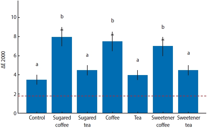
Forty-nine disc-shaped composite samples (Neo Spectra ST, Dentsply Sirona) were split into seven groups at random (n = 7). Distilled water was used to hydrate each sample for 24 hours at 37°C. After 24 hours, the first color measurements (T0) were made by using a clinical spectrophotometer (VITA Easyshade Compact; VITA Zahnfabrik). Color measurements were repeated after 7 days (T1) and 14 days (T2) of immersion in distilled water (control), tea, coffee, sugary tea, sugary coffee, tea with sweetener added, and coffee with sweetener added. After staining for 2 weeks, the specimens were bleached for 6 hours a day for a week using 16% carbamide peroxide (Opalescence Ultradent Products). Color measurements were taken again after bleaching (T3). Using CIEDE2000, color differences (ΔE) were computed. Analysis of variance (ANOVA) and repeated measures ANOVA with a Tukey post hoc test were used to evaluate the data.
Results
After 1 week, coffee-containing solutions produced significantly greater discoloration than the control (p < 0.001). By 2 weeks, tea groups exhibited similar discoloration to coffee groups (p < 0.001). The addition of sugar or sweetener had no significant effect (p > 0.05). Post-bleaching, coffee groups showed lower Whiteness Index values than the control, without statistical significance (p > 0.05).
Conclusions
Coffee and tea markedly stain resin composites, with discoloration persisting post-bleaching, while sugar or sweetener additions exert no significant effect.
- 1,212 View
- 96 Download
- Neuropeptide Y regulation of dental pulp neurogenic inflammation provoked by tooth bleaching agents: a descriptive comparative clinical study
- Javier Caviedes-Bucheli, Néstor Ríos-Osorio, Mario Pérez-Villota, Karolina Aucú-Miño, Diana Escobar-Mafla, Hernán Darío Muñoz-Alvear, José Francisco Gomez-Sosa, Luis Diaz-Barrera, Edgar Güiza – Cristancho, Hugo Roberto Munoz
- Restor Dent Endod 2026;51(1):e10. Published online February 13, 2026
- DOI: https://doi.org/10.5395/rde.2026.51.e10
-
Abstract
PDFPubReaderePub
- Objectives
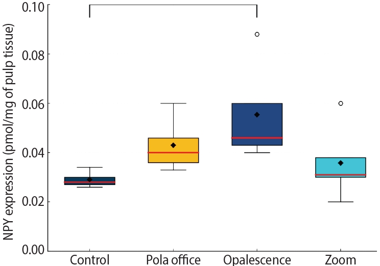
This study aimed to assess the expression of neuropeptide Y (NPY) in human dental pulp after tooth bleaching with three in-office hydrogen peroxide (H2O2)-based systems.
Methods
Forty pulps were collected from premolars scheduled for extraction and divided into four groups (n = 10): Control (no bleaching; basal NPY values); Pola Office (35% H2O2, 8 minutes); Opalescence Boost (40% H2O2, 20 minutes); and Zoom (25% H2O2 + cold blue light, 15 minutes). After extraction, pulps were fixed in 4% formaldehyde and processed. NPY levels were quantified using enzyme-linked immunosorbent assay. Data distribution was assessed with the Shapiro-Wilk test. One-way analysis of variance and Tukey post-hoc test with Bonferroni correction were applied (p < 0.05).
Results
NPY expression differed significantly among groups (p = 0.0097). The control group showed the lowest mean expression (0.026 ± 0.002 pmol/mg of pulp tissue), followed by Zoom (0.031 ± 0.005 pmol/mg), Pola Office (0.040 ± 0.004 pmol/mg), and Opalescence Boost, which exhibited the highest NPY expression (0.044 ± 0.004 pmol/mg). Post-hoc analysis revealed a statistically significant difference between the control and Opalescence Boost groups (p = 0.0122).
Conclusions
The increase in NPY expression—particularly with Opalescence Boost—indicates that in-office bleaching agents can elicit measurable neurobiological responses in pulp tissue after a single application. The significant difference between the control and Opalescence Boost groups suggests a possible H2O2 concentration- or formulation-dependent effect on pulpal neuropeptide activity, underscoring the need for further research on the biological impact of bleaching treatments.
- 1,605 View
- 90 Download
- Comparative evaluation of dentinal tubule occlusion by desensitizing agents after tooth bleaching: an in vitro study
- Dimitrios Dionysopoulos, Petros Mourouzis, Spyros Papageorgiou, Kosmas Tolidis
- Restor Dent Endod 2026;51(1):e8. Published online February 10, 2026
- DOI: https://doi.org/10.5395/rde.2026.51.e8
-
Abstract
PDFPubReaderePub
- Objectives
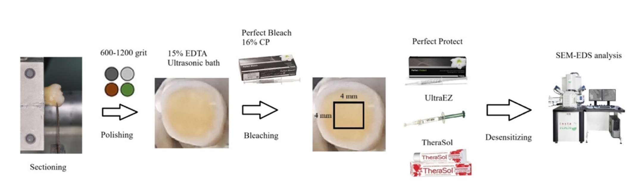
This study aimed to evaluate the efficacy of three commercially available desensitizing agents in occluding dentinal tubules, which may help reduce tooth sensitivity following a bleaching treatment.
Methods
Twenty healthy human third molars were utilized in this investigation. The samples were prepared by transversely sectioning 2.5 mm of the crowns to expose the dentin. They were initially treated with 15% ethylenediaminetetraacetic acid gel for 4 minutes, followed by application of Perfect Bleach (VOCO GmbH) bleaching agent (16% carbamide peroxide) for 2 hours. The samples were randomly allocated into four groups (n = 5), each receiving one of the following treatments: group 1: No treatment (control), group 2: treated with UltraEZ (Ultradent Products Inc.,), containing potassium nitrate and sodium fluoride, group 3: treated with Perfect Protect (VOCO GmbH), also containing potassium nitrate and sodium fluoride and group 4: treated with TheraSol Whitening & Sensitive (ABC Kinitron IKE), containing strontium acetate and sodium monofluorophosphate. Subsequently, the specimens were examined using scanning electron microscopy (SEM) and energy-dispersive X-ray spectroscopy to evaluate dentin tubule occlusion.
Results
SEM observations showed no occlusion of dentin tubules in the control group, whereas groups 2 to 4 exhibited significant occlusion. The most effective treatment was Perfect Protect (p < 0.05), while UltraEZ and TheraSol Whitening & Sensitive demonstrated similar effectiveness, with no statistically significant difference between them (p > 0.05).
Conclusions
The tested desensitizing agents effectively occluded dentin tubules to a considerable extent. Differences in their effectiveness were attributed to variations in their formulations.
- 2,206 View
- 189 Download
- Difference in light transmittance and depth of cure of flowable composite depending on tooth thickness: an in vitro experimental study
- Seong-Pyo Bae, Myung-Jin Lee, Kyung-San Min, Mi-Kyung Yu, Kwang-Won Lee
- Restor Dent Endod 2025;50(4):e39. Published online November 28, 2025
- DOI: https://doi.org/10.5395/rde.2025.50.e39
-
Abstract
PDFPubReaderePub
- Objectives
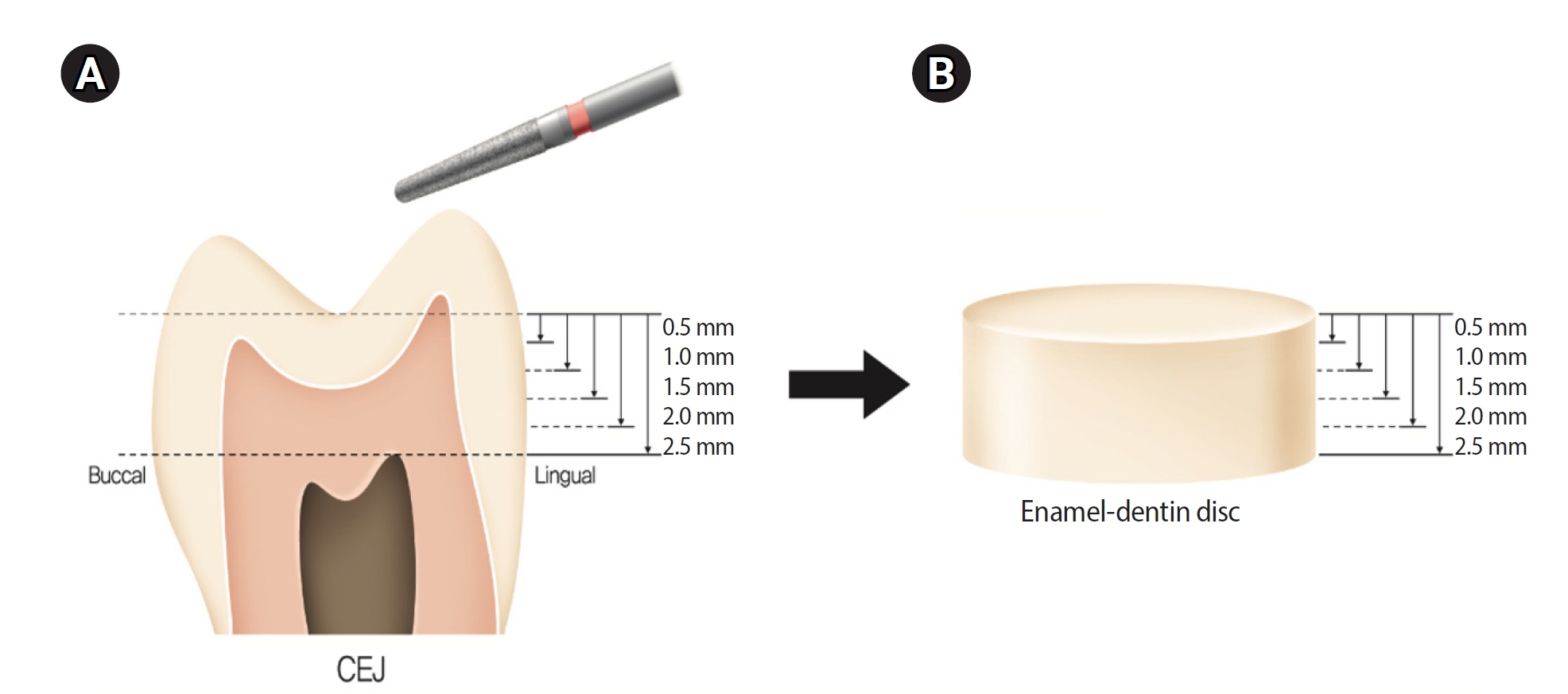
This study aimed to quantify light attenuation through varying tooth thicknesses and its impact on the depth of cure of composite resin.
Methods
Twenty extracted premolars were used to create enamel-dentin discs that were sanded progressively in 0.5 mm increments from 2.5 mm to 0.5 mm. Light irradiance was measured with and without tooth specimens to evaluate light transmittance. Resin was cured beneath different thicknesses, and the depth of cure was assessed using the Vickers hardness test.
Results
The results demonstrated that light transmittance significantly decreased as tooth thickness increased (p < 0.01), leading to reduced resin polymerization. In the 2.0-mm and 2.5-mm tooth thickness groups, the depth of cure was significantly lower than in the control group without tooth specimens (p < 0.05).
Conclusions
Ultimately, for tooth structures exceeding 2 mm, self-cure or dual-cure resin polymerization is thought to be more efficient than light polymerization.
- 2,239 View
- 153 Download
- Comparison of remineralization in caries-affected dentin using calcium silicate, glass ionomer cement, and resin-modified glass ionomer cement: an in vitro study
- Kwanchanok Youcharoen, Onwara Akkaratham, Papichaya Intajak, Pipop Saikaew, Sirichan Chiaraputt
- Restor Dent Endod 2025;50(4):e37. Published online November 14, 2025
- DOI: https://doi.org/10.5395/rde.2025.50.e37
-
Abstract
PDFPubReaderePub
- Objectives
This study evaluated the ability of calcium silicate cement (CSC) as a remineralizing agent compared with conventional glass ionomer cement (GIC) and resin-modified GIC (RMGIC) to remineralize artificial caries-affected dentin.
Methods
Twenty-five class V cavities were prepared on extracted human third molars. Twenty teeth underwent artificial caries induction. The remaining five teeth with sound dentin serve as the positive control. The twenty demineralized teeth were subdivided into four groups (n = 5): carious dentin without restoration (negative control [NC]), carious dentin restored with CSC (Biodentine, Septodont), carious dentin restored with GI (Fuji IX, GC Corporation), and carious dentin restored with RMGIC (Fuji II LC, GC Corporation). Following restoration, the specimens were stored in artificial saliva for 7 days. The elastic modulus was evaluated by a nanoindentation test. The mineral composition was analyzed by scanning electron microscopy-energy-dispersive X-ray spectroscopy (SEM-EDX), and the mineral composition at the dentin-material interface.
Results
CSC had a higher modulus of elasticity compared to GI, RMGI, and NC groups (p < 0.05). Higher calcium and phosphorus content was observed under CSC restorations, as indicated by SEM-EDX examination, which may lead to better remineralization.
Conclusions
Compared to GI and RMGI, CSC showed the best remineralization and mechanical reinforcement in caries-affected dentin, indicating CSC for use in minimally invasive restorative dentistry. -
Citations
Citations to this article as recorded by
- Comparison of mineral precipitation, elemental release, pH change and cytotoxicity of calcium-silicate cements and an experimental resin-modified glass ionomer cement containing bioactive glass
Wisitsin Potiprapanpong , Parichart Naruphontjirakul, Naruporn Monmaturapoj, Siriporn Tanodekaew, Somruethai Channasanon, Arnit Toneluck, Somying Patntirapong, Piyaphong Panpisut
Biomaterial Investigations in Dentistry.2026; 13: 337. CrossRef
- Comparison of mineral precipitation, elemental release, pH change and cytotoxicity of calcium-silicate cements and an experimental resin-modified glass ionomer cement containing bioactive glass
- 3,042 View
- 272 Download
- 1 Crossref
- The influence of bioactive glass (BGS-7) on enamel remineralization: an in vitro study
- Chaeyoung Lee, Eunseon Jeong, Kun-Hwa Sung, Su-Jung Park, Yoorina Choi
- Restor Dent Endod 2025;50(4):e33. Published online October 15, 2025
- DOI: https://doi.org/10.5395/rde.2025.50.e33
-
Abstract
PDFPubReaderePub
- Objectives
The aim of this study was to compare the remineralizing capacity of bioactive glass (BGS-7, CGBIO) with other agents.
Methods
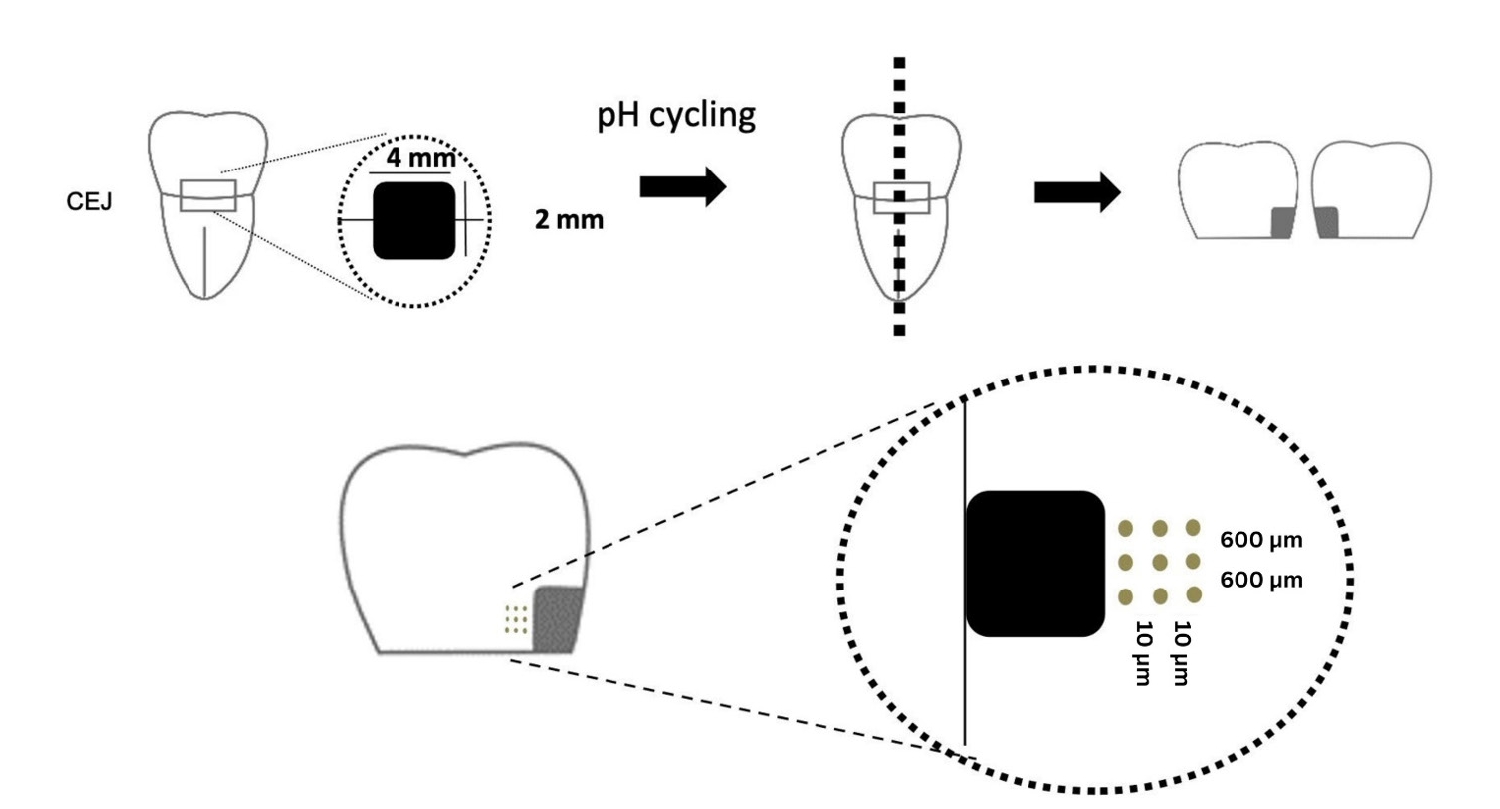
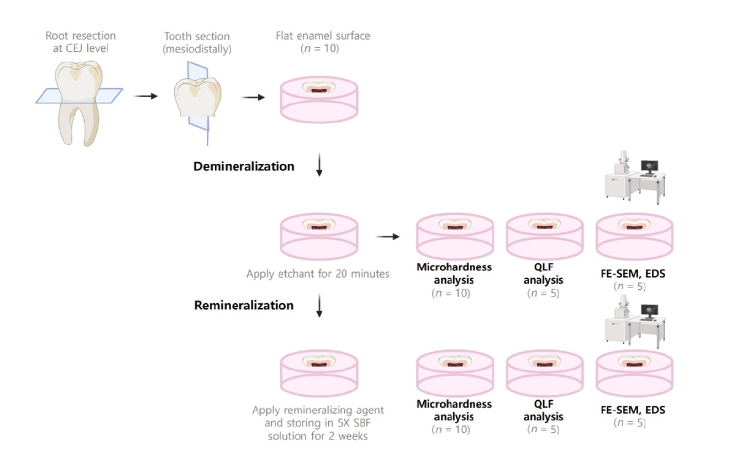
Twenty caries-free third molars were sectioned and demineralized. Specimens were divided into four groups: (1) control, (2) Clinpro XT varnish (Solventum), (3) 1.23% acidulated phosphate fluoride gel, and (4) a new type of CaO-SiO2-P2O5-B2O3 system of bioactive glass ceramics (BGS-7). Agents were applied and stored in simulated body fluid at 37℃ for 2 weeks. Microhardness was measured using the Vickers hardness testing method. Five specimens per group were analyzed using quantitative light-induced fluorescence (QLF) to assess mineral loss. Field-emission scanning electron microscopy (FE-SEM) and energy-dispersive X-ray spectroscopy (EDS) were used to examine the surface morphology and elemental composition. Data were analyzed using paired t-test and one-way analysis of variance (p < 0.05).
Results
BGS-7 showed the highest microhardness values and the greatest recovery in QLF analysis (p < 0.05). FE-SEM revealed granular precipitates on demineralized enamel in the BGS-7 group. EDS confirmed the presence of newly formed silicon and fluoride layers.
Conclusions
BGS-7 demonstrated superior remineralization capacity compared to other agents, suggesting its potential as an effective remineralizing material. -
Citations
Citations to this article as recorded by- Bacterial ghosts (BGs): A promising approach as candidate vaccine
Helal F. Hetta, Ibraheem M. Mwafey, Noura H. Abd Ellah, Fawaz E. Alanazi, Yasmin N. Ramadan
World Journal of Microbiology and Biotechnology.2026;[Epub] CrossRef
- Bacterial ghosts (BGs): A promising approach as candidate vaccine
- 2,801 View
- 255 Download
- 1 Web of Science
- 1 Crossref
Case Report
- Multidisciplinary management of an endo-perio lesion complicated by a cemental tear: a case report
- Nishanth D. Sadhak, Akshaya Pallod, Shreyas Oza
- Restor Dent Endod 2025;50(3):e31. Published online August 22, 2025
- DOI: https://doi.org/10.5395/rde.2025.50.e31
-
Abstract
PDFPubReaderePub
- Endodontic-periodontal lesions (EPLs) complicated by cemental tears present a diagnostic and therapeutic challenge. This case report describes the successful management of a 66-year-old male patient with a mandibular second molar (#18) exhibiting an EPL complicated by a cemental tear. Clinical examination revealed a draining sinus tract, deep periodontal pockets, and radiographic evidence of a “J-shaped” lesion and a radiopaque cemental fragment. The tooth had previously initiated endodontic treatment. A multidisciplinary approach involving endodontic treatment and surgical removal of the cemental tear was implemented. At 24-month follow-up, clinical and radiographic examination revealed significant improvement in periodontal health, bone regeneration, and resolution of the lesion. This case highlights the importance of considering cemental tears in the differential diagnosis of EPLs and demonstrates the efficacy of a combined endodontic-periodontal approach for achieving predictable outcomes.
- 4,694 View
- 352 Download
Research Articles
- Comparative study of the effectiveness of different bleaching agents on blood-colored extracted teeth and investigation of recoloring after bleaching: an in vitro experimental study
- Gülşen Arslan, Akın Aladağ, Ayşegül Demirbaş, Murat Türkün
- Restor Dent Endod 2025;50(3):e22. Published online July 9, 2025
- DOI: https://doi.org/10.5395/rde.2025.50.e22
-
Abstract
PDFPubReaderePub
- Objectives
This study evaluated the efficacy of three distinct bleaching agents over time on blood-stained, devitalized teeth. Furthermore, the recoloring subsequent to bleaching will be monitored.
Methods
The study was conducted on 60 caries-free, unfilled, upper human incisors. The Freccia and Peters blood staining technique was employed, and four groups (n = 15) were identified: control, 35% hydrogen peroxide-treated, 37% carbamide peroxide-treated, and sodium perborate-treated groups. Color differences were measured using ΔE00, ΔWID, L*, a*, and b* values. To investigate tooth discoloration after bleaching, 10 unbleached teeth with three groups of 10 bleached teeth were compared by vine staining. The group of bleached teeth was restored immediately, another group waited one week, and the third group had sodium ascorbate applied and analyzed using one-way analysis of variance tests (p < 0.05).
Results
Among the groups, carbamide peroxide exhibited the most significant whitening during the 6-day bleaching process, followed by hydrogen peroxide and sodium perborate. Subsequent examination of the wine recoloring of post-bleaching samples demonstrated that bleached teeth exhibited a heightened propensity for recoloration in contrast to unbleached teeth. Notably, sodium ascorbate treatments for hydrogen peroxide neutralization and the wait-and-restore approach were not statistically significant in terms of preventing recoloration.
Conclusions
Sodium perborate is less effective and more time-consuming than hydrogen peroxide or carbamide peroxide for bleaching purposes. Carbamide peroxide is the most effective bleaching agent. The sodium ascorbate treatment and the wait-and-restore approach are ineffective in preventing recoloring. Bleached teeth have more discoloration than unbleached teeth. -
Citations
Citations to this article as recorded by- The Effect of Adhesive Systems on Shade Matching of Composite Veneer
Fadak Al Marar, Raghad Aljarboua, Fatimah M. Alatiyyah, Shahad AlGhamdi, Faraz Ahmed Farooqi, Lama Almuhanna, Rasha AlSheikh, Abdul Samad Khan
Dentistry Journal.2026; 14(2): 85. CrossRef
- The Effect of Adhesive Systems on Shade Matching of Composite Veneer
- 4,534 View
- 283 Download
- 1 Web of Science
- 1 Crossref
- Surface properties and susceptibility to staining of a resin composite after brushing with different whitening toothpastes
- Aline da Silva Barros, Carolina Meneghin Barbosa, Renata Siqueira Scatolin, Waldemir Francisco Vieira Junior, Laura Nobre Ferraz
- Restor Dent Endod 2025;50(1):e6. Published online February 26, 2025
- DOI: https://doi.org/10.5395/rde.2025.50.e6
-
Abstract
PDFPubReaderePub
- Objectives
This study investigated the effects of different whitening toothpaste (WT) on the surface properties and staining susceptibility of a resin composite.
Methods
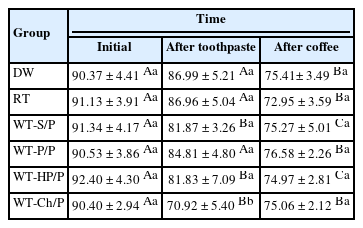
Cylindrical samples were prepared with a micro-hybrid resin composite and were randomized into groups according to the toothpaste (n = 12): distilled water (DW), regular toothpaste (RT), WT with silica + pyrophosphate (WT-S/P), WT with pentaphosphate and pyrophosphate (WT-P/P), WT with hydrogen peroxide and pyrophosphate (WT-HP/P) and WT with charcoal and pyrophosphate (WT-Ch/P). The samples were brushed for 825 cycles in an automatic brushing machine, simulating 30 days of brushing. After that, an immersion in coffee (10 mL/sample) was performed for 30 minutes for 30 days. The analyses of color, surface microhardness (SMH), and surface roughness (Ra) were performed at the initial time, after brushing with toothpaste and after immersion in coffee. The ΔL*, Δa*, Δb*, ΔEab, Δand E00 values were calculated comparing after toothpaste with initial time and after coffee with after toothpaste. Data were analyzed using a mixed linear model for repeated measures (SMH), Kruskal-Wallis, Dunn, Friedman, and Nemenyi tests, with α = 0.05.
Results
For ΔL*, the WT-Ch/P group had the lowest values and differed from the other groups comparing the after toothpaste with the initial time interval (p < 0.001). The WT-Ch/P group had the lowest SMH values in after-toothpaste time (p < 0.001). In after-toothpaste time and after coffee time, the WT-S/P group had the highest Ra values and differed from the groups except the WT-Ch/P group (p < 0.001).
Conclusions
The toothpaste composition affects the surface characteristics and susceptibility to staining of the resin composite. The charcoal-based toothpaste had the worst performance for the color analyses and SMH. -
Citations
Citations to this article as recorded by- Color Stability and Surface Roughness of Esthetic Resin Composites Following Simulated Toothbrushing with Whitening Toothpastes
Ecehan Kaplan , Ayşe Dündar, Çağatay Barutçugil
Odovtos - International Journal of Dental Sciences.2026; 1(1): 595. CrossRef - Influence of commercial mouth rinses with different formulations on enamel properties during at-home bleaching
Thalita Novello Coelho, Ana Júlia Gil, Marcos Roberto Lima Benati, Carolina Meneghin Barbosa, Tatiane Cristina Dotta, Waldemir Francisco Vieira-Junior, Renata Siqueira Scatolin, Laura Nobre Ferraz
Odontology.2026;[Epub] CrossRef
- Color Stability and Surface Roughness of Esthetic Resin Composites Following Simulated Toothbrushing with Whitening Toothpastes
- 6,699 View
- 199 Download
- 1 Web of Science
- 2 Crossref
- Development of whitening mouth rinses formulated with mushroom residues and their effect on enamel’s physical properties
- Julliana Andrade da Silva, Dayse Alexia de Carvalho de Brito, Débora Alves Nunes Leite Lima, Juliano Lemos Bicas, Gislaine Ricci Leonardi
- Restor Dent Endod 2024;49(3):e27. Published online June 27, 2024
- DOI: https://doi.org/10.5395/rde.2024.49.e27
-
Abstract
PDFPubReaderePub
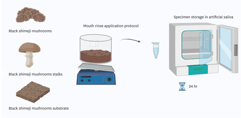
Objectives This study aimed to develop whitening mouth rinses formulated with industrial mushrooms and compare them with over-the-counter whitening mouth rinses.
Materials and Methods Formulations with black shimeji mushrooms, mushroom substrates, and mushroom stalks were developed. Bovine enamel/dentin samples were divided into 7 groups (
n = 10): Colgate Luminous White, Listerine Whitening Extreme (LWE), Listerine Cool Mint (LC), mushroom extract rinse (MEC), mushroom substrate rinse (MSB), mushroom stalk rinse (MTC), and artificial saliva. Samples were stained with black tea for 6 days, and then were immersed in 100 mL of each mouth rinse twice daily for 14 days. Color parameters (CIELAB [ΔE*], CIEDE2000 [ΔE00], whiteness index for dentistry [ΔWID]) and microhardness (Knoop hardness number [KHN]) were analyzed at T1 (initial), T2 (24 hours), and T3 (7 days). Mouth rinse pH was measured, and enamel was examined using a scanning electron microscope. Data were analyzed using generalized linear models, and KHN with the generalized linear mixed model for repeated measures (p ≤ 0.05).Results ΔE* was higher in LW and MSB groups. No significant differences were found for ΔE00 (
p = 0.0982) and ΔWID (p = 0.2536). Experimental mouth rinses did not promote enamel whitening based on ΔE00 and ΔWID. LWE and LC reduced KHN and had a more acidic pH, while MEC had higher KHN at T2. MEC, MSB, and MTC had alkaline pH, not altering the tooth surface.Conclusions Black shimeji mushrooms are promising for mouth rinse development due to their alkaline pH and non-altering effect on surface microhardness.
-
Citations
Citations to this article as recorded by- Evaluation of cytotoxicity and bleaching efficacy of gels with calcium polyphosphate and violet LED
Larissa de Jesus Gomes, Rafael Antonio de Oliveira Ribeiro, Mariangela Ivette Guanipa Ortiz, Klaus Rischka, Carlos Alberto de Souza Costa, Débora Alves Nunes Leite Lima
Brazilian Dental Journal.2025;[Epub] CrossRef
- Evaluation of cytotoxicity and bleaching efficacy of gels with calcium polyphosphate and violet LED
- 2,934 View
- 101 Download
- 1 Web of Science
- 1 Crossref
- Color stability and solubility of Biodentine and NeoPutty in contact with different irrigation solutions
- Sıla Nur Usta, Cangül Keskin
- Restor Dent Endod 2024;49(3):e25. Published online June 19, 2024
- DOI: https://doi.org/10.5395/rde.2024.49.e25
-
Abstract
PDFPubReaderePub
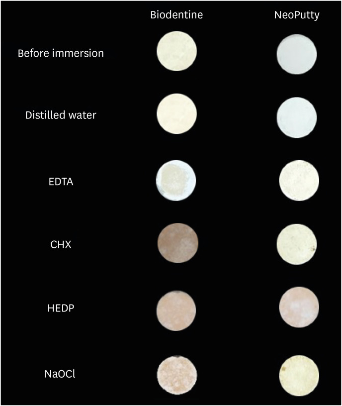
Objectives This study aimed to evaluate the color stability and solubility of Biodentine and NeoPutty in contact with different irrigation solutions.
Materials and Methods Biodentine and NeoPutty were set in cylindrical molds with 7 mm diameter and 1.5 mm high and immersed in distilled water, 17% ethylenediaminetetraacetic acid (EDTA), 2% chlorhexidine (CHX), 9% 1-hydroxyethylidene 1,1-diphosphonate (HEDP), and 5% sodium hypochlorite (NaOCl) solutions for 24 hours. The color change was measured with a spectrophotometer. The solubility values were calculated as the mass loss was expressed as a percentage of the original mass using an analytical balance with 10−4 g accuracy. Data were analyzed with Kruskal-Wallis followed by Mann-Whitney
U tests, and 2-way analysis of variance test followed by Bonferroni corrections for pairwise comparisons for solubility and color stability with a 5% significance threshold, respectively.Results Biodentine exhibited higher color changes compared to the NeoPutty contact with all solutions except distilled water (
p < 0.05). Both hydraulic cements (HCs) showed higher discoloration values immersion in CHX followed by NaOCl. No statistically significant difference was found between Biodentine and NeoPutty regardless of irrigation solution in terms of solubility (p > 0.05). Solubility values were lower in the distilled water group compared to EDTA and CHX (p < 0.05).Conclusions Tested HCs showed solubility and color changes at various rates. NeoPutty could be an appropriate material in aesthetic areas. The usage of HEDP as an irrigant solution can be considered suitable for various endodontic treatments due to its relatively lower solubility and discoloration values.
-
Citations
Citations to this article as recorded by- Sealing ability of Biodentine, zirconia reinforced glass ionomer cement and Mineral Trioxide Aggregate as furcation perforation repair materials: an in vitro analysis
Sumita Panwar, Yajuvender Singh Hada
Biomaterial Investigations in Dentistry.2026; 13: 21. CrossRef - Effect of calcium silicate-based materials on tooth discolouration in repairing root perforations of lower molars: an in-vitro study
Sevil Zırhlı, Davut Celık, Tugba Kosar
Journal of the Australian Ceramic Society.2026;[Epub] CrossRef - Influence of endodontic irrigants on hydraulic cements: solubility, color alteration and surface changes
Sıla Usta, Cangül Keskin, Ayşe Oktay, Emmanuel João Nogueira Leal Silva
European Oral Research.2026; 60(1): 230. CrossRef - Comparison of cytotoxicity and physicochemical properties of Cal Bio LC MTA, ProRoot MTA and NeoPutty MTA: an in vitro study
Behnaz Rezaei, Aysun Avşar, Cangül Keskin
Odontology.2026;[Epub] CrossRef
- Sealing ability of Biodentine, zirconia reinforced glass ionomer cement and Mineral Trioxide Aggregate as furcation perforation repair materials: an in vitro analysis
- 3,879 View
- 180 Download
- 3 Web of Science
- 4 Crossref
- Can discolored dental composites be bleached in depth?
- Luca Giachetti, Daniele Scaminaci Russo, Michele Nieri, Francesca Cinelli
- Restor Dent Endod 2024;49(3):e23. Published online June 11, 2024
- DOI: https://doi.org/10.5395/rde.2024.49.e23
-
Abstract
PDFPubReaderePub
Objectives Previous
in vitro studies determined the whitening effects of bleaching products on stained resin composite surfaces. Thisin vitro study aimed to verify the effectiveness of a whitening system on composite resin previously subjected to pigmentation, specifically examining the depth of whitening effectiveness within the material structure.Materials and Methods A commercially available nano-filled composite resin was used. Specimens were stained using a coffee-based solution and a 10% carbamide peroxide-based gel was employed as the whitening agent. The pigment’s penetration and the effect of the bleaching gel were evaluated by measuring color (CieLab values) from the outer edge to the inner part of the specimens. Color measurements were taken at 14 points, starting from 0.1 mm from the external perimeter up to 3.0 mm.
Results Analysis of variance tests showed a statistically significant difference between the Control Group (CG), Pigmentation Group, and Whitening Group. The whitening agent was effective up to 1.5 mm in depth, with Whiteness index (W) values not statistically different from those of CG up to 0.5 mm in depth.
Conclusions Whitening agents on nano-filled resin composite previously pigmented appear effective in restoring the W to values similar to the original, particularly in the superficial layers of the sample.
-
Citations
Citations to this article as recorded by- Color Stability of Tooth-Colored Restorative Materials After Exposure to Arabic Coffee and Black Tea: A Systematic Review
Abdulrhman Y Alenezi, Abdulwahab M AlEyada, Yousef H Aldhafiri, Mohammed S Alsubaie, Mohammed S Alshahrani, Mahesh Shenoy
Cureus.2025;[Epub] CrossRef - Comparative evaluation to composite resin bleaching using ozone-enhanced low-concentration hydrogen peroxide
Mahmoud K. AL-Omiri, Dania Sa’ed Hussam Abuherra, Khaled M. AL-Omiri, Ali Y. Alsaeed, Mohammad Alamri, Ali M. Alqahtani, Saleh Ali Alqahtani, Ghadeer Saleh Alwadai, Naif Abogazalah, Edward Lynch
Scientific Reports.2025;[Epub] CrossRef - The effects of mechanical and chemical degradation on the surface roughness, gloss, and color stability of bulk-fill resin composites
Merve Nezir, Hanife Altınışık, Esra Özyurt, Naz Bayar, Mediha Büyükgöze Dindar
BMC Oral Health.2025;[Epub] CrossRef
- Color Stability of Tooth-Colored Restorative Materials After Exposure to Arabic Coffee and Black Tea: A Systematic Review
- 5,132 View
- 158 Download
- 2 Web of Science
- 3 Crossref
Review Article
- Can carbamide peroxide be as effective as hydrogen peroxide for in-office tooth bleaching and cause less sensitivity? A systematic review
- Patrick Wesley Marques de Boa, Kaiza de Sousa Santos, Francisca Jennifer Duarte de Oliveira, Boniek Castillo Dutra Borges
- Restor Dent Endod 2024;49(2):e14. Published online March 20, 2024
- DOI: https://doi.org/10.5395/rde.2024.49.e14
-
Abstract
PDFPubReaderePub
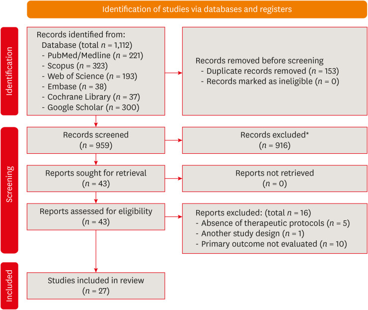
This study aimed to answer the question through a systematic review: Can carbamide peroxide be as effective as hydrogen peroxide and cause less in-office bleaching sensitivity? A literature survey was performed in PubMed/MEDLINE, Embase, Scopus, ISI Web of Science, and gray literature. Primary clinical trials that compared the efficacy or the in-office bleaching sensitivity between carbamide and hydrogen peroxides were included. The risk of bias was evaluated using the RoB2. The certainty of the evidence was assessed using the GRADE approach. DPI training significantly improved the mean scores of the dental undergraduates from 7.53 in the pre-DPI-training test to 9.01 in the post-DPI-training test (
p < 0.001). After 6 weeks, the mean scores decreased marginally to 8.87 in the retention test (p = 0.563). DPI training increased their confidence level from 5.68 pre-DPI training to 7.09 post-DPI training. The limited evidence suggests that the 37% carbamide peroxide may be similarly effective to the 35% hydrogen peroxide for bleaching teeth in-office and causes less bleaching sensitivity. However, more well-designed split-mouth clinical trials are necessary to strengthen the evidence.-
Citations
Citations to this article as recorded by- Impact of nanostructured additives in tooth bleaching agents on enhancing color change and reducing side effects: a scoping review
Patrick Wesley Marques de Boa, Kaiza de Sousa Santos, Aleph Matthews da Silva Souza, Arnóbio Antônio da Silva-Júnior, Boniek Castillo Dutra Borges
Clinical Oral Investigations.2025;[Epub] CrossRef - Quantitative and Qualitative Assessment of Enamel Surface Roughness Following High-Concentration Peroxide Bleaching: A Comparative In Vitro Study
Mamnoon Ghafir, Nida Mehmood, Leeza Bharati, Shreya Bhukal, Ritika Sethi, Aanchal Chaudhary, Seema Gupta
Cureus.2025;[Epub] CrossRef - Using violet light during in-office tooth bleaching to enhance the efficacy of carbamide peroxide without increasing bleaching sensitivity: a systematic review and meta-analysis
Mariana Silva de Bessa, Kaiza de Sousa Santos, Patrick Wesley Marques de Boa, Francisca Jennifer Duarte de Oliveira, Bárbara Faria de Sá Barbosa, Boniek Castillo Dutra Borges
Lasers in Medical Science.2025;[Epub] CrossRef - Influence of Different Light-Activated Bleaching Gels on Pulp Chamber Temperature: An In Vitro Study
Mandana Karimi, Elmira Ataee, Ladan Ranjbar Omrani, Mahdi Abbasi, Elham Ahmadi
Avicenna Journal of Dental Research.2024; 16(4): 225. CrossRef
- Impact of nanostructured additives in tooth bleaching agents on enhancing color change and reducing side effects: a scoping review
- 13,612 View
- 211 Download
- 1 Web of Science
- 4 Crossref
Research Articles
- Effect of different storage media on elemental analysis and microhardness of cervical cavity margins restored with a bioactive material
- Hoda Saleh Ismail, Brian Ray Morrow, Ashraf Ibrahim Ali, Rabab Elsayed Elaraby Mehesen, Salah Hasab Mahmoud, Franklin Garcia-Godoy
- Restor Dent Endod 2024;49(1):e6. Published online January 17, 2024
- DOI: https://doi.org/10.5395/rde.2024.49.e6
-
Abstract
PDFPubReaderePub
Objectives This study aimed to investigate the elemental analysis and microhardness of a bioactive material (Activa) and marginal tooth structure after storage in different media.
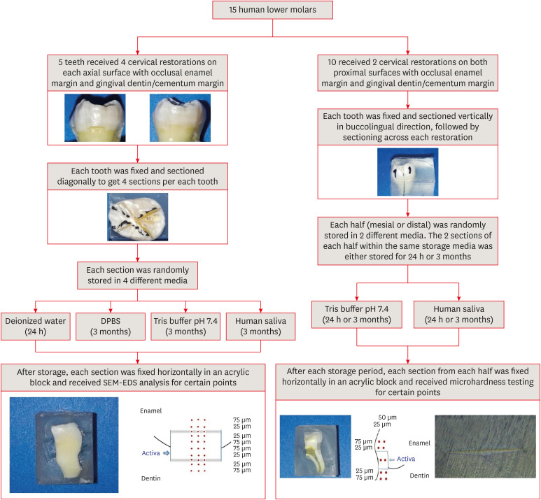
Materials and Methods Fifteen teeth received cervical restorations with occlusal enamel and gingival dentin margins using the tested material bonded with a universal adhesive, 5 of them on the 4 axial surfaces and the other 10 on only the 2 proximal surfaces. The first 5 teeth were sectioned into 4 restorations each, then stored in 4 different media; deionized water, Dulbecco's phosphate buffered saline (DPBS), Tris buffer, and saliva. The storage period for deionized water was 24 hours while it was 3 months for the other media. Each part was analyzed by scanning electron microscopy-energy dispersive spectroscopy (SEM-EDS) analysis for different substrates/distances and the wt% of calcium, phosphorus, silica, and fluoride were calculated. The other 10 teeth were sectioned across the restoration, stored in either Tris buffer or saliva for 24 hours or 3 months, and were evaluated for microhardness of different substrates/areas. Data were analyzed using analysis of variance and Tukey’s
post hoc test.Results Enamel and dentin interfaces in the DPBS group exhibited a significant increase in calcium and phosphorus wt%. Both silica and fluoride significantly increased in tooth structure up to a distance of 75 μm in the 3-month-media groups than the immediate group. Storage media did not affect the microhardness values.
Conclusions SEM-EDS analysis suggests an ion movement between Activa and tooth structure through a universal adhesive while stored in DPBS.
-
Citations
Citations to this article as recorded by- A two-year randomized clinical trial of bulk-fill and ion-releasing composites with universal adhesives in class V carious lesions
Hoda Saleh Ismail, Hanan Ahmed Nabil Soliman, Ashraf Ibrahim Ali, Ahmed Gamal Raghip, Eman H. Albelasy
Clinical Oral Investigations.2026;[Epub] CrossRef - Elemental and micromorphological analysis of ion releasing restoration/carious dentin interface
Alaa Esmat Abdelsalam, Hoda Saleh Ismail, Hamdi Hosni Hamama
Scientific Reports.2025;[Epub] CrossRef - Influence of curing mode and aging on the bonding performance of universal adhesives in coronal and root dentin
Hoda Saleh Ismail, Ashraf Ibrahim Ali, Mohamed Elshirbeny Elawsya
BMC Oral Health.2024;[Epub] CrossRef
- A two-year randomized clinical trial of bulk-fill and ion-releasing composites with universal adhesives in class V carious lesions
- 3,092 View
- 113 Download
- 3 Web of Science
- 3 Crossref
- Can different agents reduce the damage caused by bleaching gel to pulp tissue? A systematic review of basic research
- Letícia Aparecida Silva Batista, Alexandre Henrique dos Reis-Prado, Hebertt Gonzaga dos Santos Chaves, Lara Cancella de Arantes, Luís Fernando Santos Alves Morgan, Carolina Bosso André, Thaís Yumi Suzuki, Francine Benetti
- Restor Dent Endod 2023;48(4):e39. Published online November 6, 2023
- DOI: https://doi.org/10.5395/rde.2023.48.e39
-
Abstract
PDFSupplementary MaterialPubReaderePub
Objectives This study aimed to investigate the effectiveness of different topical/systemic agents in reducing the damage caused by bleaching gel to pulp tissue or cells.
Materials and Methods Electronic searches were performed in July 2023.
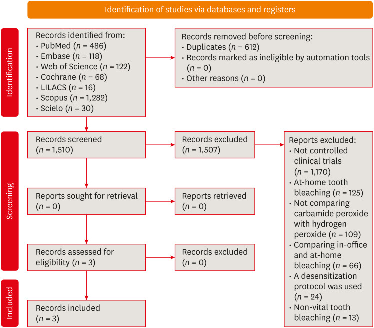
In vivo andin vitro studies evaluating the effects of different topical or systemic agents on pulp inflammation or cytotoxicity after exposure to bleaching agents were included. The risk of bias was assessed.Results Out of 1,112 articles, 27 were included. Nine animal studies evaluated remineralizing/anti-inflammatories agents in rat molars subjected to bleaching with 35%–38% hydrogen peroxide (HP). Five of these studies demonstrated a significant reduction in inflammation caused by HP when combined with bioglass or MI Paste Plus (GC America), or following KF-desensitizing or Otosporin treatment (
n = 3). However, orally administered drugs did not reduce pulp inflammation (n = 4). Cytotoxicity (n = 17) was primarily assessed using the 3-(4,5-dimethylthiazol-2-yl)-2,5-diphenyltetrazolium bromide assay on human dental pulp cells and mouse dental papilla Cell-23 cells. Certain substances, including sodium ascorbate, butein, manganese chloride, and peroxidase, were found to reduce cytotoxicity, particularly when applied prior to bleaching. The risk of bias was high in animal studies and low in laboratory studies.Conclusions Few
in vivo studies have evaluated agents to reduce the damage caused by bleaching gel to pulp tissue. Within the limitations of these studies, it was found that topical agents were effective in reducing pulp inflammation in animals and cytotoxicity. Further analyses with human pulp are required to substantiate these findings.Trial Registration PROSPERO Identifier:
CRD42022337192 -
Citations
Citations to this article as recorded by- 3D-Printed and Bioprinted Scaffolds in Regenerative Endodontics: A Systematic Review
Hebertt Gonzaga dos Santos Chaves, Diana B. Sequeira, Vilton Cardozo Moreira Dias, Alberto Cabrera-Fernández, João Peça, Francine Benetti, João Miguel Marques dos Santos
Applied Sciences.2026; 16(8): 3940. CrossRef - Clinical Study on the Efficacy of 35% Hydrogen Peroxide Gel According to Exposure Time (40 min vs. 20 min) by Spectrophotometry
Trinidad Rincón, Maria Portillo Muñoz, Maria Lobato, Ana María Martín Casado, Laryssa Mylenna Madruga Barbosa, Alessandro Loguercio, Cristina Gómez‐Polo
Journal of Esthetic and Restorative Dentistry.2026;[Epub] CrossRef - Clareamento dental e TikTok: avaliação da qualidade do conteúdo em mídia social
Rafaele T Costa, Thayna Silva do Carmo Tavares, André Walsh-Monteiro
Ciência ET Praxis.2025; 21(36): 111. CrossRef - Synthesis, characterization and evaluation of novel bleaching gels containing bioactive glass and nano-hydroxyapatite on hydrogen peroxide diffusion, bleaching efficacy and enamel protection
Adrieli Burey, Byron Carpio-Salvatierra, Michael Favoretto, María Luján Méndez Bauer, Viviane Hass, Alessandra Reis, Alessandro D. Loguercio, Paulo Vitor Farago
Clinical Oral Investigations.2025;[Epub] CrossRef - Cytotoxicity of Bleaching Products: A Systematic Review
Mireia Montaner, José Luis Sanz, Carmen Llena, María Melo, Clara Puig-Herreros, James Ghilotti
Applied Sciences.2024; 14(9): 3680. CrossRef
- 3D-Printed and Bioprinted Scaffolds in Regenerative Endodontics: A Systematic Review
- 4,449 View
- 60 Download
- 4 Web of Science
- 5 Crossref
- Evaluation of at-home bleaching protocol with application on different surfaces: bleaching efficacy and hydrogen peroxide permeability
- Heloisa Forville, Michael Willian Favoreto, Michel Wendlinger, Roberta Micheten Dias, Christiane Philippini Ferreira Borges, Alessandra Reis, Alessandro D. Loguercio
- Restor Dent Endod 2023;48(4):e33. Published online October 6, 2023
- DOI: https://doi.org/10.5395/rde.2023.48.e33
-
Abstract
PDFPubReaderePub
Objectives This study aimed to evaluate the bleaching efficacy and hydrogen peroxide permeability in the pulp chamber by the at-home bleaching gel in protocols applied on different dental surfaces.
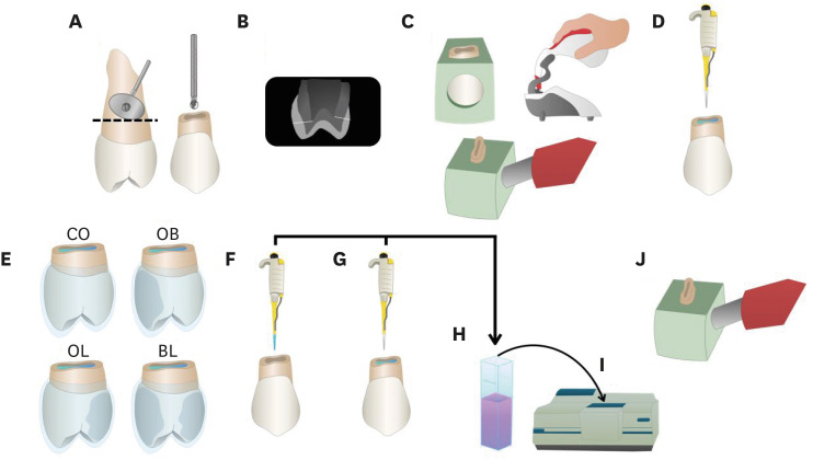
Materials and Methods Forty premolars were randomly into 4 groups: control group no bleaching, only application on the buccal surface (OB), only application on the lingual surface (OL) and application in buccal and lingual surfaces, simultaneously (BL). At-home bleaching gel (White Class 7.5%) was used for the procedure. The bleaching efficacy was evaluated with a digital spectrophotometer (color change in CIELAB [Δ
E ab] and CIEDE 2000 [ΔE 00] systems and Whitening Index for Dentistry [ΔWID]). The hydrogen peroxide permeability in the pulp chamber (µg/mL) was assessed using UV-Vis spectrophotometry and data were analyzed for a 1-way analysis of variance and Tukey’s test (α = 0.05).Results All groups submitted to bleaching procedure showed bleaching efficacy when measured with Δ
E ab and ΔE 00 (p > 0.05). Therefore, when analyzed by ΔWID, a higher bleaching efficacy were observed for the application on the groups OB and BL (p = 0.00003). Similar hydrogen peroxide permeability was found in the pulp chambers of the teeth undergoing different protocols (p > 0.05).Conclusions The application of bleaching gel exclusively on the OB is sufficient to achieve bleaching efficacy, when compared to BL. Although the OL protocol demonstrated lower bleaching efficacy based on the ΔWID values, it may still be of interest and relevant in certain clinical scenarios based on individual needs, requiring clinical trials to better understand its specificities.
-
Citations
Citations to this article as recorded by- Effect of whitening pens on hydrogen peroxide permeability in the pulp chamber, color change and surface morphology
Laryssa Mylenna Madruga Barbosa, Gabrielle Gomes Centenaro, Deisy Cristina Ferreira Cordeiro, Maria Alice de Matos Rodrigues, Letícia Condolo, Michael Willian Favoreto, Alessandra Reis, Alessandro D. Loguercio
Journal of Dentistry.2025; 154: 105595. CrossRef - Evaluation of bleaching efficiency of carbamide peroxide applied on different dental surfaces: An in vitro study
R. Gokulnath, R. S. Mohan Kumar, A. Jayasenthil, R. Anjana, G. Sree Vidya
Journal of Conservative Dentistry and Endodontics.2025; 28(4): 366. CrossRef - Characterization and effects on enamel of low-concentration bleaching gels containing hyaluronic acid, NF_TiO2 nanoparticles and irradiated with violet LED light
Marcos Roberto Lima Benati, Matheus Kury, Priscila Borges Gobbo de Melo, Iago César Ribeiro Teles Matos, Roberta Tarkany Basting, Rosanna Tarkany Basting, Fernando Luis Esteban Florez, Vanessa Cavalli
Clinical Oral Investigations.2025;[Epub] CrossRef - Impact of bleaching on white spot lesions: hydrogen peroxide permeability and color alteration
Laryssa Mylenna Madruga Barbosa, Bruno Baracco, Taynara S. Carneiro, Michael Willian Favoreto, Michel Wendlinger, Daniel Jiménez-Díez, Laura Ceballos, Alessandro D. Loguercio
Clinical Oral Investigations.2025;[Epub] CrossRef - Efficacy of a buccal and lingual at‐home bleaching protocol—A randomized, split‐mouth, single‐blind controlled trial
Heloisa Forville, Laís Giacomini Bernardi, Michael Willian Favoreto, Felipe Coppla, Taynara de Souza Carneiro, Fabiana Madalozzo Coppla, Alessandro D. Loguercio, Alessandra Reis
Journal of Esthetic and Restorative Dentistry.2024; 36(9): 1301. CrossRef - REANATOMIZAÇÃO DE DENTE CONOIDE ASSOCIADA A ESTÉTICA VERMELHA: RELATO DE CASO
Ana Karolayne Sousa de Morais, Daniele Fernanda Sousa Barros, Daniel Messias Limeira, Rhana Leticia de Oliveira Faria, Roberta Furtado Carvalho, Sandna Nolêto de Araújo, Laura Barbosa Santos Di Milhomem
Revista Contemporânea.2024; 4(10): e6299. CrossRef - Effect of the reduction in the exposure time to at-home bleaching gel on color change and tooth sensitivity: A systematic review and meta-analysis
Priscila Borges Gobbo de Melo, Letícia Vasconcelos Silva Souza, Lucianne Cople Maia, Guido Artemio Marañón-Vásquez, Matheus Kury, Vanessa Cavalli
Clinical Oral Investigations.2024;[Epub] CrossRef
- Effect of whitening pens on hydrogen peroxide permeability in the pulp chamber, color change and surface morphology
- 5,351 View
- 91 Download
- 5 Web of Science
- 7 Crossref
- Impact of combined at-home bleaching and whitening toothpaste use on the surface and color of a composite resin
- Carolina Meneghin Barbosa, Renata Siqueira Scatolin, Waldemir Francisco Vieira-Junior, Marcia Hiromi Tanaka, Laura Nobre Ferraz
- Restor Dent Endod 2023;48(3):e26. Published online July 26, 2023
- DOI: https://doi.org/10.5395/rde.2023.48.e26
-
Abstract
PDFPubReaderePub
Objective This
in vitro study aimed to evaluate the effects of different whitening toothpastes on a composite resin during at-home bleaching with 10% carbamide peroxide.Materials and Methods Sixty samples (7 mm × 2 mm) were used for color and roughness analyses, while another 60 samples (3 mm × 2 mm) were utilized to assess microhardness. The factors analyzed included toothpaste, for which 5 options with varying active agents were tested (distilled water; conventional toothpaste; whitening toothpaste with abrasive agents; whitening toothpaste with abrasive and chemical agents; and whitening toothpaste with abrasive, chemical, and bleaching agents). Brushing and application of whitening gel were performed for 14 days. Surface microhardness (SMH), surface roughness (Ra), and color (∆L*, ∆a*, ∆b, ∆E*ab, and ∆E00) were analyzed. The Ra and SMH data were analyzed using mixed generalized linear models for repeated measures, while the color results were assessed using the Kruskal-Wallis and Dunn tests.
Results Between the initial and final time points, all groups demonstrated significant increases in Ra and reductions in SMH. No significant differences were found between groups for SMH at the final time point, at which all groups differed from the distilled water group. Conventional toothpaste exhibited the lowest Ra, while whitening toothpaste with abrasive agent had the highest value. No significant differences were observed in ∆L*, ∆a*, and ∆b.
Conclusions While toothpaste composition did not affect the color stability and microhardness of resin composite, combining toothbrushing with whitening toothpaste and at-home bleaching enhanced the change in Ra.
-
Citations
Citations to this article as recorded by- Current evidence on the impact of whitening toothpastes on dental restorative materials: A comprehensive review
Soyeon Kim, Shin Hye Chung, Satoshi Yamaguchi, Taro Arima, Young-Seok Park
Journal of Prosthodontic Research.2026; 70(1): 4. CrossRef - Property changes in resin composite exposed to mouth rinses during 10% carbamide peroxide bleaching
Mariana Ferreira da Silva, Giovana Contin Germinari, Carolina Meneghin Barbosa, Tatiane Cristina Dotta, Renata Siqueira Scatolin, Waldemir Francisco Vieira Júnior, Laura Nobre Ferraz
Brazilian Journal of Oral Sciences.2026; 25: e260366. CrossRef - Influence of commercial mouth rinses with different formulations on enamel properties during at-home bleaching
Thalita Novello Coelho, Ana Júlia Gil, Marcos Roberto Lima Benati, Carolina Meneghin Barbosa, Tatiane Cristina Dotta, Waldemir Francisco Vieira-Junior, Renata Siqueira Scatolin, Laura Nobre Ferraz
Odontology.2026;[Epub] CrossRef - At‐Home and In‐Office Bleaching Protocols on the Color Match of Restorations Made With Single‐Shade Composites
Luciana Vasconcelos Ramos, Dayana Fernandes Rocha Aparicio, André Luis Faria‐e‐Silva, Maíra do Prado, Andréa Vaz Braga Pintor, Marcela Baraúna Magno
Journal of Esthetic and Restorative Dentistry.2025; 37(6): 1567. CrossRef - Surface properties and susceptibility to staining of a resin composite after brushing with different whitening toothpastes
Aline da Silva Barros, Carolina Meneghin Barbosa, Renata Siqueira Scatolin, Waldemir Francisco Vieira Junior, Laura Nobre Ferraz
Restorative Dentistry & Endodontics.2025; 50(1): e6. CrossRef - Dental Care Behaviors and Oral Health Challenges in School-Age Populations
Ahmad Mahmoud Saleh , Aishah Al Daragemeh , Asmaa Morgan Farahat Khatap , Prakash Palanivelu , Arul Vellaiyan , Elturabi Elsayed Ebrahim , Ahmad Rayan , Nermen Abdelftah Mohamed
Salud, Ciencia y Tecnología.2025; 5: 1372. CrossRef - Effect of bleaching and repolishing on whiteness change and staining susceptibility of resin-based materials
Sultan Aktuğ Karademir, Samet Atasoy, Beyza Yılmaz
BMC Oral Health.2024;[Epub] CrossRef - Influence of using different toothpaste during bleaching with violet LED light (405 nm) on the colour and roughness of dental enamel: an in vitro study
Franco Sousa Leticia, Mazzalli Redondo Victor, Ferraz Nobre Laura, Vitti Pino Rafael, Renata Siqueira Scatolin
Lasers in Medical Science.2024;[Epub] CrossRef - Effect of coffee staining and simulated oral hygiene methods on the color and translucency of a nanoceramic resin
Luiz Felipe Schneider, Bruna Mueller, Rubens Nisie Tango, Claudia Angela Maziero Volpato
Journal of Esthetic and Restorative Dentistry.2024; 36(7): 1020. CrossRef
- Current evidence on the impact of whitening toothpastes on dental restorative materials: A comprehensive review
- 6,454 View
- 70 Download
- 8 Web of Science
- 9 Crossref
Review Article
- Does photobiomodulation on the root surface decrease the occurrence of root resorption in reimplanted teeth? A systematic review of animal studies
- Theodoro Weissheimer, Karolina Frick Bischoff, Carolina Horn Troian Michel, Bruna Barcelos Só, Manoela Domingues Martins, Matheus Albino Souza, Ricardo Abreu da Rosa, Marcus Vinícius Reis Só
- Restor Dent Endod 2023;48(3):e24. Published online June 12, 2023
- DOI: https://doi.org/10.5395/rde.2023.48.e24
-
Abstract
PDFSupplementary MaterialPubReaderePub
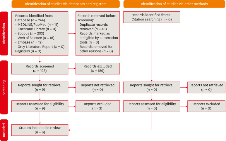
This review aimed to answer the following question “Does photobiomodulation treatment of the root surface decrease the occurrence of root resorption in reimplanted teeth?” Electronic searches were performed in the MEDLINE/PubMed, Cochrane Library, Scopus, Web of Science, Embase, and Grey Literature Report databases. Risk of bias was evaluated using SYRCLE Risk of Bias tool. The Grading of Recommendations, Assessment, Development, and Evaluations (GRADE) tool was used to assess the certainty of evidence. In total, 6 studies were included. Five studies reported a reduced occurrence of root resorption in teeth that received photobiomodulation treatment of the root surface prior to replantation. Only 1 study reported contradictory results. The photobiomodulation parameters varied widely among studies. GRADE assessment showed a low certainty of evidence. It can be inferred that photobiomodulation treatment of the root surface prior to replantation of teeth can reduce the occurrence of root resorption. Nonetheless, further clinical studies are needed.
Trial Registration PROSPERO Identifier: CRD42022349891
-
Citations
Citations to this article as recorded by- Liquid/Gel Mixed‐Phase Concentrated Growth Factors Enhance Periodontal and Pulpal Healing in Delayed Replantation of Immature Permanent Teeth: A Study in Beagle Dogs
Tiange Li, Xiaoxiao Yang, Yao Liu, Aochen Wang, Shu Zhu, Xu Chen, Zhenjiang Ding
International Endodontic Journal.2026; 59(7): 1430. CrossRef - Feasibility and Outcomes of Cell-based Regenerative Endodontic Therapy in Postautogenous Transplantation of a Mature Tooth: A Case Report
Noriaki Yoshihashi
Journal of Endodontics.2025; 51(1): 85. CrossRef - Evidence Mapping and Quality Assessment of Systematic Reviews in Dental Traumatology: A 54 Months Update
Nitesh Tewari, Pavithra Devi, Hemlata Nehta, Ekta Wadhwani, Rigzen Tamchos, Georgios Tsilingaridis, Vijay Prakash Mathur, Morankar Rahul
Dental Traumatology.2025; 41(6): 727. CrossRef - Photobiomodulation Literature Watch September 2023
James D. Carroll
Photobiomodulation, Photomedicine, and Laser Surgery.2024; 42(7): 498. CrossRef
- Liquid/Gel Mixed‐Phase Concentrated Growth Factors Enhance Periodontal and Pulpal Healing in Delayed Replantation of Immature Permanent Teeth: A Study in Beagle Dogs
- 4,119 View
- 58 Download
- 4 Web of Science
- 4 Crossref
Research Articles
- Effects of different calcium-silicate based materials on fracture resistance of immature permanent teeth with replacement root resorption and osteoclastogenesis
- Gabriela Leite de Souza, Gabrielle Alves Nunes Freitas, Maria Tereza Hordones Ribeiro, Nelly Xiomara Alvarado Lemus, Carlos José Soares, Camilla Christian Gomes Moura
- Restor Dent Endod 2023;48(2):e21. Published online May 5, 2023
- DOI: https://doi.org/10.5395/rde.2023.48.e21
-
Abstract
PDFSupplementary MaterialPubReaderePub
Objectives This study evaluated the effects of Biodentine (BD), Bio-C Repair (BCR), and mineral trioxide aggregate (MTA) plug on the fracture resistance of simulated immature teeth with replacement root resorption (RRR) and
in vitro -induced osteoclastogenesis.Materials and Methods Sixty bovine incisors simulating immature teeth and RRR were divided into 5 groups: BD and BCR groups, with samples completely filled with the respective materials; MTA group, which utilized a 3-mm apical MTA plug; RRR group, which received no root canal filling; and normal periodontal ligament (PL) group, which had no RRR and no root canal filling. All the teeth underwent cycling loading, and compression strength testing was performed using a universal testing machine. RAW 264.7 macrophages were treated with 1:16 extracts of BD, BCR, and MTA containing receptor activator of nuclear factor-kappa B ligand (RANKL) for 5 days. RANKL-induced osteoclast differentiation was assessed by staining with tartrate-resistant acid phosphatase. The fracture load and osteoclast number were analyzed using 1-way ANOVA and Tukey’s test (α = 0.05).
Results No significant difference in fracture resistance was observed among the groups (
p > 0.05). All materials similarly inhibited osteoclastogenesis (p > 0.05), except for BCR, which led to a lower percentage of osteoclasts than did MTA (p < 0.0001).Conclusions The treatment options for non-vital immature teeth with RRR did not strengthen the teeth and promoted a similar resistance to fractures in all cases. BD, MTA, and BCR showed inhibitory effects on osteoclast differentiation, with BCR yielding improved results compared to the other materials.
-
Citations
Citations to this article as recorded by- Effect of Restoration Strategy and Cavity Location on the Fracture Resistance of Teeth with External Cervical Resorption
Saadet Elpe, Öznur Sarıyılmaz
Journal of Endodontics.2026; 52(6): 980. CrossRef - Influence of Different Post-core Restorative Modalities on Fracture Characteristics of Immature Endodontically Treated Premolars
Wafa H Alaajam, Khalid M Abdelaziz, Malaz M Mustafa, Mohammed S Al-Ak'hali, Ashraf A Khalil, Mohammed M Al Moaleem, Hoda L Abouzeid
The Journal of Contemporary Dental Practice.2026; 27(4): 399. CrossRef - In vitro comparison of fracture strength of maxillary incisors with the simulated external root resorption cavities repaired with BioMTA or Biodentine
Tufan Ozasir, Birgul Ozasir, Nagihan Aribal, Derin Bugu Yuzer, Baris Kandemir, Kamran Gulsahi
Journal of Dental Sciences.2025; 20(3): 1532. CrossRef - Comparative Analysis of Gene Expression in Periodontal Ligament Stem Cells Exposed to Biodentine and Bio-C Repair: Implications for Cementogenesis—An In Vitro Study
Mahmoud M. Bakr, Mahmoud Al Ankily, Mohammed Meer, Mohamed Shamel
Oral.2025; 5(1): 19. CrossRef - Efficacy of Mineral Trioxide Aggregate Versus Biodentine as a Direct Pulp Capping Material in Carious Human Mature Permanent Teeth: A Systematic Review
Rashmi Misra, Nikita Toprani, Sumita Bhagwat, Aashaka Vaishnav, Aastha Dureja, Omkar Bhosale
Cureus.2025;[Epub] CrossRef - Evaluation of Different Techniques and Materials for Filling in 3-dimensional Printed Teeth Replicas with Perforating Internal Resorption by Means of Micro–Computed Tomography
Angelo J.S. Torres-Carrillo, Helena C. Assis, Rodrigo E. Salazar-Gamarra, Leonardo Moreira Teodosio, Alice C. Silva-Sousa, Jardel F. Mazzi-Chaves, Priscila B. Ferreira-Soares, Manoel D. Sousa-Neto, Fabiane C. Lopes-Olhê
Journal of Endodontics.2024; 50(2): 205. CrossRef
- Effect of Restoration Strategy and Cavity Location on the Fracture Resistance of Teeth with External Cervical Resorption
- 3,703 View
- 82 Download
- 4 Web of Science
- 6 Crossref
- Epigallocatechin-3-gallate prior to composite resin in abfraction lesions: a split-mouth randomized clinical trial
- Luísa Valente Gotardo Lara Alves, Lisiane Martins Fracasso, Thiago Vinicius Cortez, Aline Evangelista Souza-Gabriel, Silmara Aparecida Milori Corona
- Restor Dent Endod 2023;48(2):e13. Published online March 20, 2023
- DOI: https://doi.org/10.5395/rde.2023.48.e13
-
Abstract
PDFPubReaderePub
Objectives Natural extracts have been investigated as a biomimetic strategy to mechanically strengthen the collagen network and control the biodegradation of extracellular matrix. This study evaluated the effect of epigallocatechin-3-gallate (EGCG) on abfraction lesions prior to the composite resin.
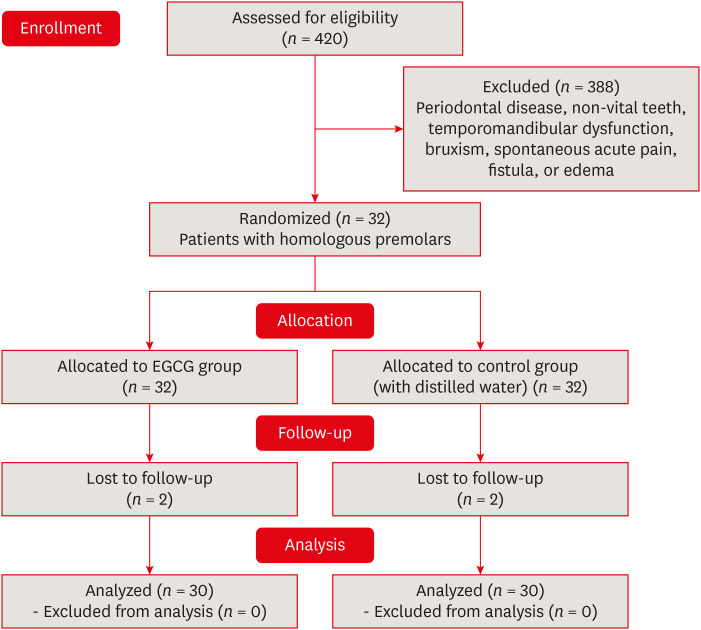
Materials and Methods The sample consisted of 30 patients (aged between 28 and 60 years) with abfraction lesions located in 2 homologous premolars. The teeth were randomly assigned according to dentin treatment: 0.02% EGCG solution or distilled water (control). After enamel acid etching, the solutions were applied immediately for 1 minute. The teeth were restored with Universal Adhesive (3M) and Filtek Z350 XT (3M). Analyzes were done by 2 independent examiners using modified USPHS (retention, secondary caries, marginal adaptation, and postoperative sensitivity) and photographic (color, marginal pigmentation, and anatomical form) criteria at baseline (7 days) and final (18 months). The data analysis used Friedman and Wilcoxon signed-rank tests (α = 0.05).
Results At baseline, all restorations were evaluated as alpha for all criteria. After 18 months, restorations were evaluated as alpha for secondary caries, color, and marginal pigmentation. There was significant difference between baseline and 18 months (
p = 0.009) for marginal adaptation and postoperative sensitivity (p = 0.029), but no significant difference were verified between treatments (p = 0.433). The EGCG group had a restoration retention rate of 93.3%, while the control group had 96.7%.Conclusions The application of EGCG solution on abfraction lesions did not significantly influence the survival of the restorations based on clinical and photographic criteria.
-
Citations
Citations to this article as recorded by- Therapeutic potential of flavonoids in erosive tooth wear management: a scoping review
Gabriel Pereira Nunes, Renata de Oliveira Alves, Geórgia Rondó Peres, Priscila Toninatto Alves de Toledo, Aline Rogéria Freire de Castilho
Clinical Oral Investigations.2025;[Epub] CrossRef
- Therapeutic potential of flavonoids in erosive tooth wear management: a scoping review
- 2,479 View
- 56 Download
- 1 Web of Science
- 1 Crossref
- Resin infiltrant protects deproteinized dentin against erosive and abrasive wear
- Ana Theresa Queiroz de Albuquerque, Bruna Oliveira Bezerra, Isabelly de Carvalho Leal, Maria Denise Rodrigues de Moraes, Mary Anne S. Melo, Vanara Florêncio Passos
- Restor Dent Endod 2022;47(3):e29. Published online July 1, 2022
- DOI: https://doi.org/10.5395/rde.2022.47.e29
-
Abstract
PDFPubReaderePub
Objectives This study aimed to investigate the anti-erosive/abrasive effect of resin infiltration of previous deproteinized dentin.
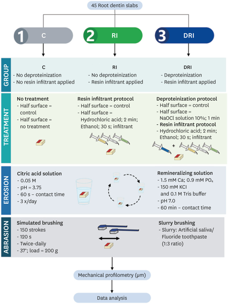
Materials and Methods Dentin slabs were randomly assigned to 3 groups (
n = 15): Control (no deproteinization; no resin infiltrant applied), RI (no deproteinization; resin infiltrant applied), and DRI (deproteinization; resin infiltrant applied). After undergoing the assigned treatment, all slabs were subjected to anin vitro cycling model for 5 days. The specimens were immersed in citric acid (0.05 M, pH = 3.75; 60 seconds; 3 times/day) and brushed (150 strokes). Between the challenges, the specimens were exposed to a remineralizing solution (60 minutes). The morphological alterations were analyzed by mechanical profilometry (µm) and scanning electron microscopy (SEM). Data were submitted to one-way analysis of variance (ANOVA) and Tukey tests (p < 0.05).Results Control and RI groups presented mineral wear and did not significantly differ from each other (
p = 0.063). DRI maintained a protective layer preserving the dentin (p < 0.001). After erosive/abrasive cycles, it was observed that in group RI, only 25% of the slabs partially evidenced the presence of the infiltrating, while, in the DRI group, 80% of the slabs presented the treated surface entirely covered by a resin-component layer protecting the dentin surface as observed in SEM images.Conclusions The removal of the organic content allows the resin infiltrant to efficiently protect the dentin surface against erosive/abrasive lesions.
-
Citations
Citations to this article as recorded by- Acidic/abrasive challenges on simulated non-carious cervical lesions development and morphology
Giovanna C. Denucci, Ian Towle, Cecilia P. Turssi, George J. Eckert, Anderson T. Hara
Archives of Oral Biology.2025; 169: 106120. CrossRef - Physio‐Mechanic and Microscopic Analyses of Bioactive Glass‐Based Resin Infiltrants
Syed Zubairuddin Ahmed, Abdul Samad Khan, Wejdan Waleed Nasser, Methayel Abdulrahman Alrushaid, Zahrah Mohammed Alfaraj, Moayad Mohammed Aljeshi, Asma Tufail Shah, Budi Aslinie Md Sabri, Sultan Akhtar, Mohamed Ibrahim Abu Hassan
Microscopy Research and Technique.2025; 88(2): 595. CrossRef - Resin Infiltration Treatment of Developmental Enamel Defects in a Patient With Hydrocephalus and Cerebral Palsy: A Case Report on the Impact on the Maternal Caregiver
Eduarda Martins Fontes Cantarella de Almeida, Anna Luísa Araujo Pimenta, Francisco Wanderley Garcia de Paula‐Silva, Fabricio Kitazono de Carvalho, Laurindo Borelli‐Neto, Susanne Effenberger, Fernanda de Carvalho Panzeri, Silmara Aparecida Milori Corona, K
Special Care in Dentistry.2025;[Epub] CrossRef
- Acidic/abrasive challenges on simulated non-carious cervical lesions development and morphology
- 3,111 View
- 52 Download
- 3 Web of Science
- 3 Crossref
Case Report
- Persistent pain after successful endodontic treatment in a patient with Wegener’s granulomatosis: a case report
- Ricardo Machado, Jorge Aleixo Pereira, Filipe Colombo Vitali, Michele Bolan, Elena Riet Correa Rivero
- Restor Dent Endod 2022;47(3):e26. Published online June 9, 2022
- DOI: https://doi.org/10.5395/rde.2022.47.e26
-
Abstract
PDFPubReaderePub
Wegener’s granulomatosis (WG) is a condition with immune-mediated pathogenesis that can present oral manifestations. This report describes the case of a patient diagnosed with WG 14 years previously, who was affected by persistent pain of non-odontogenic origin after successful endodontic treatment. A 39-year-old woman with WG was diagnosed with pulp necrosis and apical periodontitis of teeth #31, #32, and #41, after evaluation through a clinical examination and cone-beam computed tomography (CBCT). At the first appointment, these teeth were subjected to conventional endodontic treatment. At 6- and 12-month follow-up visits, the patient complained of persistent pain associated with the endodontically treated teeth (mainly in tooth #31), despite complete remission of the periapical lesions shown by radiographic and CBCT exams proving the effectiveness of the endodontic treatments, thus indicating a probable diagnostic of persistent pain of non-odontogenic nature. After the surgical procedure was performed to curette the lesion and section 3 mm of the apical third of tooth #31, the histopathological analysis suggested that the painful condition was likely associated with the patient's systemic condition. Based on clinical, radiographic, and histopathological findings, this unusual case report suggests that WG may be related to non-odontogenic persistent pain after successful endodontic treatments.
-
Citations
Citations to this article as recorded by- Toothaches of Non-odontogenic Origin
Davis C. Thomas, Tanvee Somaiya, Ahana Ajayakumar, Vaishnavi Prabhakar
Dental Clinics of North America.2026; 70(1): 209. CrossRef - Prevalence of persistente pain after endodontic treatment
Edmundo Duarte Martins, Allya Francisca Marques Borges, Lidiane Oliveira Leão, Bianca Marques de Mattos de Araujo, José Stechman Neto, Tatiana Carvalho Kowaltschuk, Camila de Castro Corrêa Corrêa, Cristiano Miranda de Araújo, Karinna Veríssimo Meira Tave
Brazilian Journal of Oral Sciences.2026; 25: e269272. CrossRef
- Toothaches of Non-odontogenic Origin
- 6,845 View
- 84 Download
- 2 Crossref
Research Articles
- In-office dental bleaching with violet light emitting diode: bleaching efficacy and pulpal temperature rise
- Brunna Katyuscia de Almeida Guanaes, Talyta Neves Duarte, Gisele Maria Correr, Marina da Rosa Kaizer, Carla Castiglia Gonzaga
- Restor Dent Endod 2022;47(1):e7. Published online February 3, 2022
- DOI: https://doi.org/10.5395/rde.2022.47.e7
-
Abstract
PDFPubReaderePub
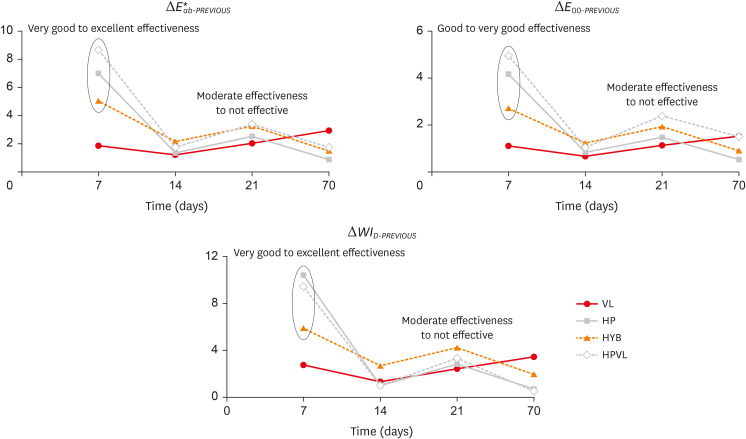
Objectives This study evaluated the bleaching efficacy of different in-office protocols associated with violet light emitting diode (V-LED), and measured the pulpal temperature rise caused by V-LED with or without gel application.
Materials and Methods Bovine incisors were distributed in 4 groups (
n = 10): VL – V-LED; HP – 35% hydrogen peroxide (control); HYB – hybrid protocol, V-LED applied without gel for 10 irradiation cycles followed by V-LED applied with gel for another 10 irradiation cycles; and HPVL – gel and V-LED applied for 20 irradiation cycles. Three bleaching sessions were performed with 7-day intervals. Bleaching efficacy was evaluated withE 00 and ΔWID . Data were recorded at baseline, 7, 14, 21 and 70 days. For pulpal temperature rise, thermocouples were placed inside the pulp chamber of human incisors. To determine intrapulpal temperature, the teeth were irradiated with V-LED with or without application of bleaching gel. Color difference data were analyzed by 2-way repeated measures ANOVA and Tukey’s test. Pulpal temperature was analyzed byt -test (α = 5%).Results VL exhibited lower color (
E 00) and whiteness changes (ΔWID ) than the other groups. HPVL presented higher color change values than HYB. HYB and HPVL showed not different ΔWID values; and HP showed the highest whiteness changes at all times. There were significant differences comparing ΔT with gel (8.9°C) and without gel application (7.2°C).Conclusions HPLV was more efficient than HYB. The 2 protocols with VL showed similar results to control. Gel application combined with VL promoted higher pulpal temperature than to the no gel group.
-
Citations
Citations to this article as recorded by- Inverse Heat Conduction Estimation of Heat Flux in Human Dentin from Dental Curing Lights Using the Conjugate Gradient Method
Ahmad Soori, Farshad Kowsary, Shadab Safarzadeh Khosroshahi, Mohammad Vahedi
International Journal of Thermophysics.2026;[Epub] CrossRef - Illuminating the evidence: A comprehensive review of light-assisted in-office tooth bleaching
Márcia V.G.B. Queiroz, Rafael Dascanio, Vinicius H. Hutemma, Diogo A. Chiovetto, Adriano F. Lima, Jorge R. Soto-Montero, Matheus Kury
Journal of Dentistry.2026; 171: 106707. CrossRef - Spectrophotometric Evaluation of Laser-Assisted Dental Bleaching Using Erbium-Doped Yttrium Aluminum Garnet (Er:YAG) and Diode Lasers at Different Wavelengths: An In Vitro Study
Esraa Ihssan Alshibli, Omar H. Hamadah, Mohammad Y. Hajeer
Cureus.2026;[Epub] CrossRef - Effect of antioxidant on tooth sensitivity after bleaching
Mohamed Nabil, Mostafa Mohamed Hasan, Eman Abd Elghany Shebl
Journal of Esthetic and Restorative Dentistry.2024; 36(3): 429. CrossRef - In-office Bleaching Activated With Violet LED: Effect on Pulpal and Tooth Temperature and Pulp Viability
NR Carlos, RT Basting, KR Kantovitz, ES Bronze-Uhle, PN Lisboa Filho, V Cavalli, RT Basting
Operative Dentistry.2024; 49(3): 262. CrossRef - Low and high hydrogen peroxide concentrations of in-office dental bleaching associated with violet light: an in vitro study
Isabela Souza Vardasca, Michael Willian Favoreto, Mylena de Araujo Regis, Taynara de Souza Carneiro, Emanuel Adriano Hul, Christiane Philippini Ferreira Borges, Alessandra Reis, Alessandro D. Loguercio, Carlos Francci
Clinical Oral Investigations.2024;[Epub] CrossRef - Bleaching efficacy of in-office bleaching with violet light using low-concentration hydrogen peroxide nanoparticulate photocatalyst gel: A randomized controlled trial
Gustavo Garcia Castro, Palena Araújo Pinto, Michael Willian Favoreto, Alessandra Reis, Maria Viviana-Mora, Rita de Cássia Mendonça de Miranda, Andres Felipe Milan Cardenas, Alessandro D. Loguercio, Rudys Rodolfo de Jesus Tavarez
Photodiagnosis and Photodynamic Therapy.2024; 50: 104410. CrossRef - Influence of Different Light-Activated Bleaching Gels on Pulp Chamber Temperature: An In Vitro Study
Mandana Karimi, Elmira Ataee, Ladan Ranjbar Omrani, Mahdi Abbasi, Elham Ahmadi
Avicenna Journal of Dental Research.2024; 16(4): 225. CrossRef - Continuous vs fractionated violet LED light protocols for dental bleaching: Evaluations of color change and temperature of the dental pulp and buccal surface
Mayanna Pacheco Trindade Najar, Luciana Hilel Rangel Barbosa, Natália Russo Carlos, Fabiana Mantovani Gomes França, Cecilia Pedroso Turssi, Waldemir Francisco Vieira-Junior, Roberta Tarkany Basting
Photodiagnosis and Photodynamic Therapy.2023; 42: 103631. CrossRef - Improved esthetic efficacy and reduced cytotoxicity are achieved with a violet LED irradiation of manganese oxide-enriched bleaching gels
Marlon Ferreira Dias, Beatriz Voss Martins, Rafael Antonio de Oliveira Ribeiro, Josimeri Hebling, Carlos Alberto de Souza Costa
Lasers in Medical Science.2022;[Epub] CrossRef
- Inverse Heat Conduction Estimation of Heat Flux in Human Dentin from Dental Curing Lights Using the Conjugate Gradient Method
- 4,486 View
- 46 Download
- 10 Web of Science
- 10 Crossref
- Effect of hydrogel-based antibiotic intracanal medicaments on crown discoloration
- Rayan B. Yaghmoor, Jeffrey A. Platt, Kenneth J. Spolnik, Tien Min Gabriel Chu, Ghaeth H. Yassen
- Restor Dent Endod 2021;46(4):e52. Published online October 5, 2021
- DOI: https://doi.org/10.5395/rde.2021.46.e52
-
Abstract
PDFPubReaderePub
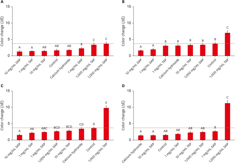
Objectives This study evaluated the effects of low and moderate concentrations of triple antibiotic paste (TAP) and double antibiotic paste (DAP) loaded into a hydrogel system on crown discoloration and explored whether application of an adhesive bonding agent prevented crown discoloration.
Materials and Methods Intact human molars (
n = 160) were horizontally sectioned 1 mm apical to the cementoenamel junction. The crowns were randomized into 8 experimental groups (calcium hydroxide, Ca[OH]2; 1, 10, and 1,000 mg/mL TAP and DAP; and no medicament. The pulp chambers in half of the samples were coated with an adhesive bonding agent before receiving the intracanal medicament. Color changes (ΔE) were detected by spectrophotometry after 1 day, 1 week, and 4 weeks, and after 5,000 thermal cycles, with ΔE = 3.7 as a perceptible threshold. The 1-samplet -test was used to determine the significance of color changes relative to 3.7. Analysis of variance was used to evaluate the effects of treatment, adhesive, and time on color change, and the level of significance wasp < 0.05.Results Ca(OH)2 and 1 and 10 mg/mL DAP did not cause clinically perceivable tooth discoloration. Adhesive agent use significantly decreased tooth discoloration in the 1,000 mg/mL TAP group up to 4 weeks. However, adhesive use did not significantly improve coronal discoloration after thermocycling when 1,000 mg/mL TAP was used.
Conclusions Ca(OH)2 and 1 and 10 mg/mL DAP showed no clinical discoloration. Using an adhesive significantly improved coronal discoloration up to 4 weeks with 1,000 mg/mL TAP.
-
Citations
Citations to this article as recorded by- Comparative analysis of tooth discoloration induced by different intracanal medicaments in regenerative endodontics: A systematic review and network meta-analysis
Ashlesha Nageshwar Madankar, Sulabha Radke, Shanmuga Priya, Darshan Dakshindas
Endodontology.2026; 38(1): 8. CrossRef - Tooth discoloration caused by nanographene oxide as an irrigant and intracanal medicament in the endodontic treatment of extracted single-rooted teeth: An ex-vivo study
Abbas Abbaszadegan, Zeinab Rafiee, Bahar Asheghi, Ahmad Gholami, Mohmed Isaqali Karobari
PLOS One.2025; 20(6): e0325430. CrossRef - Root development of immature necrotic permanent teeth following regenerative endodontic process: Case series
Abbasali Khademi, Pedram Iranmanesh, Ali Akhavan, Movahed Ghassem Yeganeh, Samira Khalifezade Esfahani
Dental Research Journal.2025;[Epub] CrossRef - Root Canal Dentin Microhardness after Contact with Antibiotic Medications: An In Vitro Study
Amanda Palmeira Arruda Nogueira, Renata Grazziotin-Soares, Adriana Marques Mesquita Leal, Sérgio Alves Guida Freitas Júnior, Bruna Laís Lins Gonçalves, José Bauer, Meire Coelho Ferreira, Ceci Nunes Carvalho
Dentistry Journal.2024; 12(7): 201. CrossRef - Potential Crown Discoloration Induced by the Combination of Various Intracanal Medicaments and Scaffolds Applied in Regenerative Endodontic Therapy
NB Altun, A Turkyilmaz
Nigerian Journal of Clinical Practice.2024; 27(7): 897. CrossRef
- Comparative analysis of tooth discoloration induced by different intracanal medicaments in regenerative endodontics: A systematic review and network meta-analysis
- 3,175 View
- 43 Download
- 4 Web of Science
- 5 Crossref
- Laboratory model to evaluate efficacy of an experimental titanium oxide nanofibers bleaching agent
- Clayton Tran, Ellin Choi, Brittany Watu, Udochukwu Oyoyo, Christopher Perry, So Ran Kwon
- Restor Dent Endod 2021;46(4):e47. Published online September 2, 2021
- DOI: https://doi.org/10.5395/rde.2021.46.e47
-
Abstract
PDFPubReaderePub
Objectives This study aimed to use a laboratory model to evaluate the efficacy of an experimental bleaching agent.
Materials and Methods The model used human extracted molars that were treated and measured for bleaching efficacy. Teeth (
n = 50) were distributed into 5 groups: Negative control (NC): immersion in water for 8 hours; Nanofibers (NFs): Experimental titanium dioxide nanofibers with stirring and light activation for 8 hours; Whitestrips (WS): Crest 3D White Glamorous White Whitestrips, 2 applications daily for 30 minutes, 14 days; 1% hydrogen peroxide (HP) standard: 1% hydrogen peroxide for 8 hours; and 30% HP standard: 30% hydrogen peroxide for 8 hours. Instrumental measurements were performed using a spectrophotometer. Results were recorded at baseline, 1-day post-bleaching, and 1-week post-bleaching. Kruskal-Wallis procedure was used to determine differences in color change. Pearson correlation was used to evaluate the relationship between visual and instrumental measurements. Tests of hypotheses were 2-sided with alpha = 0.05.Results There was no significant difference in color parameters (L1, a1, b1, and shade guide units [SGU]) at baseline (
p > 0.05). There was a significant difference among the groups for overall color change (ΔE*ab) and change in shade guide units (ΔSGU) at 1-day and 1-week post-bleaching (p < 0.05). The higher the HP concentration, the higher the color change as expressed in ΔSGU and ΔE*ab. The negative control exceeded the perceptibility threshold of ΔE* = 1.2 regardless of time point. NFs showed a decrease in chroma, but were not statistically different compared to the negative control.Conclusions The laboratory model was successful in screening an experimental bleaching agent.
-
Citations
Citations to this article as recorded by- Evaluating the Efficacy of Titanium Dioxide Nanoparticles in Combination with Commonly Used Bleaching Agents: An In Vitro Study
Rajasekhar Vemareddy, Sudhakar Naidu, Bala Raju Korrai, Shanmukha Nagadevara, Someshwar Battu, Jyotsnanjali Thati, Sivaji Kavuri
World Journal of Dentistry.2024; 15(5): 377. CrossRef
- Evaluating the Efficacy of Titanium Dioxide Nanoparticles in Combination with Commonly Used Bleaching Agents: An In Vitro Study
- 2,714 View
- 17 Download
- 1 Crossref
- Microleakage and characteristics of resin-tooth tissues interface of a self-etch and an etch-and-rinse adhesive systems
- Xuan Vinh Tran, Khanh Quang Tran
- Restor Dent Endod 2021;46(2):e30. Published online May 18, 2021
- DOI: https://doi.org/10.5395/rde.2021.46.e30
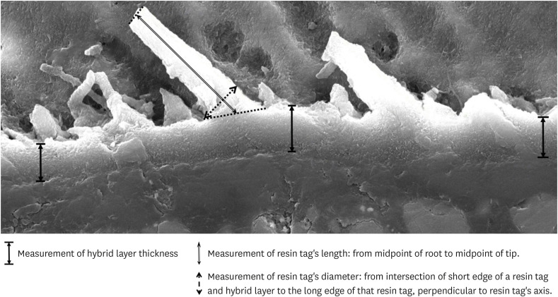
-
Abstract
PDFPubReaderePub
Objectives This study was conducted to compare the microleakage and characteristics of the resin-tooth tissue interface between self-etch and etch-and-rinse adhesive systems after 48 hours and 3 months.
Materials and Methods 40 extracted premolar teeth were randomly divided into 2 groups: 1-step self-etch adhesive system – Optibond™ All-In-One, and 2-step etch-and-rinse adhesive system - Adper™ Single Bond 2. Both groups were subjected to 500 thermocycles (5°C–55°C) before scanning electron microscope (SEM) analysis or microleakage trial at 48-hour and 3-month time periods.
Results SEM images showed the hybrid layer thickness, diameter, and length of resin tags of the self-etch adhesive (0.42 ± 0.14 µm; 1.49 ± 0.45 µm; 16.35 ± 14.26 µm) were smaller than those of the etch-and-rinse adhesive (4.39 ± 1.52 µm; 3.49 ± 1 µm; 52.81 ± 35.81 µm). In dentin, the microleakage scores of the 2 adhesives were not different in both time periods (48 hours/3 months). However, the microleakage score of etch-and-rinse adhesive increased significantly after 3 months (0.8 ± 0.63 and 1.9 ± 0.88,
p < 0.05).Conclusions The self-etch adhesive exhibited better long-term sealing ability in dentin when compared to that of the etch-and-rinse adhesive. The greater hybrid layer thickness and dimensions of resin tags did not guarantee reliable, long-lasting sealing in the bonding area.
-
Citations
Citations to this article as recorded by- A systematic review of shear bond strength of sixth- and fourth-generation adhesives in primary teeth
Maryam Hajiahmadi, Najmeh Akhlaghi, Hamid Mosleh, Ehsan Samani, Sheida Bagheri, Zohreh Salehi
Dental Research Journal.2026;[Epub] CrossRef - Comparative evaluation of shear bond strength of nano-hybrid composites to dentin: influence of universal adhesives versus two-step etch-and-rinse adhesives
Soodabeh Shoale, Mahsa Kowkabi, Artam Enayat, Hamed Manafi
Discover Applied Sciences.2026;[Epub] CrossRef - Effect of aging on microshearing bond strength of different adhesive systems
Cansu Dağdelen Ahısha, Mine Betül Üçtaşlı
BMC Oral Health.2026;[Epub] CrossRef - Clinical performance of the ethanol-wet bonding technique with different adhesive systems in noncarious cervical lesion restorations: a 6-year randomized clinical trial
Luana dos Santos Souza, Maria Fernanda Monnerat, Maurício Yugo Souza, Marcella Batista Rocha, Taciana Marco Ferraz Caneppele, Eduardo Bresciani
Clinical Oral Investigations.2026;[Epub] CrossRef - Efficacy of different adhesive systems in bonding direct resin composite restorations: a systematic review and meta-analysis
Ravinder S. Saini, Rajesh Vyas, Sunil Kumar Vaddamanu, Syed Altafuddin Quadri, Seyed Ali Mosaddad, Artak Heboyan
Evidence-Based Dentistry.2025; 26(2): 115. CrossRef - Characterisation of universal adhesive bonded resin-dentin interface after focused ultrasound smear layer conditioning
Cheryl Fu, Peta L. Clode, Amr S. Fawzy
International Journal of Adhesion and Adhesives.2025; 142: 104115. CrossRef - Effect of Dentin Pretreatment With Dimethyl Sulfoxide Solution on Interfacial Fracture Toughness of Composite Resin to Wet and Dry Dentin
Fatemeh Molaei, Mehrsima Ghavami-Lahiji, Seyedeh Maryam Tavangar, Hannah Wesley
International Journal of Dentistry.2025;[Epub] CrossRef - Resin tags formation by modified Renewal MI formulations in a carious dentine model
Nabih Alkhouri, Wendy Xia, Paul Ashley, Anne Young
Frontiers in Oral Health.2024;[Epub] CrossRef - Effect of propolis added to single‐bottle adhesives on water permeation through the hybrid layer
Lucineide Silva da Rocha, Daniela Ferreira de Oliveira, Cinthya Luna Veloso de Lima, Ticiano Gomes do Nascimento, Johnnatan Duarte de Freitas, Jeniffer Mclaine Duarte de Freitas, Isabel Cristina Celerino de Moraes Porto
European Journal of Oral Sciences.2024;[Epub] CrossRef - Exploration and preliminary clinical investigation of an adhesive approach for primary tooth restoration
Xiangqin Xu, Jiansheng Zhu, May Lei Mei, Huaying Wu, Kaipeng Xie, Shoulin Wang, Yaming Chen
The Journal of Biomedical Research.2023; 37(2): 138. CrossRef - Adhesion to enamel and dentine: an update
Rana Alkattan
Primary Dental Journal.2023; 12(3): 33. CrossRef - Effects of carbodiimide combined with ethanol–wet bonding pretreatment on dentin bonding properties: an in vitro study
Xiaoxiao You, Long Chen, Jie Xu, Sihui Li, Zhenghao Zhang, Ling Guo
PeerJ.2022; 10: e14238. CrossRef - The effects of amalgam contamination and different surface modifications on microleakage of dentin bonded to bulk fill composite when using different adhesive protocols
Nojoud Alshehri, Abdullah Aljamhan, Mohammed Bin-Shuwaish
BMC Oral Health.2022;[Epub] CrossRef - Development of low-shrinkage dental adhesives via blending with spiroorthocarbonate expanding monomer and unsaturated epoxy resin monomer
Zonghua Wang, Xiaoran Zhang, Shuo Yao, Jiaxin Zhao, Chuanjian Zhou, Junling Wu
Journal of the Mechanical Behavior of Biomedical Materials.2022; 133: 105308. CrossRef - Influence of silver nanoparticles on the resin-dentin bond strength and antibacterial activity of a self-etch adhesive system
Jia Wang, Wei Jiang, Jingping Liang, Shujun Ran
The Journal of Prosthetic Dentistry.2022; 128(6): 1363.e1. CrossRef
- A systematic review of shear bond strength of sixth- and fourth-generation adhesives in primary teeth
- 3,819 View
- 64 Download
- 13 Web of Science
- 15 Crossref
- Retrospective study of fracture survival in endodontically treated molars: the effect of single-unit crowns versus direct-resin composite restorations
- Kanet Chotvorrarak, Warattama Suksaphar, Danuchit Banomyong
- Restor Dent Endod 2021;46(2):e29. Published online May 6, 2021
- DOI: https://doi.org/10.5395/rde.2021.46.e29
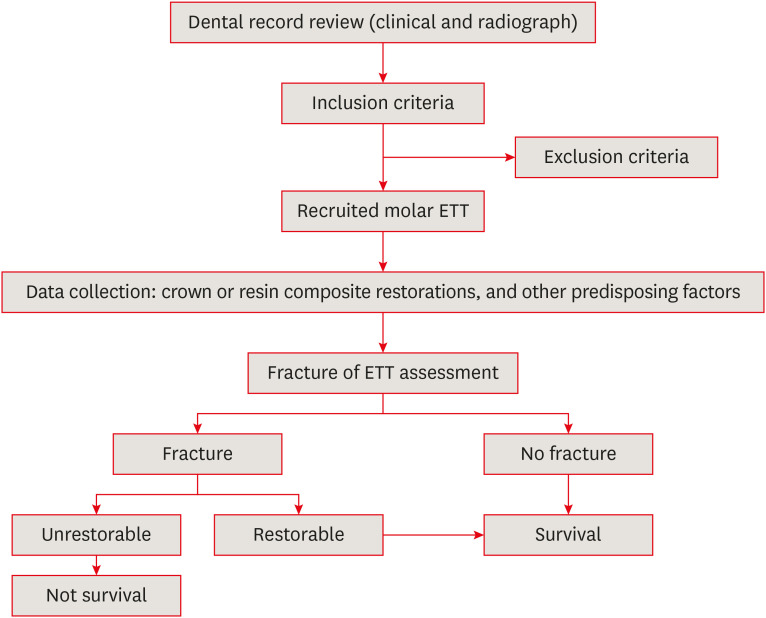
-
Abstract
PDFPubReaderePub
Objectives This study was conducted to compare the post-fracture survival rate of endodontically treated molar endodontically treated teeth (molar ETT) restored with resin composites or crowns and to identify potential risk factors, using a retrospective cohort design.
Materials and Methods Dental records of molar ETT with crowns or composite restorations (recall period, 2015–2019) were collected based on inclusion and exclusion criteria. The incidence of unrestorable fractures was identified, and molar ETT were classified according to survival. Information on potential risk factors was collected. Survival rates and potential risk factors were analyzed using the Kaplan-Meier log-rank test and Cox regression model.
Results The overall survival rate of molar ETT was 87% (mean recall period, 31.73 ± 17.56 months). The survival rates of molar ETT restored with composites and crowns were 81.6% and 92.7%, reflecting a significant difference (
p < 0.05). However, ETT restored with composites showed a 100% survival rate if only 1 surface was lost, which was comparable to the survival rate of ETT with crowns. The survival rates of ETT with composites and crowns were significantly different (97.6% vs. 83.7%) in the short-term (12–24 months), but not in the long-term (> 24 months) (87.8% vs. 79.5%).Conclusions The survival rate from fracture was higher for molar ETT restored with crowns was higher than for ETT restored with composites, especially in the first 2 years after restoration. Molar ETT with limited tooth structure loss only on the occlusal surface could be successfully restored with composite restorations.
-
Citations
Citations to this article as recorded by- Effect of Conventional Filler, Short Fiber-reinforced, and Polyethylene Woven Fiber-reinforced Composite on the Fracture Toughness of Extracted Premolar Teeth
Leeza Bharati, Chandrakar Chaman, Udai P Singh, Asib Ahmad, Siddharth Anand, Aparna Singh
The Journal of Contemporary Dental Practice.2025; 26(7): 693. CrossRef - Risk factors for the appearance of cracks and fractures of teeth according to a survey of dentists
Emilia A. Olesova, Alexander A. Ilyin, Sergey D. Arutyunov, Elena V. Glazkova, Arsen A. Popov, Svetlana P. Iarilkina
Russian Journal of Dentistry.2024; 28(6): 562. CrossRef - Performance of Bonded Lithium Disilicate Partial-coverage Crowns in the Restoration of Endodontically Treated Posterior Teeth: An Up to Seven-Year Retrospective Study
Q Jiang, Z Wang, S Zhang, X Liu, B Fu
Operative Dentistry.2024; 49(4): 365. CrossRef - In Vitro Bond Strength of Dentin Treated with Sodium Hypochlorite: Effects of Antioxidant Solutions
Guillermo Grazioli, Elisa de León Cáceres, Romina Tessore, Rafael Lund, Ana Monjarás-Ávila, Monika Lukomska-Szymanska, Louis Hardan, Rim Bourgi, Carlos Cuevas-Suárez
Antioxidants.2024; 13(9): 1116. CrossRef - Stress Analysis on Mesiolingual Cavity of Endodontically Treated Molar Restored Using Bidirectional Fiber-Reinforced Composite (Wallpapering Technique)
Harnia Neri, Dudi Aripin, Anna Muryani, Hendra Dharsono, Yolanda Yolanda, Andi Mahyuddin
Clinical, Cosmetic and Investigational Dentistry.2024; Volume 16: 75. CrossRef - Effect of Luting Cement Film Thickness on the Pull-Out Bond Strength of Endodontic Post Systems
Khalil Aleisa, Syed Rashid Habib, Abdul Sadekh Ansari, Ragad Altayyar, Shahad Alharbi, Sultan Ali S. Alanazi, Khalid Tawfik Alduaiji
Polymers.2021; 13(18): 3082. CrossRef
- Effect of Conventional Filler, Short Fiber-reinforced, and Polyethylene Woven Fiber-reinforced Composite on the Fracture Toughness of Extracted Premolar Teeth
- 4,262 View
- 65 Download
- 9 Web of Science
- 6 Crossref
Case Report
- Endodontic approach in a replanted tooth with an immature root apex and chronic apical periodontitis: a case report
- Graziela Garrido Mori, Bruna Souza Andrade, Marina Bardelli Araujo
- Restor Dent Endod 2020;45(3):e29. Published online May 21, 2020
- DOI: https://doi.org/10.5395/rde.2020.45.e29
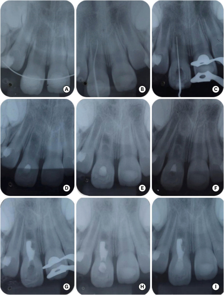
-
Abstract
PDFPubReaderePub
This study describes the clinical steps taken in the treatment of a patient who had an avulsed right upper central incisor that presented with incomplete root development and chronic apical periodontitis. A 7-year-old boy was referred from a private dentist to a dental office specializing in endodontics. The tooth had remained in a dry environment for 20 minutes, and tooth replantation was performed at an emergency appointment. After clinical and radiographic examinations, root canal decontamination was performed, followed by several changes in intracanal calcium hydroxide medication. Blood clot formation was attempted, but bleeding within the root canal was insufficient; therefore, we opted for an intracanal medication change to stimulate mineralized tissue formation in the apical region. Root obturation was performed 45 days after the last change of intracanal medication, and clinical, radiographic, and tomographic follow-up examinations were performed at 3, 6, 18, and 40 months after the endodontic intervention. The increase in thickness and length of the root structure and the absence of root resorption were verified through follow-up examinations. Therefore, it was concluded that the procedures used were successful for tooth replantation.
-
Citations
Citations to this article as recorded by- Are Extracellular Vesicles Used in Tissue Engineering Effective for Regenerative Endodontic Treatments? Scoping Review
Carolina dos Santos Santinoni, Jéssica Pestana, Lívia Apolito Rissi, Milena de Queiroz Guedes, Graziela Garrido Mori Panucci
Australian Endodontic Journal.2025; 51(3): 825. CrossRef - Intentional replantation of periodontally compromised teeth with concentrated growth factors
Nabeel Ayappali Kalluvalappil, R. Saravana Kumar, Tamer Hamed Hassan
Clinical Advances in Periodontics.2025;[Epub] CrossRef - The Impact of the Preferred Reporting Items for Case Reports in Endodontics (PRICE) 2020 Guidelines on the Reporting of Endodontic Case Reports
Sofian Youssef, Phillip Tomson, Amir Reza Akbari, Natalie Archer, Fayjel Shah, Jasmeet Heran, Sunmeet Kandhari, Sandeep Pai, Shivakar Mehrotra, Joanna M Batt
Cureus.2023;[Epub] CrossRef
- Are Extracellular Vesicles Used in Tissue Engineering Effective for Regenerative Endodontic Treatments? Scoping Review
- 4,757 View
- 138 Download
- 3 Crossref
Research Articles
- Influence of pain-relieving therapies on inflammation and the expression of proinflammatory neuropeptides after dental bleaching treatment
- Livia Maria Alves Valentim da Silva, Luciano Tavares Angelo Cintra, Marjorie de Oliveira Gallinari, Francine Benetti, Vanessa Rahal, Edilson Ervolino, Sibele de Alcântara, André Luiz Fraga Briso
- Restor Dent Endod 2020;45(2):e20. Published online February 28, 2020
- DOI: https://doi.org/10.5395/rde.2020.45.e20
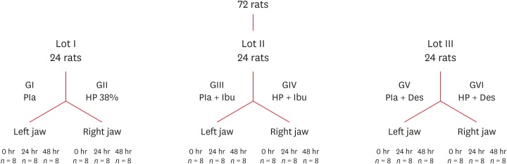
-
Abstract
PDFPubReaderePub
Objectives To minimize the tooth sensitivity caused by in-office bleaching, many dentists use non-steroidal anti-inflammatory drugs and topical desensitizing gels containing potassium nitrate and sodium fluoride. This study aimed to evaluate the influence of these substances on inflammation and the expression of substance P and calcitonin gene-related peptide in pulp nerve fibers.
Materials and Methods Seventy-two rats were divided into 6 groups as follows: GI, control; GII, only dental bleaching; GIII, only ibuprofen; GIV, ibuprofen administered 30 minutes before and after the bleaching treatment and every 12 hours until the analysis; GV, only topical application of a desensitizing agent; and GVI, topical application of a desensitizing agent before dental bleaching. Placebo gel was applied to the upper left jaw and the bleaching agent was applied to the upper right jaw in all groups. Subsequently, the groups were divided into 3 subgroups based on the time of analysis: 0, 24, and 48 hours after bleaching (
n = 8). The rats were euthanized and the maxillae were processed and evaluated by histopathological and immunohistochemical analyses. The data were analyzed using the Kruskal-Wallis test, followed by the Dunn test (p < 0.05).Results In the bleaching groups, the inflammatory process and expression of neuropeptides decreased over time. The animals in which a desensitizing agent was applied showed better results within 24 hours.
Conclusions The use of a desensitizing agent had positive effects on inflammation and pain-related neuropeptide expression, minimizing the painful effects of dental bleaching treatment.
-
Citations
Citations to this article as recorded by- Effectiveness of Analgesics in Dental Whitening Pain Management: A Systematic Review and Meta-Analysis
Gabriella Alves Julião Costa, Caio Ferreira Freire Caetano, Ravy Jucá Farias, Diana Araújo Cunha, Dayrine Silveira de Paula, Edson Luiz Cetira Filho, Paulo Goberlânio de Barros Silva
Expert Opinion on Pharmacotherapy.2025; 26(5): 639. CrossRef - The Use of Ozone Therapy in Combination with a Desensitizing Agent for Dentinal Tubules Occlusion: An In Vitro Study
Banna Alnufaiy
The Open Dentistry Journal.2025;[Epub] CrossRef - The role of neurogenic inflammation in pulp repair and the techniques used for its assessment (narrative review)
Muna Sh. Ahmed, Anas F. Mahdee
Frontiers in Dental Medicine.2025;[Epub] CrossRef - Influence of dental bleaching on the pulp tissue: A systematic review of in vivo studies
Mariana Viana Donato, Alexandre Henrique dos Reis‐Prado, Lucas Guimarães Abreu, Lara Cancella de Arantes, Juliana Goto, Hebertt Gonzaga dos Santos Chaves, Luciano Tavares Angelo Cintra, André Luiz Fraga Briso, Isabella Faria da Cunha Peixoto, Francine Ben
International Endodontic Journal.2024; 57(6): 630. CrossRef - Role of induced nitric oxide synthases in orofacial nociception/discomfort after dental tooth bleaching with hydrogen peroxide
Marcílio Rodrigues Pinto, Kirlya Isabel da Silva Medeiros, Letícia Menezes Maia, Antonio Alexandre Coelho, Ana Paula Negreiros Nunes Alves, Caio Ferreira Freire Caetano, Karine Cestaro Mesquita, Paulo Goberlânio de Barros Silva, Fabricio Bitu Sousa
Archives of Oral Biology.2024; 161: 105937. CrossRef - Can different agents reduce the damage caused by bleaching gel to pulp tissue? A systematic review of basic research
Letícia Aparecida Silva Batista, Alexandre Henrique dos Reis-Prado, Hebertt Gonzaga dos Santos Chaves, Lara Cancella de Arantes, Luís Fernando Santos Alves Morgan, Carolina Bosso André, Thaís Yumi Suzuki, Francine Benetti
Restorative Dentistry & Endodontics.2023;[Epub] CrossRef - The Effects of Different Drugs with Anti-Inflamatory Potential in Prevention of Pulp Damage During the Teeth Bleaching
Miona Glisic, Andjela Milojevic, Milica Milinkovic, Marina Rankovic
Experimental and Applied Biomedical Research (EABR).2023;[Epub] CrossRef - Bleaching gel volume influences hydrogen peroxide diffusion, inflammation, and the presence of nitric oxide in the pulp tissue: in vitro and in vivo model
Sibele de ALCÂNTARA, Francine BENETTI, Lívia Maria Alves Valentim da SILVA, Nathália Evelyn da Silva MACHADO, Isabela Joane Prado SILVA, Lara Maria Bueno ESTEVES, Edilson ERVOLINO, Luciano Tavares Angelo CINTRA, André Luiz Fraga BRISO
Journal of Applied Oral Science.2023;[Epub] CrossRef - Design of a thermosensitive ibuprofen-loaded nanogel as smart material applied as anti-inflammatory in tooth bleaching: An in vivo study
Samara K.S.C.F. Moura, Milena L.V. dos Santos, Lucas A. do Nascimento, Mariana F.A. da Silva, Glória M. de França, Lucas M. da Costa, Aldo C. Medeiros, Raimundo F. Araújo-Júnior, Aurigena A. de Araújo, Cláudia N. Oliveira, André L. Dorini, Rejane A. de Ca
Journal of Drug Delivery Science and Technology.2022; 68: 103123. CrossRef - Topical application of Otosporin® before in-office bleaching: a split mouth, triple-blind, multicenter randomized clinical trial
Michael Willian Favoreto, Laína Vochikovski, Renata Maria Oleniki Terra, Veridiana Silva Campos, Mariana Evangelista Santos, Sônia Saeger Meireles, Alessandra Reis, Alessandro D. Loguercio
Clinical Oral Investigations.2022; 26(3): 2555. CrossRef - A novel tooth bleaching gel based on peroxymonosulfate/polyphosphates advanced oxidation process: Effective whitening avoiding pulp damage and sensitivity
Su Yang, Baiyan Sui, Xin Liu, Jiao Sun, Jun Wang
Chemical Engineering Journal.2022; 429: 132525. CrossRef - Effectiveness of Violet LED alone or in association with bleaching gel during dental photobleaching: A Systematic Review
Bianca Rossi, Susana Morimoto, Tamara Kerber Tedesco, Sandra Ribeiro Cunha, Anna Carolina Ratto Tempestini Horliana, Karen Müller Ramalho
Photodiagnosis and Photodynamic Therapy.2022; 38: 102813. CrossRef
- Effectiveness of Analgesics in Dental Whitening Pain Management: A Systematic Review and Meta-Analysis
- 2,614 View
- 14 Download
- 12 Crossref
- Effect of dental bleaching on the microhardness and surface roughness of sealed composite resins
- Renan Aparecido Fernandes, Henrico Badaoui Strazzi-Sahyon, Thaís Yumi Umeda Suzuki, André Luiz Fraga Briso, Paulo Henrique dos Santos
- Restor Dent Endod 2020;45(1):e12. Published online January 10, 2020
- DOI: https://doi.org/10.5395/rde.2020.45.e12
-
Abstract
PDFPubReaderePub
Objectives The aim of this
in vitro study was to evaluate the microhardness and surface roughness of composite resins before and after tooth bleaching procedures.Materials and Methods Sixty specimens were prepared of each composite resin (Filtek Supreme XT and Opallis), and BisCover LV surface sealant was applied to half of the specimens. Thirty enamel samples were obtained from the buccal and lingual surfaces of human molars for use as the control group. The surface roughness and microhardness were measured before and after bleaching procedures with 35% hydrogen peroxide or 16% carbamide (
n = 10). Data were analyzed using 1-way analysis of variance and the Fisher test (α = 0.05).Results Neither hydrogen peroxide nor carbamide peroxide treatment significantly altered the hardness of the composite resins, regardless of surface sealant application; however, both treatments significantly decreased the hardness of the tooth samples (
p < 0.05). The bleaching did not cause any change in surface roughness, with the exception of the unsealed Opallis composite resin and dental enamel, both of which displayed an increase in surface roughness after bleaching with carbamide peroxide (p < 0.05).Conclusions The microhardness and surface roughness of enamel and Opallis composite resin were influenced by bleaching procedures.
-
Citations
Citations to this article as recorded by- Property changes in resin composite exposed to mouth rinses during 10% carbamide peroxide bleaching
Mariana Ferreira da Silva, Giovana Contin Germinari, Carolina Meneghin Barbosa, Tatiane Cristina Dotta, Renata Siqueira Scatolin, Waldemir Francisco Vieira Júnior, Laura Nobre Ferraz
Brazilian Journal of Oral Sciences.2026; 25: e260366. CrossRef - Effect of Bleaching Protocols on the Microhardness and Surface Roughness of Composite Resins with Different Filler Architectures: An In Vitro Study
Flor de Maria Celis-Jacinto , Fredy Hugo Cruzado-Oliva, Jorge Wilfredo Vera-Alvarado
Odovtos - International Journal of Dental Sciences.2026;[Epub] CrossRef - Effect of Bleaching on Surface Roughness and Color Parameters of Coffee-Stained Nanohybrid Dental Composites with Different Viscosities
Hetaf S. Redwan, Mohamed A. Hussein, Mohamed M. Abdul-Monem
European Journal of General Dentistry.2025; 14(01): 027. CrossRef - Effect of Staining and External Bleaching on the Color Stability and Surface Roughness of Universal-Shade Resin-Based Composite
AlHanouf AlHabdan, Amal Alsuhaibani, Lama Alomran, Lulwah Almutib
Clinical, Cosmetic and Investigational Dentistry.2025; Volume 17: 1. CrossRef - Comparative Analysis Between Strip and Gels Indicated for at Home Bleaching: Analysis of Color Alteration, Roughness and Microhardness of Dental Enamel
K. M. S. Aidar, L. T. A. Cintra, M. C. B. Ferreira, T. C. Fagundes, L. M. B. Esteves, J. Goto, A. Catelan, A. L. F. Briso
Journal of Esthetic and Restorative Dentistry.2025; 37(6): 1504. CrossRef - Surface properties and susceptibility to staining of a resin composite after brushing with different whitening toothpastes
Aline da Silva Barros, Carolina Meneghin Barbosa, Renata Siqueira Scatolin, Waldemir Francisco Vieira Junior, Laura Nobre Ferraz
Restorative Dentistry & Endodontics.2025; 50(1): e6. CrossRef - Degradation Resistance of Next-Generation Dental Composites Under Bleaching and Immersion: A Multiscale Investigation
Syed Zubairuddin Ahmed, Shahad Al-Qahtani, Naif H. Al-Qahtani, Hussah Al-Mulhim, Maha Al-Qahtani, Ali Albalushi, Sultan Akhtar
Prosthesis.2025; 7(3): 57. CrossRef - Effect of Over-the-Counter Whitening Dentifrices on the Color Stability and Microhardness of Composite Resins
Xinnuo Yu, Maria Pilar Melo, Sofia Folguera, Carmen Llena
Journal of Composites Science.2025; 9(7): 324. CrossRef - From Microstructure to Shade Shift: Confocal and Spectrophotometric Evaluation of Peroxide-Induced Dental Bleaching
Berivan Laura Rebeca Buzatu, Magda Mihaela Luca, Atena Galuscan, Adrian Ovidiu Vaduva, Aurora Doris Fratila, Ramona Dumitrescu, Ruxandra Sava-Rosianu, Octavia Balean, Roxana Buzatu, Daniela Jumanca
Journal of Clinical Medicine.2025; 14(13): 4642. CrossRef - In Vitro Evaluation of Chemical and Microhardness Alterations in Human Enamel Induced by Three Commercial In-Office Bleaching Agents
Berivan Laura Rebeca Buzatu, Atena Galuscan, Ramona Dumitrescu, Roxana Buzatu, Magda Mihaela Luca, Octavia Balean, Gabriela Vlase, Titus Vlase, Iasmina-Mădălina Anghel, Carmen Opris, Bianca Ioana Todor, Mihaela Adina Dumitrache, Daniela Jumanca
Dentistry Journal.2025; 13(8): 357. CrossRef - Effect of Hydrogen Peroxide Bleaching on Color Stability and Microhardness of Alkasite Restorative Materials: An In Vitro Study
Souad A Alfouzan, Rahaf A Alolayan, Asma Munir Khan
Cureus.2025;[Epub] CrossRef - Evaluation of Color Stability and Surface Roughness of Nanohybrid Resin Composites with Different Photoinitiator Systems After Staining and Home/Office Bleaching: An In Vitro Study
Fatma Yılmaz, Buse Kesgin
Meandros Medical And Dental Journal.2025; 26(3): 240. CrossRef - The Effect of Hydrogen Peroxide With Different Concentration on the Color and Surface Microhardness of the Resin Bracket
Song‐Yi Yang
Clinical and Experimental Dental Research.2025;[Epub] CrossRef - Comparative evaluation of different bleaching agents on the color stability, hardness and surface roughness of indirect esthetic restorative materials with different manufacturing methods
Ayse Atay, Defne Canpolat, Soner Sismanoglu, Aslihan Usumez
BMC Oral Health.2025;[Epub] CrossRef - Comparison of Microhardness and Surface Roughness of New Nanofiber Filled Flowable Composite
Rumeysa Hatice ENGINLER OZLEN, Zumrut Ceren OZDUMAN, Burcu OGLAKCI OZKOC, Evrim ELIGUZELOGLU DALKILIC
Bezmialem Science.2024; 12(4): 406. CrossRef - Effect of Bleaching Agents on Composite Resins with and without Bis-GMA: An In Vitro Study
María Melo, Bianca Dumitrache, James Ghilotti, José Luis Sanz, Carmen Llena
Journal of Functional Biomaterials.2024; 15(6): 144. CrossRef - Changes in physical properties of universal composites and CAD/CAM materials after bleaching and antioxidant applications: Scanning electron microscope and atomic force microscope evaluation
Oguz Kaan Tuysuz, Merve Gurses
Microscopy Research and Technique.2024; 87(5): 977. CrossRef - The Effects of Home and Over-The-Counter Whitening Agents on Surface Roughness and Microhardness of High Aesthetic Composites
Elif İpek KILIÇ DÖNMEZ, İhsan HUBBEZOĞLU
Cumhuriyet Dental Journal.2024; 27(1): 30. CrossRef - Effect of carbamide peroxide treatment on the ion release of different dental restorative materials
Merve Nur Yilmaz, Pinar Gul
BMC Oral Health.2024;[Epub] CrossRef - Inorganic Phosphate Effect in a Hydrogen Peroxide-based Bleaching Agent: Physicochemical, Mechanical, and Morphological Properties of Dental Enamel
KG Garcia, GP Nunes, ACB Delbem, PH dos Santos, GLP Fernandes, HF Robles, PBB Lemos, M Danelon
Operative Dentistry.2024; 49(4): 465. CrossRef - Effect of bleaching and repolishing on whiteness change and staining susceptibility of resin-based materials
Sultan Aktuğ Karademir, Samet Atasoy, Beyza Yılmaz
BMC Oral Health.2024;[Epub] CrossRef - Influence of Low pH on the Microhardness and Roughness Surface of Dental Composite—A Preliminary Study
Leszek Szalewski, Dorota Wójcik, Monika Sowa, Vladyslav Vivcharenko, Krzysztof Pałka
Materials.2024; 17(14): 3443. CrossRef - In Vitro Evaluation of the Effectiveness and pH Variation of Dental Bleaching Gels and Their Effect on Enamel Surface Roughness
Federica Veneri, Francesco Cavani, Giovanni Bolelli, Vittorio Checchi, Alessia Bizzi, Giacomo Setti, Luigi Generali
Dentistry Journal.2024; 12(12): 415. CrossRef - Does the combination of whitening toothpaste and hydrogen peroxide bleaching increase the surface roughness and change the morphology of a nanofilled composite?
Cecília Pereira da Silva Braga Tenório, Matheus Kury, Geyse Maria dos Santos Muniz Mota, Cecília Pedroso Turssi, Flávia Lucisano Botelho do Amaral, Vanessa Cavalli
Brazilian Journal of Oral Sciences.2024; 23: e241938. CrossRef - Effect of peroxide‐free and peroxide‐based in‐office bleaching on the surface and mechanical properties of CAD/CAM esthetic restorative materials
Majed M. Alsarani, Aftab Ahmed Khan, Leonel S. J. Bautista, Hanan Alsunbul, Jukka P. Matinlinna
European Journal of Oral Sciences.2024;[Epub] CrossRef - Effect of Repolishing on Color Stability, Translucency, and Surface Roughness of Aged Monochromatic Dental Composites
Mohamed M. Abdul-Monem, Mohamed A. Hussein, Mona G. Abdelrehim
European Journal of General Dentistry.2024; 13(03): 240. CrossRef - Color changes of nanofiller composite resin after glycerin application immersed in turmeric extract
Sukaton, Galih Sampoerno, Widyajeng Ayu Laksmi, Daradhasih Bestari Santiaji
Conservative Dentistry Journal.2023; 13(1): 37. CrossRef - Effects of Dental Bleaching Agents on the Surface Roughness of Dental Restoration Materials
Alexandru Dan Popescu, Mihaela Jana Tuculina, Oana Andreea Diaconu, Lelia Mihaela Gheorghiță, Claudiu Nicolicescu, Cristian Niky Cumpătă, Cristiana Petcu, Jaqueline Abdul-Razzak, Ana Maria Rîcă, Ruxandra Voinea-Georgescu
Medicina.2023; 59(6): 1067. CrossRef - Effect of Bleaching on the Microhardness and Modulus of Elasticity of ACTIVA BioACTIVE – RESTORATIVE: An In Vitro Study
Sushritha Sricharan, Swaroop Hegde, Narmada J., Indiresha H. Narayana, Chatura Mohan, Nithin K. Shetty
Journal of Advanced Oral Research.2023; 14(2): 190. CrossRef - The effect of bleaching on surface roughness and gloss of different CAD/CAM ceramic and hybrid ceramic materials
Ruwaida Z Alshali, Mohammed A AlQahtani, Dalea M Bukhary, Mlak A Alzahrani, Shatha S Alsoraihi, Majed A Alqahtani
Journal of Applied Biomaterials & Functional Materials.2023;[Epub] CrossRef - Effect of bleaching with 15% carbamide peroxide on color stability of microhybrid, nanohybrid, and nanofilled resin composites, each in 3 staining solutions (coffee, cola, red grape juice): A 3-phase study
Azadeh Ghaemi, Sanaz Sharifishoshtari, Mohsen Shahmoradi, Hossein Akbari, Parisa Boostanifard, Sepideh Bagheri, Mohammadreza Shokuhifar, Negin Ashoori, Vahid Rakhshan
Dental Research Journal.2023;[Epub] CrossRef - Micro-Hardness and Surface Roughness of Bulk-Fill Composite Resin: Effect of Surface Sealant Application and Two Bleaching Regimens
Reham Mohamad Attia, Eman Mohamed Sobhy, Mona El Said Abd El Hameed Essa
European Journal of General Dentistry.2023; 12(03): 169. CrossRef - Shear bond strength after using sealant before bonding: a systematic review and meta-analysis of in vitro studies
Jennifer Hoppe, Thomas Lehmann, Christoph-Ludwig Hennig, Ulrike Schulze-Späte, Collin Jacobs
Clinical Oral Investigations.2022; 26(1): 1. CrossRef - Effect of 16% Carbamide Peroxide and Activated-Charcoal-Based Whitening Toothpaste on Enamel Surface Roughness in Bovine Teeth: An In Vitro Study
Jorge Zamudio-Santiago, Marysela Ladera-Castañeda, Flor Santander-Rengifo, Carlos López-Gurreonero, Alberto Cornejo-Pinto, Ali Echavarría-Gálvez, Luis Cervantes-Ganoza, César Cayo-Rojas
Biomedicines.2022; 11(1): 22. CrossRef - Direct dentin bleaching: Would it be possible?
Camila Ferro Clemente, Sibele de Alcântara, Lívia Maria Alves Valentim da Silva, Lara Maria Bueno Esteves, Anderson Catelan, Karen Milaré Seiscento Aidar, Ticiane Cestari Fagundes, André Luiz Fraga Briso
Photodiagnosis and Photodynamic Therapy.2022; 40: 103121. CrossRef - EFFECT OF İN-OFFİCE BLEACHİNG ON THE SURFACE ROUGHNESS OF DİFFERENT COMPOSİTE RESİNS
Seher KAYA, Ozden OZEL BEKTAS
Cumhuriyet Dental Journal.2022; 25(Supplement): 78. CrossRef - Effect of Polishing on the Surface Microhardness of Nanohybrid Composite Resins Subjected to 35% Hydrogen Peroxide
Giovanna Gisella Ramírez-Vargas, Julia Elbia Medina y Mendoza, Ana Sixtina Aliaga-Mariñas, Marysela Irene Ladera-Castañeda, Luis Adolfo Cervantes-Ganoza, César Félix Cayo-Rojas
Journal of International Society of Preventive and Community Dentistry.2021; 11(2): 216. CrossRef - Intrapulpal Concentration of Hydrogen Peroxide of Teeth Restored With Bulk Fill and Conventional Bioactive Composites
DP Silva, BA Resende, M Kury, CB André, CPM Tabchoury, M Giannini, V Cavalli
Operative Dentistry.2021; 46(3): E158. CrossRef - An Environmental Scanning Electron Microscopy Evaluation on Comparison of Three Different Bleaching Agents using the Laser Activated in-Office Bleaching at Different Wavelengths
Shachi Goenka, Sushil Kumar Cirigiri, Kanika Poplai, Baig Mirza Aslam, Shalini Singh, Shweta Gangavane
Journal of Pharmacy and Bioallied Sciences.2021; 13(Suppl 2): S1478. CrossRef - Effects of Artificial Staining and Bleaching Protocols on the Surface Roughness, Color, and Whiteness Changes of an Aged Nanofilled Composite
Geyse Maria dos Santos Muniz Mota, Matheus Kury, Cecília Pereira da Silva Braga Tenório, Flávia Lucisano Botelho do Amaral, Cecília Pedroso Turssi, Vanessa Cavalli
Frontiers in Dental Medicine.2020;[Epub] CrossRef
- Property changes in resin composite exposed to mouth rinses during 10% carbamide peroxide bleaching
- 4,446 View
- 48 Download
- 40 Crossref
-
The effect of individualization of fiberglass posts using bulk-fill resin-based composites on cementation: an
in vitro study - Rodrigo Barros Esteves Lins, Jairo Matozinho Cordeiro, Carolina Perez Rangel, Thiago Bessa Marconato Antunes, Luís Roberto Marcondes Martins
- Restor Dent Endod 2019;44(4):e37. Published online October 18, 2019
- DOI: https://doi.org/10.5395/rde.2019.44.e37
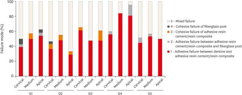
-
Abstract
PDFPubReaderePub
Objectives This study evaluated the bond strength of various fiberglass post cementation techniques using different resin-based composites.
Materials and Methods The roots from a total of 100 bovine incisors were randomly assigned to 5 treatment groups: G1, post + Scotchbond Multi-Purpose (SBMP) + RelyX ARC luting agent; G2, relined post (Filtek Z250) + SBMP + RelyX ARC; G3, individualized post (Filtek Z250) + SBMP; G4, individualized post (Filtek Bulk-Fill) + SBMP; G5, individualized post (Filtek Bulk-Fill Flow) + SBMP. The samples were subjected to the push-out (
n = 10) and pull-out (n = 10) bond strength tests. Data from the push-out bond strength test were analyzed using 2-way analysis of variance (ANOVA) with the Bonferronipost hoc test, and data from the pull-out bond strength test were analyzed using 1-way ANOVA.Results The data for push-out bond strength presented higher values for G2 and G5, mainly in the cervical and middle thirds, and the data from the apical third showed a lower mean push-out bond strength in all groups. No significant difference was noted for pull-out bond strength among all groups. The most frequent failure modes observed were adhesive failure between dentine and resin and mixed failure.
Conclusions Fiberglass post cementation using restorative and flowable bulk-fill composites with the individualization technique may be a promising alternative to existing methods of post cementation.
-
Citations
Citations to this article as recorded by- EVALUATION OF PUSH-OUT BOND STRENGTH OF GLASS FIBER POSTS USING DIFFERENT LUTING CEMENTS
Jannah Mohammed, Maha Agha
BULLETIN OF STOMATOLOGY AND MAXILLOFACIAL SURGERY.2025; : 274. CrossRef - EVALUATION OF PUSH-OUT BOND STRENGTH OF GLASS FIBER POSTS USING DIFFERENT LUTING CEMENTS
Jannah Mohammed, Jannah Mohammed
BULLETIN OF STOMATOLOGY AND MAXILLOFACIAL SURGERY.2025; : 274. CrossRef - Effects of a relined fiberglass post with conventional and self-adhesive resin cement
Wilton Lima dos Santos Junior, Marina Rodrigues Santi, Rodrigo Barros Esteves Lins, Luís Roberto Marcondes Martins
Restorative Dentistry & Endodontics.2024;[Epub] CrossRef - Fracture resistance of weakened roots restored with relined or milled CAD-CAM glass fiber posts
Belizane das Graças Oliveira MAIA, Thais da Silva Alves SANTOS, Cláudio Antonio Talge CARVALHO, Francielle Silvestre VERNER, Rafael Binato JUNQUEIRA
Dental Materials Journal.2023; 42(1): 92. CrossRef - Evaluation of pretreatments on intra‐radicular dentin bond strength of self‐adhesive resin cements
Marina Rodrigues Santi, Rodrigo Barros Esteves Lins, Beatriz Ometto Sahadi, Luís Roberto Marcondes Martins, Jorge Rodrigo Soto‐Montero
Journal of Esthetic and Restorative Dentistry.2022; 34(7): 1051. CrossRef - Comparison of the Mechanical Properties and Push-out Bond Strength of Self-adhesive and Conventional Resin Cements on Fiber Post Cementation
MR Santi, RBE Lins, BO Sahadi, JR Soto-Montero, LRM Martins
Operative Dentistry.2022; 47(3): 346. CrossRef - Glass fiber posts
Renata Pereira, Rodrigo Barros Esteves Lins, Victória Castelan Rodrigues, Débora Alves Nunes Leite Lima, Luís Roberto Marcondes Martins, Flávio Henrique Baggio Aguiar
Brazilian Journal of Oral Sciences.2020; 19: e207508. CrossRef
- EVALUATION OF PUSH-OUT BOND STRENGTH OF GLASS FIBER POSTS USING DIFFERENT LUTING CEMENTS
- 2,166 View
- 14 Download
- 7 Crossref
Case Report
- Pulp revascularization with and without platelet-rich plasma in two anterior teeth with horizontal radicular fractures: a case report
- Edison Arango-Gómez, Javier Laureano Nino-Barrera, Gustavo Nino, Freddy Jordan, Henry Sossa-Rojas
- Restor Dent Endod 2019;44(4):e35. Published online August 20, 2019
- DOI: https://doi.org/10.5395/rde.2019.44.e35

-
Abstract
PDFPubReaderePub
Pulp revascularization is an alternative treatment in immature traumatized teeth with necrotic pulp. However, this procedure has not been reported in horizontal root fractures. This is a case report of a 9-year-old patient with multiple horizontal root fractures in 2 upper central incisors that were successfully treated with pulp revascularization. The patient presented for treatment 2 years after the initial trauma, and revascularization was attempted after the initial treatment with calcium hydroxide had failed. Prior to pulp revascularization, cone-beam computed tomography and autoradiograms demonstrated multiple horizontal fractures in the middle and apical thirds of the roots of the 2 affected teeth. Revascularization was performed in both teeth; platelet-rich plasma (PRP) was used in one tooth (#11) and the conventional method (blood clot) was used in the other tooth (#21). Clinical and radiographic follow-up over 4 years demonstrated pulp calcification in the PRP-treated tooth. Neither of the 2 teeth were lost, and the root canal calcification of tooth #11 was greater than that of tooth #21. This case suggests that PRP-based pulp revascularization may be an alternative for horizontal root fractures.
-
Citations
Citations to this article as recorded by- Regenerative Endodontic Treatment in Permanent Incisors: Two Case Reports with 6 Years of Follow-Up
María Biedma-Perea, Marcela Arenas-González, María José Barra-Soto, Carolina Caleza-Jiménez, David Ribas-Pérez
Children.2026; 13(2): 246. CrossRef - Platelet-Rich Plasma and Platelet-Rich Fibrin in Endodontics: A Scoping Review
Simão Rebimbas Guerreiro, Carlos Miguel Marto, Anabela Paula, Joana Rita de Azevedo Pereira, Eunice Carrilho, Manuel Marques-Ferreira, Siri Vicente Paulo
International Journal of Molecular Sciences.2025; 26(12): 5479. CrossRef - Dental pulp mesenchymal stem cells-response to fibrin hydrogel reveals ITGA2 and MMPs expression
David Tong, Stéphanie Gobert, Alicia Reuzeau, Jean-Christophe Farges, Marianne Leveque, Marie Bolon, Arthur Costantini, Marielle Pasdeloup, Jérôme Lafont, Maxime Ducret, Mourad Bekhouche
Heliyon.2024; 10(13): e32891. CrossRef - Pulp regeneration treatment using different bioactive materials in permanent teeth of pediatric subjects
Dina Abdellatif, Alfredo Iandolo, Giuseppina De Benedetto, Francesco Giordano, Davide Mancino, Edouard Euvrard, Massimo Pisano
Journal of Conservative Dentistry and Endodontics.2024; 27(5): 458. CrossRef - Retreatment of a Failed Regenerative Endodontic Treatment in an Immature Tooth with a Horizontal Root Fracture: A Case Report
Zaher Marjy, Iris Slutzky-Goldberg
International Journal of Clinical Pediatric Dentistry.2024; 17(10): 1168. CrossRef - The Impact of the Preferred Reporting Items for Case Reports in Endodontics (PRICE) 2020 Guidelines on the Reporting of Endodontic Case Reports
Sofian Youssef, Phillip Tomson, Amir Reza Akbari, Natalie Archer, Fayjel Shah, Jasmeet Heran, Sunmeet Kandhari, Sandeep Pai, Shivakar Mehrotra, Joanna M Batt
Cureus.2023;[Epub] CrossRef - Evaluation of postoperative pain and healing following regenerative endodontics using platelet‐rich plasma versus conventional endodontic treatment in necrotic mature mandibular molars with chronic periapical periodontitis. A randomized clinical trial
Yassmin Elsayed Ahmed, Geraldine Mohamed Ahmed, Angie Galal Ghoneim
International Endodontic Journal.2023; 56(4): 404. CrossRef - Regenerative endodontic procedures for two traumatized mature anterior teeth with transverse root fractures
Jing Lu, Bill Kahler
BMC Oral Health.2022;[Epub] CrossRef - Are platelet concentrate scaffolds superior to traditional blood clot scaffolds in regeneration therapy of necrotic immature permanent teeth? A systematic review and meta-analysis
Qianwei Tang, Hua Jin, Song Lin, Long Ma, Tingyu Tian, Xiurong Qin
BMC Oral Health.2022;[Epub] CrossRef - Platelet-Rich Fibrin Used as a Scaffold in Pulp Regeneration: Case Series
Ceren ÇİMEN, Selin ŞEN, Elif ŞENAY, Tuğba BEZGİN
Cumhuriyet Dental Journal.2021; 24(1): 113. CrossRef - Plasma rico en plaquetas en Odontología: Revisión de la literatura
Hugo Anthony Rosas Rozas, Hugo Leoncio Rosas Cisneros
Yachay - Revista Científico Cultural.2021; 10(1): 536. CrossRef
- Regenerative Endodontic Treatment in Permanent Incisors: Two Case Reports with 6 Years of Follow-Up
- 3,699 View
- 49 Download
- 11 Crossref
Review Article
- Effect of calcium hydroxide on inflammatory root resorption and ankylosis in replanted teeth compared with other intracanal materials: a review
- Maryam Zare Jahromi, Mahmood Reza Kalantar Motamedi
- Restor Dent Endod 2019;44(3):e32. Published online August 1, 2019
- DOI: https://doi.org/10.5395/rde.2019.44.e32
-
Abstract
PDFPubReaderePub
Calcium hydroxide (CH) is the gold-standard intracanal dressing for teeth subjected to traumatic avulsion. A common complication after the replantation of avulsed teeth is root resorption (RR). The current review was conducted to compare the effect of CH with that of other intracanal medications and filling materials on inflammatory RR and replacement RR (ankylosis) in replanted teeth. The PubMed and Scopus databases were searched through June 2018 using specific keywords related to the title of the present article. The materials that were compared to CH were in 2 categories: 1) mineral trioxide aggregate (MTA) and endodontic sealers as permanent filling materials for single-visit treatment, and 2) Ledermix, bisphosphonates, acetazolamide, indomethacin, gallium nitrate, and enamel matrix-derived protein (Emdogain) as intracanal medicaments for multiple-visit management of avulsed teeth prior to the final obturation. MTA can be used as a single-visit root filling material; however, there are limited data on its efficacy due to a lack of clinical trials. Ledermix and acetazolamide were comparable to CH in reducing RR. Emdogain seems to be an interesting material, but the data supporting its use as an intracanal medication remain very limited. The conclusions drawn in this study were limited by the insufficiency of clinical trials.
-
Citations
Citations to this article as recorded by- Endodontic Intracanal Medicaments and Agents
Anu Priya Guruswamy Pandian, Depti Bellani, Ritya Mary Jibu, Varsha Agnihotri
Dental Clinics of North America.2026; 70(1): 45. CrossRef - Comparison of 2 kinds of endodontic treatments for pulp necrosis and infection in immature teeth following avulsion: A retrospective study
Weiwei Zhou, Xin Zhang, Meng Wang, Liang Xue, Lili Zhang
Medicine.2026; 105(14): e48250. CrossRef - Knockout guide: avulsion injury management in general practice
Numra Maryam, Laura Warrilow, Sohaila Elyoussfi
Dental Update.2026; 53(6): 450. CrossRef - Efficacy of Simvastatin in Inhibiting Bone Resorption and Promoting Healing in Delayed Tooth Avulsion: A Case Series
Rajesh Kumar, Supraja N Atluri, Alekhya Achanta, Chittaranjan Bogishetty, Tejaswini R Chunduri, Tejaswini PSS, Ramakrishna Ravi
Cureus.2025;[Epub] CrossRef - Interdisziplinäre Lösung nach dentalem Trauma mit Avulsion und Wurzelresorption
Eva Maier, Julia Lubauer, Kerstin M. Galler
Oralprophylaxe & Kinderzahnmedizin.2025; 47(3): 161. CrossRef - Bioactive potential of Bio-C Temp demonstrated by systemic mineralization markers and immunoexpression of bone proteins in the rat connective tissue
Camila Soares Lopes, Mateus Machado Delfino, Mário Tanomaru-Filho, Estela Sasso-Cerri, Juliane Maria Guerreiro-Tanomaru, Paulo Sérgio Cerri
Journal of Materials Science: Materials in Medicine.2024;[Epub] CrossRef - The use of mineral trioxide aggregate for treatment of children with complications of dental trauma
L.Yu. Kharkova, M.V. Korolenkova
Stomatology.2024; 103(4): 59. CrossRef - Instant Re-Implantation of Avulsed Teeth
Smita Paul, Sambarta Das, Nirmal Debbarma, Barun Dasgupta, Bidyut Seal, Ayesha Satapathy
Journal of Pharmacy and Bioallied Sciences.2024; 16(Suppl 4): S3461. CrossRef - Interpretation by literature review of the use of calcium hydroxide as an intra-ductal medication
María Belén Muñoz Padilla, Verónica Alicia Vega Martínez, Camila Alejandra Villafuerte Moya
Salud, Ciencia y Tecnología.2024; 4: 924. CrossRef - Evaluation of the physicochemical properties of intracanal medications used in traumatized teeth
Patricia Almeida da Silva de Macedo, Walbert de Andrade Vieira, Paulo Henrique Gabriel, Karla de Faria Vasconcelos, Francisco Haiter Neto, Ana Carolina Correia Laurindo de Cerqueira Neto, Brenda Paula Figueiredo de Almeida Gomes, Marcos Roberto dos Santo
Brazilian Journal of Oral Sciences.2024; 23: e242997. CrossRef - Treatment of Teeth with Root Resorptions: A Case Report and Systematic Review
Damla Erkal, Abdullah Başoğlu, Damla Kırıcı, Nezahat Arzu Kayar, Simay Koç, Kürşat Er
Galician Medical Journal.2023;[Epub] CrossRef - Successful outcome of permanent maxillary incisor reimplanted after 30 hours of extra‐oral time—a case report with 5‐year follow‐up
Ibadat Preet Kaur, Ashok Kumar, Mukul Kumar, Kanistika Jha
Clinical Case Reports.2023;[Epub] CrossRef - Replantation of an Avulsed Tooth: A Case Report
Nishad Kadulkar, Rubi Kataki, Adrija Deka, Salouno Thonai
Cureus.2023;[Epub] CrossRef - Avulsion of Permanent Mandibular Incisors: A Report of Two Cases with Pertinent Literature
Ibadat Preet Kaur, Jitendra Sharan, Pallawi Sinha, Ashok Kumar, Anand Marya, Leandro Napier de Souza
Case Reports in Dentistry.2023; 2023: 1. CrossRef - The Impact of Autologous Platelet Concentrates on the Periapical Tissues and Root Development of Replanted Teeth: A Systematic Review
Zohaib Khurshid, Faris Yahya I. Asiri, Shariq Najeeb, Jithendra Ratnayake
Materials.2022; 15(8): 2776. CrossRef
- Endodontic Intracanal Medicaments and Agents
- 5,354 View
- 109 Download
- 15 Crossref
Research Articles
- Discoloration of teeth due to different intracanal medicaments
- Farzaneh Afkhami, Sadaf Elahy, Alireza Mahmoudi Nahavandi, Mohamad Javad Kharazifard, Aidin Sooratgar
- Restor Dent Endod 2019;44(1):e10. Published online February 12, 2019
- DOI: https://doi.org/10.5395/rde.2019.44.e10
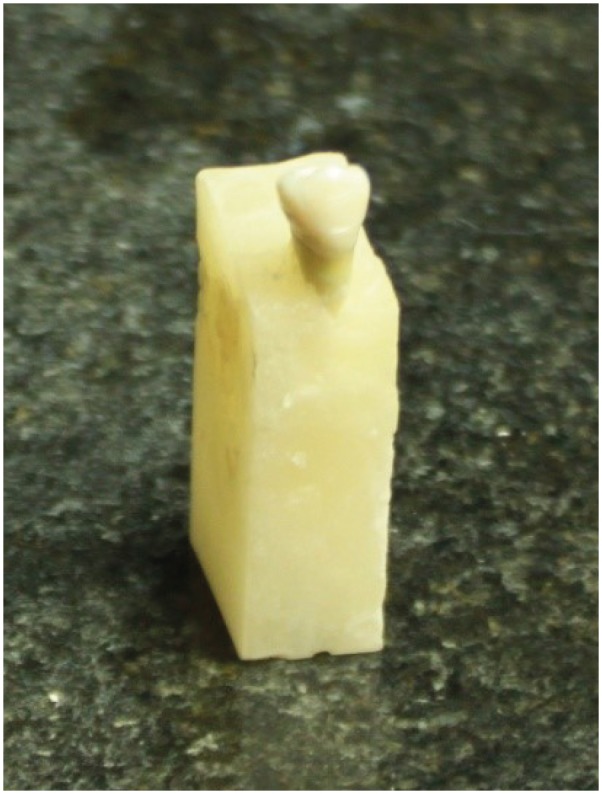
-
Abstract
PDFPubReaderePub
Objectives The objective of this study was to assess coronal discoloration induced by the following intracanal medicaments: calcium hydroxide (CH), a mixture of CH paste and chlorhexidine gel (CH/CHX), and triple antibiotic paste (3Mix).
Materials and Methods Seventy extracted single-canal teeth were selected. Access cavities were prepared and each canal was instrumented with a rotary ProTaper system. The specimens were randomly assigned to CH, CH/CHX, and 3Mix paste experimental groups (
n = 20 each) or a control group (n = 10). Each experimental group was randomly divided into 2 subgroups (A and B). In subgroup A, medicaments were only applied to the root canals, while in subgroup B, the root canals were completely filled with medicaments and a cotton pellet dipped in medicament was also placed in the pulp chamber. Spectrophotometric readings were obtained from the mid-buccal surface of the tooth crowns immediately after placing the medicaments (T1) and at 1 week (T2), 1 month (T3), and 3 months (T4) after filling. The ∆E was then calculated. Data were analyzed using 2-way analysis of variance (ANOVA), 3-way ANOVA, and the Scheffépost hoc test.Results The greatest color change (ΔE) was observed at 3 months (
p < 0.0001) and in 3Mix subgroup B (p = 0.0057). No significant color change occurred in the CH (p = 0.7865) or CH/CHX (p = 0.1367) groups over time, but the 3Mix group showed a significant ΔE (p = 0.0164).Conclusion Intracanal medicaments may induce tooth discoloration. Use of 3Mix must be short and it must be carefully applied only to the root canals; the access cavity should be thoroughly cleaned afterwards.
-
Citations
Citations to this article as recorded by- Time-dependent Tooth Color Changes Following Conventional, Silver-based, and Photodynamic Root Canal Irrigants: An In Vitro Study
Laila Mohamed Mohamed Kenawi, Mohamed Fattouh, Khaled Abid Althaqafi, Abla Arafa
The Open Dentistry Journal.2026;[Epub] CrossRef - Comparative analysis of tooth discoloration induced by different intracanal medicaments in regenerative endodontics: A systematic review and network meta-analysis
Ashlesha Nageshwar Madankar, Sulabha Radke, Shanmuga Priya, Darshan Dakshindas
Endodontology.2026; 38(1): 8. CrossRef - Effects of Intra-canal Medicaments on Infrared Light Energy Transmission Through Enamel and Dentin During Photobiomodulation: An In Vitro Study
Sachin Kulkarni, Laurence J. Walsh, Yash Bhurani, Roy George
Journal of Endodontics.2025; 51(5): 616. CrossRef - Tooth discoloration caused by nanographene oxide as an irrigant and intracanal medicament in the endodontic treatment of extracted single-rooted teeth: An ex-vivo study
Abbas Abbaszadegan, Zeinab Rafiee, Bahar Asheghi, Ahmad Gholami, Mohmed Isaqali Karobari
PLOS One.2025; 20(6): e0325430. CrossRef - Investigation of Discoloration of Anterior Teeth With Three Types of Substances Used in Endodontic Treatment
Sahar Soltani, Eshagh Ali Saberi, Nazanin Shahradnia, Pedram Abdollahzade Sangrodi, Elham Majidi
Clinical and Experimental Dental Research.2025;[Epub] CrossRef - A New Disinfection Approach Using a Chitosan-Based Endodontic Irrigant
Alejandra Itzel Lopez-Flores, Ulises Velazquez-Enriquez, Rogelio Jose Scougall-Vilchis, Laura Susana Acosta-Torres, Laura Emma Rodriguez-Vilchis, Rosalía Contreras-Bulnes, Paloma Netzayeli Serrano-Diaz, Rene Garcia-Contreras
Materials.2025; 18(24): 5552. CrossRef - Spectrophotometric Analysis of Intracoronal Bleaching on Crown Discoloration Induced by Various Antibiotic Pastes: An In Vitro Study
Avneet Kaur, Dileep Soni, Poorva R Sharma
International Journal of Clinical Pediatric Dentistry.2025; 18(12): 1443. CrossRef - Effect of Calcium Hydroxide Versus Double Antibiotic Paste on Endodontic Treatment Outcomes in Teeth With Large Periapical Lesions: A Triple‐Blind Randomized Clinical Trial
Afsaneh Rahmati, Farshad Seyedein, Omid Dianat, Sara Saedi, Golriz Rostami, Alireza Akbarzadeh Baghban, Shima Sabertahan, Majid Kazem, Kee Y. Kum
International Journal of Dentistry.2024;[Epub] CrossRef - The effect of different intracanal irrigants on the push-out bond strength of dentin in damaged anterior primary teeth
Leila Bassir, Shirin Taravati, Farzad Nouri, Saeide Rahimi
Journal of Medicine and Life.2024; 17(5): 536. CrossRef - In Vıtro Evaluatıon of Dıscoloratıon Caused by Root Canal Sealers and Color Changes after Bleachıng
Emre Bodrumlu, Esma Dinger
Annals of Dental Specialty.2024; 12(1): 77. CrossRef - Assessment of Discoloration Induced by Root Canal Sealers and Color Alterations Post-Bleaching
T.P. Van der Burgt, T.P. Mullaney, A.J.M. Plasschaert
International Journal of Dental Research and Allied Sciences.2024; 4(1): 1. CrossRef - The effect of four different intracanal medicaments on the push-out bond strength of root canal sealers
Shalu Maan, Vijaya Dhar Bhatt, Rohit Singh, Sayak Gupta, Syed Alay Noorain, Aashna Gill, Pradeep Kumar, Sushil Yadav, Preeti Sharma
Journal of Medicine and Life.2022; 15(4): 448. CrossRef - Effect of hydrogel-based antibiotic intracanal medicaments on crown discoloration
Rayan B. Yaghmoor, Jeffrey A. Platt, Kenneth J. Spolnik, Tien Min Gabriel Chu, Ghaeth H. Yassen
Restorative Dentistry & Endodontics.2021;[Epub] CrossRef
- Time-dependent Tooth Color Changes Following Conventional, Silver-based, and Photodynamic Root Canal Irrigants: An In Vitro Study
- 4,364 View
- 70 Download
- 13 Crossref
- Evaluation of the effects of whitening mouth rinses combined with conventional tooth bleaching treatments
- Jaqueline Costa Favaro, Omar Geha, Ricardo Danil Guiraldo, Murilo Baena Lopes, Andreza Maria Fábio Aranha, Sandrine Bittencourt Berger
- Restor Dent Endod 2019;44(1):e6. Published online January 30, 2019
- DOI: https://doi.org/10.5395/rde.2019.44.e6
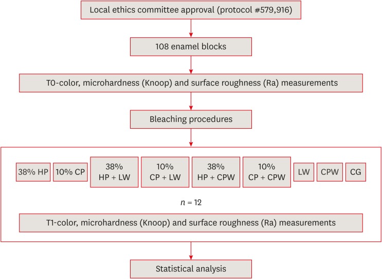
-
Abstract
PDFPubReaderePub
Objectives The aim of the present study was to evaluate the effect of whitening mouth rinses alone and in combination with conventional whitening treatments on color, microhardness, and surface roughness changes in enamel specimens.
Materials and Methods A total of 108 enamel specimens were collected from human third molars and divided into 9 groups (
n = 12): 38% hydrogen peroxide (HP), 10% carbamide peroxide (CP), 38% HP + Listerine Whitening (LW), 10% CP + LW, 38% HP + Colgate Plax Whitening (CPW), 10% CP + CPW, LW, CPW, and the control group (CG). The initial color of the specimens was measured, followed by microhardness and roughness tests. Next, the samples were bleached, and their color, microhardness, and roughness were assessed. Data were analyzed through 2-way analysis of variance (ANOVA; microhardness and roughness) and 1-way ANOVA (color change), followed by the Tukeypost hoc test. The Dunnett test was used to compare the roughness and microhardness data of the CG to those of the treated groups.Results Statistically significant color change was observed in all groups compared to the CG. All groups, except the LW group, showed statistically significant decreases in microhardness. Roughness showed a statistically significant increase after the treatments, except for the 38% HP group.
Conclusions Whitening mouth rinses led to a whitening effect when they were used after conventional treatments; however, this process caused major changes on the surface of the enamel specimens.
-
Citations
Citations to this article as recorded by- Property changes in resin composite exposed to mouth rinses during 10% carbamide peroxide bleaching
Mariana Ferreira da Silva, Giovana Contin Germinari, Carolina Meneghin Barbosa, Tatiane Cristina Dotta, Renata Siqueira Scatolin, Waldemir Francisco Vieira Júnior, Laura Nobre Ferraz
Brazilian Journal of Oral Sciences.2026; 25: e260366. CrossRef - Effect of Over-the-counter Whitening Products on Postbleaching Enamel Surface Roughness and Shade Recovery: An In Vitro Study
Rocío Llancari-Alonzo, Jorge Manrique-Guzmán, Jorge Manrique-Chávez, Leonor Castro-Ramirez, Carlos López-Gurreonero, Alberto Cornejo-Pinto, César Cayo-Rojas
The Journal of Contemporary Dental Practice.2026; 27(2): 148. CrossRef - Influence of commercial mouth rinses with different formulations on enamel properties during at-home bleaching
Thalita Novello Coelho, Ana Júlia Gil, Marcos Roberto Lima Benati, Carolina Meneghin Barbosa, Tatiane Cristina Dotta, Waldemir Francisco Vieira-Junior, Renata Siqueira Scatolin, Laura Nobre Ferraz
Odontology.2026;[Epub] CrossRef - Which Whitening Mouthwash With Different Ingredients Is More Effective on Color and Bond Strength of Enamel?
Elif Varli Tekingur, Fatih Bedir, Muhammet Karadas, Rahime Zeynep Erdem
Journal of Esthetic and Restorative Dentistry.2025; 37(4): 960. CrossRef - Do Different Tooth Bleaching–Remineralizing Regimens Affect the Bleaching Effectiveness and Enamel Microhardness In Vitro?
Hamideh Sadat Mohammadipour, Parnian Shokrollahi, Sima Gholami, Hosein Bagheri, Fatemeh Namdar, Salehe Sekandari, Cesar Rogério Pucci
International Journal of Dentistry.2024;[Epub] CrossRef - Effect of hydrogen peroxide versus charcoal-based whitening mouthwashes on color, surface roughness, and color stability of enamel
Mayada S. Sultan
BMC Oral Health.2024;[Epub] CrossRef - Effects of online marketplace-sourced over-the-counter tooth whitening products on the colour, microhardness, and surface topography of enamel: an in vitro study
Radhika Agarwal, Nikki Vasani, Urmila Sachin Mense, Niharika Prasad, Aditya Shetty, Srikant Natarajan, Arindam Dutta, Manuel S. Thomas
BDJ Open.2024;[Epub] CrossRef - Effect of Whitening Mouthwashes on Color Change and Enamel Mineralization: An In Vitro Study
Rosa Josefina Roncal Espinoza, José Alberto Castañeda Vía, Alexandra Mena-Serrano, Lidia Yileng Tay
World Journal of Dentistry.2023; 14(9): 739. CrossRef - Effectiveness and Adverse Effects of Over-the-Counter Whitening Products on Dental Tissues
Maiara Rodrigues de Freitas, Marynara Mathias de Carvalho, Priscila Christiane Suzy Liporoni, Ana Clara Borges Fort, Rodrigo de Morais e Moura, Rayssa Ferreira Zanatta
Frontiers in Dental Medicine.2021;[Epub] CrossRef - Renklendirilmiş kompozit rezinin renk değişimine ve yüzey pürüzlülüğüne beyazlatıcı ağız gargarasının etkisi
Şeref Nur MUTLU, Makbule Tuğba TUNCDEMIR
Selcuk Dental Journal.2020; 7(3): 435. CrossRef
- Property changes in resin composite exposed to mouth rinses during 10% carbamide peroxide bleaching
- 2,415 View
- 17 Download
- 10 Crossref
Case Report
- Endodontic management of central incisor associated with large periapical lesion and fused supernumerary root: a conservative approach
- Gautam P. Badole, Pratima R. Shenoi, Ameya Parlikar
- Restor Dent Endod 2018;43(4):e44. Published online October 26, 2018
- DOI: https://doi.org/10.5395/rde.2018.43.e44
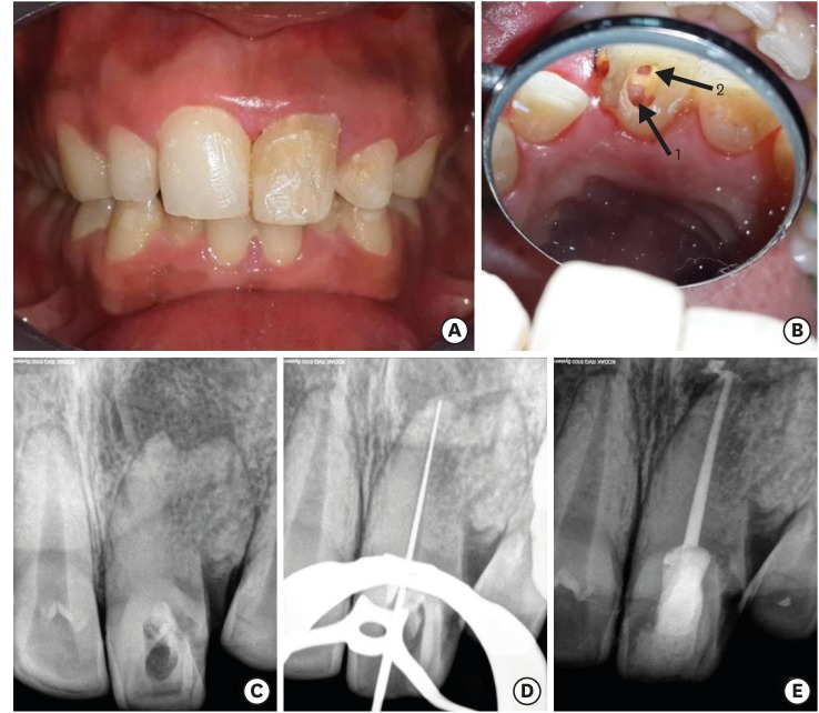
-
Abstract
PDFPubReaderePub
Fusion and gemination are developmental anomalies of teeth that may require endodontic treatment. Fusion may cause various clinical problems related to esthetics, tooth spacing, and other periodontal complications. Additional diagnostic tools are required for the diagnosis and the treatment planning of fused tooth. The present case report describes a case of unilateral fusion of a supernumerary root to an upper permanent central incisor with large periapical lesion in which a conservative approach was used without extraction of supernumerary tooth and obturated with mineral trioxide aggregate to reach a favorable outcome.
-
Citations
Citations to this article as recorded by- A rare case of fusion between a third molar and a distomolar: case report
G. M. Almeida, M.A. H. Duarte, J. R. Carvalho-Junior, R.M. C. Travassos, G. F. Silva, M.F. V. Marceliano-Alves, A. G. Limoeiro, M. P. Alcalde
Endodontics Today.2026; 24(1): 151. CrossRef - Nonsurgical Endodontics and Decompression-Based Management of Extensive Periapical Cystic-Like Lesions: A Comparative and Radiological Study with A Two-Year Follow-Up
Roxana Talpoș-Niculescu, Ioana Veja, Carina Sonia Neagu, Laura Cristina Rusu, Șerban Talpoș-Niculescu, Mălina Popa, Luminița Maria Nica
Journal of Clinical Medicine.2025; 14(17): 6127. CrossRef - Fusion of a Tooth with a Supernumerary Tooth: A Case Report and Literature Review of 35 Cases
Tatsuya Akitomo, Satoru Kusaka, Momoko Usuda, Mariko Kametani, Ami Kaneki, Taku Nishimura, Masashi Ogawa, Chieko Mitsuhata, Ryota Nomura
Children.2023; 11(1): 6. CrossRef - Approche multidisciplinaire d’un cas de fusion incisive centrale maxillaire avec un « talon cusp »
Sonia Terbeche, Kheira Yousfi, Samia Saddat, Souad Larbi Messaoudi, Noureddine Ahmed Fouatih, G. Mer, O. Weissenbach
Revue d'Orthopédie Dento-Faciale.2022; 56(2): 205. CrossRef
- A rare case of fusion between a third molar and a distomolar: case report
- 3,893 View
- 33 Download
- 4 Crossref
Research Articles
- Effects of a bleaching agent on properties of commercial glass-ionomer cements
- Fernanda Lúcia Lago de Camargo, Ailla Carla Lancellotti, Adriano Fonseca de Lima, Vinícius Rangel Geraldo Martins, Luciano de Souza Gonçalves
- Restor Dent Endod 2018;43(3):e32. Published online July 5, 2018
- DOI: https://doi.org/10.5395/rde.2018.43.e32
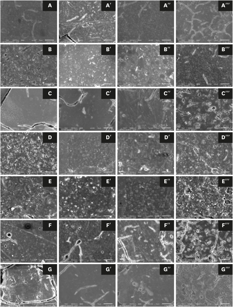
-
Abstract
PDFPubReaderePub
Objectives This study evaluated the effects of a bleaching agent on the composition, mechanical properties, and surface topography of 6 conventional glass-ionomer cements (GICs) and one resin-modified GIC.
Materials and Methods For 3 days, the specimens were subjected to three 20-minute applications of a 37% H2O2-based bleaching agent and evaluated for water uptake (WTK), weight loss (WL), compressive strength (CS), and Knoop hardness number (KHN). Changes in surface topography and chemical element distribution were also analyzed by energy-dispersive X-ray spectroscopy and scanning electron microscopy. For statistical evaluation, the Kruskal-Wallis and Wilcoxon paired tests (
α = 0.05) were used to evaluate WTK and WL. CS specimens were subjected to 2-way analysis of variance (ANOVA) and the Tukeypost hoc test (α = 0.05), and KH was evaluated by one-way ANOVA, the Holm-Sidakpost hoc test (α = 0.05), and thet -test for independent samples (α = 0.05).Results The bleaching agent increased the WTK of Maxxion R, but did not affect the WL of any GICs. It had various effects on the CS, KHN, surface topography, and the chemical element distribution of the GICs.
Conclusions The bleaching agent with 37% H2O2 affected the mechanical and surface properties of GICs. The extent of the changes seemed to be dependent on exposure time and cement composition.
-
Citations
Citations to this article as recorded by- Multidisciplinary conservative management of a severely discolored nonvital tooth
Álvaro Ferrando Cascales, Francesc Abella Sans, Rubén Agustín-Panadero, José Amengual Lorenzo
The Journal of Prosthetic Dentistry.2025; 133(4): 941. CrossRef - The effects of bleaching products on the color stability of ion-releasing restoratives
Jian Sheng Lee, Noor Azlin Yahya, Azwatee Abdul Aziz, Adrian U-Jin Yap
BMC Oral Health.2025;[Epub] CrossRef - Physical-mechanical, chemical and biological properties of graphene-reinforced glass ionomer cements
Tatiane Ramos dos Santos Jordão, Laura Soares Viana Fernandes, Karla Lorene de França Leite, Adílis Alexandria, Emmanuel João Nogueira Leal Silva, Lucianne Cople Maia, Tatiana Kelly da Silva Fidalgo
Restorative Dentistry & Endodontics.2024;[Epub] CrossRef - An In Vitro Exploration of Interaction Mechanisms of Intracoronal Bleaching on the Compressive Strength of Conventional and Calcium Silicate–Based Self‐Adhesive Resins and Their Bonding to Composite Resin Restorative Material
Fereshteh Shafiei, Paria Dehghanian, Shadi Tivay, Yasamin Ghahramani, Luca Testarelli
International Journal of Dentistry.2024;[Epub] CrossRef - Éclaircissement dentaire
V. Pilliol, B. Ballester, T. Baudinet, G. Aboudharama, E. Terrer
EMC - Odontologie.2023; 39(2): 1. CrossRef - The Effect of Home and In-Office Bleaching on Microhardness and Color of Different CAD/CAM Ceramic Materials
Ruwaida Z. Alshali, Mohammed A. Alqahtani
Materials.2022; 15(17): 5948. CrossRef - Éclaircissement dentaire
V. Pilliol, B. Ballester, T. Baudinet, G. Aboudharama, E. Terrer
EMC - Médecine buccale.2022; 15(4): 1. CrossRef - Éclaircissement dentaire
V. Pilliol, B. Ballester, T. Baudinet, G. Aboudharam, E. Terrer
EMC - Orthopédie dentofaciale.2022; 34(1): 1. CrossRef - Comparative Evaluation of Two Glass Polyalkenoate Cements: An In Vivo Pilot Study Using a Sheep Model
Leyla Hasandoost, Daniella Marx, Paul Zalzal, Oleg Safir, Mark Hurtig, Cina Mehrvar, Stephen D. Waldman, Marcello Papini, Mark R. Towler
Journal of Functional Biomaterials.2021; 12(3): 44. CrossRef - The Effect of Simulated Field Storage Conditions on Dental Restorative Materials for Military Field Use
David J Lemon, Wen Chen, Trevor Smith, April A Ford, Steven X Moffett, Jeffrey T Hoyle, Nicholas J Hamlin, Yoon Y Hwang
Military Medicine.2020; 185(5-6): e831. CrossRef
- Multidisciplinary conservative management of a severely discolored nonvital tooth
- 2,310 View
- 5 Download
- 10 Crossref
- Effect of various bleaching treatments on shear bond strength of different universal adhesives and application modes
- Fatma Dilsad Oz, Zeynep Bilge Kutuk
- Restor Dent Endod 2018;43(2):e20. Published online April 16, 2018
- DOI: https://doi.org/10.5395/rde.2018.43.e20
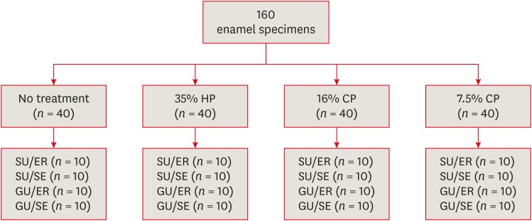
-
Abstract
PDFPubReaderePub
Objectives The aim of this
in vitro study was to evaluate the bond strength of 2 universal adhesives used in different application modes to bleached enamel.Materials and Methods Extracted 160 sound human incisors were used for the study. Teeth were divided into 4 treatment groups: No treatment, 35% hydrogen peroxide, 16% carbamid peroxide, 7.5% carbamid peroxide. After bleaching treatments, groups were divided into subgroups according to the adhesive systems used and application modes (
n = 10): 1) Single Bond Universal, etch and rinse mode; 2) Single Bond Universal, self-etch mode; 3) Gluma Universal, etch and rinse mode; 4) Gluma Universal, self-etch mode. After adhesive procedures nanohybrid composite resin cylinders were bonded to the enamel surfaces. All specimens were subjected to shear bond strength (SBS) test after thermocycling. Data were analyzed using a 3-way analysis of variance (ANOVA) and Tukeypost hoc test.Results No significant difference were found among bleaching groups (35% hydrogen peroxide, 16% carbamid peroxide, 7.5% carbamid peroxide, and no treatment groups) in the mean SBS values. There was also no difference in SBS values between Single Bond Universal and Gluma Universal at same application modes, whereas self-etch mode showed significantly lower SBS values than etch and rinse mode (
p < 0.05).Conclusions The bonding performance of the universal adhesives was enhanced with the etch and rinse mode application to bleached enamel and non-bleached enamel.
-
Citations
Citations to this article as recorded by- Antioxidant effect on shear bond strength of resin composite to in-office versus home bleached enamel surface
Maha Mosaad Mohamed, Magda E. -A. Shalaby, Eman A. E. -G. Shebl
Tanta Dental Journal.2025; 22(3): 409. CrossRef - Effects of Time-Elapsed Bleaching on the Surface and Mechanical Properties of Dentin Substrate Using Hydrogen Peroxide-Free Nanohydroxyapatite Gel
Aftab Khan, Abdulaziz AlKhureif, Manal Almutairi, Abrar Nooh, Saeed Hassan, Yasser Alqahtani
International Journal of Nanomedicine.2024; Volume 19: 10307. CrossRef - Effect of sodium ascorbate on the shear bond strength of orthodontic brackets to bleached enamel using universal dental adhesive
Saeid Sadeghian, Kamyar Fathpour, Mahshid Biglari
Dental Research Journal.2023;[Epub] CrossRef - Quantitative Measurements of the Depth of Enamel Demineralization before and after Bleach: An In Vitro Study
Sara Naim, Gianrico Spagnuolo, Essam Osman, Syed Sarosh Mahdi, Gopi Battineni, Syed Saad B. Qasim, Mariangela Cernera, Hasna Rifai, Nada Jaafar, Elie Maalouf, Carina Mehanna Zogheib, Konstantinos Michalakis
BioMed Research International.2022;[Epub] CrossRef - DİŞ BEYAZLATMA İŞLEMİNİN LİTYUM DİSİLİKAT SERAMİĞİN BAĞLANMA DAYANIMINA ETKİSİ
Merve YILDIRAK, Rıfat GÖZNELİ
Atatürk Üniversitesi Diş Hekimliği Fakültesi Dergisi.2020; : 1. CrossRef - The Effect of Different Bleaching Protocols, Used with and without Sodium Ascorbate, on Bond Strength between Composite and Enamel
Maroun Ghaleb, Giovanna Orsini, Angelo Putignano, Sarah Dabbagh, Georges Haber, Louis Hardan
Materials.2020; 13(12): 2710. CrossRef - Influence of phototherapy on adhesive strength and microleakage of bleached enamel bonded to orthodontic brackets: An in-vitro study
Erum Khan, Ibrahim Alshahrani, Muhammad Abdullah Kamran, Abdulaziz Samran, Ali Alqerban, Saad Abdul Rehman
Photodiagnosis and Photodynamic Therapy.2019; 25: 344. CrossRef - Effect of Er: YAG Laser on Microtensile Bond Strength of Bleached Dentin to Composite
Mohsen Rezaei, Elham Aliasghar, Mohammad Bagher Rezvani, Nasim Chiniforush, Zohreh Moradi
Journal of Lasers in Medical Sciences.2019; 10(2): 117. CrossRef
- Antioxidant effect on shear bond strength of resin composite to in-office versus home bleached enamel surface
- 3,133 View
- 31 Download
- 8 Crossref
- Anatomical analysis of the resected roots of mandibular first molars after failed non-surgical retreatment
- Jiyoung Yoon, Byeong-Hoon Cho, Jihyun Bae, Yonghoon Choi
- Restor Dent Endod 2018;43(2):e16. Published online March 5, 2018
- DOI: https://doi.org/10.5395/rde.2018.43.e16
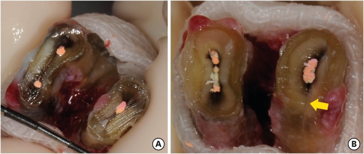
-
Abstract
PDFPubReaderePub
Objectives Understanding the reason for an unsuccessful non-surgical endodontic treatment outcome, as well as the complex anatomy of the root canal system, is very important. This study examined the cross-sectional root canal structure of mandibular first molars confirmed to have failed non-surgical root canal treatment using digital images obtained during intentional replantation surgery, as well as the causative factors of the failed conventional endodontic treatments.
Materials and Methods This study evaluated 115 mandibular first molars. Digital photographic images of the resected surface were taken at the apical 3 mm level and examined. The discolored dentin area around the root canal was investigated by measuring the total surface area, the treated areas as determined by the endodontic filling material, and the discolored dentin area.
Results Forty 2-rooted teeth showed discolored root dentin in both the mesial and distal roots. Compared to the original filled area, significant expansion of root dentin discoloration was observed. Moreover, the mesial roots were significantly more discolored than the distal roots. Of the 115 molars, 92 had 2 roots. Among the mesial roots of the 2-rooted teeth, 95.7% of the roots had 2 canals and 79.4% had partial/complete isthmuses and/or accessory canals.
Conclusions Dentin discoloration that was not visible on periapical radiographs and cone-beam computed tomography was frequently found in mandibular first molars that failed endodontic treatment. The complex anatomy of the mesial roots of the mandibular first molars is another reason for the failure of conventional endodontic treatment.
-
Citations
Citations to this article as recorded by- In vitro evaluation of the sealing ability of combined use of iRoot BP Plus and iRoot SP for root-end filling
Xu Dong, Qian Xie, Xin Xu
Clinical Oral Investigations.2023; 27(6): 2969. CrossRef - The Impact of the Preferred Reporting Items for Case Reports in Endodontics (PRICE) 2020 Guidelines on the Reporting of Endodontic Case Reports
Sofian Youssef, Phillip Tomson, Amir Reza Akbari, Natalie Archer, Fayjel Shah, Jasmeet Heran, Sunmeet Kandhari, Sandeep Pai, Shivakar Mehrotra, Joanna M Batt
Cureus.2023;[Epub] CrossRef - Clinical diagnostic approach in the treatment of chronic periodontitis in mandibular molars: Clinical cases
M. A. Postnikov, A. M. Golovachev, S. E. Chigarina, D. N. Kudryashov, I. A. Zakharova, S. A. Burakshaev
Kuban Scientific Medical Bulletin.2023; 30(5): 100. CrossRef - Evaluation of interorifice distance in permanent mandibular first molar with middle mesial canal in Bengaluru city, Karnataka: A cone-beam computed tomography study
Shruthika Mahajan, N. Meena, Anithakumari Rangappa, Ali Mohammed Mashood, Chethana Murthy, M. Lokapriya
Endodontology.2023; 35(2): 100. CrossRef - A comparative study of the effects of gutta‐percha solvents on human osteoblasts and murine fibroblasts
Gul Ipek Gundogan, Sare Durmus, Gulgun Cansu Ozturk, Nazmi Kucukyesil, Yasin Talat Acar, Rumeysa Balaban, Cenk Kig
Australian Endodontic Journal.2021; 47(3): 569. CrossRef - Endodontic retreatment of curved root canals using the dual wavelength erbium, chromium:yttrium, scandium, gallium, garnet, and diode 940-nm lasers and the XP-Endoshaper/finisher technique
Riman Nasher, Ralf-Dieter Hilgers, Norbert Gutknecht
Lasers in Dental Science.2020; 4(4): 211. CrossRef - Evaluation of gutta-percha removal from the dentinal tubules using different instrumentation techniques with or without solvent: An In vitro study
MukeshKumar Hasija, Babita Meena, Deepti Wadhwa, KulvinderKaur Wadhwani, Virender Yadav
Journal of the International Clinical Dental Research Organization.2020; 12(1): 27. CrossRef
- In vitro evaluation of the sealing ability of combined use of iRoot BP Plus and iRoot SP for root-end filling
- 2,398 View
- 10 Download
- 7 Crossref
Case Report
- Management of dental erosion induced by gastro-esophageal reflux disorder with direct composite veneering aided by a flexible splint matrix
- Sherin Jose Chockattu, Byathnal Suryakant Deepak, Anubhav Sood, Nandini T. Niranjan, Arun Jayasheel, Mallikarjun K. Goud
- Restor Dent Endod 2018;43(1):e13. Published online February 6, 2018
- DOI: https://doi.org/10.5395/rde.2018.43.e13
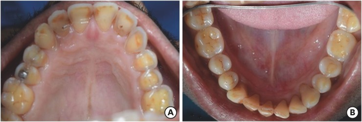
-
Abstract
PDFPubReaderePub
Dental erosion is frequently overlooked in clinical practice. The management of erosion-induced damage to the dentition is often delayed, such that extensive occlusal rehabilitation is required. These cases can be diagnosed by a careful clinical examination and a thorough review of the patient's medical history and/or lifestyle habits. This case report presents the diagnosis, categorization, and management of a case of gastro-esophageal reflux disease-induced palatal erosion of the maxillary teeth. The early management of such cases is of utmost importance to delay or prevent the progression of damage both to the dentition and to occlusal stability. Non-invasive adhesively bonded restorations aid in achieving this goal.
-
Citations
Citations to this article as recorded by- Effect of Acidic Media on Surface Topography and Color Stability of Two Different Glass Ceramics
Fatma Makkeyah, Nesrine A. Elsahn, Mahmoud M. Bakr, Mahmoud Al Ankily
European Journal of Dentistry.2025; 19(01): 173. CrossRef - Mechanical Performance and Surface Roughness of Lithium Disilicate and Zirconia-Reinforced Lithium Silicate Ceramics Before and After Exposure to Acidic Challenge
Ahmed Elsherbini, Salma M. Fathy, Walid Al-Zordk, Mutlu Özcan, Amal A. Sakrana
Dentistry Journal.2025; 13(3): 117. CrossRef - Biomechanical reinforcement by CAD-CAM materials affects stress distributions of posterior composite bridges: 3D finite element analysis.
Alaaeldin Elraggal, Islam M. Abdelraheem, David C. Watts, Sandipan Roy, Vamsi Krishna Dommeti, Abdulrahman Alshabib, Khaled Abid Althaqafi, Rania R. Afifi
Dental Materials.2024; 40(5): 869. CrossRef - Surface Properties and Wear Resistance of Injectable and Computer-Aided Design/Computer Aided Manufacturing–Milled Resin Composite Thin Occlusal Veneers
Nesrine A. Elsahn, Hatem M. El-Damanhoury, Zainab Shirazi, Abdul Rahman M. Saleh
European Journal of Dentistry.2023; 17(03): 663. CrossRef - Effect of acidic media on flexural strength and fatigue of CAD-CAM dental materials
Alaaeldin Elraggal, Rania. R Afifi, Rasha A. Alamoush, Islam Abdel Raheem, David C. Watts
Dental Materials.2023; 39(1): 57. CrossRef - Three-year Follow-up of Conservative Direct Composite Veneers on Eroded Teeth
RQ Ramos, NF Coelho, GC Lopes
Operative Dentistry.2022; 47(2): 131. CrossRef - The effects of intrinsic and extrinsic acids on nanofilled and bulk fill resin composites: Roughness, surface hardness, and scanning electron microscopy analysis
Milena F. Alencar, Mirella T. Pereira, Maria D. R. De‐Moraes, Sérgio L. Santiago, Vanara F. Passos
Microscopy Research and Technique.2020; 83(2): 202. CrossRef
- Effect of Acidic Media on Surface Topography and Color Stability of Two Different Glass Ceramics
- 3,253 View
- 28 Download
- 7 Crossref
Research Article
- Influence of size and insertion depth of irrigation needle on debris extrusion and sealer penetration
- Emel Uzunoglu-Özyürek, Hakan Karaaslan, Sevinç Aktemur Türker, Bahar Özçelik
- Restor Dent Endod 2018;43(1):e2. Published online December 22, 2017
- DOI: https://doi.org/10.5395/rde.2018.43.e2
-
Abstract
PDFPubReaderePub
Objectives To determine the effect of size and insertion depth of irrigation needle on the amount of apical extruded debris and the amount of penetration depth of sealer using a confocal laser scanning microscope (CLSM).
Materials and Methods Twenty maxillary premolars were assigned to 2 groups (
n = 10), according to the size of needle tip, 28 G or 30 G. Buccal roots of samples were irrigated with respective needle type inserted 1 mm short of the working length (WL), while palatal roots were irrigated with respective needle type inserted 3 mm short of the WL. Prepared teeth were removed from the pre-weighed Eppendorf tubes. Canals were filled with F3 gutta-percha cone and rhodamine B dye-labeled AH 26 sealer. Teeth were transversally sectioned at 1 and 3 mm levels from the apex and observed under a CLSM. Eppendorf tubes were incubated to evaporate the irrigant and were weighed again. The difference between pre- and post-weights was calculated, and statistical evaluation was performed.Results Inserting needles closer to the apex and using needles with wider diameters were associated with significantly more debris extrusion (
p < 0.05). The position of needles and level of sections had statistically significant effects on sealer penetration depth (p < 0.05 for both).Conclusions Following preparation, inserting narrower needles compatible with the final apical diameter of the prepared root canal at 3 mm short of WL during final irrigation might prevent debris extrusion and improve sealer penetration in the apical third.
-
Citations
Citations to this article as recorded by- Effect of laser-induced pulpal anesthesia of single-rooted teeth with irreversible pulpitis treated by single-visit root canal therapy - A randomized clinical trial
Geeta Asthana, Dhwani Morakhia, Ravina Parmar, Rajashree Tamuli
Endodontology.2025; 37(3): 244. CrossRef - Efficacy of different irrigation needles used in endodontics: an in silico and an in vitro investigation
Maulee Sheth, Ankit Arora, Sonali Kapoor, Balraj Shukla
Biomaterial Investigations in Dentistry.2025; 12: 264. CrossRef - Preliminary insights: exploring irrigation practices during endodontic treatment among general dental practitioners in Malaysia
Kai Qi Chiew, Xin Ni Lim, Shekhar Bhatia, Naveen Chhabra
British Dental Journal.2024;[Epub] CrossRef - Efficiency of diode laser in control of post-endodontic pain: a randomized controlled trial
Hend H. Ismail, Maram Obeid, Ehab Hassanien
Clinical Oral Investigations.2023; 27(6): 2797. CrossRef - Endodontic management of an aberrant germinated composite odontome: A case report
Ankit Arora, Kavina Desai, Sonali Kapoor, Seema Gajera
Australian Endodontic Journal.2023; 49(3): 684. CrossRef - Potentials of 3D-Modeling in the Preclinical Stage of Root Needle Research
Aleksandr V. Kuligin, Larisa N. Kazakova, Oksana S. Tereshchuk, Vadim V. Bokov
I.P. Pavlov Russian Medical Biological Herald.2022; 30(1): 95. CrossRef - Effect of root canal geometry and needle type on apical extrusion of irrigant: an ex vivo study
Büşra SERÇE FİKİRLİ, Bülent ALTUNKAYNAK, Güven KAYAOĞLU
Acta Odontologica Turcica.2022; 39(3): 58. CrossRef - An in vitro radiological evaluation of irrigant penetration in the root canals using three different irrigation systems: Waterpik WP-100 device, passive irrigation, and manual dynamic irrigation systems
Suragani Hemalatha, Archana Srinivasan, A Srirekha, Lekha Santhosh, C Champa, Ashwija Shetty
Journal of Conservative Dentistry.2022; 25(4): 403. CrossRef - Preparation Ability of ProTaper Next and XP-endo Shaper Instruments in Isthmus-containing Root Canal System
Mustafa Sarıkahya, Tayfun Alaçam
Conservative Dentistry and Endodontic Journal.2021; 5(2): 28. CrossRef - Penetration depth of irrigants into root dentine after sonic, ultrasonic and photoacoustic activation
K. M. Galler, V. Grubmüller, R. Schlichting, M. Widbiller, A. Eidt, C. Schuller, M. Wölflick, K.‐A. Hiller, W. Buchalla
International Endodontic Journal.2019; 52(8): 1210. CrossRef
- Effect of laser-induced pulpal anesthesia of single-rooted teeth with irreversible pulpitis treated by single-visit root canal therapy - A randomized clinical trial
- 2,592 View
- 25 Download
- 10 Crossref
Review Article
- Survival rates against fracture of endodontically treated posterior teeth restored with full-coverage crowns or resin composite restorations: a systematic review
- Warattama Suksaphar, Danuchit Banomyong, Titalee Jirathanyanatt, Yaowaluk Ngoenwiwatkul
- Restor Dent Endod 2017;42(3):157-167. Published online July 31, 2017
- DOI: https://doi.org/10.5395/rde.2017.42.3.157
-
Abstract
PDFPubReaderePub
This systematic review aims to summarize the current clinical studies that investigated survival rates against fracture of endodontically treated posterior teeth restored with crowns or resin composite restorations. Literature search were performed using keywords. Publications from 1980 to 2016 were searched in PubMed, ScienceDirect, Web of Science, MEDLINE, and SCOPUS. Included studies were selected based on inclusion and exclusion criteria. Three clinical studies were included: 1 randomized controlled trial and 1 prospective and 1 retrospective cohort studies. Pooled survival rates ranged from 94%–100% and 91.9%–100% for crowns and resin composite, respectively. The majority of teeth had no more than 3 surface loss of tooth structure. The studies included were heterogeneous, and were not appropriate for further meta-analysis. Current evidence suggested that the survival rates against the fracture of endodontically treated posterior teeth restored with crowns or resin composites were not significantly different in the teeth with minimum to moderate loss of tooth structure.
-
Citations
Citations to this article as recorded by- Clinical Outcomes and Complication Rates of Crown Restorations with Various Endodontic Posts: A Retrospective Analysis
Ali Alenezi, Hanin Alsalhi
Journal of Functional Biomaterials.2026; 17(2): 84. CrossRef - Restoration of the fractured or heavily restored premolar
Chris Redman, Ewen McColl, Graeme Bryce
Dental Update.2026; 53(5): 323. CrossRef - Comparison of fracture resistance among restorative techniques for endodontically treated mandibular premolars: an in vitro study
Dinie Qurratuaini Zulkifly, Mohd Haikal Muhamad Halil, Ubadah Zamry, Siti Nabilah Huda Mohamad Nor
The Saudi Dental Journal.2026;[Epub] CrossRef - Effect of using different materials and restorative techniques on cuspal deflection and microleakage in endodontically treated teeth
Ceyda Sari, Oya Bala, Sinem Akgul, Cemile Kedici Alp
BMC Oral Health.2025;[Epub] CrossRef - Direct restorations versus full crowns in endodontically treated molar teeth: A three-year randomized clinical trial
Motasum Abu-Awwad, Ruba Halasa, Laila Haikal, Ahmad El-Ma'aita, Mohammad Hammad, Haralampos Petridis
Journal of Dentistry.2025; 156: 105699. CrossRef - Is the use of an intraradicular post essential for reducing failures in restoring endodontically treated teeth? A systematic review and meta-analysis
Jacqueline Salomão Jardim, Vinicius de Menezes Félix Ferreira, Hiskell Francine Fernandes e Oliveira, Daniele Sorgatto Faé, Cleidiel Aparecido Araujo Lemos
Journal of Dentistry.2025; 159: 105739. CrossRef - Systematic Reviews Comparing Direct and Indirect Restorations: An Umbrella Review That Examines Restoration Type and Confidence in Results
Mona Kimmel, Clovis Mariano Faggion
Clinical and Experimental Dental Research.2025;[Epub] CrossRef - Knowledge, Awareness, and Perceptions on Root Canal Treatment among Patients Reporting with Dental Pain to Conservative Dentistry and Endodontics Department: An Institution-based Survey
Abdu Semeer Palottil, Moopil Midhun Mohanan, N. T. Nishad, S. Jayasree
Journal of Primary Care Dentistry and Oral Health.2025; 6(2): 99. CrossRef - One-year clinical performance of restorations with and without a bulk-fill flowable base in endodontically treated premolars: a pilot randomized controlled trial
Brenda Leyton, Jullyana Dezanetti, Rodrigo Rached, Sérgio Ignácio, Evelise Souza
BMC Oral Health.2025;[Epub] CrossRef - One-piece endodontic crown fixed partial denture: Is it possible?
João Paulo M. Tribst, Amanda Maria de O. Dal Piva, Joris Muris, Cornelis J. Kleverlaan, Albert J. Feilzer
The Journal of Prosthetic Dentistry.2024; 131(6): 1118. CrossRef - Survival Rate Against Fracture of Endodontically Treated Premolars Restored with Crowns and Resin Composites: A Retrospective Study
Enas Khamakhim, Farida Alsayeh
AlQalam Journal of Medical and Applied Sciences.2024; : 398. CrossRef - Knowledge and Awareness of Root Canal Treatment among Patients in Tripoli: A Survey-Based Study
Sumaya Aghila
AlQalam Journal of Medical and Applied Sciences.2024; : 532. CrossRef - Clinical performance of polyethylenefiber reinforced resin composite restorations in endodontically treated teeth: (a randomized controlled clinical trial)
Ahmed Abdelsattar Metwaly, Amira Farid Elzoghby, Rawda Hesham Abd ElAziz
BMC Oral Health.2024;[Epub] CrossRef - Direct Versus Indirect Treatment Options of Endodontically Treated Posterior Teeth: A Narrative Review
Mai M Alhamdan, Rodina F Aljamaan, Munira M Abuthnain, Shahd A Alsumikhi, Ghada S Alqahtani, Reem A Alkharaiyef
Cureus.2024;[Epub] CrossRef - Single crown vs. composite for glass fiber post-retained restorations: An 8-year randomized clinical trial
Victório Poletto-Neto, Luiz Alexandre Chisini, Wietske Fokkinga, Cees Kreulen, Bas Loomans, Maximiliano Sérgio Cenci, Tatiana Pereira-Cenci
Journal of Dentistry.2024; 142: 104837. CrossRef - Factors influencing the clinical performance of the restoration of endodontically treated teeth: An assessment of systematic reviews of clinical studies
Lara Dotto, Luiza Paloma S. Girotto, Yara Teresinha Correa Silva Sousa, Gabriel Kalil Rocha Pereira, Ataís Bacchi, Rafael Sarkis-Onofre
The Journal of Prosthetic Dentistry.2024; 131(6): 1043. CrossRef - Influence of technical quality and coronal restoration on periapical health of root canal treatment performed by Malaysian undergraduate students
Norazlina Mohammad, Faizah Abdul Fatah, Azlan Jaafar, Siti Hajar Omar, Aimi Amalina Ahmad, Abdul Azim Asy Abdul Aziz, Aws Hashim Ali Al-Kadhim
Saudi Endodontic Journal.2023; 13(1): 63. CrossRef - The success rate of indirect adhesive restorations in the distal dentition fabricated with chairside CAD/CAM system
Marek Šupler, Andrej Jenča, Michal Straka, Juraj Deglovič, Janka Jenčová
Stomatológ.2023; 33(2): 10. CrossRef - A Comparative Study of the Marginal Fit of Endocrowns Fabricated From Three Different Computer-Aided Design/Computer-Aided Manufacturing (CAD/CAM) Ceramic Materials: An In Vitro Study
Esraa Attar, Shatha Alshali, Tariq Abuhaimed
Cureus.2023;[Epub] CrossRef - Evaluation of titanium mesh and fibers in reinforcing endodontically treated molars: An in vitro study
Hemalatha Hiremath, Devansh Verma, Sheetal Khandelwal, AishwaryaSingh Solanki, Sonam Patidar
Journal of Conservative Dentistry.2022; 25(2): 189. CrossRef - Effect of surface treatment, ferrule height, and luting agent type on pull-out bond strength of monolithic zirconia endocrowns
Emine B. Buyukerkmen, Durmuş A. Bozkurt, Arslan Terlemez
Journal of Oral Science.2022; 64(4): 279. CrossRef - An Umbrella Review of Systematic Reviews and Meta‐Analyses Evaluating the Success Rate of Prosthetic Restorations on Endodontically Treated Teeth
Amirhossein Fathi, Behnaz Ebadian, Sara Nasrollahi Dezaki, Nahal Mardasi, Ramin Mosharraf, Sabire Isler, Shiva Sadat Tabatabaei, Stefano Pagano
International Journal of Dentistry.2022;[Epub] CrossRef - Survival and success of endocrowns: A systematic review and meta-analysis
Raghad A. Al-Dabbagh
The Journal of Prosthetic Dentistry.2021; 125(3): 415.e1. CrossRef - Fracture strength of non-invasively reinforced MOD cavities on endodontically treated teeth
René Daher, Stefano Ardu, Enrico Di Bella, Giovanni T. Rocca, Albert J. Feilzer, Ivo Krejci
Odontology.2021; 109(2): 368. CrossRef - Retrospective study of fracture survival in endodontically treated molars: the effect of single-unit crowns versus direct-resin composite restorations
Kanet Chotvorrarak, Warattama Suksaphar, Danuchit Banomyong
Restorative Dentistry & Endodontics.2021;[Epub] CrossRef - An insight into patient's perceptions regarding root canal treatment: A questionnaire-based survey
Ramta Bansal, Aditya Jain
Journal of Family Medicine and Primary Care.2020; 9(2): 1020. CrossRef - Endodontically treated posterior teeth restored with or without crown restorations: A 5‐year retrospective study of survival rates from fracture
Titalee Jirathanyanatt, Warattama Suksaphar, Danuchit Banomyong, Yaowaluk Ngoenwiwatkul
Journal of Investigative and Clinical Dentistry.2019;[Epub] CrossRef - Fracture resistance, gap and void formation in root‐filled mandibular molars restored with bulk‐fill resin composites and glass‐ionomer cement base
Nathamon Thongbai‐on, Kanet Chotvorrarak, Danuchit Banomyong, Michael F. Burrow, Sittichoke Osiri, Nattha Pattaravisitsate
Journal of Investigative and Clinical Dentistry.2019;[Epub] CrossRef - Current options concerning the endodontically-treated teeth restoration with the adhesive approach
Marco Aurélio de Carvalho, Priscilla Cardoso Lazari, Marco Gresnigt, Altair Antoninha Del Bel Cury, Pascal Magne
Brazilian Oral Research.2018;[Epub] CrossRef
- Clinical Outcomes and Complication Rates of Crown Restorations with Various Endodontic Posts: A Retrospective Analysis
- 7,154 View
- 85 Download
- 29 Crossref
Case Reports
- Microsurgical re-treatment of an endodontically treated tooth with an apically located incomplete vertical root fracture: a clinical case report
- Silvio Taschieri, Massimo Del Fabbro, Ahmed El Kabbaney, Igor Tsesis, Eyal Rosen, Stefano Corbella
- Restor Dent Endod 2016;41(4):316-321. Published online June 21, 2016
- DOI: https://doi.org/10.5395/rde.2016.41.4.316
-
Abstract
PDFPubReaderePub
Although it is challenging, the early diagnosis of a vertical root fracture (VRF) is crucial in order to ensure tooth preservation. The purpose of this clinical case report was to describe reparative surgery performed to treat a tooth affected by an incomplete VRF. A 26 year old male patient was suspected to have a VRF in a maxillary left central incisor, and an exploratory flap was performed in order to confirm the diagnosis. After detecting the fracture, the lesion was surgically treated, the fracture and the infected root-end were removed, and a platelet-rich plasma membrane was used to cover the defect in order to prevent bacterial migration. A 24 month clinical and radiological follow-up examination showed that the tooth was asymptomatic and that the healing process was in progress. The surgical approach described here may be considered an effective treatment for a combined endodontic-periodontal lesion originating from an incomplete VRF and a recurrent periapical lesion.
-
Citations
Citations to this article as recorded by- Biomechanical perspectives on dentine cracks and fractures: Implications in their clinical management
Sishi Chen, Dwayne Arola, Domenico Ricucci, Brian E. Bergeron, John A. Branton, Li-sha Gu, Franklin R. Tay
Journal of Dentistry.2023; 130: 104424. CrossRef - Efficacy of Autologous Platelet Concentrates in Regenerative Endodontic Treatment: A Systematic Review of Human Studies
Joanna Metlerska, Irini Fagogeni, Alicja Nowicka
Journal of Endodontics.2019; 45(1): 20. CrossRef - The preservation of teeth with root-originated fractures
Eyal Rosen, Ilan Beitlitum, Igor Tsesis
Evidence-Based Endodontics.2018;[Epub] CrossRef
- Biomechanical perspectives on dentine cracks and fractures: Implications in their clinical management
- 3,362 View
- 29 Download
- 3 Crossref
- Esthetic enhancement of a traumatized anterior tooth with a combination of forced eruption and tooth alignment: a case report
- So-Hee Kang, Jung-Hong Ha, Myoung-Uk Jin, Sung-Kyo Kim, Young-Kyung Kim
- Restor Dent Endod 2016;41(3):210-217. Published online June 1, 2016
- DOI: https://doi.org/10.5395/rde.2016.41.3.210
-
Abstract
PDFPubReaderePub
Exposing sound structure of a subgingivally fractured tooth using orthodontic extrusion is considered to be a conservative way to re-establish biologic width without sacrificing esthetics or jeopardizing periodontal support of neighboring teeth. When a misaligned tooth is traumatically involved, a more comprehensive approach combining tooth extrusion and re-alignment may be necessary for a successful restorative outcome. This case report describes a successful esthetic management of a patient with complicated crown-root fracture on the maxillary right central incisor and pre-existing malocclusion in the maxillary anterior region. Forced eruption along with re-alignment of teeth by orthodontic movement seems to allow re-positioning of the fracture line to a favorable position and correction of crowding, providing a better esthetic result.
-
Citations
Citations to this article as recorded by- Effects of systematic bisphosphonate use in patients under orthodontic treatment: a systematic review
Vasileios F Zymperdikas, Maria P Yavropoulou, Eleftherios G Kaklamanos, Moschos A Papadopoulos
European Journal of Orthodontics.2020; 42(1): 60. CrossRef - In vitro retention efficiency of temporary type zinc oxide cement for orthodontic forced eruption
Renato Nieto-Aguilar, Deyanira Serrato-Ochoa, Rafael Medina-Navarro, Asdrúbal Aguilera-Méndez, Karina Denisse Morales-Soto, Juan Pablo Loyola-Rodriguez, Antonio Campos, Miguel Alaminos
International Orthodontics.2019; 17(1): 96. CrossRef
- Effects of systematic bisphosphonate use in patients under orthodontic treatment: a systematic review
- 2,997 View
- 22 Download
- 2 Crossref
Research Article
- Antioxidant therapy enhances pulpal healing in bleached teeth
- Adriano Fonseca Lima, Marcelo Rocha Marques, Diana Gabriela Soares, Josimeri Hebling, Giselle Maria Marchi, Carlos Alberto de Souza Costa
- Restor Dent Endod 2016;41(1):44-54. Published online February 1, 2016
- DOI: https://doi.org/10.5395/rde.2016.41.1.44
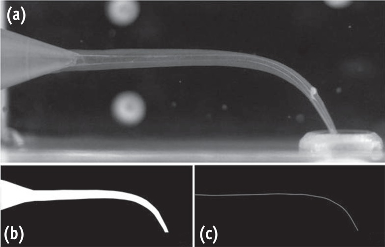
-
Abstract
PDFPubReaderePub
Objectives The purpose of this study was to evaluate the histopathological effects of an antioxidant therapy on the pulp tissue of rat teeth exposed to a bleaching gel with 35% hydrogen peroxide.
Materials and Methods Forty rats were subjected to oral ingestion by gavage of distilled water (DW) or ascorbic acid (AA) 90 min before the bleaching therapy. For the bleaching treatment, the agent was applied twice for 5 min each to buccal surfaces of the first right mandibular molars. Then, the animals were sacrificed at 6 hr, 24 hr, 3 day, or 7 day post-bleaching, and the teeth were processed for microscopic evaluation of the pulp tissue.
Results At 6 hr, the pulp tissue showed moderate inflammatory reactions in all teeth of both groups. In the DW and AA groups, 100% and 80% of teeth exhibited pulp tissue with significant necrosis and intense tissue disorganization, respectively. At 24 hr, the AA-treated group demonstrated a greater regenerative capability than the DW group, with less intense inflammatory reaction and new odontoblast layer formation in 60% of the teeth. For up to the 7 day period, the areas of pulpal necrosis were replaced by viable connective tissue, and the dentin was underlined by differentiated odontoblast-like cells in most teeth of both groups.
Conclusions A slight reduction in initial pulpal damage during post-bleaching was promoted by AA therapy. However, the pulp tissue of AA-treated animals featured faster regenerative potential over time.
-
Citations
Citations to this article as recorded by- Clinical Study on the Efficacy of 35% Hydrogen Peroxide Gel According to Exposure Time (40 min vs. 20 min) by Spectrophotometry
Trinidad Rincón, Maria Portillo Muñoz, Maria Lobato, Ana María Martín Casado, Laryssa Mylenna Madruga Barbosa, Alessandro Loguercio, Cristina Gómez‐Polo
Journal of Esthetic and Restorative Dentistry.2026;[Epub] CrossRef - Influence of dental bleaching on the pulp tissue: A systematic review of in vivo studies
Mariana Viana Donato, Alexandre Henrique dos Reis‐Prado, Lucas Guimarães Abreu, Lara Cancella de Arantes, Juliana Goto, Hebertt Gonzaga dos Santos Chaves, Luciano Tavares Angelo Cintra, André Luiz Fraga Briso, Isabella Faria da Cunha Peixoto, Francine Ben
International Endodontic Journal.2024; 57(6): 630. CrossRef - ANALYSIS OF THE INFLUENCE OF THE TEETH WHITENING PROCEDURE ON THE GUM AND CYTOKINE PROFILE OF ORAL FLUID (LITERATURE REVIEW)
S. S. Bozhik, N. V. Hasyuk, V. B. Radchuk
Bulletin of Problems Biology and Medicine.2024; 1(3): 17. CrossRef - Assessing the Viability of Laser-Activated Dental Bleaching Compared to Conventional In-Office Bleaching Methods: A Systematic Review of Clinical and In Vitro Studies
Eugenia Anagnostaki, Valina Mylona, Steven Parker, Mark Cronshaw, Martin Grootveld
Applied Sciences.2023; 13(22): 12459. CrossRef - Effects of the application of sodium ascorbate after in-office bleaching on the penetration of hydrogen peroxide, color change, and microtensile bond strength
Alexandra Mena-Serrano, María G. Granda-Albuja, Jenny Naranjo, Eliana Aldás Fierro, Michael Willian Favoreto, Alessandro D. Loguercio, Alessandra Reis
Brazilian Dental Journal.2023; 34(5): 87. CrossRef - Can different agents reduce the damage caused by bleaching gel to pulp tissue? A systematic review of basic research
Letícia Aparecida Silva Batista, Alexandre Henrique dos Reis-Prado, Hebertt Gonzaga dos Santos Chaves, Lara Cancella de Arantes, Luís Fernando Santos Alves Morgan, Carolina Bosso André, Thaís Yumi Suzuki, Francine Benetti
Restorative Dentistry & Endodontics.2023;[Epub] CrossRef - Design of a thermosensitive ibuprofen-loaded nanogel as smart material applied as anti-inflammatory in tooth bleaching: An in vivo study
Samara K.S.C.F. Moura, Milena L.V. dos Santos, Lucas A. do Nascimento, Mariana F.A. da Silva, Glória M. de França, Lucas M. da Costa, Aldo C. Medeiros, Raimundo F. Araújo-Júnior, Aurigena A. de Araújo, Cláudia N. Oliveira, André L. Dorini, Rejane A. de Ca
Journal of Drug Delivery Science and Technology.2022; 68: 103123. CrossRef - Effect of bleaching gel volume on color change and postoperative sensitivity: a randomized clinical study
Lara Maria Bueno Esteves, Paulo Henrique dos Santos, Ticiane Cestari Fagundes, Marjorie de Oliveira Gallinari, Giulia Bessa de Mello Antonaccio, Luciano Tavares Ângelo Cintra, André Luiz Fraga Briso
Clinical Oral Investigations.2022; 26(3): 2527. CrossRef - Human Dental Pulp Stem Cells Exhibit Different Biological Behaviours in Response to Commercial Bleaching Products
Carmen Llena, Mar Collado-González, Christopher Joseph Tomás-Catalá, David García-Bernal, Ricardo Elías Oñate-Sánchez, Francisco Javier Rodríguez-Lozano, Leopoldo Forner
Materials.2018; 11(7): 1098. CrossRef - Concentration‐dependent effect of bleaching agents on the immunolabelling of interleukin‐6, interleukin‐17 and CD5‐positive cells in the dental pulp
F. Benetti, J. E. Gomes‐Filho, L. L. Ferreira, G. Sivieri‐Araújo, E. Ervolino, A. L. F. Briso, L. T. A. Cintra
International Endodontic Journal.2018; 51(7): 789. CrossRef
- Clinical Study on the Efficacy of 35% Hydrogen Peroxide Gel According to Exposure Time (40 min vs. 20 min) by Spectrophotometry
- 2,462 View
- 6 Download
- 10 Crossref
Case Report
- Surgical management with intentional replantation on a tooth with palato-radicular groove
- Jorge Forero-López, Luis Gamboa-Martínez, Laura Pico-Porras, Javier Laureano Niño-Barrera
- Restor Dent Endod 2015;40(2):166-171. Published online December 22, 2014
- DOI: https://doi.org/10.5395/rde.2015.40.2.166
-
Abstract
PDFPubReaderePub
A palato-radicular groove (PRG) is a developmental anomaly primarily found in the maxillary lateral incisors. It is a potential communication path between the root canal and the periodontium that decreases the survival prognosis of the affected tooth, therefore compromising the stability of the dental structure in the oral cavity. The aim of this case report is to present an original technique where a PRG was treated by means of intracanal disinfection, PRG sealing with glass ionomer, replantation with intentional horizontal 180 degree rotation of the tooth, and an aesthetic veneer placed to provide adequate tooth morphology. The clinical and biological benefits of this novel technique are presented and discussed.
-
Citations
Citations to this article as recorded by- Retrospective Analysis of Clinical Characteristics and Therapeutic Effect of Intentional Replantation
Jiani Zhu, Xiangfen Li, Qin Su
Journal of Endodontics.2026; 52(7): 1114. CrossRef - A classification of radicular grooves from the perspective of periodontology
Huxiao Li, Zhaowei Tai, Jiachen Dong, Zhongchen Song
BMC Oral Health.2025;[Epub] CrossRef - Diagnostic Approaches of Palatogingival Groove: A Systematic Review
Greta Venskutė
Journal of Dental Health and Oral Research.2024; : 1. CrossRef - Palatogingival Groove: A Plaque Trap Leading to Bone Loss in a Maxillary Lateral Incisor – A Rare Case Report
Gayathri Priyadharshini Elangovan, Indra Kumar Periyasamy, Saravana Kumar R, Gopinath Vivekanandhan
Dental Journal of Indira Gandhi Institute of Medical Sciences.2024; 3: 104. CrossRef - Palatogingival Groove: The Known–unknown Devourer
Sumedha Gupta, Sandeep Tandon, Ambika S Rathore, Rinku Mathur, Tripti S Rai, Kanchan Kumari Dhaker
International Journal of Clinical Pediatric Dentistry.2024; 17(S1): S95. CrossRef - Palatal groove associated with periodontal lesions: a systematic review illustrated by a decisional tree for management
Yvan Gaudex, Vianney Gandillot, Isabelle Fontanille, Philippe Bouchard, Stephane Kerner, Maria Clotilde Carra
BMC Oral Health.2024;[Epub] CrossRef - Prevalence of palatogingival groove affecting maxillary anterior teeth in Saudi subpopulation: A cone-beam computed tomographic study with literature review
Ali Ibrahim Aljuailan, Roqayah Aljuailan, Rahul N. Gaikwad, Shaul Hameed Kolarkodi, Nasser Rufaydan Alamri
The Saudi Dental Journal.2023; 35(8): 1039. CrossRef - Interdisciplinary approach for diagnosis and management of the tooth with type III palatogingival groove
Harakh Chand Baranwal, Jyoti Yadav
Saudi Endodontic Journal.2023; 13(2): 211. CrossRef - Management of Palatogingival Groove in Maxillary Lateral Incisor: A Report of a Rare Case With a Brief Review of Literature
Irfan Ansari, Sanjay Miglani, Vijay Yadav, Shamimul Hasan
Cureus.2023;[Epub] CrossRef - Intentional replantation combined root resection therapy for the treatment of type III radicular groove with two roots: A case report
Dan Tan, Shi-Ting Li, Hao Feng, Zhong-Chao Wang, Cai Wen, Min-Hai Nie
World Journal of Clinical Cases.2022; 10(20): 6991. CrossRef - The incidence of radicular groove on maxillary lateral incisors of Saudi population: CBCT evaluation
Sarah M. Alkahtany, Fatemah Alrwais, Asma Altamimi, Sundus M. Bukhary, Amani Mirdad
BMC Oral Health.2022;[Epub] CrossRef - Retrospective Study of Intentional Replantation for Type IIIb Dens Invaginatus with Periapical Lesions
Na Li, Huihui Xu, Cunhui Kan, Jing Zhang, Song Li
Journal of Endodontics.2022; 48(3): 329. CrossRef - Combined Periodontal and Endodontic Management of Palatal Radicular Groove with Platelet‐Rich Fibrin and Biodentine®
Arjun Hari Rijal, Bhageshwar Dhami, Pratistha Ghimire, Konstantinos Michalakis
Case Reports in Dentistry.2022;[Epub] CrossRef - Intentional Replantation of Single-Rooted and Multi-Rooted Teeth: A Systematic Review
Massimo Pisano, Federica Di Spirito, Stefano Martina, Giuseppe Sangiovanni, Francesco D’Ambrosio, Alfredo Iandolo
Healthcare.2022; 11(1): 11. CrossRef - Management of the palato-radicular groove with a periodontal regenerative procedure and prosthodontic treatment: A case report
Dan-Hua Ling, Wei-Ping Shi, Yan-Hong Wang, Dan-Ping Lai, Yan-Zhen Zhang
World Journal of Clinical Cases.2022; 10(17): 5732. CrossRef - Prevalence and Periodontal Conditions of Developmental Grooves in an Italian School of Dentistry and Dental Hygiene: A Cross-Sectional Study
Giovanna Laura Di Domenico, Simone Fabrizi, Paolo Capparè, Maria Teresa Sberna, Massimo de Sanctis
International Journal of Environmental Research and Public Health.2022; 19(7): 4047. CrossRef - Clinical Observation and Research Progress of Comprehensive Treatment of Palatogingival Groove
华姣 胡
Advances in Clinical Medicine.2021; 11(03): 846. CrossRef - Palato-gingival groove: A silent nidus. Recognition and an innovative management approach: A report of 3 cases
Purushothama Rangaswamy, Sri Harsha Tummala, Samrat R Magarvalli, Sujith Ramachandra, Kavitha Govindappa, Shwetha E
IP Indian Journal of Conservative and Endodontics.2021; 6(2): 114. CrossRef - Periodontal Regenerative Treatment of Intrabony Defects Associated with Palatal Grooves: A Report of Two Cases
Stefano Corbella, Alice Alberti, Beatrice Zotti, Luca Francetti, Jiiang H. Jeng
Case Reports in Dentistry.2019;[Epub] CrossRef - Recognition and management of palatogingival groove for tooth survival: a literature review
Hee-Jin Kim, Yoorina Choi, Mi-Kyung Yu, Kwang-Won Lee, Kyung-San Min
Restorative Dentistry & Endodontics.2017; 42(2): 77. CrossRef - The use of Intentional Replantation to Repair an External Cervical Resorptive Lesion not am Enable to Conventional Surgical Repair
Kreena Pa Tel, Federico Foschi, Ioana Pop, Shanon Patel, Francesco Mannocci
Primary Dental Journal.2016; 5(2): 78. CrossRef - Management of apicomarginal defect in esthetic region associated with a tooth with anomalies
Vinayak Venkoosa Meharwade, Dipali Yogesh Shah, Pradyna Prabhakar Mali, Vidya Vinayak Meharwade
Restorative Dentistry & Endodontics.2015; 40(4): 314. CrossRef
- Retrospective Analysis of Clinical Characteristics and Therapeutic Effect of Intentional Replantation
- 3,046 View
- 16 Download
- 22 Crossref
Research Article
- Effects of applying antioxidants on bond strength of bleached bovine dentin
- Hyo-Jin Whang, Dong-Hoon Shin
- Restor Dent Endod 2015;40(1):37-43. Published online October 13, 2014
- DOI: https://doi.org/10.5395/rde.2015.40.1.37
-
Abstract
PDFPubReaderePub
Objectives Some antioxidants are believed to restore dentin bond strength after dental bleaching. This study was done to evaluate the influence of antioxidants on the bond strength of bleached bovine dentin.
Materials and Methods Thirty incisors were randomly assigned to 10 groups (two unbleached control and eight bleached groups: immediate bonding IB, 4 wk delayed bonding DB, 10% sodium ascorbate treated SA, 10% α-tocopherol treated TP groups). Teeth in half of groups were subjected to thermal stress, whereas the remaining groups were not. Resin-dentin rods with a cross-sectional area of 2.25 mm2 were obtained and microtensile bond strength was determined at a crosshead speed of 1 mm/min. Fifteen specimens were prepared for SEM to compare the surface characteristics of each group. The change in dentin bond strength from thermal stress and antioxidant treatment was evaluated using two-way analysis of variance (ANOVA) and Sheffe's
post hoc test at a significance level of 95%.Results The control group exhibited the highest bond strength values, whereas IB group showed the lowest value before and after thermocycling. The DB group recovered its bond strength similar to that of the control group. The SA and TP groups exhibited similar bond strength values with those of the control and DB groups before thermocycling. However, The TP group did not maintain bond strength with thermal stress, whereas the SA group did.
Conclusions Applying a 10% sodium ascorbate solution rather than 10% α-tocopherol solution for 60 sec is recommended to maintain dentin bond strength when restoring non-vitally bleached teeth.
-
Citations
Citations to this article as recorded by- Shear bond strength analysis of self-etch and total-etch adhesive systems on antioxidant-treated dentin in post-endodontic restoration
Suvarna Kavya Harish, Shruthi H. Attavar
Frontiers in Dental Medicine.2026;[Epub] CrossRef - Preliminary In Vitro Evaluation of Vitamin E (α‐Tocopherol) Incorporation on Cytotoxicity, Antifungal Activity, and Surface Roughness of Denture Acrylic Soft Liner
Neven S. Aref, Hannah Wesley
International Journal of Dentistry.2026;[Epub] CrossRef - Evaluation of the effect of the application of Quercus cerris extract and the use of fluoride bonding material on the bonding strength of orthodontic brackets after tooth bleaching with hydrogen peroxide
Ezgi Ay, Derya Dursun
PeerJ.2025; 13: e19335. CrossRef - Antioxidant effect on shear bond strength of resin composite to in-office versus home bleached enamel surface
Maha Mosaad Mohamed, Magda E. -A. Shalaby, Eman A. E. -G. Shebl
Tanta Dental Journal.2025; 22(3): 409. CrossRef - Comparative analysis of the impact of modern cavity disinfectants on dentin bond strength
Simge Gümüş Ayaz, Ezgi Sonkaya, Gökçe Keçeci
Frontiers in Materials.2025;[Epub] CrossRef - Effect of glutathione on bond strength of composite resin to enamel following extracoronal bleaching
Nair Devika, Chandrasekaran Charanya, K Athira, James Vandana, Sundaresan Balagopal
Journal of Conservative Dentistry and Endodontics.2024; 27(11): 1110. CrossRef - Effect of Chitosan Nanoparticle as an Antioxidant Material on Shear Bond Strength of Composite Resin to Enamel after External Bleaching
Diatri Nari Ratih, Shintatika Erlagista, Tunjung Nugraheni
Open Access Macedonian Journal of Medical Sciences.2024; 12: 1. CrossRef - Effects of alpha‐tocopherol antioxidant on fracture strength and adhesion of endodontically treated teeth restored after dental bleaching
Natália Marcomini, Maria Carolina da Costa Albaricci, Joatan Lucas de Sousa Gomes Costa, João Felipe Besegato, Eduardo Fernández Godoy, Andréa Abi Rached Dantas, Milton Carlos Kuga
European Journal of Oral Sciences.2024;[Epub] CrossRef - Alpha-tocopherol: An alternative solution for the adverse effects of dental bleaching on dentin adhesion
Maria Carolina da Costa Albaricci, Natália Marcomini, Joatan Lucas de Sousa Gomes Costa, Antonia Patricia Oliveira Barros, Lucas David Galvani, Milton Carlos Kuga, Andréa Abi Rached Dantas
International Journal of Adhesion and Adhesives.2024; 131: 103655. CrossRef - Efficacy of organic and antioxidant agents to regain bond strength to bleached enamel in different dental adhesive solvents
Satheesh B Haralur, Renad Mohammed Al-Ibrahim, Faten Abdullah Al-Shahrani, Rahaf Abdullah Al-Qahtani, Saurabh Chaturvedi, Naseer M Alqahtani
Journal of Applied Biomaterials & Functional Materials.2023;[Epub] CrossRef - Present status and future directions – Managing discoloured teeth
Bill Kahler
International Endodontic Journal.2022; 55(S4): 922. CrossRef - Comparative evaluation of the postbleaching application of sodium ascorbate, alpha‐tocopherol, and quercetin on shear bond strength of composite resin to enamel
Marzieh Moradian, Maryam Saadat, Mohammad Hossein S. Shiri, Fatemeh Sohrabniya
Clinical and Experimental Dental Research.2022; 8(6): 1598. CrossRef - Use of antioxidants to restore bond strength after tooth bleaching with peroxides
Dorcas E. R. P. Olmedo, Matheus Kury, Bruna A. Resende, Vanessa Cavalli
European Journal of Oral Sciences.2021;[Epub] CrossRef - Elemental and morphological analysis of enamel following the application of two bleaching systems with amorphous calcium phosphate: effect on enamel erosion susceptibility
Shaymaa M. Nagi, Shahinaz H. Nabil, Mohamed H. Zaazou
Bulletin of the National Research Centre.2021;[Epub] CrossRef - Pomegranate (Punica granatum L.) gel extract as an antioxidant on the shear bond strength of a resin composite post-bleaching application with 40% hydrogen peroxide
Indes Rosmalisa Suratno, Irfan Dwiandhono, Ryana Budi Purnama
Dental Journal.2021; 54(2): 87. CrossRef - In Vitro Re-Hardening of Bleached Enamel Using Mineralizing Pastes: Toward Preventing Bacterial Colonization
Andrea Scribante, Claudio Poggio, Simone Gallo, Paolo Riva, Antonella Cuocci, Manuel Carbone, Carla Arciola, Marco Colombo
Materials.2020; 13(4): 818. CrossRef - DİŞ BEYAZLATMA İŞLEMİNİN LİTYUM DİSİLİKAT SERAMİĞİN BAĞLANMA DAYANIMINA ETKİSİ
Merve YILDIRAK, Rıfat GÖZNELİ
Atatürk Üniversitesi Diş Hekimliği Fakültesi Dergisi.2020; : 1. CrossRef - The improvement of biocompatibility of adhesives
Cigdem Atalayin, Huseyin Tezel, Zeynep Ergucu, Nimet Unlu, Guliz Armagan, Taner Dagci, Timur Kose
Clinical Oral Investigations.2019; 23(8): 3213. CrossRef - Dentin bond strength and nanoleakage of the adhesive interface after intracoronal bleaching
Vanessa Cavalli, Maicon Sebold, Mirela Sanae Shinohara, Patrícia Nóbrega Rodrigues Pereira, Marcelo Giannini
Microscopy Research and Technique.2018; 81(4): 428. CrossRef - Composite resin shear bond strength on bleached dentin increased by 35% sodium ascorbate application
Tunjung Nugraheni, N Nuryono, Siti Sunarintyas, Ema Mulyawati
Dental Journal (Majalah Kedokteran Gigi).2017; 50(4): 178. CrossRef - Comparative Evaluation of Immediate Bond Strength to Bleached Enamel Following Application of Various Antioxidant Solutions
Anshu Minocha, Ashu K. Gupta, Alisha Dhingra, Nayantara Sen
Dental Journal of Advance Studies.2017; 5(2): 84. CrossRef - Effects of Erbium Family Laser on Shear Bond Strength of Composite to Dentin After Internal Bleaching
Nazanin Kiomarsi, Yasaman Arjmand, Mohammad Javad Kharrazi Fard, Nasim Chiniforush
Journal of Lasers in Medical Sciences.2017; 9(1): 58. CrossRef - Antioxidant therapy enhances pulpal healing in bleached teeth
Adriano Fonseca Lima, Marcelo Rocha Marques, Diana Gabriela Soares, Josimeri Hebling, Giselle Maria Marchi, Carlos Alberto de Souza Costa
Restorative Dentistry & Endodontics.2016; 41(1): 44. CrossRef - Effects of alpha-tocopherol on fracture resistance after endodontic treatment, bleaching and restoration
Keren Cristina Fagundes JORDÃO-BASSO, Milton Carlos KUGA, Andrea Abi Rached DANTAS, Mateus Rodrigues TONETTO, Suellen Nogueira Linhares LIMA, Matheus Coêlho BANDÉCA
Brazilian Oral Research.2016;[Epub] CrossRef - Resin Bonding of Self-Etch Adhesives to Bovine Dentin Bleached from Pulp Chamber
Akiko Haruyama, Atsushi Kameyama, Junji Kato, Shinji Takemoto, Yutaka Oda, Eiji Kawada, Toshiyuki Takahashi, Masahiro Furusawa
BioMed Research International.2016; 2016: 1. CrossRef
- Shear bond strength analysis of self-etch and total-etch adhesive systems on antioxidant-treated dentin in post-endodontic restoration
- 2,249 View
- 8 Download
- 25 Crossref
Case Reports
- Clinical management of a fused upper premolar with supernumerary tooth: a case report
- Kyu-Min Cho, Ji-Hyun Jang, Sang-Hyuk Park
- Restor Dent Endod 2014;39(4):319-323. Published online July 17, 2014
- DOI: https://doi.org/10.5395/rde.2014.39.4.319
-
Abstract
PDFPubReaderePub
In dentistry, the term 'fusion' is used to describe a developmental disorder of dental hard tissues. In the permanent dentition, fusion of a normal tooth and a supernumerary tooth usually involves the incisors or canines. However, a few cases of fusion involving premolars have also been reported to date. We present a rare case in which fusion of the maxillary left second premolar and a supernumerary tooth in a 13-year-old girl was diagnosed using cone beam computed tomography (CBCT, Alphard-3030, Asahi Roentgen Ind. Co., Ltd.). The tooth was bicuspidized after routine nonsurgical root canal treatment, and the separated teeth underwent appropriate restoration procedures. The second premolar and supernumerary tooth remained asymptomatic without any signs of inflammation after a follow-up period of 9 years. Identification of anatomical anomalies is important for treatment in cases involving fusion with supernumerary tooth, and therefore the microscopic examinations and CBCT are essential for the diagnosis. Fused teeth can be effectively managed by the comprehensive treatment which includes both endodontic and periodontal procedures.
-
Citations
Citations to this article as recorded by- Fusion of a Tooth with a Supernumerary Tooth: A Case Report and Literature Review of 35 Cases
Tatsuya Akitomo, Satoru Kusaka, Momoko Usuda, Mariko Kametani, Ami Kaneki, Taku Nishimura, Masashi Ogawa, Chieko Mitsuhata, Ryota Nomura
Children.2023; 11(1): 6. CrossRef - Malformed Teeth and Their Endodontic Implications
Annapoorna Annapoorna, Manjunatha M, Shubhashini N, Swetha H. B.
Journal of Evolution of Medical and Dental Sciences.2020; 9(04): 245. CrossRef - Endodontic management of central incisor associated with large periapical lesion and fused supernumerary root: a conservative approach
Gautam P. Badole, Pratima R. Shenoi, Ameya Parlikar
Restorative Dentistry & Endodontics.2018;[Epub] CrossRef - Common dental diseases in children and malocclusion
Jing Zou, Mingmei Meng, Clarice S Law, Yale Rao, Xuedong Zhou
International Journal of Oral Science.2018;[Epub] CrossRef - Endodontic Management of Dilacerated Maxillary Central Incisor fused to a Supernumerary Tooth using Cone Beam Computed Tomography: An Unusual Clinical Presentation
Thilla S Vinothkumar, Deivanayagam Kandaswamy, Ganesh Arathi, Sathishkumar Ramkumar, Gnanasekaran Felsypremila
The Journal of Contemporary Dental Practice.2017; 18(6): 522. CrossRef - Nonsurgical endodontic retreatment of fused teeth with transposition: a case report
Miguel Agostinho Beco Pinto Cardoso, Rita Brandão Noites, Miguel André Duarte Martins, Manuel Pedro da Fonseca Paulo
Restorative Dentistry & Endodontics.2016; 41(2): 148. CrossRef
- Fusion of a Tooth with a Supernumerary Tooth: A Case Report and Literature Review of 35 Cases
- 2,998 View
- 28 Download
- 6 Crossref
- The use of platelet rich plasma in the treatment of immature tooth with periapical lesion: a case report
- Günseli Güven Polat, Ceren Yıldırım, Özlem Martı Akgün, Ceyhan Altun, Didem Dinçer, Cansel Köse Özkan
- Restor Dent Endod 2014;39(3):230-234. Published online June 2, 2014
- DOI: https://doi.org/10.5395/rde.2014.39.3.230
-
Abstract
PDFPubReaderePub
This study describes the treatment of an immature permanent tooth with periapical lesion which was treated with regenerative approach using platelet rich plasma (PRP). The root canal of immature human permanent tooth with periapical lesion was gently debrided of necrotic tissue and disinfected with 2.5% NaOCl, and then medicated with triple antibiotic paste comprised of ciprofloxacin, metronidazole, and tetracycline. When the tooth was asymptomatic, PRP and mineral trioxide aggregate (MTA) were placed into the root canal. Six months after PRP treatment, radiographical examination revealed resolution of the radiolucency and progressive thickening of the root wall and apical closure. Our findings suggest that PRP can be used for the treatment of immature permanent teeth with periapical lesion, as part of a regenerative endodontic treatment procedure.
-
Citations
Citations to this article as recorded by- Evaluation of postoperative pain and healing following regenerative endodontics using platelet‐rich plasma versus conventional endodontic treatment in necrotic mature mandibular molars with chronic periapical periodontitis. A randomized clinical trial
Yassmin Elsayed Ahmed, Geraldine Mohamed Ahmed, Angie Galal Ghoneim
International Endodontic Journal.2023; 56(4): 404. CrossRef - Different Approaches to the Regeneration of Dental Tissues in Regenerative Endodontics
Anna M. Krupińska, Katarzyna Skośkiewicz-Malinowska, Tomasz Staniowski
Applied Sciences.2021; 11(4): 1699. CrossRef - Coronal tooth discoloration induced by regenerative endodontic treatment using different scaffolds and intracanal coronal barriers: a 6-month ex vivo study
Noushin Shokouhinejad, Hassan Razmi, Maryam Farbod, Marzieh Alikhasi, Josette Camilleri
Restorative Dentistry & Endodontics.2019;[Epub] CrossRef - Efficacy of Autologous Platelet Concentrates in Regenerative Endodontic Treatment: A Systematic Review of Human Studies
Joanna Metlerska, Irini Fagogeni, Alicja Nowicka
Journal of Endodontics.2019; 45(1): 20. CrossRef - Bone, Periodontal and Dental Pulp Regeneration in Dentistry: A Systematic Scoping Review
Luiz Alexandre Chisini, Marcus Cristian Muniz Conde, Guillermo Grazioli, Alissa Schmidt San Martin, Rodrigo Varella de Carvalho, Letícia Regina Morello Sartori, Flávio Fernando Demarco
Brazilian Dental Journal.2019; 30(2): 77. CrossRef - Mineral trioxide aggregate and other bioactive endodontic cements: an updated overview – part II: other clinical applications and complications
M. Torabinejad, M. Parirokh, P. M. H. Dummer
International Endodontic Journal.2018; 51(3): 284. CrossRef - Alternative to Avoid Tooth Discoloration after Regenerative Endodontic Procedure: A Systematic Review
Luciane Geanini Pena dos Santos, Luiz Alexandre Chisini, Camila Guerner Springmann, Beatriz Dulcineia Mendes de Souza, Fernanda Geraldo Pappen, Flávio Fernando Demarco, Mara Cristina Santos Felippe, Wilson Tadeu Felippe
Brazilian Dental Journal.2018; 29(5): 409. CrossRef - Influence of Apical Diameter on the Outcome of Regenerative Endodontic Treatment in Teeth with Pulp Necrosis: A Review
Yanjun Fang, Xinhuan Wang, Jingjing Zhu, Chaonan Su, Ying Yang, Liuyan Meng
Journal of Endodontics.2018; 44(3): 414. CrossRef - A scoping review of root canal revascularization: relevant aspects for clinical success and tissue formation
M. C. M. Conde, L. A. Chisini, R. Sarkis‐Onofre, H. S. Schuch, J. E. Nör, F. F. Demarco
International Endodontic Journal.2017; 50(9): 860. CrossRef - Effects of Epigallocatechin Gallate, an Antibacterial Cross-linking Agent, on Proliferation and Differentiation of Human Dental Pulp Cells Cultured in Collagen Scaffolds
Young-Sun Kwon, Hee-Jin Kim, Yun-Chan Hwang, Vinicius Rosa, Mi-Kyung Yu, Kyung-San Min
Journal of Endodontics.2017; 43(2): 289. CrossRef - Regenerative Endodontics
Kristina Feigin, Bonnie Shope
Journal of Veterinary Dentistry.2017; 34(3): 161. CrossRef - Regenerative Endodontic Treatment or Mineral Trioxide Aggregate Apical Plug in Teeth with Necrotic Pulps and Open Apices: A Systematic Review and Meta-analysis
Mahmoud Torabinejad, Ali Nosrat, Prashant Verma, Oyoyo Udochukwu
Journal of Endodontics.2017; 43(11): 1806. CrossRef - Platelet concentrates for revitalization of immature necrotic teeth: a systematic review of the clinical studies
Alessandra Lolato, Cristina Bucchi, Silvio Taschieri, Ahmed El Kabbaney, Massimo Del Fabbro
Platelets.2016; 27(5): 383. CrossRef - Regenerative endodontics—Creating new horizons
Harnoor Dhillon, Mamta Kaushik, Roshni Sharma
Journal of Biomedical Materials Research Part B: Applied Biomaterials.2016; 104(4): 676. CrossRef - The impact of autologous platelet concentrates on endodontic healing: a systematic review
Nastaran Meschi, Ana B. Castro, Katleen Vandamme, Marc Quirynen, Paul Lambrechts
Platelets.2016; 27(7): 613. CrossRef - Pulp and Periodontal Regeneration of an Avulsed Permanent Mature Incisor Using Platelet-rich Plasma after Delayed Replantation: A 12-month Clinical Case Study
Harini Priya M, Pavan B. Tambakad, Jaya Naidu
Journal of Endodontics.2016; 42(1): 66. CrossRef - Platelet preparations in dentistry: How? Why? Where? When?
Luigi Fabrizio Rodella
World Journal of Stomatology.2015; 4(2): 39. CrossRef
- Evaluation of postoperative pain and healing following regenerative endodontics using platelet‐rich plasma versus conventional endodontic treatment in necrotic mature mandibular molars with chronic periapical periodontitis. A randomized clinical trial
- 2,527 View
- 11 Download
- 17 Crossref
 First
First Prev
Prev

