
Search
- Page Path
- HOME > Search
Research Articles
- Determination of optimal horizontal beam angulations for canal separation in mandibular molars using cone-beam computed tomography: a retrospective image-based analysis
- Benedikt Schneider, Tamina Tepe, Daniel Rapp, Wilhelm Frank, Maria Lessani, Constantin von See, Sebastian Fitzek, Jörg Philipp Tchorz
- Restor Dent Endod 2026;51(1):e9. Published online February 26, 2026
- DOI: https://doi.org/10.5395/rde.2026.51.e9
-
 Abstract
 PDF
PDF PubReader
 ePub
ePub - Objectives
Two-dimensional intraoral radiographs often obscure canals due to superimposition, especially in mandibular molars with complex anatomy. This cone-beam computed tomography (CBCT) study identified the horizontal beam angles at which first and second molar canals overlap and derived clinically applicable angulations for enhanced canal separation.
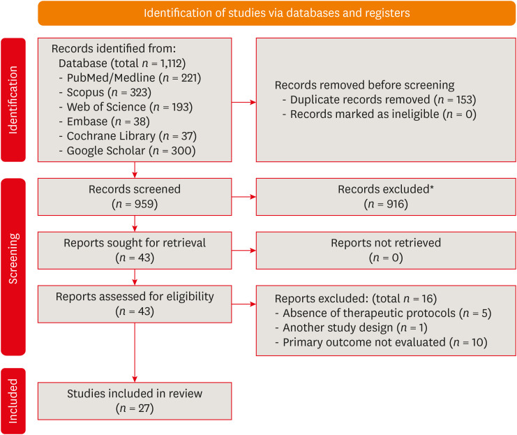
Methods
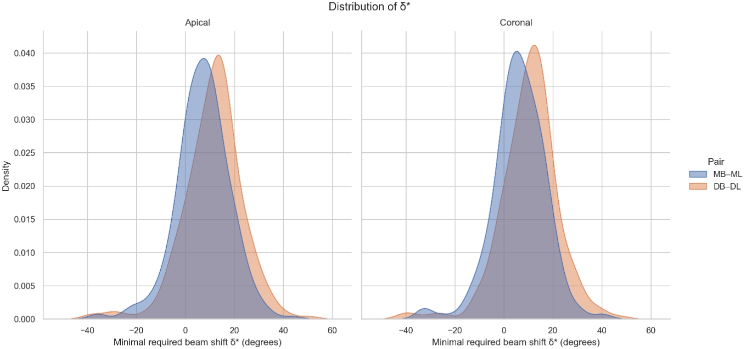
Eighty-five CBCT datasets from 100 patients met the inclusion criteria, yielding 318 mandibular molars (160 first, 158 second). Using ImageJ, absolute horizontal overlap angles (α) were measured to determine the corresponding theoretical separation angles defined as δ* = 90° – α. Separability was modeled across horizontal beam angulation increments from −45° to +45° in five steps, and Wilson’s 95% confidence intervals were computed. Group comparisons used the Mann-Whitney U and independent t-tests (p ≤ 0.05)
Results
Minimal mesial beam angulations for effective canal separability (δ* = 90° − α) ranged from approximately 7° to 15° for mesial roots and approximately 10° to 13° for distal roots. No significant mesial differences were observed between first and second molars (p > 0.30). Distal roots of second molars exhibited significantly higher angulations (p = 0.003 coronal, p < 0.001 apical). Mesial canals achieved ≥95% separability at approximately 25° and ≥99% at approximately 35°; distal canals required approximately 30° and approximately 40°.
Conclusions
A mesial beam angulation of 30° to 35° provides probable canal differentiation in mandibular molars, separating mesial canals in ≥99% and distal canals in ≥95% of cases. This range refines previous recommendations and supports the as low as reasonably achievable (ALARA) principle.
- 1,411 View
- 36 Download

- Neuropeptide Y regulation of dental pulp neurogenic inflammation provoked by tooth bleaching agents: a descriptive comparative clinical study
- Javier Caviedes-Bucheli, Néstor Ríos-Osorio, Mario Pérez-Villota, Karolina Aucú-Miño, Diana Escobar-Mafla, Hernán Darío Muñoz-Alvear, José Francisco Gomez-Sosa, Luis Diaz-Barrera, Edgar Güiza – Cristancho, Hugo Roberto Munoz
- Restor Dent Endod 2026;51(1):e10. Published online February 13, 2026
- DOI: https://doi.org/10.5395/rde.2026.51.e10
-
Abstract
PDFPubReaderePub
- Objectives
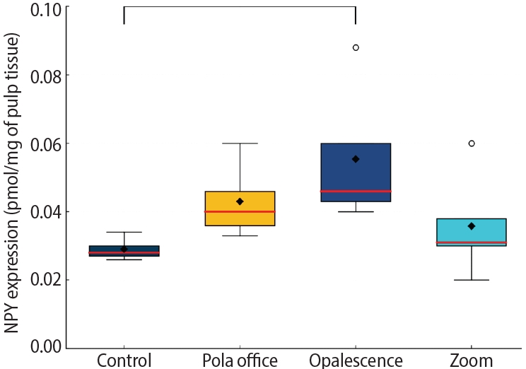
This study aimed to assess the expression of neuropeptide Y (NPY) in human dental pulp after tooth bleaching with three in-office hydrogen peroxide (H2O2)-based systems.
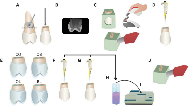
Methods
Forty pulps were collected from premolars scheduled for extraction and divided into four groups (n = 10): Control (no bleaching; basal NPY values); Pola Office (35% H2O2, 8 minutes); Opalescence Boost (40% H2O2, 20 minutes); and Zoom (25% H2O2 + cold blue light, 15 minutes). After extraction, pulps were fixed in 4% formaldehyde and processed. NPY levels were quantified using enzyme-linked immunosorbent assay. Data distribution was assessed with the Shapiro-Wilk test. One-way analysis of variance and Tukey post-hoc test with Bonferroni correction were applied (p < 0.05).
Results
NPY expression differed significantly among groups (p = 0.0097). The control group showed the lowest mean expression (0.026 ± 0.002 pmol/mg of pulp tissue), followed by Zoom (0.031 ± 0.005 pmol/mg), Pola Office (0.040 ± 0.004 pmol/mg), and Opalescence Boost, which exhibited the highest NPY expression (0.044 ± 0.004 pmol/mg). Post-hoc analysis revealed a statistically significant difference between the control and Opalescence Boost groups (p = 0.0122).
Conclusions
The increase in NPY expression—particularly with Opalescence Boost—indicates that in-office bleaching agents can elicit measurable neurobiological responses in pulp tissue after a single application. The significant difference between the control and Opalescence Boost groups suggests a possible H2O2 concentration- or formulation-dependent effect on pulpal neuropeptide activity, underscoring the need for further research on the biological impact of bleaching treatments.
- 1,595 View
- 90 Download
- Analysis of the reciprocating kinematics of the VDW Silver Reciproc, E-Connect Pro, Ecom, and Endopen endodontic motors: an in vitro experimental study
- Cristielly França, Juliana D. Bronzato, Dieimes Braambati, Adriana de-Jesus-Soares, Carla C. R. B. Félix, Michelle A. N. S. Ferreira, Marcos Frozoni
- Restor Dent Endod 2026;51(1):e5. Published online January 20, 2026
- DOI: https://doi.org/10.5395/rde.2026.51.e5
-
Abstract
PDFPubReaderePub
- Objectives
This study aimed to evaluate the actual parameters of four endodontic motors, each adjusted for reciprocating motion, and compare them to the manufacturers’ declared values.
Methods
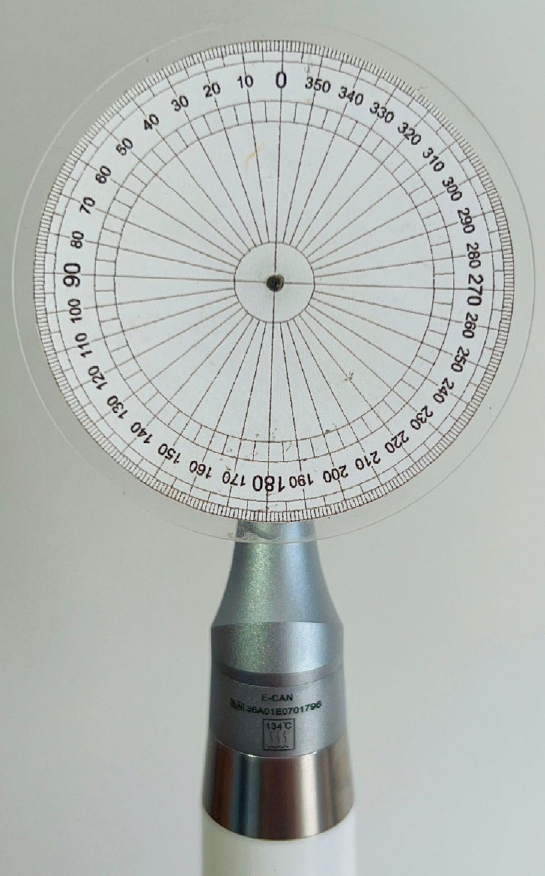
The motors used were the VDW Silver Reciproc (VDW GmbH), E-Connect Pro (MK Life), Ecom (Woodpecker), and Endopen (Schuster Woodpecker). A custom optical target was attached to the motor contra-angle, the movements were recorded with a high-resolution camera, and the images were analyzed. Engagement, disengagement, net angles, and speed for each operation cycle, duration of clockwise (CW) and counter-clockwise (CCW) movement, duration of standstill after CW and CCW movement, and the number of cycles to complete a full rotation were analyzed. The data were statistically analyzed at a significance level of 5%. The replicability of all reciprocal parameters analyzed was statistically different from that reported by the manufacturers.
Results
There was no statistically significant difference between the VDW Silver Reciproc, Ecom, and Endopen for the engagement angle. The E-Connect Pro was the least reliable at the 150°/30° settings for both angle parameters. There was no significant difference between the set and actual cycle net angles for the VDW Silver Reciproc (p = 0.493). While the actual values for the Ecom and E-Connect Pro were significantly higher than the set (p < 0.001), the actual values for the Endopen were significantly lower than the set (p < 0.001).
Conclusions
Experiments on four commercially available reciprocating endodontic motors revealed that the actual motor values differed significantly from the set values.
- 2,213 View
- 90 Download
- Enhancing antimicrobial properties of a resin-based material via incorporation of a powdered phytotherapeutic extract: an in vitro experimental study
- Rodolfo Xavier de Sousa-Lima, Maria Eduarda Lima do Nascimento Marinho, Janielly Cristina Costa da Silva, Moan Jéfter Fernandes Costa, Pedro Henrique Sette-de-Souza, Giana da Silveira Lima, Boniek Castillo Dutra Borges
- Restor Dent Endod 2026;51(1):e2. Published online January 20, 2026
- DOI: https://doi.org/10.5395/rde.2026.51.e2
-
Abstract
PDFPubReaderePub
- Objectives
This study aimed to evaluate the degree of conversion (DC), immediate enamel bond strength (IEBS), antimicrobial activity, and release of the active principle of a resin-based material (RBM) enriched with the powdered Schinopsis brasiliensis (Braúna) stem antibacterial extract.
Methods
The RBM was enriched with 0, 1.25, 2.5, 5, 10, and 20 wt% powdered Braúna extract. The DC (n = 7) was assessed using micro-Raman spectroscopy. The IEBS (n = 7) was determined through the microshear test until failure, and failure modes were examined under a stereomicroscope. The antimicrobial activity (n = 15) was assessed by quantifying colony-forming units, and the release of the active principle was determined using ultra-high-performance liquid chromatography. One-way analysis of variance/Tukey and Kruskal-Wallis/Dunn tests were utilized to analyze the data (p < 0.05).
Results
Materials with 10 wt% and 20 wt% extract showed the lowest DC statistically. However, for IEBS, there were no statistically significant differences among the different groups. All materials released the active principle, but only those with 20 wt% and 10 wt% extract could inhibit biofilm formation similarly to 0.12% chlorhexidine.
Conclusions
Adding powdered Braúna extract between 10 wt% and 20 wt% is a promising alternative to provide an antimicrobial function to RBMs.
- 2,314 View
- 234 Download
- Effect of moisture and pH on setting time and microhardness of three premixed calcium silicate-based root canal sealers: an in vitro experimental study
- Sooyoun Kim
- Restor Dent Endod 2025;50(4):e41. Published online November 28, 2025
- DOI: https://doi.org/10.5395/rde.2025.50.e41
-
Abstract
PDFPubReaderePub
- Objectives
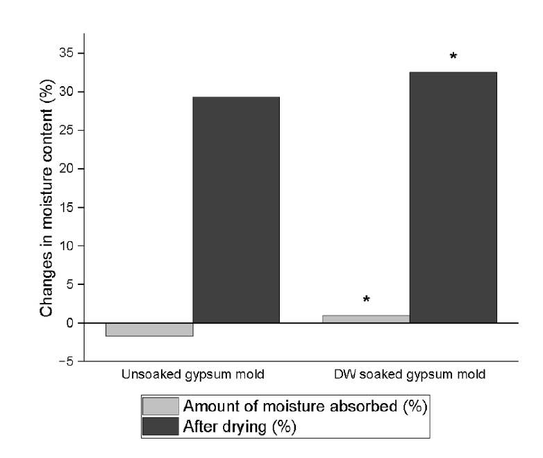
The study aimed to investigate how environmental conditions impact the setting time and microhardness of premixed calcium silicate-based sealers.
Methods
The setting time and microhardness of three sealers (Endoseal MTA [MARUCHI], One-Fil [MEDICLUS], and Well-Root ST [VERICOM]) were evaluated under four environmental conditions: unsoaked, distilled water-soaked, phosphate-buffered saline-soaked, and pH 5-soaked gypsum molds (n = 12/group/condition). The setting time was measured with Gilmore needles, and microhardness was assessed using a Vickers tester after 3 days. Welch’s analysis of variance and Games-Howell post hoc tests were used for statistical analysis.
Results
The sealer type and environmental conditions significantly influenced setting time and microhardness (p < 0.001). The initial and final setting times were the shortest in the unsoaked samples. For Endoseal MTA and One-Fil, the unsoaked condition exhibited significantly shorter setting times than the soaked conditions. Well-Root ST exhibited significantly longer setting times in acidic conditions. Surface microhardness was highest in the unsoaked group (p < 0.001). Among the soaked groups, the phosphate-buffered saline-soaked group had the lowest hardness for Endoseal MTA, whereas the pH 5-soaked group exhibited the lowest hardness for One-Fil and Well-Root ST. Endoseal MTA consistently demonstrated a lower microhardness than the other sealers (p < 0.001).
Conclusions
Moisture, pH, and solution chemistry influenced the setting time and microhardness of premixed calcium silicate sealers. Although acidic conditions generally prolong the setting time and reduce hardness, the effects vary based on the sealers used and the setting environment. -
Citations
Citations to this article as recorded by
- Setting Characteristics, Solubility, Bioactivity and Interaction with Dentin of Four Calcium Silicate-Based Endodontic Sealers
Areti Dimitra Vrochari, Anastasia Agrafioti, Maria Dimitriadi, George Eliades
Journal of Functional Biomaterials.2026; 17(4): 192. CrossRef
- Setting Characteristics, Solubility, Bioactivity and Interaction with Dentin of Four Calcium Silicate-Based Endodontic Sealers
- 2,603 View
- 118 Download
- 1 Web of Science
- 1 Crossref
- Resolvin E1 incorporated carboxymethyl chitosan scaffold accelerates repair of dental pulp stem cells under inflammatory conditions: a laboratory investigation
- Hemalatha P Balasubramanian, Nandini Suresh, Vishnupriya Koteeswaran, Velmurugan Natanasabapathy
- Restor Dent Endod 2025;50(4):e40. Published online November 28, 2025
- DOI: https://doi.org/10.5395/rde.2025.50.e40
-
Abstract
PDF
 Supplementary MaterialPubReaderePub
 - Objectives
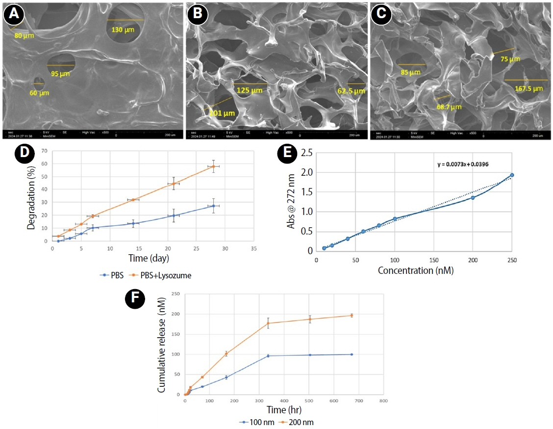
This study fabricated and characterized a resolvin E1 (RvE1)-loaded carboxymethyl chitosan (CMC) scaffold and determined its cytotoxicity and mineralization potential on inflamed human dental pulp stem cells (hDPSCs).
Methods
CMC scaffold incorporated with two concentrations of RvE1 (100 and 200 nM) was fabricated and characterized. The scaffolds’ porosity, drug release kinetics, and degradation were assessed. The impact of RvE1 on inflamed hDPSCs proliferation, proinflammatory gene expression (tumor necrosis factor alpha [TNF-α]), alkaline phosphatase activity, and alizarin red S staining was evaluated.
Results
Scanning electron microscopy analysis demonstrated a highly porous interconnected microstructure. Release kinetics showed gradual RvE1 release peaking at day 14. Cumulative degradation of the CMC scaffold at 28 days was 57.35%. Inflamed hDPSCs exposed to 200 nM RvE1-CMC scaffold exhibited significantly improved viability compared to 100 nM. Both RvE1-CMC scaffolds significantly suppressed the expression of TNF-α at 7 days. Alkaline phosphatase activity was enhanced by both RvE1 concentrations on days 7 and 14. Alizarin red staining revealed superior mineralization potential of 200 nM RvE1 on days 14 and 21.
Conclusions
This study concludes 200 nM RvE1-CMC scaffold is a promising therapy for inflamed pulp conditions, enhancing cell proliferation and biomineralization potential in inflamed hDPSCs.
- 1,489 View
- 63 Download
- The influence of bioactive glass (BGS-7) on enamel remineralization: an in vitro study
- Chaeyoung Lee, Eunseon Jeong, Kun-Hwa Sung, Su-Jung Park, Yoorina Choi
- Restor Dent Endod 2025;50(4):e33. Published online October 15, 2025
- DOI: https://doi.org/10.5395/rde.2025.50.e33
-
Abstract
PDFPubReaderePub
- Objectives
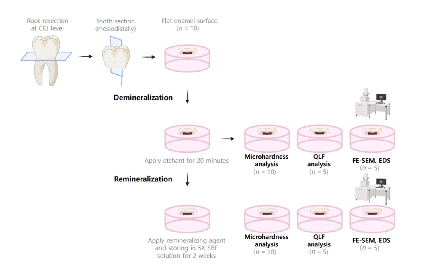
The aim of this study was to compare the remineralizing capacity of bioactive glass (BGS-7, CGBIO) with other agents.
Methods
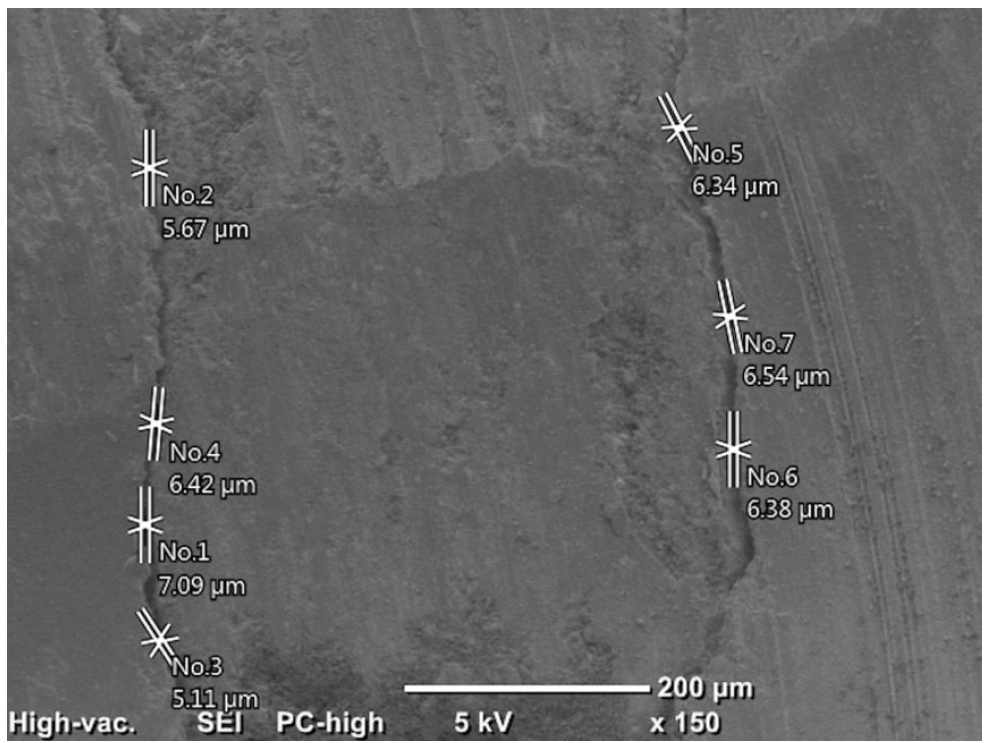
Twenty caries-free third molars were sectioned and demineralized. Specimens were divided into four groups: (1) control, (2) Clinpro XT varnish (Solventum), (3) 1.23% acidulated phosphate fluoride gel, and (4) a new type of CaO-SiO2-P2O5-B2O3 system of bioactive glass ceramics (BGS-7). Agents were applied and stored in simulated body fluid at 37℃ for 2 weeks. Microhardness was measured using the Vickers hardness testing method. Five specimens per group were analyzed using quantitative light-induced fluorescence (QLF) to assess mineral loss. Field-emission scanning electron microscopy (FE-SEM) and energy-dispersive X-ray spectroscopy (EDS) were used to examine the surface morphology and elemental composition. Data were analyzed using paired t-test and one-way analysis of variance (p < 0.05).
Results
BGS-7 showed the highest microhardness values and the greatest recovery in QLF analysis (p < 0.05). FE-SEM revealed granular precipitates on demineralized enamel in the BGS-7 group. EDS confirmed the presence of newly formed silicon and fluoride layers.
Conclusions
BGS-7 demonstrated superior remineralization capacity compared to other agents, suggesting its potential as an effective remineralizing material. -
Citations
Citations to this article as recorded by- Bacterial ghosts (BGs): A promising approach as candidate vaccine
Helal F. Hetta, Ibraheem M. Mwafey, Noura H. Abd Ellah, Fawaz E. Alanazi, Yasmin N. Ramadan
World Journal of Microbiology and Biotechnology.2026;[Epub] CrossRef
- Bacterial ghosts (BGs): A promising approach as candidate vaccine
- 2,789 View
- 254 Download
- 1 Web of Science
- 1 Crossref
- Marginal adaptation of three root-end filling materials in cavities prepared with laser and ultrasonic tips: an in vitro comparative study
- Busra Zengin, Seda Aydemir, Nicholas Paul Chandler
- Restor Dent Endod 2025;50(4):e32. Published online September 9, 2025
- DOI: https://doi.org/10.5395/rde.2025.50.e32
-
Abstract
PDFPubReaderePub
- Objectives
This study evaluated the marginal adaptation of ProRoot MTA (Dentsply Tulsa Dental), Biodentine (Septodont), and TotalFill BC RRM (FKG) placed in root-end cavities prepared with ultrasonic or Er,Cr:YSGG laser tips, using scanning electron microscopy.
Methods
The canals of 90 extracted maxillary central incisors were prepared and obturated and their roots resected. Six groups of 15 specimens were allocated as follows: ultrasonic + ProRoot MTA, ultrasonic + Biodentine, ultrasonic + TotalFill, laser + ProRoot MTA, laser + Biodentine, and laser + TotalFill. Roots were sectioned longitudinally to expose the filling material. Apical and coronal micrographs were taken, and the greatest distance between dentin and filling material was measured. The total gap area was also calculated using further micrographs.
Results
Cavities prepared with the ultrasonic tips and filled with Biodentine showed significantly greater gap dimensions compared with TotalFill (p < 0.001) and ProRoot MTA (p = 0.007) in the apical region. The ultrasonic group showed significantly higher void values compared to the laser group for ProRoot MTA (p = 0.026), when comparing the total values of void. The Biodentine group was significantly higher than the TotalFill group in root-end cavities prepared with ultrasonic tips (p < 0.001). The Biodentine group was significantly higher than the ProRoot MTA group in root-end cavities prepared with the laser tip (p = 0.002).
Conclusions
Under the conditions of this study, it was determined that the root-end cavity preparation technique had an effect on the amount of gaps formed between the dentin and the three filling materials. -
Citations
Citations to this article as recorded by- Marginal Adaptability of Harvard MTA and Biodentine Used as Root-End Filling Material: A Comparative SEM Study
Yaneta Kouzmanova, Ivanka Dimitrova
Materials.2025; 18(19): 4598. CrossRef
- Marginal Adaptability of Harvard MTA and Biodentine Used as Root-End Filling Material: A Comparative SEM Study
- 4,157 View
- 299 Download
- 1 Web of Science
- 1 Crossref
Case Report
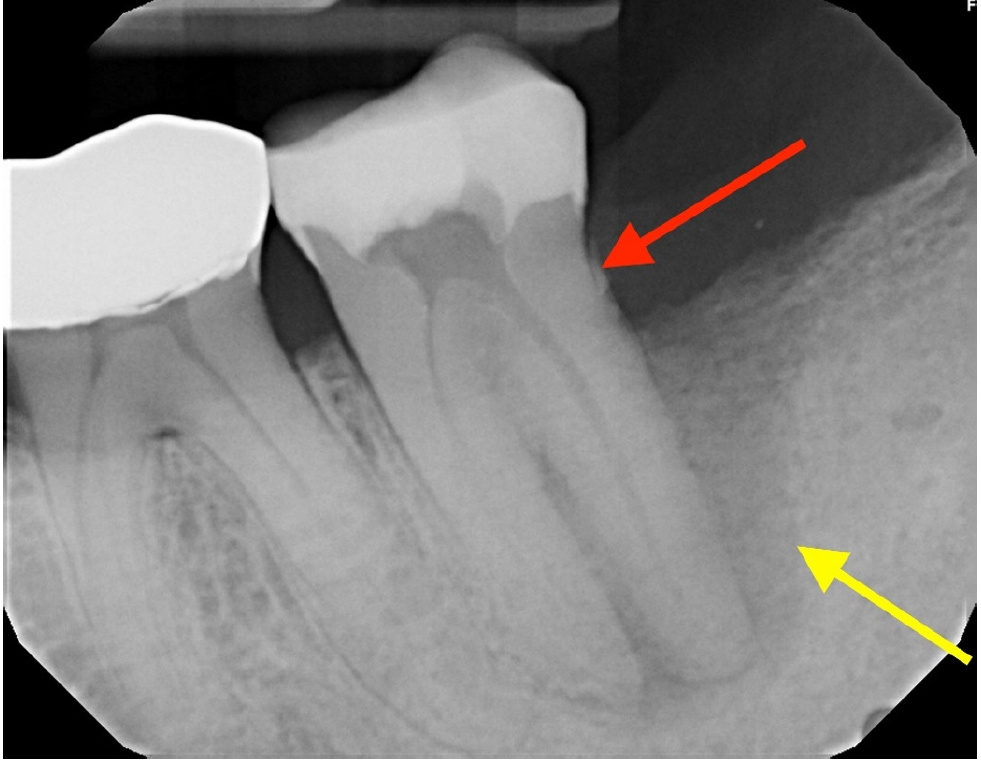
- Multidisciplinary management of an endo-perio lesion complicated by a cemental tear: a case report
- Nishanth D. Sadhak, Akshaya Pallod, Shreyas Oza
- Restor Dent Endod 2025;50(3):e31. Published online August 22, 2025
- DOI: https://doi.org/10.5395/rde.2025.50.e31
-
Abstract
PDFPubReaderePub
- Endodontic-periodontal lesions (EPLs) complicated by cemental tears present a diagnostic and therapeutic challenge. This case report describes the successful management of a 66-year-old male patient with a mandibular second molar (#18) exhibiting an EPL complicated by a cemental tear. Clinical examination revealed a draining sinus tract, deep periodontal pockets, and radiographic evidence of a “J-shaped” lesion and a radiopaque cemental fragment. The tooth had previously initiated endodontic treatment. A multidisciplinary approach involving endodontic treatment and surgical removal of the cemental tear was implemented. At 24-month follow-up, clinical and radiographic examination revealed significant improvement in periodontal health, bone regeneration, and resolution of the lesion. This case highlights the importance of considering cemental tears in the differential diagnosis of EPLs and demonstrates the efficacy of a combined endodontic-periodontal approach for achieving predictable outcomes.
- 4,674 View
- 349 Download
Research Articles
- Structural and morphological characterization of silver nanoparticles intruded mineral trioxide aggregate admixture as a chair-side restorative medicament: an in vitro experimental study
- H. Murali Rao, Rajkumar Krishnan, Chitra Shivalingam, Ramya Ramadoss
- Restor Dent Endod 2025;50(3):e30. Published online August 8, 2025
- DOI: https://doi.org/10.5395/rde.2025.50.e30

-
Abstract
PDFPubReaderePub
- Objectives
The aim of this study was to create a rapid admixture of mineral trioxide aggregate (MTA) and silver nanoparticles (AgNPs) for chairside use in clinical settings to remediate the challenges associated with root canal treatment and pulp capping.
Methods
Synthesized AgNPs at ratios of 10 and 25% were added to commercially available MTA to create an admixture. The admixture was subjected to structural and morphological assessment using X-ray diffraction analysis (XRD), Fourier transform infrared (FT-IR) analysis, Raman spectroscopy, and scanning electron microscopy. Antioxidant activity was measured using the hydroxyl radical scavenging assay. A significance level of 0.05 was applied to determine statistical differences.
Results
The addition of AgNPs decreased the carbonate peak intensity in XRD and FT-IR. The rod-like morphology of MTA was changed to a flake-like morphology with the addition of AgNPs. Antibacterial efficacy enhanced proportionally with the augmentation of AgNPs concentration.
Conclusions
The creation of rapid admixture of MTA and AgNPs during chairside use in clinical settings can deliver beneficial characteristics of enhanced morphological features favoring mineralization and profound antibacterial effects to overcome the challenges associated with root canal treatment and pulp capping.
- 2,820 View
- 98 Download
- 1 Web of Science
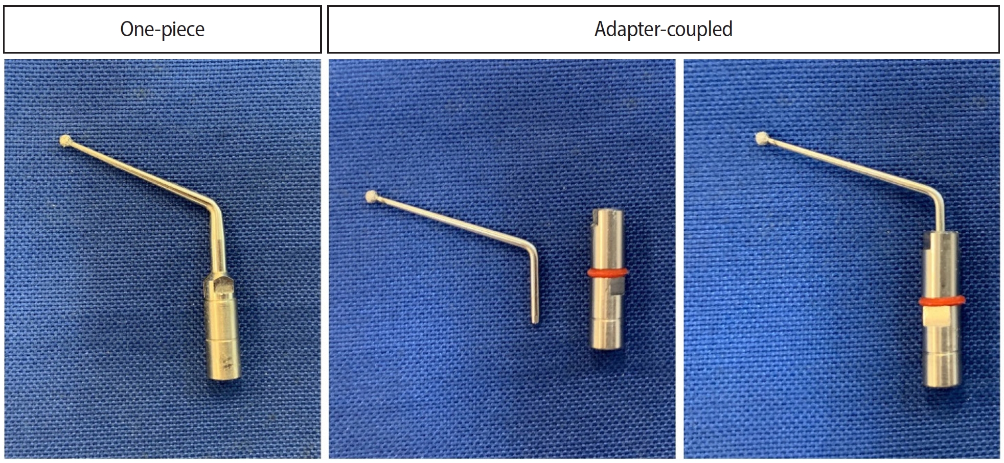
- Analysis of thermal profiles on tooth structure and insert during one-piece or adapter-coupled ultrasonic insert use: an in vitro experimental study
- Gabriela Loewen Brotto, Bruno Monguilhott Crozeta, Bruno Marques-da-Silva, Alysson Nunes Diógenes, Emmanuel João Nogueira Leal da Silva, Flávia Sens Fagundes Tomazinho
- Restor Dent Endod 2025;50(3):e24. Published online July 11, 2025
- DOI: https://doi.org/10.5395/rde.2025.50.e24
-
Abstract
PDFPubReaderePub
- Objectives
This in vitro study aimed to evaluate temperature variation on the external surface of mandibular molars and within ultrasonic inserts when using adapter-coupled versus one-piece inserts.
Methods
Twenty-four extracted human mandibular molars were divided into two groups based on the type of ultrasonic insert used: adapter-coupled and one-piece inserts. Temperature on the external surface of each tooth was measured with a thermocouple probe positioned in the furcation area, capturing data continuously. The temperature of the ultrasonic inserts was monitored in real-time using a thermal imaging camera. Measurements were taken in a controlled environment without cooling for over 120 seconds. Statistical analysis was conducted using analysis of variance (ANOVA) and two-way ANOVA with repeated measures to evaluate temperature variations between groups and over time, with significance set at 5%.
Results
In the external tooth surface temperature measurements, no significant differences were observed between the groups during the initial 15 seconds (p = 0.185) and 30 seconds (p = 0.067). However, significant differences emerged at 60 seconds (p = 0.025), 90 seconds (p = 0.024), and 120 seconds (p = 0.020), with the one-piece insert group demonstrating higher temperatures in the furcation region. Thermal imaging of the inserts revealed a significant difference at all time points (p < 0.001), with adapter-coupled inserts showing greater heating.
Conclusions
The use of ultrasonic inserts leads to a gradual rise in temperature on the external tooth surface. One-piece inserts generated higher temperatures on the tooth, while adapter-coupled inserts exhibited greater heating within the insert.
- 2,727 View
- 109 Download
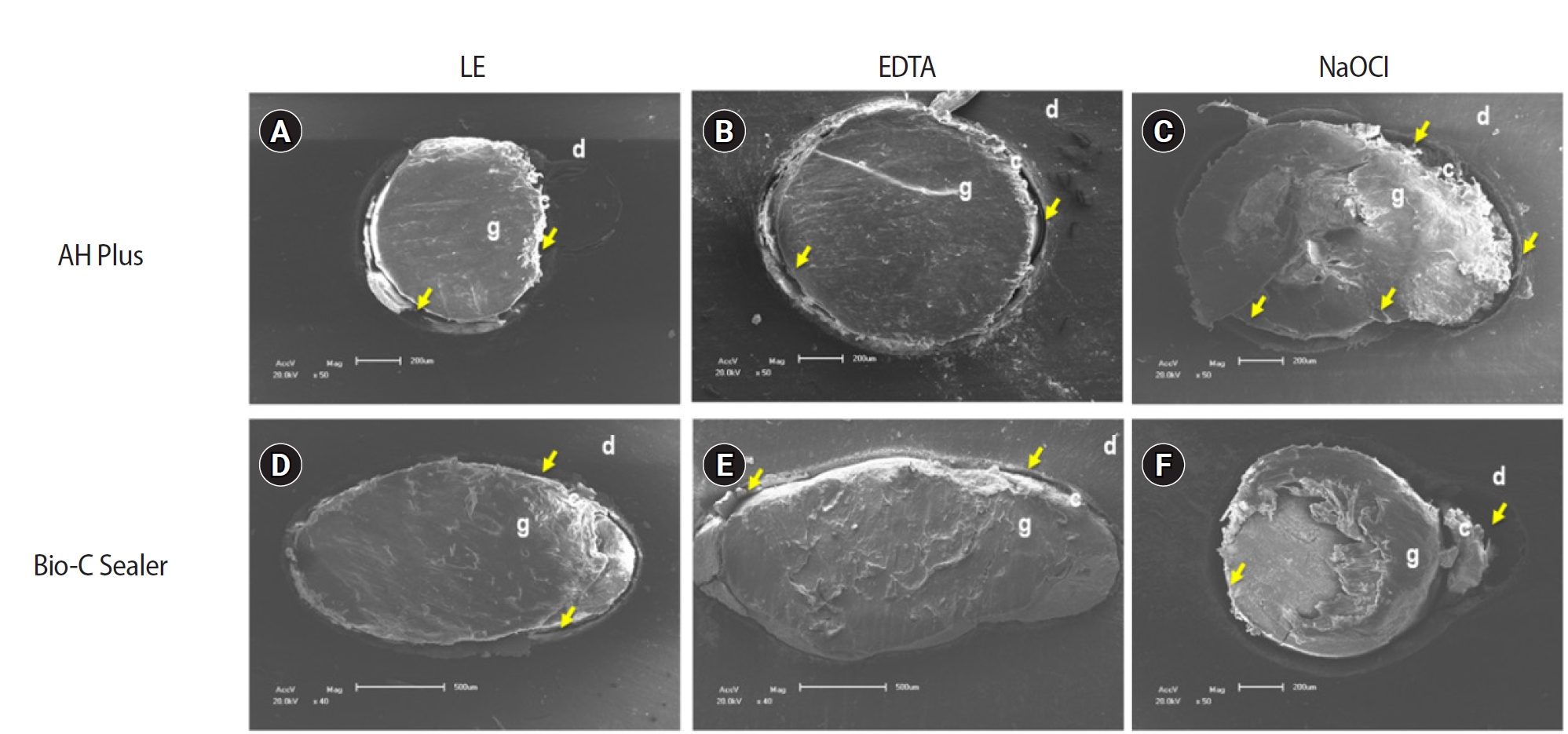
- The effect of limonene extract on the adhesion of different endodontic cements to root dentin: an in vitro experimental study
- Nayara Lima Ferraz Aguiar, Eduardo José Soares, Guilherme Nilson Alves dos Santos, Anna Luísa Araújo Pimenta, Laryssa Karla Romano, Ricardo Gariba Silva, Fernanda de Carvalho Panzeri
- Restor Dent Endod 2025;50(2):e16. Published online May 12, 2025
- DOI: https://doi.org/10.5395/rde.2025.50.e16
-
Abstract
PDFPubReaderePub
- Objectives
The study aimed to evaluate the effect of limonene extract (LE) on push-out bond strength (BS) to root dentin in endodontically treated teeth.
Methods
Single-rooted teeth were selected and instrumented using the reciprocating technique, then divided into three groups based on the final irrigating solution: 2.5% sodium hypochlorite (NaOCl), 17% ethylenediaminetetraacetic acid (EDTA), and 5% LE. The roots were further divided (n = 12) and obturated using the single-cone technique with epoxy resin-based (ERB) or bioceramic sealer (Bio-C). After 3 days, the roots were sectioned into 2-mm slices, obtaining two slices from each root third. Push-out BS testing was conducted at 0.5 mm/min, followed by failure pattern and adhesive interface analysis using scanning electron microscopy. Push-out BS data were analyzed by three-way analysis of variance and Tukey post-hoc test (p < 0.05).
Results
ERB showed higher BS when irrigated with EDTA (5.0 ± 2.3 MPa) compared to NaOCl (1.8 ± 1.1 MPa) (p = 0.0005), particularly in the cervical third. LE yielded intermediate values without significant differences from the other irrigants (3.5 ± 1.9 MPa) (p > 0.05). For Bio-C, the highest BS was observed in the apical third, especially with LE (9.4 ± 5.0 MPa), differing from other thirds and final irrigating solutions (p < 0.05). Mixed failure patterns were most prevalent, regardless of the irrigant solutions.
Conclusions
The combination of LE with Bio-C demonstrated superior BS in the apical third, suggesting its potential as a final irrigating solution in endodontic treatments.
- 3,381 View
- 242 Download
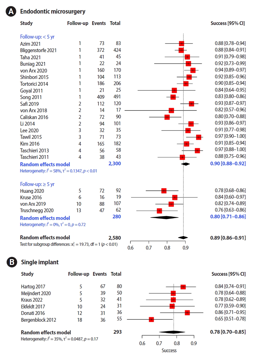
- Success rates comparison of endodontic microsurgery and single implants with comprehensive and explicit criteria: a systematic review and meta-analysis
- Min Jung Ko, Ju Hyun Park, Na Rae Lee, Joon-Ho Yoon, Young-Taek Kim, Sin-Yeon Cho
- Restor Dent Endod 2025;50(1):e8. Published online February 19, 2025
- DOI: https://doi.org/10.5395/rde.2025.50.e8
-
Abstract
PDFSupplementary MaterialPubReaderePub
- Objectives
While the success criteria of endodontic microsurgery (EMS) have been consistently defined and widely accepted, the success criteria of dental implants are outdated and focus only on the implant fixture and surrounding bone. This study aimed to evaluate the outcomes of EMS and single implants (SIs) with explicit criteria.
Methods
We searched for articles published from January 2010 to February 2022 and discussed them and consulted with a clinical advisory committee composed of four dental specialists and one epidemiologist during article selection and data extraction.
Results
Twenty-two EMS studies and six SI studies were included in the meta-analysis. Teeth treated using EMS had a pooled success rate of 89% (90% at <5-year follow-up and 80% at ≥5-year follow-up) and the pooled success rate of SI was 78%.
Conclusions
The success rates of the two procedures with similar follow-up periods were comparable. Subgroup analysis found no other variable that significantly influenced study heterogeneity. Considering the treatment sequence and the similar success rates, it would be advantageous to consider EMS, rather than implants, first in a situation where both procedures are applicable. -
Citations
Citations to this article as recorded by- Surgical Management of a Separated Instrument and Radicular Cyst: A Nine-Month Cone Beam Computed Tomography (CBCT) Follow-up
Dipti Chauhan, Hemant Yadav, Anshu Minocha, Vishal Sharma
Cureus.2025;[Epub] CrossRef - Cost-effectiveness of Endodontic Retreatment vs Implants: A 5-year Retrospective Analysis in India
Pramod Kumar, Himanshu Sharma
Journal of Clinical Insights and Research in Dentistry.2025; 1(3): 121. CrossRef
- Surgical Management of a Separated Instrument and Radicular Cyst: A Nine-Month Cone Beam Computed Tomography (CBCT) Follow-up
- 10,911 View
- 227 Download
- 2 Crossref
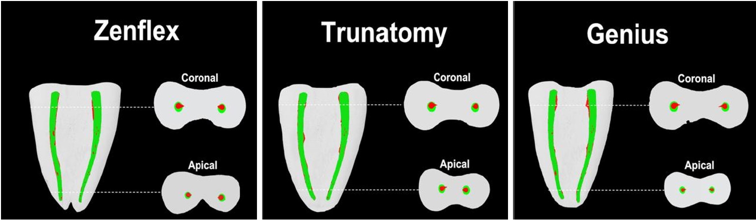
- Shaping ability and cyclic fatigue resistance between Genius ProFlex, ZenFlex, and TruNatomy rotary systems: an experimental study
- Raimundo Sales de Oliveira Neto, Murilo Priori Alcalde, Pedro Cesar Gomes Titato, Pedro Henrique Souza Calefi, Carlos Alberto Spironelli Ramos, Guilherme Ferreira da Silva, Rodrigo Ricci Vivan, Marco Antonio Hungaro Duarte
- Restor Dent Endod 2025;50(1):e9. Published online February 13, 2025
- DOI: https://doi.org/10.5395/rde.2025.50.e9
-
Abstract
PDFPubReaderePub
- Objectives
The aim of this study was to investigate the efficacy of three newly introduced rotary endodontic systems: Genius ProFlex (Medidenta), TruNatomy (Dentsply Maillefer), and ZenFlex (Kerr).
Methods
Forty-five mandibular molars with root canal curvatures <5° were utilized. Micro-computed tomography scans were performed pre- and post-preparation to assess apical transportation, centralization, percentage of dentin wear, and canal volume alterations. Eight instruments of each diameter underwent cyclic fatigue testing.
Results
The percentage of dentin wear on mesial and distal walls showed no significant differences among ZenFlex, TruNatomy, and Genius ProFlex at 1, 2, 3, and 4 mm from the apical foramen and root canal orifice (p > 0.05). Centering ability varied in the mesiolingual canal (p < 0.05). No notable differences were observed in transportation (p > 0.05). Genius ProFlex demonstrated lower volumetric changes (p < 0.05). There were significant differences in cyclic fatigue, with higher values for Genius ProFlex and lower values for TruNatomy (p < 0.05).
Conclusions
The three nickel-titanium rotary instruments are safe and efficient for root canal preparation, with Genius ProFlex exhibiting superior cyclic fatigue resistance. -
Citations
Citations to this article as recorded by- Influence of kinematic motion and instrumentation strategy on apical debris extrusion during root canal preparation: An in vitro study
Amira Alghazaly, Jumanah Aljohani, Khadijah Mohabat, Rafah Ghous
Journal of Conservative Dentistry and Endodontics.2026; 29(7): 741. CrossRef - Comparison of Shaping Ability and Apical Debris Extrusion Using 4 Different Nickel–Titanium Single‐File Systems
Siyu Li, Mengzhen Tang, Xi Wang, Jian Yang, Hyun-Do Jung
International Journal of Biomaterials.2025;[Epub] CrossRef
- Influence of kinematic motion and instrumentation strategy on apical debris extrusion during root canal preparation: An in vitro study
- 4,287 View
- 190 Download
- 1 Web of Science
- 2 Crossref
-
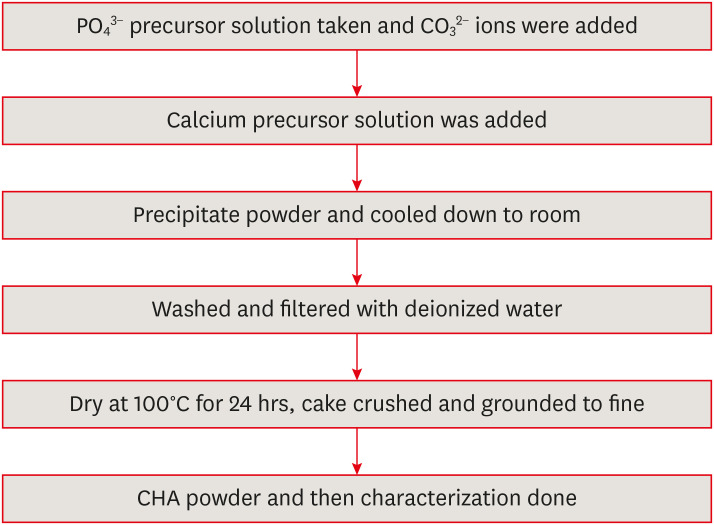
Evaluation of mineral induction ability and cytotoxicity of carbonated hydroxyapatite for pulp tissue regeneration: an
in vitro study - S. Swathi Priyadharshini, Chinnasamy Ragavendran, Anand Sherwood, J. Ramana Ramya, Jogikalmat Krithikadatta
- Restor Dent Endod 2024;49(4):e40. Published online October 29, 2024
- DOI: https://doi.org/10.5395/rde.2024.49.e40
-
Abstract
PDFPubReaderePub
Objectives This study aimed to evaluate carbonated hydroxyapatite (CHA)’s ability for mineral induction and its
in vitro cytotoxicity with human dental pulp cells.Materials and Methods Precursors for the study include di-ammonium hydrogen phosphate and calcium nitrate tetrahydrate, with sodium hydrogen carbonate added to achieve different levels of carbonate substitution. The synthesized CHA samples are characterized using X-ray diffraction, Fourier transform infrared spectroscopy, and Raman spectroscopy. Scanning electron microscopy (SEM) was used to observe morphology. For 14 days at 37°C, samples were submerged in simulated body fluid to assess their mineral induction capabilities. SEM was used to confirm apatite formation on sample surfaces. The cytotoxicity assay was used to assess the vitality of the cells following their exposure to various concentrations of CHA.
Results The Joint Committee on Powder Diffraction Standards data for HA aligned well with the results from X-ray diffraction analysis of CHA across 3 different concentrations, indicating strong agreement. Fourier transform infrared spectra indicated the presence of phosphate, hydroxyl, and carbonate groups within the samples. SEM and Energy-dispersive X-ray analysis show agglomerated and flaky nanoparticles. All the samples are bioactive, but the formation of apatite differs from one another.
In vitro cytotoxicity assay showed that over 70% of cells maintain viability.Conclusions The results of this study may provide insight into the potential use of carbonated HA as a dental pulp-capping material for vital pulp therapy.
-
Citations
Citations to this article as recorded by- Smart Nanomaterials: Current State and Future Prospects in Drug Delivery and Tissue Engineering
E. Elizabeth Rani, D. Sakthi Sanjana, E. Karthikeyan, J. Nandhini
Biomedical Materials & Devices.2026; 4(2): 1455. CrossRef - Thermoresponsive Nanomaterials: Revolutionizing Cancer Theranostics
Bellarmin Michael, Mohanakrishnan Srinivasan, Karthikeyan Elumalai, Lokeshwar Ravikumar, Sivaprakash Kathiresan, Nandhini Jayaprakash
Biomedical Materials & Devices.2026; 4(3): 2697. CrossRef - Physicochemical and antibacterial evaluation of novel nano α-TCP–AgNPs biocomposites for direct pulp-capping applications
Selviana Wulansari, Hendra Dian Adhita Dharsono, Nasrul Wathoni, Rosalina Tjandrawinata, Arief Cahyanto, Moehamad Orliando Roeslan
Frontiers in Oral Health.2026;[Epub] CrossRef - Physicochemical effects of nano type-B bone substitute on pulp protective cement formulations
Njwan Fadhel SHEHAB
Dental Materials Journal.2026; 45(1): 92. CrossRef - Recycling waste for sustainability: The green synthesis of silver nanoparticles from Bougainvillea glabra green waste, and the evaluation of their antioxidant, cytotoxic, catalytic, antibacterial and in-silico molecular docking properties
Hafsa Naleem, Mathivathani Kandiah, Beneli Gunaratne, Ominda Perera
Next Research.2026; 11: 101990. CrossRef - Comparative evaluation of compressive strength and morphological interface of carbonated hydroxyapatite with other pulp capping materials: An in vitro analysis
S. Swathi Priyadharshini, Chinnasamy Ragavendran, I. Anand Sherwood, Ramanaramya Jeyapalan
Endodontology.2025; 37(1): 90. CrossRef - Bioactive Dioxo-Phosphobetaines derived from the reaction of Dichlorodinitrobenzofuroxane with various phosphines
Irina V. Galkina, Haiyan Fan, Semen R. Romanov, Dmitriy I. Bakhtiyarov, Luisa M. Usupova, Svetlana N. Egorova, Yulia V. Bakhtiyarova, Enrico Benassi
Bioorganic Chemistry.2025; 163: 108695. CrossRef - Near-infrared laser-activated PLGA-PDA core-shell nanohybrids for synergistic photothermal antibacterial therapy and sustained ion release in orthodontic white spot lesions prevention
Zezhou Feng, Yujiang Liu, Silu Sun, Minmin Si, Di Huang, Zhiyuan Feng
Journal of Dentistry.2025; 162: 106078. CrossRef - Formation and utilization of soluble microbial products in denitrifying biofilters at different carbon-to-nitrogen ratios: Microbial community characteristics
Fangyuan Jiang, Xianyang Shi
Journal of Environmental Chemical Engineering.2025; 13(6): 119554. CrossRef - Bioactivity and biocompatibility of bioceramic-based pulp capping materials in laboratory and animal models
Rafiqul Islam, Md. Refat Readul Islam, Kenta Tsuchiya, Yu Toida, Hidehiko Sano, Monica Yamauti, Hany Mohamed Aly Ahmed, Atsushi Tomokiyo
Journal of Materials Science: Materials in Medicine.2025;[Epub] CrossRef - Physical, Chemical, and Biological Properties of Graphene Nanoparticle-added Tricalcium Silicate Formulations: A Systematic Review
Soundaria Srinivasan, Deepa Gurunathan, Lakshmi Thangavelu
Journal of International Oral Health.2025; 17(6): 453. CrossRef - Advanced structural and compositional profiling of mineral trioxide aggregate incorporated with nano-carbonated hydroxyapatite: a comprehensive X-ray diffraction and energy dispersive X-ray investigation
Njwan Fadhel Shehab, Nadia Hameed Hasan, Alaa Edrees Dawood, Nawal Atiya Khalaf
Biomaterial Investigations in Dentistry.2025; 12: 216. CrossRef
- Smart Nanomaterials: Current State and Future Prospects in Drug Delivery and Tissue Engineering
- 4,775 View
- 156 Download
- 9 Web of Science
- 12 Crossref
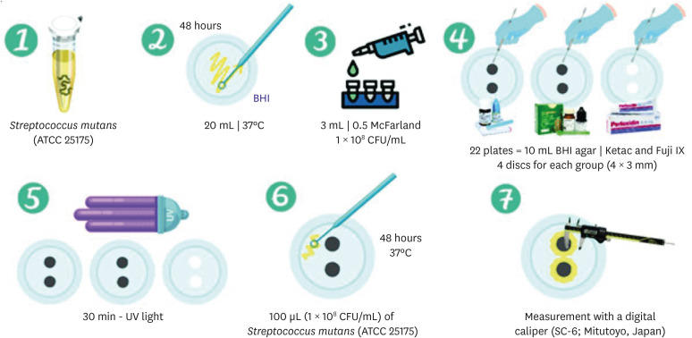
- Physical-mechanical, chemical and biological properties of graphene-reinforced glass ionomer cements
- Tatiane Ramos dos Santos Jordão, Laura Soares Viana Fernandes, Karla Lorene de França Leite, Adílis Alexandria, Emmanuel João Nogueira Leal Silva, Lucianne Cople Maia, Tatiana Kelly da Silva Fidalgo
- Restor Dent Endod 2024;49(4):e37. Published online October 10, 2024
- DOI: https://doi.org/10.5395/rde.2024.49.e37
-
Abstract
PDFPubReaderePub
Objectives This study aimed to evaluate the physical-mechanical, chemical, and biological properties of graphene-reinforced glass ionomer cements (GICs).
Materials and Methods Different proportions of graphene powder were incorporated into 2 high-viscosity self-curing GIC, Ketac Molar (GKetac) and Fuji IX (GFuji), in 4 different concentrations: 0.5%, 1%, 2%, and 5%. The control groups included the GICs without graphene. Experiments were performed to analyze linear (Ra) and volumetric roughness (Sa), antimicrobial activity, radiopacity, fluoride release, microhardness, solubility, and water sorption. Data were analyzed using Kruskal-Wallis, Mann-Whitney, Wilcoxon, analysis of variance, and Tukey’s test (
p ≤ 0.05).Results The GKetac 0% and GFuji0% groups presented higher Ra (4.05 and 2.72) and Sa (4.76 and 5.16), respectively. No inhibition zone was observed, and the incorporation of graphene reduced radiopacity. Moreover, there was no influence on the solubility and water sorption after 21 days. A greater fluoride release was observed in the period of 7 days for most of the groups. After 21 days, GKetac 5%, 2%, and 1% presented higher releasing than 0% and 0.5% (
p ≤ 0.05).Conclusions The graphene incorporation improved the microhardness of GICs in lower concentrations. Graphene incorporation to GICs modified some physical-mechanical, and chemical, but not affected biological properties.
-
Citations
Citations to this article as recorded by- Laboratory-based additive modifications in glass ionomer cements: A scoping review using a systematic data mining and trend analysis framework (2015-2024)
Kenta Tsuchiya, Sharanbir K Sidhu, Salvatore Sauro, Jukka P. Matinlinna, Hidehiko Sano, Monica Yamauti, Shuhei Hoshika, James Kit Hon Tsoi, Atsushi Tomokiyo
Journal of Dentistry.2026; 166: 106349. CrossRef - Physicomechanical and Antibacterial Properties of Resin-Based Dental Sealants Modified with Graphene Oxide Nanoparticles
Ploypim Kraisintu, Suparaksa Yamockul, Tool Sriamporn, Niyom Thamrongananskul, Awiruth Klaisiri, Theerapat Chanamuangkon, Somphob Thompho, Thanchanok Suriyapongprapai, Guang Hong
European Journal of Dentistry.2026;[Epub] CrossRef - Influence of expanded graphene on physical and chemical properties, and in vitro toxicity of glass ionomer cements for luting
Sarah Pereira Martins, Carolina Mara Geraldino Monteiro, Renan Rocha Da Silva, Andrea Vaz Braga Pintor, Marcela Baraúna Magno, Maria Augusta Visconti, Maria Teresa Villela Romanos, Livia Rodrigues De Menezes, Lucianne Cople Maia, Matheus Melo Pithon
Biomaterial Investigations in Dentistry.2026; 13: 440. CrossRef - Graphene Oxide Incorporation Enhances Biocompatibility and Surface Stability of Conventional Glass Ionomer Cements
Mayara Silva de Santana, Lucas dos Santos Silva, Joicy Cortez de Sá Sousa, Thalita Santana Conceição, Barbara Emanoele Costa Oliveira, Ceci Nunes Carvalho, Edilausson Moreno Carvalho, Luís Cláudio Nascimento da Silva
Pesquisa Brasileira em Odontopediatria e Clínica Integrada.2026;[Epub] CrossRef - Impact of graphene incorporation on the mechanical and optical properties of glass ionomer cements
Sarah Pereira Martins, Carolina Mara Geraldino Monteiro, Kenderson Santos Silva, Renan Rocha da Silva, Cássia Almeida Brito, Lívia Rodrigues de Menezes, Lucianne Cople Maia, Matheus Melo Pithon
Brazilian Journal of Oral Sciences.2026; 25: e269444. CrossRef - Potential of Nano-Gum Arabic on the Physical, Mechanical, Adhesive, Optical, and Biological Performance of Glass Ionomer Cement: A Comprehensive In Vitro Study
Marwa Beleidy, Soha A. Hassan, Rania Rashad Omar Taha, Yousra Nashaat, Yasmine Alaa El-din
BMC Oral Health.2026;[Epub] CrossRef
- Laboratory-based additive modifications in glass ionomer cements: A scoping review using a systematic data mining and trend analysis framework (2015-2024)
- 4,011 View
- 198 Download
- 5 Web of Science
- 6 Crossref
Case Report
- Straightforward replication of digital wax-up design into direct composite resin restorations in adolescents using a custom 3-dimensionally printed index
- Ra’fat Ibrahim Farah, Sanaa Najeh Al-Haj Ali, Abdullah Alharbi, Bandar Alresheedi
- Restor Dent Endod 2024;49(4):e36. Published online October 10, 2024
- DOI: https://doi.org/10.5395/rde.2024.49.e36
-
Abstract
PDFPubReaderePub
This case report introduces a straightforward, noninvasive approach for the esthetic rehabilitation of malformed anterior teeth in adolescents using direct composite restorations. The universal composite resin restorations are applied within a transparent 3-dimensionally printed rigid-resin index, which is individually customized from a digital wax-up. Compared to other methods, this technique streamlines the restoration process, significantly reducing chairside time while enhancing the predictability, accuracy, and patient acceptance of the aesthetic outcome.
-
Citations
Citations to this article as recorded by- Diastema closure and esthetic rehabilitation with peg-shaped laterals: A case series
Afsana Ansari, Dipika Yadav
The Saint's International Dental Journal.2024; 8(2): 48. CrossRef
- Diastema closure and esthetic rehabilitation with peg-shaped laterals: A case series
- 5,990 View
- 281 Download
- 1 Crossref
Research Articles
- Effects of different curing methods on the color stability of composite resins
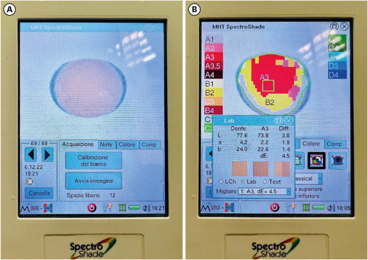
- Massimo Pisano, Alfredo Iandolo, Dina Abdellatif, Andrea Chiacchio, Marzio Galdi, Stefano Martina
- Restor Dent Endod 2024;49(4):e33. Published online September 5, 2024
- DOI: https://doi.org/10.5395/rde.2024.49.e33
-
Abstract
PDFPubReaderePub
Objectives The aim of this study was to compare the effects of different polymerization strategies and the effectiveness of finishing and polishing procedures of composite resins on color stability.
Materials and Methods The samples were divided into 4 main groups according to the polymerization strategy, and all groups except the control group received surface treatment. Each group was subsequently divided into 3 subgroups respectively: Kuraray Clearfil Majesty ES-2 Classic, Premium and Universal. Approximately 24 hours after preparation of the samples, they were immersed for 7 days in a coffee solution. A first color measurement was performed after the preparation of the samples, the second measurement was performed after 7 days in the coffee solution. All measurements were carried out using a dental spectrophotometer to assess the CIE
L *a *b * color parameters.Results There was a statistically significant difference between ΔE values for different procedures (
p = 0.003); in particular, the differences were found only between the groups that received surface treatment and the control group. In addition, a statistically significant difference was observed between the values of ΔE for different composites in the different procedure groups.Conclusions Spectrophotometric analysis showed that the additional photopolymerization and oxygen inhibition procedures did not yield better results in relation to color stability. In addition, finishing and polishing provided better color stability compared to not performing these procedures.
-
Citations
Citations to this article as recorded by- Color Stability Under Challenge: Effects of Thermo-Aging and Mouthrinse Exposure on Anterior Teeth and Esthetic Composites
Gökçe Keçeci, Zehra Güner, Süleyman Ziya Şenyurt, Kamile Erciyas
European Journal of Therapeutics.2026; 32(1): 94. CrossRef - Color stability and degree of conversion of conventional, amine-free, and self-adhesive resin cements polymerized under different conditions
Su Young Lee, Yasushi Shimada, Jaeyoung Edwin Han, Seung-Hoon Han
Dental Materials.2026;[Epub] CrossRef - Abrasiveness and Bleaching Level of Toothpastes on Composite Resins: A Quantitative Analysis Using a Novel Brushing Simulator
Simge Meseli, Elif Alkan, Bora Korkut, Ozlem Kanar, Dilek Tagtekin
Applied Sciences.2025; 15(5): 2314. CrossRef - Comparative Evaluation of Direct and Indirect Composite Restorations in Class II Tooth Preparations - An In vivo Study
Akshun Gupta, Garima Arora, Aprajita Mehta, Satish Sane, Siddhi Nevrekar, Apurva Nagrale
Advances in Human Biology.2025; 15(4): 550. CrossRef - Micro- and Nanoplastics and the Oral Cavity: Implications for Oral and Systemic Health, Dental Practice, and the Environment—A Narrative Review
Federica Di Spirito, Veronica Folliero, Maria Pia Di Palo, Giuseppina De Benedetto, Leonardo Aulisio, Stefano Martina, Luca Rinaldi, Gianluigi Franci
Journal of Functional Biomaterials.2025; 16(9): 332. CrossRef
- Color Stability Under Challenge: Effects of Thermo-Aging and Mouthrinse Exposure on Anterior Teeth and Esthetic Composites
- 7,549 View
- 389 Download
- 4 Web of Science
- 5 Crossref
- Effect of surface sealant on the color stability and whiteness index of single-shade resin composites after staining and bleaching
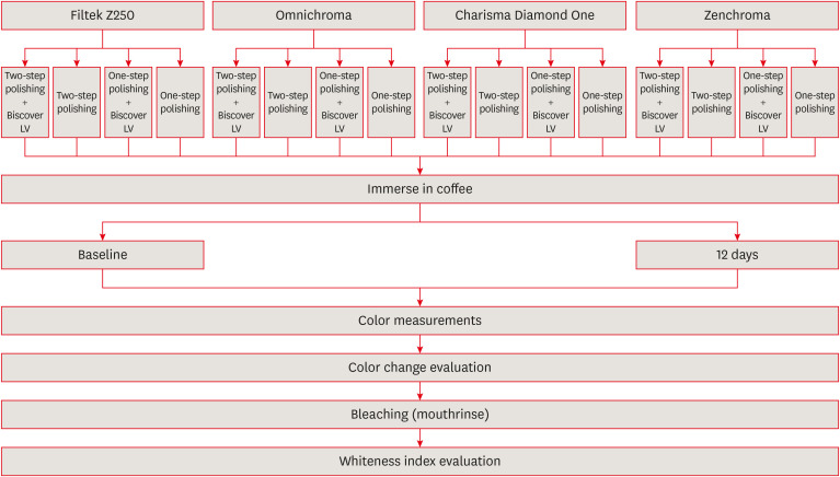
- Muhammet Fidan, Özhan Yağcı
- Restor Dent Endod 2024;49(3):e30. Published online July 11, 2024
- DOI: https://doi.org/10.5395/rde.2024.49.e30
-
Abstract
PDFPubReaderePub
Objectives The aim of the current study was to evaluate the effect of polishing systems and surface sealant on the color stability and whiteness index of single-shade resin composites after staining and bleaching.
Materials and Methods Three single-shade (Omnichroma, Charisma Diamond One, Zenchroma) and one multi-shade (Filtek Z250) materials were tested. From each resin composite, 40 specimens were prepared. The specimens were divided into 4 subgroups (
n = 10) according to the surface treatments: 1-step polishing, 1-step + Biscover LV, 2-step polishing, and 2-step polishing + Biscover LV. Color differences (ΔE00) were calculated after being immersed in the coffee solution for 12 days. After the staining, the specimens were immersed in a whitening mouthrinse (Crest-3D White) for 12 hours. Whiteness index differences (∆WID = WID after staining − WID after bleaching) values were recorded. The generalized linear model was used for analysis (p < 0.05).Results The lowest and highest ΔE00 values were found for Zenchroma and Charisma Diamond One respectively. Sealed groups indicated higher ΔE00 values than nonsealed groups with significant differences (
p = 0.008). The lowest and highest ΔWID values were found for Zenchroma and Charisma Diamond One respectively. Sealed groups indicated lower ΔWID values than nonsealed groups with significant differences (p = 0.022).Conclusions The use of surface sealant increased the discoloration and showed less whiteness change in resin materials. When the 1-step was compared with the 2-step polishing, the effects on the color stability and whiteness index values of the resin materials were similar.
-
Citations
Citations to this article as recorded by- Color and surface properties of conventional, injectable, and 3D-printed resin composites for anterior restorations: influence of a surface sealant
Soner Sismanoglu
Odontology.2026;[Epub] CrossRef - Effect of Different Surface Finishing Processes on the Optical Properties of Zirconium Oxide Ceramics
Nurşen Şahin, Elif Nazli Tekin, Çağrı Ural
Online Türk Sağlık Bilimleri Dergisi.2026; 11(1): 1. CrossRef - Integrative review of resin-based dental pit and fissure sealants: Composition analysis and a novel categorization proposal
Paweł J. Piszko, Paulina Drapiewska, Julia Kurczyk, Natalia Stelmaszczyk, Michał J. Kulus, Aleksandra Piszko, Maciej Dobrzyński
Materials Science-Poland.2026; 44(1): 83. CrossRef - Influence of Surface Sealants and Chromogenic Dietary Agents on the Color Stability of Composite Resin Restorations: An In Vitro Study
Jorge Ferreira-Coelho, Maria do Carmo Vilas-Boas, Orlanda Torres, Virgínia M. F. Gonçalves, Lígia Lopes-Rocha
Applied Sciences.2026; 16(12): 5960. CrossRef - Evaluating the effects of bleaching on color stability and surface roughness in single-shade and multi-shade resin composites
Hatice Tepe, Özge Çeliksöz, Zeynep Biçer, Batucan Yaman
Anatolian Current Medical Journal.2024; 6(6): 372. CrossRef
- Color and surface properties of conventional, injectable, and 3D-printed resin composites for anterior restorations: influence of a surface sealant
- 4,082 View
- 105 Download
- 3 Web of Science
- 5 Crossref
- Endodontic characteristics of mandibular premolar with dens evaginatus: a retrospective study
- Minjin Kim, Sujin Jeon, Min-Seock Seo
- Restor Dent Endod 2024;49(3):e28. Published online July 11, 2024
- DOI: https://doi.org/10.5395/rde.2024.49.e28
-
Abstract
PDFPubReaderePub
Objectives This study aimed to investigate the endodontic characteristics of mandibular premolars with dens evaginatus (DE) that require endodontic treatment.
Materials and Methods Patients who underwent endodontic treatment were enrolled. The inclusion criteria were patients who underwent root canal treatment in the lower permanent teeth with DE and were followed up for at least 1 year. Preoperative clinical and radiographic variables were obtained. The frequency distribution of the preoperative variables was compared using the χ2 or Fisher’s exact tests. The significance of the change in periapical health index (PAI) and root development stages before and after treatment was examined using the Wilcoxon signed-rank test.
Results A total of 150 teeth of 134 patients with an average age of 15.3 years were included. The percentage distribution comparison of the preoperative variables and obturation techniques revealed significant differences in pulpal and periapical diagnosis, and percussion, and especially regarding age, root development stage, and PAI. Age was the only statistically significant preoperative variable associated with root growth (
p < 0.05).Conclusions Approximately, 60% of DEs requiring endodontic treatment had immature roots. Age being the most significant predisposing factor, early treatment provides the greatest opportunity for full root development.
-
Citations
Citations to this article as recorded by- A tooth with multiple supernumerary cusps and taurodontism concurrently accompanied with other taurodont teeth: a rare case report
Zihui Tang, Hongchen Zhang, Rongrong Dang, Qiushi Zhang, Yan Huang, Yanwei Yang
Surgical and Radiologic Anatomy.2025;[Epub] CrossRef
- A tooth with multiple supernumerary cusps and taurodontism concurrently accompanied with other taurodont teeth: a rare case report
- 4,502 View
- 127 Download
- 1 Web of Science
- 1 Crossref
- Alkasite restorative material for endodontically treated teeth: a randomized controlled pilot study
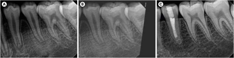
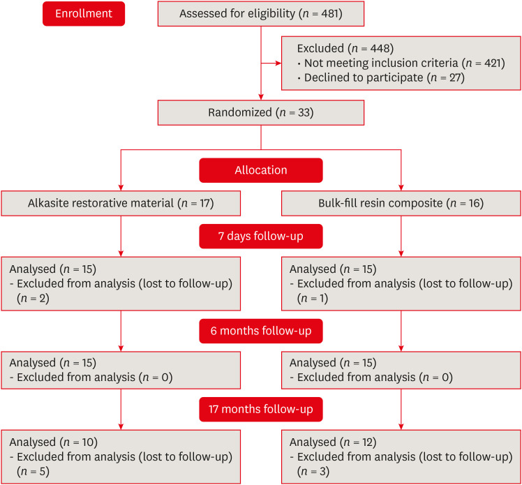
- Davi Ariel Nobuo Bepu, Renata Siqueira Scatolin, Natalia Saud Junqueira Franco, Luiza Pejon Sanchez, Aline Evangelista Souza-Gabriel, Silmara Aparecida Milori Corona
- Restor Dent Endod 2024;49(3):e24. Published online June 11, 2024
- DOI: https://doi.org/10.5395/rde.2024.49.e24
-
Abstract
PDFPubReaderePub
Objectives This study aimed to evaluate the clinical performance of an alkasite restorative material in molars that had undergone root canal treatment.
Materials and Methods The research was registered in Brazilian Registry of Clinical Trials. The randomized clinical trial involved 33 patients, each with at least 1 mandibular molar requiring restoration after receiving endodontic treatment. Patients were randomly assigned to receive either bulk-fill resin composite (Tetric N Ceram Bulk Fill, Ivoclar Vivadent) or the alkasite restorative material (Cention N, Ivoclar Vivadent). Upon completion of the restorations, 3 calibrated professionals utilized the United States Public Health Service criteria to assess various factors, including retention, secondary caries, marginal adaptation, restoration color, marginal pigmentation, and anatomical form. Evaluations were conducted at intervals of 7 days, 6 months, and 17 months. Additionally, the assessment encompassed the presence of radiolucent lines adjacent to the restoration, material deficiencies or excess, contact points, and caries recurrence. The data underwent analysis using the Friedman and Mann-Whitney tests (α = 0.05).
Results After 17 months, the results revealed that the alkasite restorative material exhibited greater wear of anatomical shape compared to the bulk-fill resin composite (
p = 0.0189). Furthermore, the alkasite restorative material significantly differed from the natural tooth color in most cases (p = 0.0000). However, no other criteria displayed significant differences between the materials or over time (p > 0.05).Conclusions The alkasite restorative material (Cention N) emerges as a viable option for restoring endodontically treated teeth, displaying clinically acceptable alterations after a 17-month evaluation period.
Trial Registration Brazilian Registry of Clinical Trials (ReBEC) Identifier:
RBR-97kx5jv -
Citations
Citations to this article as recorded by- The Effect of Intraorifice Barrier Materials on the Fracture Resistance of Endodontically Treated Teeth: A Systematic Review and Network Meta-Analysis
Sevilay Karahan, Zeynep Buket Dağ, Emel Uzunoğlu Özyürek
Journal of Endodontics.2026; 52(5): 696. CrossRef - A Systematic Review and Meta-Analysis on the Clinical Performance and Longevity of Bioactive Composite Resin Restorations
Ahmed A. Holiel, Mounir M. Al Nakouzi, Rim Bourgi, Carlos Enrique Cuevas-Suárez, Iván Olivares Acosta, Louis Hardan, Naji Kharouf, Youssef Haikel
Journal of Composites Science.2026; 10(1): 39. CrossRef - Evaluation of Clinical Performance of Alkasite Restorative Materials: A Systematic Review and Meta-Analysis
Chloé Laporte, Rim Bourgi, Carlos Enrique Cuevas-Suárez, Naji Kharouf, Louis Hardan, Miguel Ángel Fernández-Barrera, Anh Tuan Dang, Youssef Haikel, Abigailt Flores-Ledesma
Journal of Functional Biomaterials.2026; 17(2): 93. CrossRef - 48-month clinical performance of an Alkasite restorative material versus resin composite in class II restorations: a randomized controlled trial
Ece Meral, Betül Kesim, Fatma Dilşad Öz, Sevil Gürgan
Journal of Dentistry.2026; 173: 106792. CrossRef - Alkasites in restorative dentistry: a review of their performance and properties
Alexander Bonchev, Ralitsa Bogovska-Gigova
Journal of Dentistry.2025; 160: 105916. CrossRef - Comparative Analysis of Flexural and Compressive Strengths of Bioactive Alkasite Compared to Other Ion-Releasing Restorative Materials
Hanin E. Yeslam, Fatin A. Hasanain
Biomimetics.2025; 10(11): 751. CrossRef
- The Effect of Intraorifice Barrier Materials on the Fracture Resistance of Endodontically Treated Teeth: A Systematic Review and Network Meta-Analysis
- 5,367 View
- 159 Download
- 6 Web of Science
- 6 Crossref
- Single-session associative protocol for dentin hypersensitivity management: a 1-year randomized, blinded clinical study
- Thayna Carolina Zeni, Poliana Maria de Faveri Cardoso, Rafael da Silva Vanolli, Márcio José Mendonça, Julio Katuhide Ueda, Veridiana Camilotti
- Restor Dent Endod 2024;49(2):e15. Published online March 20, 2024
- DOI: https://doi.org/10.5395/rde.2024.49.e15
-
Abstract
PDFPubReaderePub
Objectives This study aimed to establish a single-session associative protocol for non-restorative management of dentin hypersensitivity (DH).
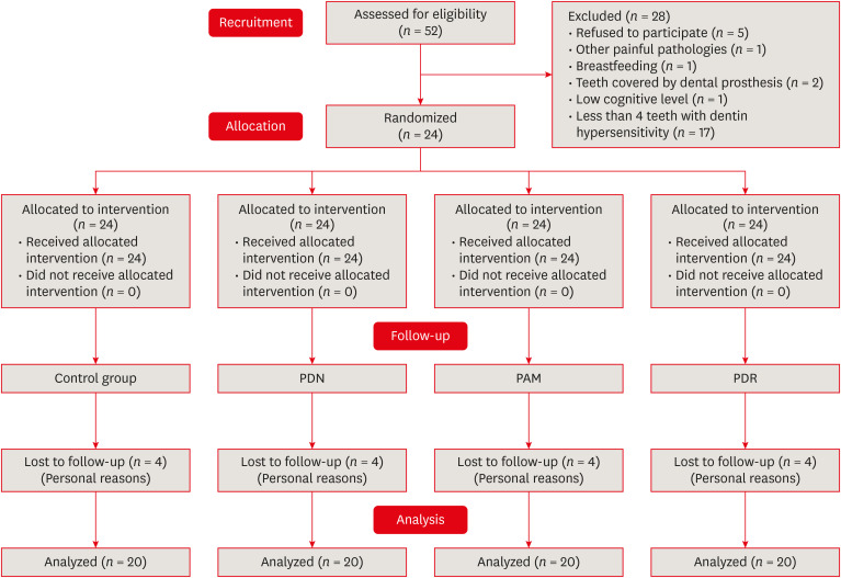
Materials and Methods Twenty-four individuals with DH and a minimum sensitivity level of 4 on the visual analog scale (VAS) were selected. The study was conducted in a split-mouth design, with each participant (
n = 20) having at least 1 affected tooth in all quadrants. The management protocols consisted of control group: universal adhesive, Neural Desensitizing Protocol group: 5% potassium nitrate, Mixed Desensitizing Protocol (PAM) group: 5% sodium fluoride and 5% potassium nitrate, Remineralizing Desensitizing Protocol (PDR) group: surface-partially reacted glass technology photopolymerizable varnish. Evaluations were performed immediately after application, at 1 week, 1 month, 2 months, and 12 months using the VAS sensitivity test.Results The scores were subjected to statistical analysis using the Friedman test (
p < 0.05), Durbin-Conover test (p < 0.05), and Wilcoxon test (p < 0.05). At the 12-month evaluation, all groups showed statistically significant differences compared to the initial assessment. For the evaluation after 12 months, there was a statistically significant difference between the PAM group, the control group, and the PDR group.Conclusions It can be concluded that all groups were effective in controlling DH, but there were significant results in the control group and PDR group. The clinical relevance of this study is to demonstrate that the application of single-session desensitizing protocols can be effective in controlling DH for up to 12 months.
Trial Registration Brazilian Clinical Trials Registry Identifier:
RBR-4r63d7s -
Citations
Citations to this article as recorded by- Mesoporous Bioactive Glass: Preparation, Characterisation, and Emerging Applications in Regenerative Medicine and Dentistry
Bakhtawar Mobeen, Nawshad Muhammad, Minati Choudhury, Ayesha Feroz, Sandleen Feroz
International Dental Journal.2026; 76(2): 109454. CrossRef - Effect of different material protocols on the control of dentin hypersensitivity: a split-mouth randomized controlled clinical trial
Júlia Marques Martins, Maria Fernanda Ferreira Nogueira, Guilherme José Pimentel Lopes de Oliveira, Alexandre Coelho Machado, Paulo César de Freitas Santos Filho, Hugo Lemes Carlo, Carlos José Soares, Gisele Rodrigues da Silva
Clinical Oral Investigations.2026;[Epub] CrossRef - In vivo and in situ evaluation of innovative approaches in dentin hypersensitivity treatment
Heba Abd El-Fattah Mohamed, Dina Ezzeldin Mohamed, Elhassan Hassanein, Heba El-din Salah El-din Hamza
BMC Oral Health.2025;[Epub] CrossRef - Publication trends and scientific profile of clinical trials on universal adhesives in dentistry: A metrics-based review
Aurélio de Oliveira Rocha, Lucas Menezes dos Anjos, Michael Willian Favoreto, Michely Cristina Goebel, Bruno Henriques, Alessandra Reis, Alessandro D. Loguercio, Mariane Cardoso
Journal of Dentistry.2025; 161: 105965. CrossRef - EVALUATION OF PUSH-OUT BOND STRENGTH OF GLASS FIBER POSTS USING DIFFERENT LUTING CEMENTS
Jannah Mohammed, Maha Agha
BULLETIN OF STOMATOLOGY AND MAXILLOFACIAL SURGERY.2025; : 274. CrossRef - EVALUATION OF PUSH-OUT BOND STRENGTH OF GLASS FIBER POSTS USING DIFFERENT LUTING CEMENTS
Jannah Mohammed, Jannah Mohammed
BULLETIN OF STOMATOLOGY AND MAXILLOFACIAL SURGERY.2025; : 274. CrossRef - CLINICAL AND BEHAVIORAL DETERMINANTS OF DENTIN SENSITIVITY AMONG DENTAL STUDENTS: AN INSTITUTIONAL CROSS-SECTIONAL STUDY
Giuseppe Eliseo ALLOCCA, Alexandrina MUNTEAN , Cristian Doru OLTEANU , Sorana Maria BUCUR
Medicine and Materials.2025; 5(2): 73. CrossRef - Desensitizing efficacy of a universal dentin adhesive containing mesoporous bioactive glass on dentin hypersensitivity: a randomized clinical trial with a split-mouth model
Hyun-Jung Kim, Soram Oh, Jiyoung Kwon, Kyoung-Kyu Choi, Ji-Hyun Jang, Duck-Su Kim
Scientific Reports.2024;[Epub] CrossRef
- Mesoporous Bioactive Glass: Preparation, Characterisation, and Emerging Applications in Regenerative Medicine and Dentistry
- 8,122 View
- 169 Download
- 5 Web of Science
- 8 Crossref
- YouTube as a source of information about rubber dam: quality and content analysis
- Gülsen Kiraz, Arzu Kaya Mumcu, Safa Kurnaz
- Restor Dent Endod 2024;49(1):e10. Published online February 5, 2024
- DOI: https://doi.org/10.5395/rde.2024.49.e10
-
Abstract
PDFPubReaderePub
Objectives This study aimed to evaluate the content, quality and demographics of YouTube videos about rubber dam as an information source for clinicians and dental students.
Materials and Methods “Rubber dam,” “rubber dam application,” “dental isolation,” “rubber dam isolation,” and “dental dam” were determined as keywords for the detection of YouTube videos related to rubber dam. Seventy 3 videos were evaluated and a total of 34 videos met the inclusion criteria. All selected videos were evaluated according to 8 parameters. The videos were scored 1 if the videos contained information about the selected parameter, but if the videos did not contain enough information, they were scored 0. The data were statistically analyzed with the analysis of variance and
post hoc Tukey test (p < 0.05).Results We found that 41% of the videos have poor, 47% have moderate, and 12% have good information. There is a statistically significant difference in time between poor and good information content (
p < 0.05). There is a statistically significant difference between the poor and good information in the video information and quality index 1.Conclusions Rubber dam-related videos available on YouTube are generally moderately informed and insufficient. YouTube is currently not sufficient as a source of information for patients and clinicians at the moment. The YouTube platform should be developed and enriched with quality information on current and dental issues.
-
Citations
Citations to this article as recorded by- YouTube™ as an information source for non-surgical root canal retreatment: quality and content analysis
Betül Uslu, Arzu Kaya Mumcu
BMC Oral Health.2026;[Epub] CrossRef - Formulation and Evaluation of Elephant Ginger Essential Oil-Chitosan Nanospray: Antibacterial Activity against P. gingivalis ATCC 33277 and Cytotoxicity on Gingival Fibroblasts
Indira Larasati Dewi, Archadian Nuryanti, Indah Titien Soeprihati, Sri Raharjo, Qurrotul A'yun
Cakradonya Dental Journal.2026; 18(1): 19. CrossRef - Assessing the Quality of YouTube® Videos on Nitrous Oxide/Oxygen Inhalation: A Multi-Dimensional Approach for Pediatric Dentists
Sanaa N. Al-Haj Ali, Nehal AlHarbi, Hessah H. Almutairi
Pesquisa Brasileira em Odontopediatria e Clínica Integrada.2025;[Epub] CrossRef - Assessing the reliability and educational value of YouTube videos on computer-controlled local anesthesia in dentistry
Hulya Cerci Akcay, Erdal Cem Kargu, Nefise Seker, Tanay Chaubal
PLOS One.2025; 20(8): e0329291. CrossRef - Evaluation of Endodontic Retreatment Videos on The Youtube Platform: Quality and Content Analysis
Birgül Özaşır, Tufan Özaşır, Derin Buğu Yüzer, Deniz İmamoğlu, Kamran Gülşahı
European Annals of Dental Sciences.2025; 52(2): 103. CrossRef - Assessing the usefulness of educational videos on endodontic irrigation for dental students: a pilot study
Jin Wey Kock, Shahmin Kar Sze Yeap, Naveen Chhabra, Philip Yuan-Ho Chien, Shekhar Bhatia
BMC Medical Education.2025;[Epub] CrossRef
- YouTube™ as an information source for non-surgical root canal retreatment: quality and content analysis
- 3,995 View
- 67 Download
- 5 Web of Science
- 6 Crossref
- Effect of Dental Practicality Index training using an online video on decision-making and confidence level in treatment planning by dental undergraduates
- Zhai Wei See, Ming Sern Lee, Abhishek Parolia, Shalini Kanagasingam, Shilpa Gunjal, Shanon Patel
- Restor Dent Endod 2024;49(1):e8. Published online January 24, 2024
- DOI: https://doi.org/10.5395/rde.2024.49.e8
-
Abstract
PDFPubReaderePub
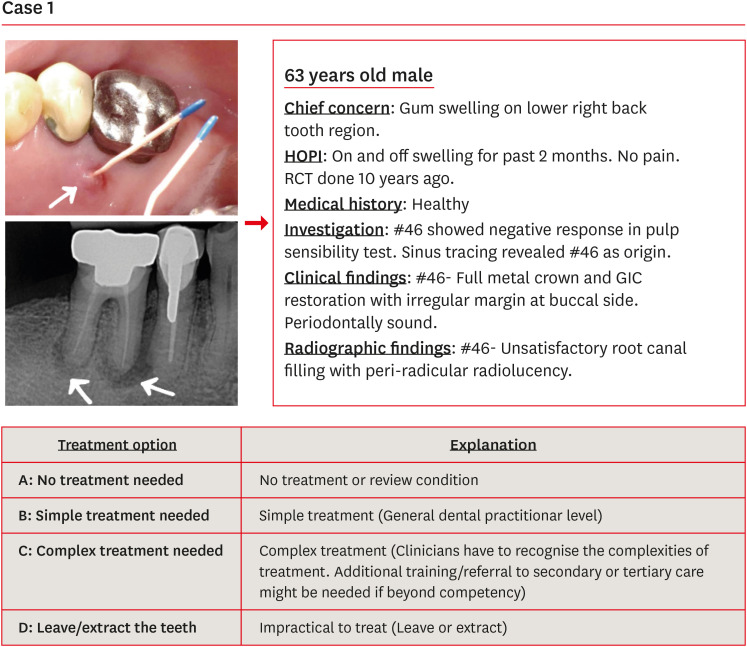
Objectives The purpose of this study was to evaluate the effect of Dental Practicality Index (DPI) training using an online video on the treatment planning decisions and confidence level of dental undergraduates (DUs).
Materials and Methods Ninety-four DUs were shown 15 clinical case scenarios and asked to decide on treatment plans based on 4 treatment options. The most appropriate treatment plan had been decided by a consensus panel of experienced dentists. DUs then underwent DPI training using an online video. In a post-DPI-training test, DUs were shown the same clinical case scenarios and asked to assign the best treatment option. After 6 weeks, DUs were retested to assess their knowledge retention. In all 3 tests, DUs completed the confidence level scale questionnaire. Data were analyzed using the related-samples Wilcoxon signed rank test and the independent-samples Mann-Whitney
U test with the level of significance set atp < 0.05.Results DPI training significantly improved the mean scores of the DUs from 7.53 in the pre-DPI-training test to 9.01 in the post-DPI-training test (
p < 0.001). After 6 weeks, the mean scores decreased marginally to 8.87 in the retention test (p = 0.563). DPI training increased their confidence level from 5.68 pre-DPI training to 7.09 post-DPI training.Conclusions Training DUs using DPI with an online video improved their decision-making and confidence level in treatment planning.
-
Citations
Citations to this article as recorded by- STUDY OF THE EFFECTIVENESS OF THE USE OF DIRECT AND INDIRECT RESTORATION OVER TIME IN THE TREATMENT OF DEFECTS OF HARD DENTAL TISSUES AFTER ENDODONTIC INTERVENTION
V. V. Fedoriuk, М. М. Rozhko
Art of Medicine.2025; : 94. CrossRef
- STUDY OF THE EFFECTIVENESS OF THE USE OF DIRECT AND INDIRECT RESTORATION OVER TIME IN THE TREATMENT OF DEFECTS OF HARD DENTAL TISSUES AFTER ENDODONTIC INTERVENTION
- 3,800 View
- 79 Download
- 1 Crossref
- Color discrepancy of single-shade composites at different distances from the interface measured using cell phone images
- Márcia Luciana Carregosa Santana, Gabriella de Jesus Santos Livi, André Luis Faria-e-Silva
- Restor Dent Endod 2024;49(1):e7. Published online January 24, 2024
- DOI: https://doi.org/10.5395/rde.2024.49.e7
-
Abstract
PDFPubReaderePub
Objectives This study aimed to evaluate the impact of substrate color and interface distance on the color adjustment of 2 single-shade composites, Vittra APS Unique and Charisma Diamond One.
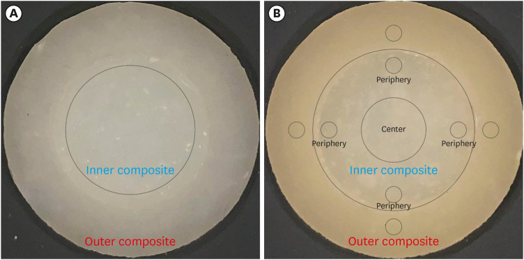
Materials and Methods Dual disc-shaped specimens were created using Vittra APS Unique or Charisma Diamond One as the center composite, surrounded by shaded composites (A1 or A3). Color measurements were taken with a spectrophotometer against a gray background, recording the color coordinates in the CIELAB color space. Illumination with a light-correcting device and image acquisition using a polarizing filter-equipped cell phone were performed on specimens over the same background. Image processing software was used to measure the color coordinates in the center and periphery of the inner composite and in the outer composite. The color data were then converted to CIELAB coordinates and adjusted using data from the spectrophotometer. Color differences (ΔE00) between the center/periphery of single-shade and outer composites were calculated, along with color changes in single-shade composites caused by different outer composites. Color differences for the inner composites surrounded by A1 and A3 were also calculated. Data were analyzed using repeated-measures analysis of variance (α = 0.05).
Results The results showed that color discrepancies were lowest near the interface and when the outer composite was whiter (A1). Additionally, Charisma Diamond One exhibited better color adjustment ability than Vittra APS Unique.
Conclusions Color discrepancies between the investigated single-shade composites diminished towards the interface with the surrounding composite, particularly when the latter exhibited a lighter shade.
-
Citations
Citations to this article as recorded by- Evaluation of color stability in single-shade composite resins using spectrophotometer and cross-polarized mobile photography
Hatice Tepe, Ozge Celiksoz, Batu Can Yaman
BMC Oral Health.2025;[Epub] CrossRef - Comparative Evaluation of the Staining Resistance of Two Single-Shade Composites in Coffee and Chlorhexidine: A Spectrophotometric Analysis
Unmesh Khanvilkar, Shrinath D Kulkarni, Siddhesh Bandekar, Ved M Talathi, Oshin Baghel, Priyanka Razdan, Seema Gupta
Cureus.2025;[Epub] CrossRef - Clinical Implications of Color Adjustment in Single-Shade Resins Post-Dental Bleaching: A Systematic Review
Samille Biasi Miranda, Caroline de Farias Charamba Leal, Rodrigo Barros Esteves Lins, Marcos Antonio Japiassu Resende Montes
Journal of Clinical Medicine.2025; 14(9): 3194. CrossRef - Accuracy and Reliability of Smartphone Versus Mirrorless Camera Images-Assisted Digital Shade Guides: An In Vitro Study
Soo Teng Chew, Suet Yeo Soo, Mohd Zulkifli Kassim, Khai Yin Lim, In Meei Tew
Applied Sciences.2025; 15(14): 8070. CrossRef
- Evaluation of color stability in single-shade composite resins using spectrophotometer and cross-polarized mobile photography
- 3,273 View
- 89 Download
- 3 Web of Science
- 4 Crossref
- Prevalence of salivary microbial load and lactic acid presence in diabetic and non-diabetic individuals with different dental caries stages
- Monika Mohanty, Shashirekha Govind, Shakti Rath
- Restor Dent Endod 2024;49(1):e4. Published online January 12, 2024
- DOI: https://doi.org/10.5395/rde.2024.49.e4
-
Abstract
PDFPubReaderePub
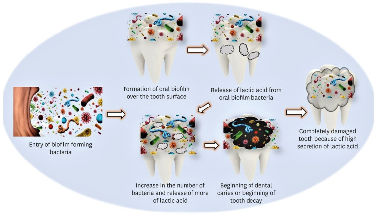
Objectives This study aims to correlate caries-causing microorganism load, lactic acid estimation, and blood groups to high caries risk in diabetic and non-diabetic individuals and low caries risk in healthy individuals.
Materials and Methods This study includes 30 participants divided into 3 groups: Group A, High-risk caries diabetic individuals; Group B, High-risk caries non-diabetic individuals; and Group C, Low-risk caries individuals. The medical condition, oral hygiene, and caries risk assessment (American Dental Association classification and International Caries Detection and Assessment System scoring) were documented. Each individual’s 3 mL of saliva was analyzed for microbial load and lactic acid as follows: Part I: 2 mL for microbial quantity estimation using nutrient agar and blood agar medium, biochemical investigation, and carbohydrate fermentation tests; Part II: 0.5 mL for lactic acid estimation using spectrophotometric analysis. Among the selected individuals, blood group correlation was assessed. The χ2 test, Kruskal-Wallis test, and
post hoc analysis were done using Dunn’s test (p < 0.05).Results Group A had the highest microbial load and lactic acid concentration, followed by Groups B and C. The predominant bacteria were
Lactobacilli (63.00 ± 15.49) andStreptococcus mutans (76.00 ± 13.90) in saliva. Blood Group B is prevalent in diabetic and non-diabetic high-risk caries patients but statistically insignificant.Conclusions Diabetic individuals are more susceptible to dental caries due to high microbial loads and increased lactic acid production. These factors also lower the executing tendency of neutrophils, which accelerates microbial accumulation and increases the risk of caries in diabetic individuals.
-
Citations
Citations to this article as recorded by- Oral Health Disparities in Type 2 Diabetes: Examining the Elevated Risk for Dental Caries—A Comparative Study
José Frias-Bulhosa, Maria Conceição Manso, Carla Lopes Mota, Paulo Melo
Dentistry Journal.2025; 13(6): 258. CrossRef - Exploring the photosensitizing potential of Nanoliposome Loaded Improved Toluidine Blue O (NLITBO) Against Streptococcus mutans: An in-vitro feasibility study
Swagatika Panda, Lipsa Rout, Neeta Mohanty, Anurag Satpathy, Bhabani Sankar Satapathy, Shakti Rath, Divya Gopinath, Geelsu Hwang
PLOS ONE.2024; 19(10): e0312521. CrossRef - Altered salivary microbiota associated with high-sugar beverage consumption
Xiaozhou Fan, Kelsey R. Monson, Brandilyn A. Peters, Jennifer M. Whittington, Caroline Y. Um, Paul E. Oberstein, Marjorie L. McCullough, Neal D. Freedman, Wen-Yi Huang, Jiyoung Ahn, Richard B. Hayes
Scientific Reports.2024;[Epub] CrossRef
- Oral Health Disparities in Type 2 Diabetes: Examining the Elevated Risk for Dental Caries—A Comparative Study
- 4,290 View
- 101 Download
- 3 Web of Science
- 3 Crossref
- Can different agents reduce the damage caused by bleaching gel to pulp tissue? A systematic review of basic research
- Letícia Aparecida Silva Batista, Alexandre Henrique dos Reis-Prado, Hebertt Gonzaga dos Santos Chaves, Lara Cancella de Arantes, Luís Fernando Santos Alves Morgan, Carolina Bosso André, Thaís Yumi Suzuki, Francine Benetti
- Restor Dent Endod 2023;48(4):e39. Published online November 6, 2023
- DOI: https://doi.org/10.5395/rde.2023.48.e39
-
Abstract
PDFSupplementary MaterialPubReaderePub
Objectives This study aimed to investigate the effectiveness of different topical/systemic agents in reducing the damage caused by bleaching gel to pulp tissue or cells.
Materials and Methods Electronic searches were performed in July 2023.
In vivo andin vitro studies evaluating the effects of different topical or systemic agents on pulp inflammation or cytotoxicity after exposure to bleaching agents were included. The risk of bias was assessed.Results Out of 1,112 articles, 27 were included. Nine animal studies evaluated remineralizing/anti-inflammatories agents in rat molars subjected to bleaching with 35%–38% hydrogen peroxide (HP). Five of these studies demonstrated a significant reduction in inflammation caused by HP when combined with bioglass or MI Paste Plus (GC America), or following KF-desensitizing or Otosporin treatment (
n = 3). However, orally administered drugs did not reduce pulp inflammation (n = 4). Cytotoxicity (n = 17) was primarily assessed using the 3-(4,5-dimethylthiazol-2-yl)-2,5-diphenyltetrazolium bromide assay on human dental pulp cells and mouse dental papilla Cell-23 cells. Certain substances, including sodium ascorbate, butein, manganese chloride, and peroxidase, were found to reduce cytotoxicity, particularly when applied prior to bleaching. The risk of bias was high in animal studies and low in laboratory studies.Conclusions Few
in vivo studies have evaluated agents to reduce the damage caused by bleaching gel to pulp tissue. Within the limitations of these studies, it was found that topical agents were effective in reducing pulp inflammation in animals and cytotoxicity. Further analyses with human pulp are required to substantiate these findings.Trial Registration PROSPERO Identifier:
CRD42022337192 -
Citations
Citations to this article as recorded by- 3D-Printed and Bioprinted Scaffolds in Regenerative Endodontics: A Systematic Review
Hebertt Gonzaga dos Santos Chaves, Diana B. Sequeira, Vilton Cardozo Moreira Dias, Alberto Cabrera-Fernández, João Peça, Francine Benetti, João Miguel Marques dos Santos
Applied Sciences.2026; 16(8): 3940. CrossRef - Clinical Study on the Efficacy of 35% Hydrogen Peroxide Gel According to Exposure Time (40 min vs. 20 min) by Spectrophotometry
Trinidad Rincón, Maria Portillo Muñoz, Maria Lobato, Ana María Martín Casado, Laryssa Mylenna Madruga Barbosa, Alessandro Loguercio, Cristina Gómez‐Polo
Journal of Esthetic and Restorative Dentistry.2026;[Epub] CrossRef - Clareamento dental e TikTok: avaliação da qualidade do conteúdo em mídia social
Rafaele T Costa, Thayna Silva do Carmo Tavares, André Walsh-Monteiro
Ciência ET Praxis.2025; 21(36): 111. CrossRef - Synthesis, characterization and evaluation of novel bleaching gels containing bioactive glass and nano-hydroxyapatite on hydrogen peroxide diffusion, bleaching efficacy and enamel protection
Adrieli Burey, Byron Carpio-Salvatierra, Michael Favoretto, María Luján Méndez Bauer, Viviane Hass, Alessandra Reis, Alessandro D. Loguercio, Paulo Vitor Farago
Clinical Oral Investigations.2025;[Epub] CrossRef - Cytotoxicity of Bleaching Products: A Systematic Review
Mireia Montaner, José Luis Sanz, Carmen Llena, María Melo, Clara Puig-Herreros, James Ghilotti
Applied Sciences.2024; 14(9): 3680. CrossRef
- 3D-Printed and Bioprinted Scaffolds in Regenerative Endodontics: A Systematic Review
- 4,441 View
- 60 Download
- 4 Web of Science
- 5 Crossref
- Evaluation of at-home bleaching protocol with application on different surfaces: bleaching efficacy and hydrogen peroxide permeability
- Heloisa Forville, Michael Willian Favoreto, Michel Wendlinger, Roberta Micheten Dias, Christiane Philippini Ferreira Borges, Alessandra Reis, Alessandro D. Loguercio
- Restor Dent Endod 2023;48(4):e33. Published online October 6, 2023
- DOI: https://doi.org/10.5395/rde.2023.48.e33
-
Abstract
PDFPubReaderePub
Objectives This study aimed to evaluate the bleaching efficacy and hydrogen peroxide permeability in the pulp chamber by the at-home bleaching gel in protocols applied on different dental surfaces.
Materials and Methods Forty premolars were randomly into 4 groups: control group no bleaching, only application on the buccal surface (OB), only application on the lingual surface (OL) and application in buccal and lingual surfaces, simultaneously (BL). At-home bleaching gel (White Class 7.5%) was used for the procedure. The bleaching efficacy was evaluated with a digital spectrophotometer (color change in CIELAB [Δ
E ab] and CIEDE 2000 [ΔE 00] systems and Whitening Index for Dentistry [ΔWID]). The hydrogen peroxide permeability in the pulp chamber (µg/mL) was assessed using UV-Vis spectrophotometry and data were analyzed for a 1-way analysis of variance and Tukey’s test (α = 0.05).Results All groups submitted to bleaching procedure showed bleaching efficacy when measured with Δ
E ab and ΔE 00 (p > 0.05). Therefore, when analyzed by ΔWID, a higher bleaching efficacy were observed for the application on the groups OB and BL (p = 0.00003). Similar hydrogen peroxide permeability was found in the pulp chambers of the teeth undergoing different protocols (p > 0.05).Conclusions The application of bleaching gel exclusively on the OB is sufficient to achieve bleaching efficacy, when compared to BL. Although the OL protocol demonstrated lower bleaching efficacy based on the ΔWID values, it may still be of interest and relevant in certain clinical scenarios based on individual needs, requiring clinical trials to better understand its specificities.
-
Citations
Citations to this article as recorded by- Effect of whitening pens on hydrogen peroxide permeability in the pulp chamber, color change and surface morphology
Laryssa Mylenna Madruga Barbosa, Gabrielle Gomes Centenaro, Deisy Cristina Ferreira Cordeiro, Maria Alice de Matos Rodrigues, Letícia Condolo, Michael Willian Favoreto, Alessandra Reis, Alessandro D. Loguercio
Journal of Dentistry.2025; 154: 105595. CrossRef - Evaluation of bleaching efficiency of carbamide peroxide applied on different dental surfaces: An in vitro study
R. Gokulnath, R. S. Mohan Kumar, A. Jayasenthil, R. Anjana, G. Sree Vidya
Journal of Conservative Dentistry and Endodontics.2025; 28(4): 366. CrossRef - Characterization and effects on enamel of low-concentration bleaching gels containing hyaluronic acid, NF_TiO2 nanoparticles and irradiated with violet LED light
Marcos Roberto Lima Benati, Matheus Kury, Priscila Borges Gobbo de Melo, Iago César Ribeiro Teles Matos, Roberta Tarkany Basting, Rosanna Tarkany Basting, Fernando Luis Esteban Florez, Vanessa Cavalli
Clinical Oral Investigations.2025;[Epub] CrossRef - Impact of bleaching on white spot lesions: hydrogen peroxide permeability and color alteration
Laryssa Mylenna Madruga Barbosa, Bruno Baracco, Taynara S. Carneiro, Michael Willian Favoreto, Michel Wendlinger, Daniel Jiménez-Díez, Laura Ceballos, Alessandro D. Loguercio
Clinical Oral Investigations.2025;[Epub] CrossRef - Efficacy of a buccal and lingual at‐home bleaching protocol—A randomized, split‐mouth, single‐blind controlled trial
Heloisa Forville, Laís Giacomini Bernardi, Michael Willian Favoreto, Felipe Coppla, Taynara de Souza Carneiro, Fabiana Madalozzo Coppla, Alessandro D. Loguercio, Alessandra Reis
Journal of Esthetic and Restorative Dentistry.2024; 36(9): 1301. CrossRef - REANATOMIZAÇÃO DE DENTE CONOIDE ASSOCIADA A ESTÉTICA VERMELHA: RELATO DE CASO
Ana Karolayne Sousa de Morais, Daniele Fernanda Sousa Barros, Daniel Messias Limeira, Rhana Leticia de Oliveira Faria, Roberta Furtado Carvalho, Sandna Nolêto de Araújo, Laura Barbosa Santos Di Milhomem
Revista Contemporânea.2024; 4(10): e6299. CrossRef - Effect of the reduction in the exposure time to at-home bleaching gel on color change and tooth sensitivity: A systematic review and meta-analysis
Priscila Borges Gobbo de Melo, Letícia Vasconcelos Silva Souza, Lucianne Cople Maia, Guido Artemio Marañón-Vásquez, Matheus Kury, Vanessa Cavalli
Clinical Oral Investigations.2024;[Epub] CrossRef
- Effect of whitening pens on hydrogen peroxide permeability in the pulp chamber, color change and surface morphology
- 5,345 View
- 91 Download
- 5 Web of Science
- 7 Crossref
Case Report
- Ingestion and surgical retrieval of an endodontic file: a case report
- Devon Marta Ptak, Elinor Alon, Robert Bruce Amato, Julia Tassinari, Adrian Velasquez
- Restor Dent Endod 2023;48(4):e32. Published online September 2, 2023
- DOI: https://doi.org/10.5395/rde.2023.48.e32
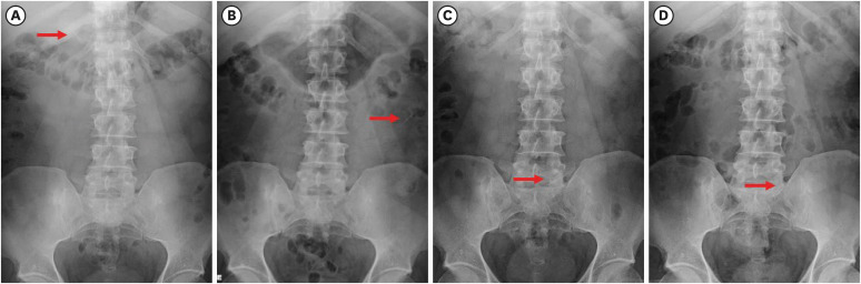
-
Abstract
PDFPubReaderePub
Ingestions and aspirations of foreign bodies are rare, but do occasionally occur during dental treatment. Although reports exist, few include photos demonstrating the extensive surgical intervention that may be necessary to manage such events. Perhaps this lack of visualization, and associated lack of awareness, is one of the reasons some clinicians still provide non-surgical root canal therapy (NSRCT) without a rubber dam. This case report outlines the medical treatment of a 30-year-old male who initially presented to a general dentist’s office (not associated with the authors) for NSRCT of their mandibular right first molar. A rubber dam was not used for this procedure, during which the accidental ingestion of an endodontic K-file occurred. The patient was subsequently hospitalized for evaluation and treatment, consisting of numerous imaging studies, endoscopic evaluation, and surgical removal of the file from his small intestine. The ingestion of foreign bodies, and the associated complications, can be reduced through the routine use of a rubber dam, which is considered the standard of care for NSRCT. This case graphically illustrates the potential consequences associated with deviating from the standard of care and should remind clinicians that a rubber dam is necessary for all cases of NSRCT.
-
Citations
Citations to this article as recorded by- Advantages and limitations of the rubber dam. Part 2: advantages of rubber dam use
Artemy Vadimovich Karmanov, Alena Sergeevna Silkina, Natalia Alexandrovna Sokolovich
Dental Update.2026; 53(3): 198. CrossRef - Anesthesiological Management of a Sewing Needle Impacted in the Larynx in an Adult: A Case Report
Stefanie R. Senn, Felix C. Jansen, Caveh Madjdpour, Isabel Besozzi, Daniel A. Button
Clinical Case Reports.2026;[Epub] CrossRef - Dental Dam Isolation for Crown Removal, Atraumatic Tooth Extraction, Immediate Implant Placement, and Restoration Cementation: A Case Study
G Guzman-Perez, S Rojas-Rueda, F Floriani, A Unnadkat, C-C Fu, CA Jurado
Operative Dentistry.2025; 50(1): 5. CrossRef - Patient and Operator Experiences with Conventional Rubber Dam and OptiDam: A Randomized Clinical Trial
Rashed F. Binqali, Abdulwahab M. Alghamdi, Mishal S. Aloufi, Suliman A. Alharbi, Omair M. Bukhari, Reham M. Alsamman
Journal of International Society of Preventive and Community Dentistry.2025; 15(6): 554. CrossRef
- Advantages and limitations of the rubber dam. Part 2: advantages of rubber dam use
- 6,050 View
- 128 Download
- 2 Web of Science
- 4 Crossref
Research Articles
- Incidence of interappointment emergencies in multiple-visit root canal treatments performed with or without intracanal medicament by undergraduate students
- Annemarie Baaij, Corine Mirjam Visscher, Manon Jansen, Ahmet Rifat Özok
- Restor Dent Endod 2023;48(3):e31. Published online August 14, 2023
- DOI: https://doi.org/10.5395/rde.2023.48.e31
-
Abstract
PDFPubReaderePub
Objectives This retrospective cohort study examined the incidence of interappointment emergencies during multiple-visit molar root canal treatments conducted by undergraduate students. Treatments performed without the use of intracanal medicament were compared to treatments that incorporated calcium hydroxide as an intracanal medicament.
Materials and Methods Interappointment emergencies, defined as instances of pain or swelling that required the patient to make an unscheduled follow-up visit, were recorded for up to 2 months after the intervention. To avoid the influence of obturation on the observed incidence of emergency visits, only unscheduled visits occurring between the start and end of the root canal treatment were included.
Results Of the 719 patients included in this study, 77 (10.7%) were recorded as experiencing interappointment emergencies. Of these emergencies, 62% occurred within 2 weeks following the most recent intervention. In the group of patients who did not receive intracanal medicament, the incidence of interappointment emergencies was 11.9% (46 of 385 patients). In comparison, this rate was 9.3% (31 of 334 patients) among those who received calcium hydroxide as an intracanal medicament (odds ratio, 1.33; 95% confidence interval, 0.82–2.15;
p = 0.249).Conclusions Interappointment emergencies may arise at any point during root canal treatment, but they most commonly occur within the first 2 weeks following intervention. The omission of intracanal medicament in multiple-visit molar root canal treatments, performed by undergraduate students, did not significantly increase the incidence of these emergencies.
-
Citations
Citations to this article as recorded by- An Observational Study on Pain Occurrence After Root Canal Treatment: Role of Operator Experience When Using a Bioceramic Sealer
Mihai Merfea, Ioana Sofia Pop-Ciutrila, Mindra Eugenia Badea, Ada Gabriela Delean, Oana Cimponeriu, Razvan Corneliu Pop, Maria Peter, Iulia Clara Badea, Sanda Ileana Cimpean
Journal of Clinical Medicine.2025; 14(13): 4558. CrossRef
- An Observational Study on Pain Occurrence After Root Canal Treatment: Role of Operator Experience When Using a Bioceramic Sealer
- 5,720 View
- 54 Download
- 1 Web of Science
- 1 Crossref
- Tip and taper compatibility of accessory gutta-percha points with rotary and reciprocating instruments
- Júlia Niero Zanatta Streck, Sabrina Arcaro, Renan Antônio Ceretta, Eduardo Antunes Bortoluzzi, Lucas da Fonseca Roberti Garcia, Josiane de Almeida, Patrícia Maria Poli Kopper, Anarela Vassen Bernardi
- Restor Dent Endod 2023;48(3):e22. Published online June 8, 2023
- DOI: https://doi.org/10.5395/rde.2023.48.e22
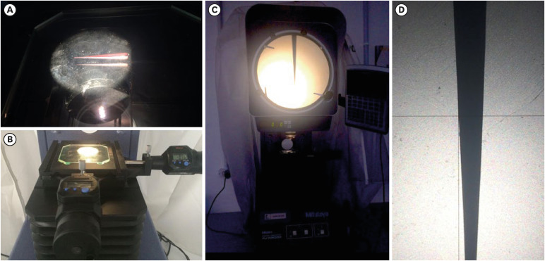
-
Abstract
PDFPubReaderePub
Objectives This study was conducted to evaluate and compare the tip and taper compatibility of accessory gutta-percha points (AGPs) with various rotary and reciprocating instruments.
Materials and Methods Using a profile analyzer, tip and taper measurements were taken of 10 AGPs of each of the 14 models available from Odous de Deus and the 4 models available from Dentsply-Maillefer. Diameter measurements were taken at 1-mm intervals, from 3 mm from the tip (D3) to 16 mm.
Results Based on the mean values obtained, 3-dimensional (3D) models of the AGPs were drawn in Autodesk Fusion 360 and superimposed on 3D models of each instrument selected (Mtwo, Reciproc, RaCe, K3, and ProDesign Logic) to determine the compatibility between the instrument and the AGP. Data corresponding to the tips and tapers of the various AGPs, as well as the tip and taper differences between the AGPs and the instruments, were analyzed using descriptive statistics. The tapers of the AGPs were subject to the American National Standards Institute/American Dental Association No. 57 standard. The Odous de Deus extra-long medium and extra-long extra-medium AGPs were shown to be compatible with Mtwo, K3, and ProDesign Logic instruments with taper 0.06 and tip sizes 25 and 30, while the Dentsply fine and fine medium cones were compatible with Mtwo, RaCe, and K3 instruments with conicity of 0.04 and tip sizes 35 and 40.
Conclusions Both the Odous de Deus and Dentsply commercial brands included 2 AGP models with tip (D3) and taper compatibility with Mtwo, RaCe, K3, and/or Prodesign Logic instruments.
- 3,774 View
- 53 Download
Review Article
- Stem cell-derived exosomes for dentin-pulp complex regeneration: a mini-review
- Dina A. Hammouda, Alaa M Mansour, Mahmoud A. Saeed, Ahmed R. Zaher, Mohammed E. Grawish
- Restor Dent Endod 2023;48(2):e20. Published online May 3, 2023
- DOI: https://doi.org/10.5395/rde.2023.48.e20
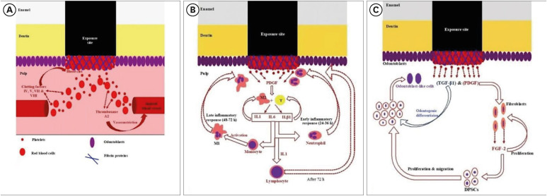
-
Abstract
PDFPubReaderePub
This mini-review was conducted to present an overview of the use of exosomes in regenerating the dentin-pulp complex (DPC). The PubMed and Scopus databases were searched for relevant articles published between January 1, 2013 and January 1, 2023. The findings of basic
in vitro studies indicated that exosomes enhance the proliferation and migration of mesenchymal cells, as human dental pulp stem cells, via mitogen-activated protein kinases and Wingless-Int signaling pathways. In addition, they possess proangiogenic potential and contribute to neovascularization and capillary tube formation by promoting endothelial cell proliferation and migration of human umbilical vein endothelial cells. Likewise, they regulate the migration and differentiation of Schwann cells, facilitate the conversion of M1 pro-inflammatory macrophages to M2 anti-inflammatory phenotypes, and mediate immune suppression as they promote regulatory T cell conversion. Basicin vivo studies have indicated that exosomes triggered the regeneration of dentin-pulp–like tissue, and exosomes isolated under odontogenic circumstances are particularly strong inducers of tissue regeneration and stem cell differentiation. Exosomes are a promising regenerative tool for DPC in cases of small pulp exposure or for whole-pulp tissue regeneration.-
Citations
Citations to this article as recorded by- Extracellular vesicles derived from dental mesenchymal stem cells for regenerative medicine: a scoping review
Maria Emília Mota, Márcia Martins Marques, Thaís Gimenez, Suely Kunimi Kubo Ariga, Tiago Góss dos Santos, Fábio Abreu Alves, Maria Stella Moreira
Molecular Biology Reports.2026;[Epub] CrossRef - Stem cell-derived exosomes in tissue regeneration of oral and maxillofacial region: A systematic review
Qianlei Dai, Qianfen Dai
Medicine.2026; 105(1): e46948. CrossRef - Stem Cell-Derived Exosomes in Wound Healing and Skin Regeneration: Emerging Therapeutic Strategies and Mechanisms
Nithin Vidiyala, Pavani Sunkishala, Prashanth Reddy Parupathi, Dinesh Nyavanandi
Cells.2026; 15(10): 872. CrossRef - REGENERATIVE POTENTIAL OF DENTAL STEM CELLS IN DENTISTRY: FROM BIOCHEMICAL MECHANISMS TO CLINICAL APPLICATIONS
A.Ye. Demkovych, O.Z. Yaremchuk, M.I. Kulitska, N.B. Hlyvka, V.Ya. Krupei, O.Ya. Lavrin
Ukrainian Dental Almanac.2026; (2): 5. CrossRef - Dental stem cell-derived extracellular vesicles for oral tissue regeneration: Mechanisms, engineering strategies, and clinical translation
Yang Liu, Peiyao Ge, Yuzhuo Ma, Haizhou Fu
Archives of Oral Biology.2026; 190: 106676. CrossRef - Cell Homing Strategies in Regenerative Endodontic Therapy
David Kim, Sahng G. Kim
Cells.2025; 14(3): 201. CrossRef - Impact of dental pulp cells-derived small extracellular vesicles on the properties and behavior of dental pulp cells: an in-vitro study
Dina A. Hammouda, Alaa M. Mansour, Ahmed R. Zaher, Mohammed E. Grawish
BMC Oral Health.2025;[Epub] CrossRef - Methodological Approaches for Economic Comparison of Mesenchymal Stem Cell and Exosome-based Therapies with Conventional Endodontic Treatments in Regenerative Endodontics
Madina A. Kurmanalina Kurmanalina, Nadiar M. Mussin, Aigul M. Sumanova, Violetta R. Detochkina, Maryam Mardani, Nader Tanideh, Amin Tamadon
West Kazakhstan Medical Journal.2025; 67(2): 188. CrossRef - Exosomal circ_0003057 promotes osteo/odontogenic differentiation of hDPSCs by binding with EIF4A3 through upregulated parental gene ANKH
Bingtao Wang, Yuanyuan Kong, Huixian Dong, Feng Lai, Zixin Guo, Liecong Lin, Jingyi Xu, Jingkun Zhang, Yiguo Jiang, Qianzhou Jiang
International Endodontic Journal.2025; 58(9): 1433. CrossRef - Mechanistic insights into dental stem cells‐derived exosomes in regenerative endodontics
Paras Ahmad, Nathan Estrin, Nima Farshidfar, Yufeng Zhang, Richard J. Miron
International Endodontic Journal.2025; 58(9): 1384. CrossRef - Development and characterization of an exosome-loaded biomimetic hydroxyapatite/gelatin scaffold for enhanced dental pulp regeneration
Yuen-Shan Tsai, Shih-Jung Cheng, Tsao-Li Chuang, Shu-Fang Chang, Feng-Huei Lin, Chun-Pin Lin
Journal of Dental Sciences.2025;[Epub] CrossRef - Exosomes as Promising Therapeutic Tools for Regenerative Endodontic Therapy
Qingyue Kong, Yujie Wang, Nan Jiang, Yifan Wang, Rui Wang, Xiaohan Hu, Jing Mao, Xin Shi
Biomolecules.2024; 14(3): 330. CrossRef - Role and Molecular Mechanism of miR-586 in the Differentiation of Dental Pulp Stem Cells into Odontoblast-like Cells
Gang Pan, Qianwen Zhou, Chenhua Pan, Yingxue Zhang
Cell Biochemistry and Biophysics.2024; 83(1): 507. CrossRef
- Extracellular vesicles derived from dental mesenchymal stem cells for regenerative medicine: a scoping review
- 11,393 View
- 150 Download
- 10 Web of Science
- 13 Crossref
Research Articles
- Effects of CTHRC1 on odontogenic differentiation and angiogenesis in human dental pulp stem cells
- Jong-soon Kim, Bin-Na Lee, Hoon-Sang Chang, In-Nam Hwang, Won-Mann Oh, Yun-Chan Hwang
- Restor Dent Endod 2023;48(2):e18. Published online April 28, 2023
- DOI: https://doi.org/10.5395/rde.2023.48.e18
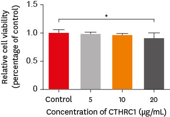
-
Abstract
PDFPubReaderePub
Objectives This study aimed to determine whether collagen triple helix repeat containing-1 (CTHRC1), which is involved in vascular remodeling and bone formation, can stimulate odontogenic differentiation and angiogenesis when administered to human dental pulp stem cells (hDPSCs).
Materials and Methods The viability of hDPSCs upon exposure to CTHRC1 was assessed with the WST-1 assay. CTHRC1 doses of 5, 10, and 20 µg/mL were administered to hDPSCs. Reverse-transcription polymerase reaction was used to detect dentin sialophosphoprotein, dentin matrix protein 1, vascular endothelial growth factor, and fibroblast growth factor 2. The formation of mineralization nodules was evaluated using Alizarin red. A scratch wound assay was conducted to evaluate the effect of CTHRC1 on cell migration. Data were analyzed using 1-way analysis of variance followed by the Tukey
post hoc test. The threshold for statistical significance was set atp < 0.05.Results CTHRC1 doses of 5, 10, and 20 µg/mL had no significant effect on the viability of hDPSCs. Mineralized nodules were formed and odontogenic markers were upregulated, indicating that CTHRC1 promoted odontogenic differentiation. Scratch wound assays demonstrated that CTHRC1 significantly enhanced the migration of hDPSCs.
Conclusions CTHRC1 promoted odontogenic differentiation and mineralization in hDPSCs.
- 2,497 View
- 40 Download
- Effects of surrounding and underlying shades on the color adjustment potential of a single-shade composite used in a thin layer
- Mariana Silva Barros, Paula Fernanda Damasceno Silva, Márcia Luciana Carregosa Santana, Rafaella Mariana Fontes Bragança, André Luis Faria-e-Silva
- Restor Dent Endod 2023;48(1):e7. Published online December 29, 2022
- DOI: https://doi.org/10.5395/rde.2023.48.e7
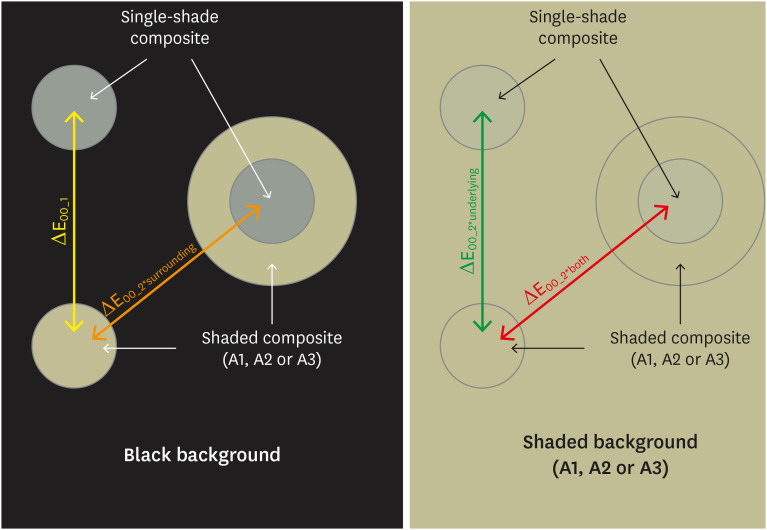
-
Abstract
PDFPubReaderePub
Objectives This study aimed to evaluate the surrounding and underlying shades’ effect on the color adjustment potential (CAP) of a single-shade composite used in a thin layer.
Materials and Methods Cylinder specimens (1.0 mm thick) were built with the Vittra APS Unique composite, surrounded (dual specimens) or not (simple specimens) by a control composite (shade A1, A2, or A3). Simple specimens were also built only with the control composites. Each specimen’s color was measured against white and black backgrounds or the simple control specimens with a spectrophotometer (CIELAB system). The whiteness index for dentistry (WID) and translucency parameters (TP00) were calculated for simple specimens. Differences (ΔE00) in color between the simple/dual specimens and the controls were calculated. The CAP was calculated based on the ratios between data from simple and dual specimens.
Results The Vittra APS Unique composite showed higher WID and TP00 values than the controls. The highest values of ΔE00 were observed among simple specimens. The color measurements of Vittra APS Unique (simple or dual) against the control specimens presented the lowest color differences. Only surrounding the single-shade composite with a shaded composite barely impacted the ΔE00. The highest CAP values were obtained using a shaded composite under simple or dual specimens.
Conclusions The CAP of Vittra APS Unique was strongly affected by the underlying shade, while surrounding this composite with a shaded one barely affected its color adjustment.
-
Citations
Citations to this article as recorded by- Impact of water absorption on the translucency of single-shade and conventional resin composites: an in vitro comparative study
Ceyda Sari, Elifnur Aydemir Aydın
Odontology.2026;[Epub] CrossRef - Baseline color-matching in anterior non-carious cervical lesions of patients of two single-shade resin composites: a randomized clinical trial
Ayşe Nur Doğan, Soley Arslan
Odontology.2026;[Epub] CrossRef - Evaluation of color blending effect of a single-shade resin composite with hybrid ceramics: an in vitro study
Jongchan Lee, Jinsoo Ahn, Sun-Young Kim
BMC Oral Health.2026;[Epub] CrossRef - Color Change and Compressive Strength of Novel Single-Shade Composite Resins Exposed to Staining Beverages
Juan P. Molina-Gantiva, Midian C. Castillo-Pedraza, Jorge H. Wilches-Visbal
Odovtos - International Journal of Dental Sciences.2026;[Epub] CrossRef - At‐Home and In‐Office Bleaching Protocols on the Color Match of Restorations Made With Single‐Shade Composites
Luciana Vasconcelos Ramos, Dayana Fernandes Rocha Aparicio, André Luis Faria‐e‐Silva, Maíra do Prado, Andréa Vaz Braga Pintor, Marcela Baraúna Magno
Journal of Esthetic and Restorative Dentistry.2025; 37(6): 1567. CrossRef - Evaluation of color matching of three single-shade composites employing simulated 3D printed cavities with different thicknesses using CIELAB and CIEDE2000 color difference formulae
Engin Kariper, Aylin Cilingir
REVIEWS ON ADVANCED MATERIALS SCIENCE.2025;[Epub] CrossRef - Impact of kombucha, coffee, and turmeric beverages on the color stability of a single-shade versus a multi-shade resin-based composite
Hanin E. Yeslam, Abdulaziz F. Bakhsh
PeerJ.2025; 13: e19759. CrossRef - Comparative Study of Esthetic Outcome of Pedo Shades of Composite Resin—A Randomized Controlled Trial: In Vivo and In Vitro Study
Priyanka Raj, Shikha Choubey, Divya Doneria, Diksha Bhat, Shivani Mathur, Shailja Sinha
International Journal of Clinical Pediatric Dentistry.2025; 18(S1): S22. CrossRef - Influence of cavity wall thickness on the color adjustment potential of single-shade resin composites
Fabrício Luscino Alves de Castro, Letícia Brandão Durand
The Journal of the American Dental Association.2024; 155(7): 605. CrossRef - Assessing color mismatch in single-shade composite resins for enamel replacement
Rafaella Mariana Fontes de Bragança, Diana Leyva Del Rio, Luiz Alves Oliveira-Neto, William Michael Johnston
The Journal of Prosthetic Dentistry.2024; 132(3): 613.e1. CrossRef - Color discrepancy of single-shade composites at different distances from the interface measured using cell phone images
Márcia Luciana Carregosa Santana, Gabriella de Jesus Santos Livi, André Luis Faria-e-Silva
Restorative Dentistry & Endodontics.2024;[Epub] CrossRef - Is It Possible for Single-shade Composites to Mimic the Color, Lightness, Chroma, and Hue of Other Single-shade Composites? An In Vitro Study
M Buldur, G Ayan
Operative Dentistry.2024; 49(6): 691. CrossRef - Color evaluation of a one-shade used for restoration of non-carious cervical lesions: an equivalence randomized clinical trial
Michael Willian Favoreto, Amanda de Oliveira de Miranda, Thalita P. Matos, Andrea dos Santos de Castro, Mylena de Abreu Cardoso, Julia Beatriz, Jenny Collantes-Acuña, Alessandra Reis, Alessandro Dourado Loguercio
BMC Oral Health.2024;[Epub] CrossRef - Influence of Thickness on the Translucency Parameter and Whiteness Index of Single-Shade Resin Composites
Ö Yağcı, M Fidan
Operative Dentistry.2024; 49(2): 189. CrossRef - A Comparative Study of the Sensitivity and Specificity of the Ishihara Test With Various Displays
Thomas Klinke, Wolfgang Hannak, Klaus Böning, Holger Jakstat
International Dental Journal.2024; 74(4): 892. CrossRef - Color match evaluation using instrumental method for three single-shade resin composites before and after in-office bleaching
Aylin Cilingir, Engin Kariper
REVIEWS ON ADVANCED MATERIALS SCIENCE.2023;[Epub] CrossRef - The role of interface distance and underlying substrate on the color adjustment potential of single‐shade composites
Gabriella Jesus Santos de Livi, Tauan Rosa Santana, Rafaella Mariana Fontes Bragança, Rosa Maria Viana de Bragança Garcez, André Luis Faria‐e‐Silva
Journal of Esthetic and Restorative Dentistry.2023; 35(8): 1279. CrossRef
- Impact of water absorption on the translucency of single-shade and conventional resin composites: an in vitro comparative study
- 5,944 View
- 132 Download
- 17 Web of Science
- 17 Crossref
- Comparative analysis of torsional and cyclic fatigue resistance of ProGlider, WaveOne Gold Glider, and TruNatomy Glider in simulated curved canal
- Pedro de Souza Dias, Augusto Shoji Kato, Carlos Eduardo da Silveira Bueno, Rodrigo Ricci Vivan, Marco Antonio Hungaro Duarte, Pedro Henrique Souza Calefi, Rina Andréa Pelegrine
- Restor Dent Endod 2023;48(1):e4. Published online December 8, 2022
- DOI: https://doi.org/10.5395/rde.2023.48.e4
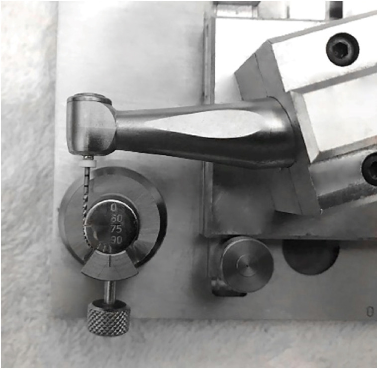
-
Abstract
PDFPubReaderePub
Objectives This study aimed to compare the torsional and cyclic fatigue resistance of ProGlider (PG), WaveOne Gold Glider (WGG), and TruNatomy Glider (TNG).
Materials and Methods A total of 15 instruments of each glide path system (
n = 15) were used for each test. A custom-made device simulating an angle of 90° and a radius of 5 millimeters was used to assess cyclic fatigue resistance, with calculation of number of cycles to failure. Torsional fatigue resistance was assessed by maximum torque and angle of rotation. Fractured instruments were examined by scanning electron microscopy (SEM). Data were analyzed with Shapiro-Wilk and Kruskal-Wallis tests, and the significance level was set at 5%.Results The WGG group showed greater cyclic fatigue resistance than the PG and TNG groups (
p < 0.05). In the torsional fatigue test, the TNG group showed a higher angle of rotation, followed by the PG and WGG groups (p < 0.05). The TNG group was superior to the PG group in torsional resistance (p < 0.05). SEM analysis revealed ductile morphology, typical of the 2 fracture modes: cyclic fatigue and torsional fatigue.Conclusions Reciprocating WGG instruments showed greater cyclic fatigue resistance, while TNG instruments were better in torsional fatigue resistance. The significance of these findings lies in the identification of the instruments’ clinical applicability to guide the choice of the most appropriate instrument and enable the clinician to provide a more predictable glide path preparation.
-
Citations
Citations to this article as recorded by- Mechanical Behaviour of Three Heat‐Treated NiTi Glide Path Systems at Simulated Intracanal Temperature
Ahmad Jamleh, Taher Omari, Abdulmohsen Alfadley, Abdullah Alqedairi, Hussam Alfawaz
Australian Endodontic Journal.2026;[Epub] CrossRef - Buckling resistance of various pathfinding endodontic instruments: An in vitro study
Ujjwal Das, Rajesh Kumar Das, Kallol Kumar Saha, Lugu Buru Murmu, Srimanta Banerjee, Rishila Nag
Journal of Conservative Dentistry and Endodontics.2025; 28(4): 384. CrossRef - Comparative evaluation of the remaining dentin volume following instrumentation with rotary, reciprocating, and hand files during root canal treatment in primary molars: An ex vivo study
İrem Eren, Berkant Sezer
Journal of Dental Sciences.2024; 19(4): 2126. CrossRef - Screw-in force, torque generation, and performance of glide-path files with three rotation kinetics
Jee-Yeon Woo, Ji-Hyun Jang, Seok Woo Chang, Soram Oh
Odontology.2024; 112(3): 761. CrossRef - Evaluation of shaping ability of different glide path instruments: a micro-computed tomography study
Merve Yeniçeri Özata, Seda Falakaloğlu, Ali Keleş, Özkan Adıgüzel, Mustafa Gündoğar
BMC Oral Health.2023;[Epub] CrossRef
- Mechanical Behaviour of Three Heat‐Treated NiTi Glide Path Systems at Simulated Intracanal Temperature
- 3,978 View
- 82 Download
- 4 Web of Science
- 5 Crossref
- Physicochemical properties of a calcium aluminate cement containing nanoparticles of zinc oxide
- Amanda Freitas da Rosa, Thuany Schmitz Amaral, Maria Eduarda Paz Dotto, Taynara Santos Goulart, Hebert Luís Rossetto, Eduardo Antunes Bortoluzzi, Cleonice da Silveira Teixeira, Lucas da Fonseca Roberti Garcia
- Restor Dent Endod 2023;48(1):e3. Published online December 8, 2022
- DOI: https://doi.org/10.5395/rde.2023.48.e3
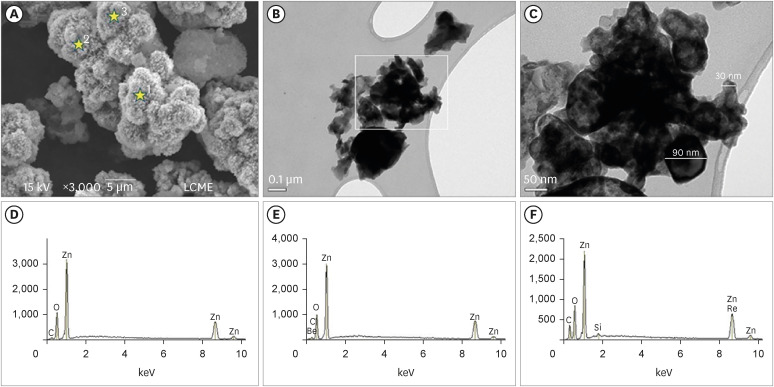
-
Abstract
PDFPubReaderePub
Objectives This study evaluated the effect of different nanoparticulated zinc oxide (nano-ZnO) and conventional-ZnO ratios on the physicochemical properties of calcium aluminate cement (CAC).
Materials and Methods The conventional-ZnO and nano-ZnO were added to the cement powder in the following proportions: G1 (20% conventional-ZnO), G2 (15% conventional-ZnO + 5% nano-ZnO), G3 (12% conventional-ZnO + 3% nano-ZnO) and G4 (10% conventional-ZnO + 5% nano-ZnO). The radiopacity (Rad), setting time (Set), dimensional change (Dc), solubility (Sol), compressive strength (Cst), and pH were evaluated. The nano-ZnO and CAC containing conventional-ZnO were also assessed using scanning electron microscopy, transmission electron microscopy, and energy-dispersive X-ray spectroscopy. Radiopacity data were analyzed by the 1-way analysis of variance (ANOVA) and Bonferroni tests (
p < 0.05). The data of the other properties were analyzed by the ANOVA, Tukey, and Fisher tests (p < 0.05).Results The nano-ZnO and CAC containing conventional-ZnO powders presented particles with few impurities and nanometric and micrometric sizes, respectively. G1 had the highest Rad mean value (
p < 0.05). When compared to G1, groups containing nano-ZnO had a significant reduction in the Set (p < 0.05) and lower values of Dc at 24 hours (p < 0.05). The Cst was higher for G4, with a significant difference for the other groups (p < 0.05). The Sol did not present significant differences among groups (p > 0.05).Conclusions The addition of nano-ZnO to CAC improved its dimensional change, setting time, and compressive strength, which may be promising for the clinical performance of this cement.
-
Citations
Citations to this article as recorded by- Histopathologic responses of the dental pulp to an experimental calcium aluminate–calcium silicate based capping material in comparison to mineral trioxide aggregate
Reham S. Saleh, Eyad A. Elbattawy, Shymaa Hamza, Yousra Aly
Scientific Reports.2026;[Epub] CrossRef - Calcium aluminate cement: a study on the effect of additives for dental applications
Sara Ghorbani, Rahim Naghizadeh, Ebrahim Ghasemi, Hamidreza Rezaie
Advances in Cement Research.2025; 37(4): 269. CrossRef - Experimental Study on Cement-Based Materials Modified by Nano-Zinc Oxide and Nano-Zirconia Based on Response Surface Optimization Design
Hongyin Hu, Fufei Wu, Jiao Chen, Shuangshuang Guan, Peng Qu, Hongqin Zhang, Yuyi Chen, Zirun Xu, Chuanteng Huang, Shuang Pu
Materials.2025; 18(7): 1515. CrossRef - Radiographic, mechanical, and chemical properties of mineral trioxide aggregate from nanosilica and clam shell calcium carbonate
Leny Yuliatun, Muhammad Adly Rahandi Lubis, Muhammad Khaliim Jati Kusala, Lia Destiarti, Ratna Betriani, Jolang Budiarta, Mariyam Mariyam
Polyhedron.2025; 278: 117590. CrossRef - Application of Calcium Aluminate-Based Materials for Direct Pulp Capping – In Vivo Study
Ognjenka Janković, Smiljana Paraš, Tijana Adamović, Ljiljana Tadić Latinović, Radmila Arbutina, Igor Đukić, Saša Marin, Marko Bulajić, Karolina Vukoje, Vukoman Jokanović, Verica Pavlić
Acta Veterinaria.2025; 75(2): 212. CrossRef - Nanotechnology for calcium aluminate cement: thematic analysis
Lapyote Prasittisopin
REVIEWS ON ADVANCED MATERIALS SCIENCE.2025;[Epub] CrossRef
- Histopathologic responses of the dental pulp to an experimental calcium aluminate–calcium silicate based capping material in comparison to mineral trioxide aggregate
- 2,967 View
- 55 Download
- 7 Web of Science
- 6 Crossref
- Cytotoxicity of two self-adhesive resin cements and their interference in the phagocytic activity of murine macrophages
- Danilo Couto da Silva, Leonardo Gomes Vaz, Warley Luciano Fonseca Tavares, Leda Quercia Vieira, Ricardo Reis de Oliveira, Antônio Paulino Ribeiro Sobrinho
- Restor Dent Endod 2022;47(3):e31. Published online July 14, 2022
- DOI: https://doi.org/10.5395/rde.2022.47.e31
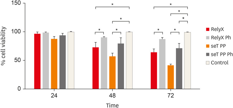
-
Abstract
PDFPubReaderePub
Objectives This study aimed to evaluate
in vitro the effects of the self-adhesive resin cements RelyX U200 (3M ESPE) and seT PP (SDI Limited) on murine macrophages and the interference of the photoactivation.Materials and Methods Cell viability assays, cell adherence, yeast phagocytosis of
Saccharomyces boulardii and production of reactive oxygen species (ROS) were performed in the presence of capillaries containing the respective self-adhesive cement when photoactivated or not.Results After long periods of contact, both types of cements, when not photoactivated, are more cytotoxic for macrophages. The seT PP cement when only chemically activated seems to interfere more negatively in the process of phagocytosis of yeasts
S. boulardii. Both types of cements interfere in the cell adhesion process, independent of photoactivation. None of the types of cements tested was able to induce the production of ROS.Conclusions Our results highlight the great importance of the photoactivation of self-adhesive resin cements in the dental clinic, since RelyX U200, when photoactivated, presented the best results within the evaluated parameters.
-
Citations
Citations to this article as recorded by- Influence of Preheating Self-Adhesive Cements on the Degree of Conversion, Cell Migration, and Cell Viability
Henrique Cantarelli, Fernando Antonio Costa Xavier, Fernando Freitas Portella, Keiichi Hosaka, Eduardo Galia Reston, Louis Hardan, Rim Bourgi, Celso Afonso Klein-Junior
Applied Mechanics.2024; 5(3): 553. CrossRef - Dental Luting Cements: An Updated Comprehensive Review
Artak Heboyan, Anna Vardanyan, Mohmed Isaqali Karobari, Anand Marya, Tatevik Avagyan, Hamid Tebyaniyan, Mohammed Mustafa, Dinesh Rokaya, Anna Avetisyan
Molecules.2023; 28(4): 1619. CrossRef
- Influence of Preheating Self-Adhesive Cements on the Degree of Conversion, Cell Migration, and Cell Viability
- 2,791 View
- 26 Download
- 5 Web of Science
- 2 Crossref
- How do imaging protocols affect the assessment of root-end fillings?
- Fernanda Ferrari Esteves Torres, Reinhilde Jacobs, Mostafa EzEldeen, Karla de Faria-Vasconcelos, Juliane Maria Guerreiro-Tanomaru, Bernardo Camargo dos Santos, Mário Tanomaru-Filho
- Restor Dent Endod 2022;47(1):e2. Published online December 15, 2021
- DOI: https://doi.org/10.5395/rde.2022.47.e2
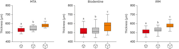
-
Abstract
PDFPubReaderePub
Objectives This study investigated the impact of micro-computed tomography (micro-CT)-based voxel size on the analysis of material/dentin interface voids and thickness of different endodontic cements.
Materials and Methods Following root-end resection and apical preparation, maxillary premolars were filled with mineral trioxide aggregate (MTA), Biodentine, and intermediate restorative material (IRM) (
n = 24). The samples were scanned using micro-CT (SkyScan 1272; Bruker) and the cement/dentin interface and thickness of materials were evaluated at voxel sizes of 5, 10, and 20 µm. Analysis of variance and the Tukey test were conducted, and the degree of agreement between different voxel sizes was evaluated using the Bland and Altman method (p < 0.05).Results All materials showed an increase in thickness from 5 to 10 and 20 µm (
p < 0.05). When evaluating the interface voids, materials were similar at 5 µm (p > 0.05), while at 10 and 20 µm Biodentine showed the lowest percentage of voids (p < 0.05). A decrease in the interface voids was observed for MTA and IRM at 20 µm, while Biodentine showed differences among all voxel sizes (p < 0.05). The Bland-Altman plots for comparisons among voxel sizes showed the largest deviations when comparing images between 5 and 20 µm.Conclusions Voxel size had an impact on the micro-CT evaluation of thickness and interface voids of endodontic materials. All cements exhibited an increase in thickness and a decrease in the void percentage as the voxel size increased, especially when evaluating images at 20 µm.
-
Citations
Citations to this article as recorded by- Effect of ultrasonic activation of endodontic sealers on root canal filling quality during the single-cone obturation procedure: a systematic review and meta-analysis of laboratory-based studies
Shuting Feng, Weiqing Zhou, Xiaojun Chu, Shuaimei Xu, Xiongqun Zeng
Odontology.2025; 113(4): 1380. CrossRef - Marginal Adaptation and Porosity of a Novel MTA Brand Applied as Root-End Filling Material: A Micro-CT Study
Yaneta Kouzmanova, Ivanka Dimitrova
Applied Sciences.2024; 14(7): 2758. CrossRef - Supplementary methods for filling material removal: A systematic review and meta-analysis of micro-CT imaging studies
Bruna Venzke Fischer, Taynara Santos Goulart, Filipe Colombo Vitali, Diego Leonardo de Souza, Cleonice da Silveira Teixeira, Lucas da Fonseca Roberti Garcia
Journal of Dentistry.2024; 151: 105445. CrossRef
- Effect of ultrasonic activation of endodontic sealers on root canal filling quality during the single-cone obturation procedure: a systematic review and meta-analysis of laboratory-based studies
- 3,045 View
- 30 Download
- 3 Web of Science
- 3 Crossref
- Spectrophotometric evaluation of restorative composite shades and their match with a classical shade guide
- Rafael Melara, Luciana Mendonça, Fábio Herrmann Coelho-de-Souza, Juliana Nunes Rolla, Luciano de Souza Gonçalves
- Restor Dent Endod 2021;46(4):e60. Published online November 12, 2021
- DOI: https://doi.org/10.5395/rde.2021.46.e60
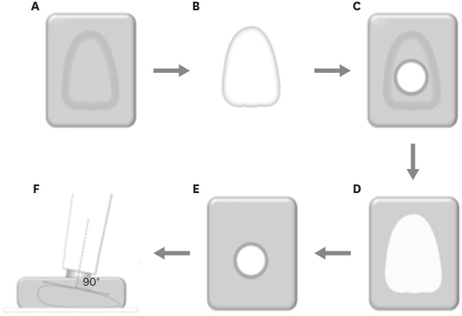
-
Abstract
PDFPubReaderePub
Objectives The aim of this study was to verify the match between 5 shades of composites from different manufacturers with a shade guide and among the systems using a portable spectrophotometer.
Materials and Methods Shade measurements were performed on specimens of Z350 XT (3M ESPE), Charisma Diamond (Heraeus Kulzer GmbH), Esthet X-HD (Dentsply Caulk), and Empress Direct (Ivoclar-Vivadent) for shades A1, A2, A3, B1, and C3 using a Vita Easyshade spectrophotometer (Vita Zahnfabrik) against a white background. Corresponding shades of Vitapan Classical (Vita Zahnfabrik) guide were measured likewise and shade variation (ΔE) was calculated based on International Commission on Illumination L*a*b* parameters. The ΔE of the composites in each shade was compared by one-way analysis of variance and Tukey's
post hoc test (α = 0.05).Results All composites presented ΔE > 3.7 compared with the shade guide. Variation in shades A3, B1, and C3 was significantly different for all composites. ΔE of Z350 XT was significantly lower for A1 than for the other shades, whereas ΔE of Z350 XT and Charisma Diamond were significantly lower for A2 than for the other shades.
Conclusions No composite shade matched with the shade guide. Equivalent shades of the restorative composite from different manufacturers may show clinically noticeable ΔE.
-
Citations
Citations to this article as recorded by- Influence of different finishing and polishing protocols of composite CAD CAM blocks on surface roughness and biological response of gingival mesenchymal stem cells
Mohamed F. Haridy, Mohamed Shamel, Raghda A. Khalil, Ahmed Refaat Mohamed, Hoda Fouda, Hend S. Ahmed
Odontology.2026; 114(3): 1320. CrossRef - Análise espectrofotométrica da cor de resinas compostas e sua correspondência com a escala de referência VITA Classical
Gardenia Mascarenhas de Oliveira, Marta Conceição Oliveira Silva, Natan dos Anjos Nery de Oliveira, Maria Fernanda Moreira Carvalho Caxico, Lucas Gomes Pereira, Leandro do Rozário Teixeira, Marcus Vinícius Santos da Silva, Iuri Muniz Pepe
Cuadernos de Educación y Desarrollo.2026; 18(1): e10592. CrossRef - Baseline color-matching in anterior non-carious cervical lesions of patients of two single-shade resin composites: a randomized clinical trial
Ayşe Nur Doğan, Soley Arslan
Odontology.2026;[Epub] CrossRef - Evaluation of color blending effect of a single-shade resin composite with hybrid ceramics: an in vitro study
Jongchan Lee, Jinsoo Ahn, Sun-Young Kim
BMC Oral Health.2026;[Epub] CrossRef - A Multidimensional Analysis of Shade Selection Difficulty for Indirect Restorations Among Dental Students and Professionals
Roxana-Ionela Vasluianu, Andreas Katsonis, Monica Silvia Tatarciuc, Anca Mihaela Vitalariu, Adina Oana Armencia, Andrea-Simoni Katsoni, Panagiotis Perperidis, Catalina Cioloca Holban, Irina Gradinaru, Ovidiu Stamatin, Magda Ecaterina Antohe
Dentistry Journal.2026; 14(4): 234. CrossRef - Assessment of Color Accuracy in A2-Shade Composite Resins Using CIEDE2000: Conventional and 3D-Printed Materials
Gülçin Cagay Sevencan, Hilal Gülgezen Aydın
Journal of International Dental Sciences.2026; 12(1): 34. CrossRef - Shade Matching and Color Stability of Chameleon Effect Composites in Class V Restorations: An In-Vitro Colorimetric Study after Cola Immersion
Youssef Znait, Karim Corbani, Carlos Khairallah, Carina Mehanna, Louis Hardan, Fadi Hammoud, Rim Bourgi, Cynthia Kassis
Journal of Dental Health and Oral Research.2026; 7(2): 1. CrossRef - Instrumental and visual evaluation of the color adjustment potential of a recently introduced single‑shade composite resin versus multishade composite resins
Jiakang Zhu, Yue Xu, Mengxun Li, Cui Huang
The Journal of Prosthetic Dentistry.2025; 134(3): 832. CrossRef - Evaluation of the roughness, color match, and color stability of two monochromatic composite resins: a randomized controlled laboratory study
Iara Campos Santana, Sabrina Sobral de Oliveira, Karolina Pena Botelho, Renan Leonardi de Oliveira Rigotti, José Cristiano Ramos Glória, Adriana Maria Botelho, Dhelfeson Willya Douglas-de-Oliveira, Karine Taís Aguiar Tavano
BMC Oral Health.2025;[Epub] CrossRef - Instrumental and Visual Evaluation of the Chameleon Effect of Single-shaded Composite Resins
RM Adiguzel, LK Kose, N Arhun
Operative Dentistry.2024; 49(4): 432. CrossRef - Color Stability of Bioactive Restorative Material vs Nanohybrid Resin Composite: An In Vitro Study
Esraa H Saber, Mohsen H Abielhassan, Yasser A Abed, Shereen E Fahim
The Journal of Contemporary Dental Practice.2024; 25(3): 221. CrossRef - A system for reliable composite shade matching: Custom shade tabs and an intra‐oral mockup
Adamo Notarantonio, Amanda Seay
Journal of Esthetic and Restorative Dentistry.2023; 35(5): 787. CrossRef - Color Stability of Bioactive Restorative Materials After Immersion in Various Media
Shara I Sajini, Ali B Mushayt, Talal A Almutairi, Roaa Abuljadayel
Journal of International Society of Preventive and Community Dentistry.2022; 12(4): 418. CrossRef
- Influence of different finishing and polishing protocols of composite CAD CAM blocks on surface roughness and biological response of gingival mesenchymal stem cells
- 2,908 View
- 41 Download
- 11 Web of Science
- 13 Crossref
- Combination of a new ultrasonic tip with rotary systems for the preparation of flattened root canals
- Karina Ines Medina Carita Tavares, Jáder Camilo Pinto, Airton Oliveira Santos-Junior, Fernanda Ferrari Esteves Torres, Juliane Maria Guerreiro-Tanomaru, Mario Tanomaru-Filho
- Restor Dent Endod 2021;46(4):e56. Published online October 27, 2021
- DOI: https://doi.org/10.5395/rde.2021.46.e56
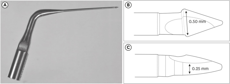
-
Abstract
PDFPubReaderePub
Objectives This study evaluated 2 nickel-titanium rotary systems and a complementary protocol with an ultrasonic tip and a small-diameter instrument in flattened root canals.
Materials and Methods Thirty-two human maxillary second premolars with flattened canals (buccolingual diameter ≥4 times larger than the mesiodistal diameter) at 9 mm from the radiographic apex were selected. The root canals were prepared by ProDesign Logic (PDL) 30/0.01 and 30/0.05 or Hyflex EDM (HEDM) 10/0.05 and 25/0.08 (
n = 16), followed by application of the Flatsonic ultrasonic tip in the cervical and middle thirds and a PDL 25/0.03 file in the apical third (FPDL). The teeth were scanned using micro-computed tomography before and after the procedures. The percentage of volume increase, debris, and uninstrumented surface area were analyzed using the Kruskal-Wallis, Dunn, Wilcoxon, analysis of variance/Tukey, and paired and unpairedt -tests (α = 0.05).Results No significant difference was found in the volume increase and uninstrumented surface area between PDL and HEDM (
p > 0.05). PDL had a higher percentage of debris than HEDM in the middle and apical thirds (p < 0.05). The FPDL protocol resulted in less debris and uninstrumented surface area for PDL and HEDM (p < 0.05). This protocol, with HEDM, reduced debris in the middle and apical thirds and uninstrumented surface area in the apical third (p < 0.05).Conclusions High percentages of debris and uninstrumented surface area were observed after preparation of flattened root canals. The HEDM, Flatsonic tip, and 25/0.03 instrument protocol enhanced cleaning in flattened root canals.
-
Citations
Citations to this article as recorded by- Kök Kanal Tedavisi Yenilemelerinde Ultrasonik Uç Kullanımı
Ayşenur Kızıltaş Gül, Turan Mert Hisar, Seniha Miçooğulları
Selcuk Dental Journal.2025; 12(1): 157. CrossRef - Flatsonic Ultrasonic Tip Optimizes the Removal of Remaining Filling Material in Flattened Root Canals: A Micro–computed Tomographic Analysis
Airton Oliveira Santos-Junior, Karina Ines Medina Carita Tavares, Jáder Camilo Pinto, Fernanda Ferrari Esteves Torres, Juliane Maria Guerreiro-Tanomaru, Mário Tanomaru-Filho
Journal of Endodontics.2024; 50(5): 612. CrossRef - The Effect of Combined Ultrasonic Tip and Mechanized Instrumentation on the Reduction of the Percentage of Non-Instrumented Surfaces in Oval/Flat Root Canals: A Systematic Review and Meta-Analysis
Marcella Dewes Cassal, Pedro Cardoso Soares, Marcelo dos Santos
Cureus.2023;[Epub] CrossRef - Heat-treated NiTi instruments and final irrigation protocols for biomechanical preparation of flattened canals
Kleber Kildare Teodoro CARVALHO, Igor Bassi Ferreira PETEAN, Alice Corrêa SILVA-SOUSA, Rafael Verardino CAMARGO, Jardel Francisco MAZZI-CHAVES, Yara Terezinha Corrêa SILVA-SOUSA, Manoel Damião SOUSA-NETO
Brazilian Oral Research.2022;[Epub] CrossRef
- Kök Kanal Tedavisi Yenilemelerinde Ultrasonik Uç Kullanımı
- 2,678 View
- 37 Download
- 4 Web of Science
- 4 Crossref
- Errors in light-emitting diodes positioning when curing bulk fill and incremental composites: impact on properties after aging
- Abdulrahman A. Balhaddad, Isadora M. Garcia, Haifa Maktabi, Maria Salem Ibrahim, Qoot Alkhubaizi, Howard Strassler, Fabrício M. Collares, Mary Anne S. Melo
- Restor Dent Endod 2021;46(4):e51. Published online September 24, 2021
- DOI: https://doi.org/10.5395/rde.2021.46.e51
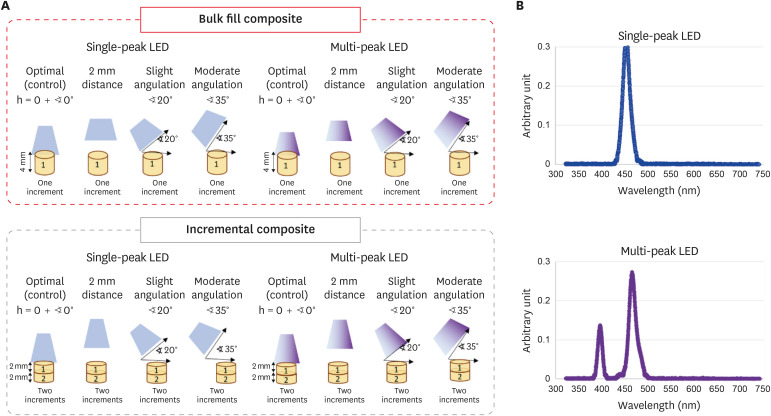
-
Abstract
PDFPubReaderePub
Objectives This study aimed to evaluate the effect of improper positioning single-peak and multi-peak lights on color change, microhardness of bottom and top, and surface topography of bulk fill and incremental composites after artificial aging for 1 year.
Materials and Methods Bulk fill and incremental composites were cured using multi-peak and single-peak light-emitting diode (LED) following 4 clinical conditions: (1) optimal condition (no angulation or tip displacement), (2) tip-displacement (2 mm), (3) slight tip angulation (α = 20°) and (4) moderate tip angulation (α = 35°). After 1-year of water aging, the specimens were analyzed for color changes (ΔE), Vickers hardness, surface topography (Ra, Rt, and Rv), and scanning electron microscopy.
Results For samples cured by single-peak LED, the improper positioning significantly increases the color change compared to the optimal position regardless of the type of composite (
p < 0.001). For multi-peak LED, the type of resin composite and the curing condition displayed a significant effect on ΔE (p < 0.001). For both LEDs, the Vickers hardness and bottom/top ratio of Vickers hardness were affected by the type of composite and the curing condition (p < 0.01).Conclusions The bulk fill composite presented greater resistance to wear, higher color stability, and better microhardness than the incremental composite when subjected to improper curing. The multi-peak LED improves curing under improper conditions compared to single-peak LED. Prevention of errors when curing composites requires the attention of all personnel involved in the patient's care once the clinical relevance of the appropriate polymerization reflects on reliable long-term outcomes.
-
Citations
Citations to this article as recorded by- A clinical survey of the output intensity of 50 light-curing units in dental clinics across Davangere and Mangalore region using a spectrometer system
Elizbeth Christy Jose, Sakshi Jha, Prema Shantagouda Biradar, J Arun, TN Nandini, Thushara Mohanan
International Journal of Oral Health Sciences.2025; 15(1): 41. CrossRef - The demineralization resistance and mechanical assessments of different bioactive restorative materials for primary and permanent teeth: an in vitro study
Maria Salem Ibrahim, Fahad Rakad Aldhafeeri, Abdullah Sami Banaemah, Mana S. Alhaider, Yousif A. Al-Dulaijan, Abdulrahman A. Balhaddad
BDJ Open.2024;[Epub] CrossRef - Inorganic Compounds as Remineralizing Fillers in Dental Restorative Materials: Narrative Review
Leena Ibraheem Bin-Jardan, Dalal Ibrahim Almadani, Leen Saleh Almutairi, Hadi A. Almoabid, Mohammed A. Alessa, Khalid S. Almulhim, Rasha N. AlSheikh, Yousif A. Al-Dulaijan, Maria S. Ibrahim, Afnan O. Al-Zain, Abdulrahman A. Balhaddad
International Journal of Molecular Sciences.2023; 24(9): 8295. CrossRef
- A clinical survey of the output intensity of 50 light-curing units in dental clinics across Davangere and Mangalore region using a spectrometer system
- 2,552 View
- 22 Download
- 2 Web of Science
- 3 Crossref
- The effectiveness of the supplementary use of the XP-endo Finisher on bacteria content reduction: a systematic review and meta-analysis
- Ludmila Smith de Jesus Oliveira, Rafaella Mariana Fontes de Bragança, Rafael Sarkis-Onofre, André Luis Faria-e-Silva
- Restor Dent Endod 2021;46(3):e37. Published online June 18, 2021
- DOI: https://doi.org/10.5395/rde.2021.46.e37
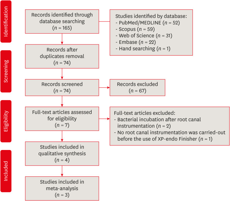
-
Abstract
PDFSupplementary MaterialPubReaderePub
Objectives This systematic review evaluated the efficacy of the supplementary use of the XP-endo Finisher on bacteria content reduction in the root canal system.
Materials and Methods In-vitro studies evaluating the use of the XP-endo Finisher on bacteria content were searched in four databases in July 2020. Two authors independently screened the studies for eligibility. Data were extracted, and risk of bias was assessed. Data were meta-analyzed by using random-effects model to compare the effect of the supplementary use (experimental) or not (control) of the XP-endo Finisher on bacteria counting reduction, and results from different endodontic protocols were combined. Four studies met the inclusion criteria while 1 study was excluded from the meta-analysis due to its high risk of bias and outlier data. The 3 studies that made it to the meta-analysis had an unclear risk of bias for at least one criterion.Results No heterogeneity was observed among the results of the studies included in the meta-analysis. The study excluded from the meta-analysis assessing the bacteria counting deep in the dentin demonstrated further bacteria reduction upon the use of the XP-endo Finisher.
Conclusions This systematic review found no evidence supporting the supplementary use of the XP-endo Finisher on further bacteria counting the reduction in the root canal.
-
Citations
Citations to this article as recorded by- Mapping risk of bias criteria in systematic reviews of in vitro endodontic studies: an umbrella review
Rafaella Rodrigues da Gama, Lucas Peixoto de Araújo, Evandro Piva, Leandro Perello Duro, Adriana Fernandes da Silva, Wellington Luiz de Oliveira da Rosa
Evidence-Based Dentistry.2025; 26(4): 179. CrossRef - Characteristics and Effectiveness of XP‐Endo Files and Systems: A Narrative Review
Sarah M. Alkahtany, Rana Alfadhel, Aseel AlOmair, Sarah Bin Durayhim, Kee Y. Kum
International Journal of Dentistry.2024;[Epub] CrossRef - Impact XP-endo finisher on the 1-year follow-up success of posterior root canal treatments: a randomized clinical trial
Ludmila Smith de Jesus Oliveira, Fabricio Eneas Diniz de Figueiredo, Janaina Araújo Dantas, Maria Amália Gonzaga Ribeiro, Carlos Estrela, Manoel Damião Sousa-Neto, André Luis Faria-e-Silva
Clinical Oral Investigations.2023; 27(12): 7595. CrossRef - Comparative analysis of the effectiveness of modern irrigants activation techniques in the process of mechanical root canal system treatment (Literature review)
Anatoliy Potapchuk, Vasyl Almashi, Arsenii Horzov, Victor Buleza
InterConf.2023; (34(159)): 200. CrossRef - Comparative analysis of the effectiveness of modern irrigants activation techniques in the protocol of chemomechanical root canal system treatment (literature review)
A. Potapchuk, V. Almashi, Y. Rak, Y. Melnyk, V. Buleza, A. Horzov
SUCHASNA STOMATOLOHIYA.2023; 114(3): 4. CrossRef - Methodological quality assessment criteria for the evaluation of laboratory‐based studies included in systematic reviews within the specialty of Endodontology: A development protocol
Venkateshbabu Nagendrababu, Paul V. Abbott, Christos Boutsioukis, Henry F. Duncan, Clovis M. Faggion, Anil Kishen, Peter E. Murray, Shaju Jacob Pulikkotil, Paul M. H. Dummer
International Endodontic Journal.2022; 55(4): 326. CrossRef
- Mapping risk of bias criteria in systematic reviews of in vitro endodontic studies: an umbrella review
- 3,491 View
- 26 Download
- 4 Web of Science
- 6 Crossref
- Evaluation of the relation between the pulp stones and direct restorations using cone beam computed tomography in a Turkish subpopulation
- Güzide Pelin Sezgin, Sema Sönmez Kaplan, Tuna Kaplan
- Restor Dent Endod 2021;46(3):e34. Published online June 8, 2021
- DOI: https://doi.org/10.5395/rde.2021.46.e34
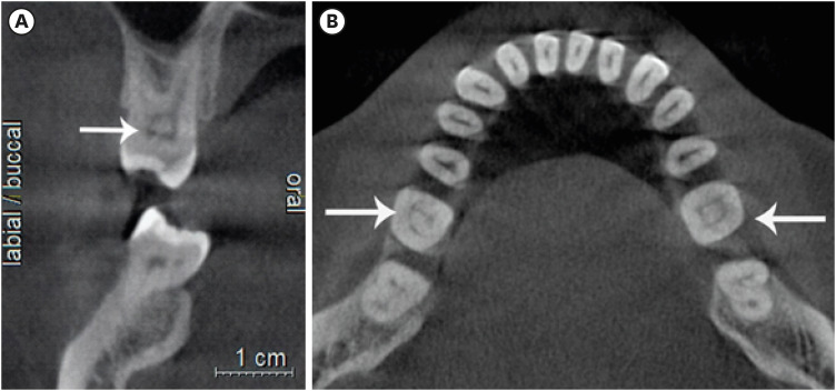
-
Abstract
PDFPubReaderePub
Objectives This study aimed to assess the presence of pulp stones through an examination of cone beam computed tomography images and correlate their prevalence with age, sex, dental arch and side, tooth type, and restoration type and depth.
Materials and Methods Cone beam computed tomography images obtained from 673 patients and archival data on 11,494 teeth were evaluated. The associations of pulp stones with age, sex, dental arch and side, tooth type, and restoration type and depth were noted. All the measurements were subjected to a χ2 test and one sample χ2 test (
p < 0.05).Results In the study group, 163 (24.2%) patients and 379 (3.3%) teeth had at least one pulp stone. The pulp stone frequency in those aged 30–39 years was significantly greater than in those aged 18–29 and ≥ 60 years, and the frequency was higher in females than in males (
p < 0.05). The highest prevalence of pulp stones was found in maxillary dental arches and molar teeth (p < 0.05). Pulp stones were significantly more common in medium-depth restorations (p < 0.05).Conclusions Maxillary molar teeth, medium-depth restorations, individuals aged 30–39 years and females had a greater percentage of pulp stones.
-
Citations
Citations to this article as recorded by- Comparative Detection and Inter-Modality Agreement of Pulp Stones Using Digital Periapical Radiography and CBCT at Two Voxel Sizes: An Ex Vivo Study
Hassan Hamed Kaabi, Sarah Saeed Binhassan, Sultan Hamad Alrumaih, Mohammed Jamal Alotaibi, Abdullah Khalid Bakarman, Nawaf Abdulaziz Alghamdi, Hamad Abdullah Almuhaythif, Qamar Mohammadziad Hashem, Abdulfatah Samih Alazmah
Diagnostics.2026; 16(7): 961. CrossRef - Cone-Beam Computed Tomography Assessment of the Prevalence and Association of Pulp Calcification with Dental and Periodontal Pathology: A Descriptive Study
José Luis Sanz, Lucía Callado, Stefana Mantale, Jenifer Nicolás, James Ghilotti, Carmen Llena
Journal of Clinical Medicine.2025; 14(4): 1373. CrossRef - Prevalence of mineralization in the pulp chamber in patients according to CBCT data
V. A. Molokova, I. N. Antonova, V. A. Osipova
Endodontics Today.2025; 23(2): 188. CrossRef - Could carotid artery calcifications and pulp stones be an alarm sign for diabetes mellitus? A retrospective observational study
Motahare Baghestani, Mohadese Faregh, Seyed Hossein Razavi, Fatemeh Owlia
BMC Endocrine Disorders.2025;[Epub] CrossRef - Distribution and influencing factors of pulp stones based on CBCT: a retrospective observational study from southwest China
Wantong Zhang, Yao Wang, Lin Ye, Yan Zhou
BMC Oral Health.2024;[Epub] CrossRef - Prevalence and Association of Calcified Pulp Stones with Periodontitis: A Cone-Beam Computed Tomography Study in Saudi Arabian Population
Abdullah Saad Alqahtani
Journal of Pharmacy and Bioallied Sciences.2024; 16(Suppl 1): S644. CrossRef - The Prevalence And Distribution Of Pulp Stones: A Cone-Beam Computed Tomography Study İn A Group Of Turkish Patients
Mujgan Firincioglulari, Seçil Aksoy, Melis Gülbeş, Umut Aksoy, Kaan Orhan
ADO Klinik Bilimler Dergisi.2024; 13(3): 496. CrossRef - Radiographical examination of pulp stone distribution by cone beam computed tomography
Fatma Tunç, Emre Çulha, Muazzez Naz Baştürk
Journal of Health Sciences and Medicine.2024; 7(4): 472. CrossRef - Cone-Beam Computed Tomography-Based Investigation of the Prevalence and Distribution of Pulp Stones and Their Relation to Local and Systemic Factors in the Makkah Population: A Cross-Sectional Study
Laila M Kenawi, Haytham S Jaha, Mashael M Alzahrani, Jihan I Alharbi, Shahad F Alharbi, Taif A Almuqati, Rehab A Alsubhi, Wahdan M Elkwatehy
Cureus.2024;[Epub] CrossRef - Cone beam computed tomography assessment of the prevalence and association of pulp calcification with periodontitis
Lingling Xiang, Botao Wang, Yuan Zhang, Jintao Wang, Peipei Wu, Jian Zhang, Liangjun Zhong, Rui He
Odontology.2023; 111(1): 248. CrossRef - Three-dimensional analysis for detection of pulp stones in a Saudi population using cone beam computed tomography
Hassan H. Kaabi, Abdullah M. Riyahi, Nassr S. Al-Maflehi, Saleh F. Alrumayyan, Abdullah K. Bakrman, Yazeed A. Almutaw
Journal of Oral Science.2023; 65(4): 257. CrossRef
- Comparative Detection and Inter-Modality Agreement of Pulp Stones Using Digital Periapical Radiography and CBCT at Two Voxel Sizes: An Ex Vivo Study
- 3,090 View
- 36 Download
- 12 Web of Science
- 11 Crossref
- Cryopreservation of mesenchymal stem cells derived from dental pulp: a systematic review
- Sabrina Moreira Paes, Yasmine Mendes Pupo, Bruno Cavalini Cavenago, Thiago Fonseca-Silva, Carolina Carvalho de Oliveira Santos
- Restor Dent Endod 2021;46(2):e26. Published online April 29, 2021
- DOI: https://doi.org/10.5395/rde.2021.46.e26
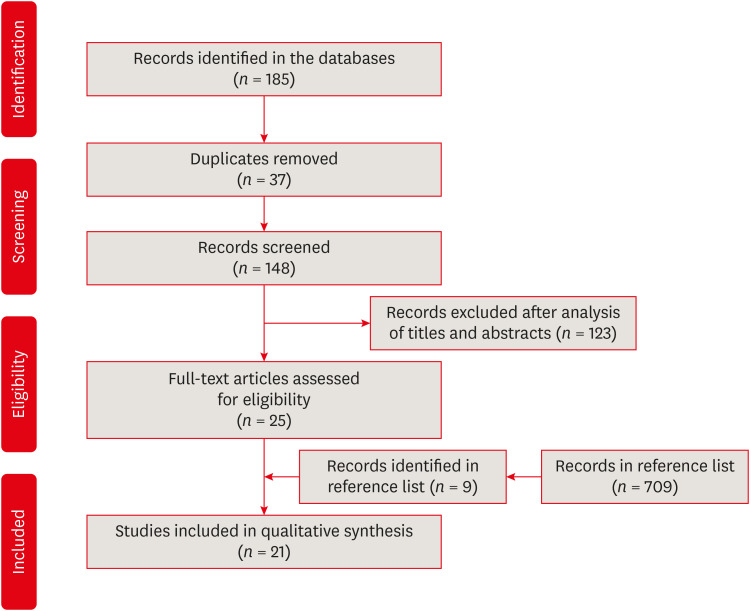
-
Abstract
PDFPubReaderePub
Objectives The aim of the present systematic review was to investigate the cryopreservation process of dental pulp mesenchymal stromal cells and whether cryopreservation is effective in promoting cell viability and recovery.
Materials and Methods This systematic review was developed in accordance with the Preferred Reporting Items for Systematic Reviews and Meta-Analyses (PRISMA) statement and the research question was determined using the population, exposure, comparison, and outcomes strategy. Electronic searches were conducted in the PubMed, Cochrane Library, Science Direct, LILACS, and SciELO databases and in the gray literature (dissertations and thesis databases and Google Scholar) for relevant articles published up to March 2019. Clinical trial studies performed with dental pulp of human permanent or primary teeth, containing concrete information regarding the cryopreservation stages, and with cryopreservation performed for a period of at least 1 week were included in this study.
Results The search strategy resulted in the retrieval of 185 publications. After the application of the eligibility criteria, 21 articles were selected for a qualitative analysis.
Conclusions The cryopreservation process must be carried out in 6 stages: tooth disinfection, pulp extraction, cell isolation, cell proliferation, cryopreservation, and thawing. In addition, it can be inferred that the use of dimethyl sulfoxide, programmable freezing, and storage in liquid nitrogen are associated with a high rate of cell viability after thawing and a high rate of cell proliferation in both primary and permanent teeth.
-
Citations
Citations to this article as recorded by- Comparative study on biological characteristics of dental mesenchymal stem cells isolated from gingiva, periodontal ligament, and dental follicle and their derived conditioned medium
Xianyi He, Yichen Gao, Haiyin Wan, Xia Wang, Jie Shen, Yun He, Junliang Chen
Annals of Anatomy - Anatomischer Anzeiger.2026; 263: 152751. CrossRef - Effect of the pulp harvesting method on the viability of human dental pulp stem cells and their odontogenic differentiation potential
Justine De Visscher, Lore Vermeir, Natasja Van den Vreken, Liesbeth Temmerman, Noëmi De Roo, Jolanda van Hengel, Guy De Pauw
Cell and Tissue Banking.2025;[Epub] CrossRef - The Antimicrobial Effect of the Incorporation of Inorganic Substances into Heat-Cured Denture Base Resins—A Systematic Review
Mariana Lima, Helena Salgado, André Correia, Patrícia Fonseca
Prosthesis.2024; 6(5): 1189. CrossRef - Sphingosine-1-phosphate Treatment Improves Cryopreservation Efficiency in Human Mesenchymal Stem Cells
Seong-Ju Oh, Chan-Hee Jo, Tae-Seok Kim, Chae-Yeon Hong, Sung-Lim Lee, Young-Hoon Kang, Gyu-Jin Rho
Life.2023; 13(6): 1286. CrossRef - Time- and Concentration-Dependent Effects of the Stem Cells Derived from Human Exfoliated Deciduous Teeth on Osteosarcoma Cells
Razieh Alipour, Batool Hashemibeni, Vajihe Asgari, Hamid Bahramian
Advanced Biomedical Research.2023;[Epub] CrossRef
- Comparative study on biological characteristics of dental mesenchymal stem cells isolated from gingiva, periodontal ligament, and dental follicle and their derived conditioned medium
- 3,968 View
- 38 Download
- 4 Web of Science
- 5 Crossref
- Color assessment of resin composite by using cellphone images compared with a spectrophotometer
- Rafaella Mariana Fontes de Bragança, Rafael Ratto Moraes, André Luis Faria-e-Silva
- Restor Dent Endod 2021;46(2):e23. Published online April 5, 2021
- DOI: https://doi.org/10.5395/rde.2021.46.e23
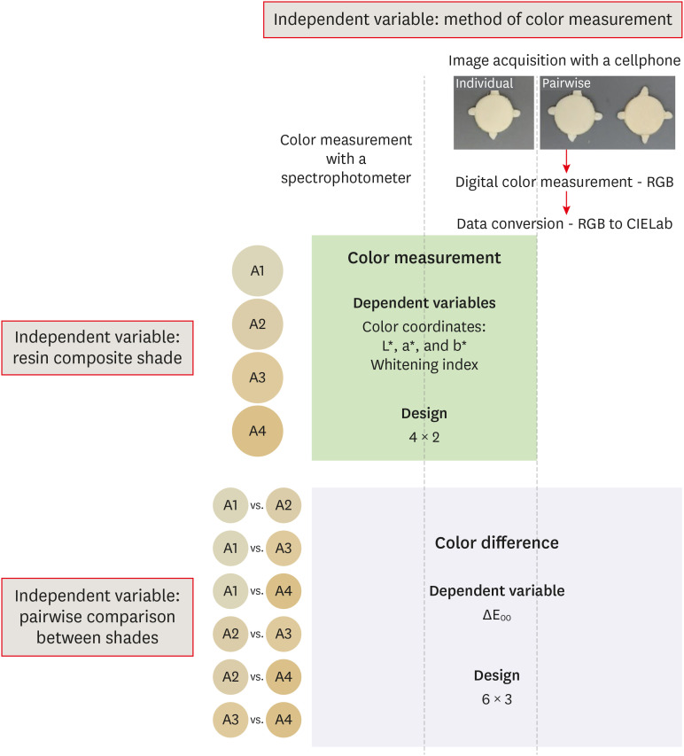
-
Abstract
PDFPubReaderePub
Objectives This study assessed the reliability of digital color measurements using images of resin composite specimens captured with a cellphone.
Materials and Methods The reference color of cylindrical specimens built-up with the use of resin composite (shades A1, A2, A3, and A4) was measured with a portable spectrophotometer (CIELab). Images of the specimens were obtained individually or pairwise (compared shades in the same photograph) under standardized parameters. The color of the specimens was measured in the images using RGB system and converted to CIELab system using image processing software. Whiteness index (WID) and color differences (ΔE00) were calculated for each color measurement method. For the cellphone, the ΔE00 was calculated between the pairs of shades in separate images and in the same image. Data were analyzed using 2-way repeated-measures analysis of variance (α = 0.05). Linear regression models were used to predict the reference ΔE00 values of those calculated using color measured in the images.
Results Images captured with the cellphone resulted in different WID values from the spectrophotometer only for shades A3 and A4. No difference to the reference ΔE00 was observed when individual images were used. In general, a similar ranking of ΔE00 among resin composite shades was observed for all methods. Stronger correlation coefficients with the reference ΔE00 were observed using individual than pairwise images.
Conclusions This study showed that the use of cellphone images to measure the color difference seems to be a feasible alternative providing outcomes similar to those obtained with the spectrophotometer.
-
Citations
Citations to this article as recorded by- Reliability of Three Image‐Based Instrumental Color‐Measurement Methods
Eloah Nunes de Almeida, Ido Luiz de Azevedo Feiten, Luis Felipe J. Schneider, Larissa Maria Assad Cavalcante, André Luis Faria‐e‐Silva
Journal of Esthetic and Restorative Dentistry.2026; 38(8): 1598. CrossRef - Evaluation of color stability in single-shade composite resins using spectrophotometer and cross-polarized mobile photography
Hatice Tepe, Ozge Celiksoz, Batu Can Yaman
BMC Oral Health.2025;[Epub] CrossRef - Color discrepancy of single-shade composites at different distances from the interface measured using cell phone images
Márcia Luciana Carregosa Santana, Gabriella de Jesus Santos Livi, André Luis Faria-e-Silva
Restorative Dentistry & Endodontics.2024;[Epub] CrossRef - How the Translucency and Color Stability of Single-Shade Universal Resin Composites Are Affected by Coffee?
Büşra Özdemir, Betül Kübra Kurucu Karadeniz, Seyit Bilal Özdemir, Ömer Akbulut
Current Research in Dental Sciences.2024; 34(4): 270. CrossRef - Color Image Expression through CIE L*a*b* System in Foods
Hyun-Woong Choi, Seong-Eun Park, Hong-Seok Son
Journal of the Korean Society of Food Science and Nutrition.2023; 52(2): 223. CrossRef - Comparative Evaluation of VITA Shade Guide and Various Composite Shades Using Spectrophotometer, Digital Single-lens Reflex, and Cellphone: An In Vitro Study
Aman Verma, Sonali Taneja, Surabhi Ghosh
World Journal of Dentistry.2023; 14(9): 803. CrossRef - Comparison of instrumental methods for color change assessment of Giomer resins
Luiza de Almeida Queiroz Ferreira, Rogéli Tibúrcio Ribeiro da Cunha Peixoto, Cláudia Silami de Magalhães, Tassiana Melo Sá, Monica Yamauti, Francisca Daniele Moreira Jardilino
Restorative Dentistry & Endodontics.2022;[Epub] CrossRef
- Reliability of Three Image‐Based Instrumental Color‐Measurement Methods
- 3,539 View
- 39 Download
- 3 Web of Science
- 7 Crossref
- Can silver diamine fluoride or silver nanoparticle-based anticaries agents to affect enamel bond strength?
- Jaqueline Costa Favaro, Yana Cosendey Toledo de Mello Peixoto, Omar Geha, Flaviana Alves Dias, Ricardo Danil Guiraldo, Murilo Baena Lopes, Sandrine Bittencourt Berger
- Restor Dent Endod 2021;46(1):e7. Published online January 12, 2021
- DOI: https://doi.org/10.5395/rde.2021.46.e7
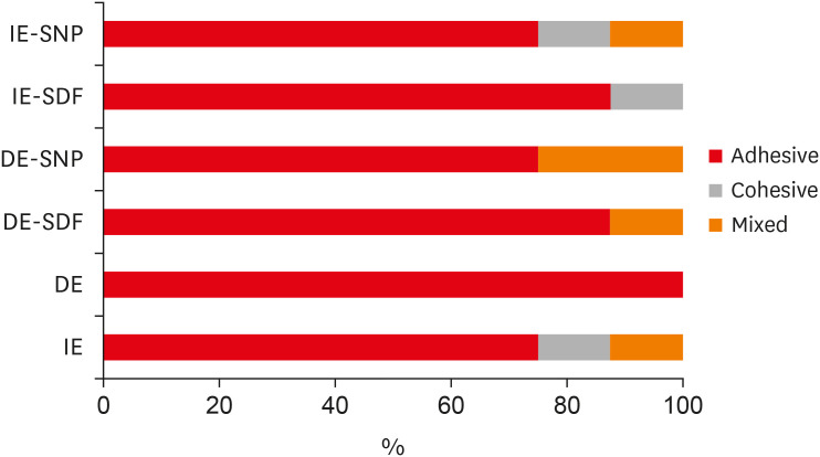
-
Abstract
PDFPubReaderePub
Objectives The aim of the current study is to investigate the effect of different anticaries agents, such as experimental agents based on silver nanoparticles (SNPs) and silver diamine fluoride (SDF), on the micro-shear bond strength (μ-SBS) of composite resin applied to intact enamel (IE) or demineralized enamel (DE).
Materials and Methods Sixty dental enamel fragments were collected from human third molars and categorized into 6 groups (
n = 10): positive control (IE), negative control (DE), IE + SDF, DE + SDF, IE + SNP and DE + SNP. Samples from DE, DE + SDF and DE + SNP groups were subjected to pH cycling; superficial microhardness test was performed to confirm demineralization. Resin composite build-ups were applied to the samples (0.75-mm diameter and 1-mm height) after the treatments (except for IE and DE groups); μ-SBS was also evaluated. Samples were analyzed under a stereomicroscope at 40× magnification to identify failure patterns. Data were subjected to one-way analysis of variance, followed by Tukey's and Dunnett's tests (p < 0.05).Results There was no significant difference among the IE, IE + SNP, DE + SDF, and DE + SNP groups. The IE + SDF and DE groups recorded the highest and the lowest μ-SBS values, respectively. Adhesive-type failures were the most frequent for all treatments.
Conclusions Anticaries agents did not have a negative effect on the μ-SBS of composite resin when it was used on IE or DE.
-
Citations
Citations to this article as recorded by- Impact of Incorporating Nanoparticles to Adhesive Resin on the Demineralization of Enamel: A Systematic Review
Naif Almosa
Dentistry Journal.2025; 13(3): 89. CrossRef - Preventing white spot lesions around orthodontic brackets: efficacy of pre-reacted glass-ionomer barrier coat versus silver diamine fluoride: an in vitro study
Enas A. Elshenawy, Safa B. Alawy, Wafaa Yahia Alghonemy, Ahmed Ibrahime El dosoky
BDJ Open.2025;[Epub] CrossRef - Research Status of Silver Nanoparticles for Dental Applications
Yanyan Guo, Xiaomei Hou, Sanjun Fan, Chanyuan Jin
Inorganics.2025; 13(5): 168. CrossRef - The use of silver diamine fluoride to prevent/treat enamel carious lesions: a narrative review
Rasha N. AlSheikh
PeerJ.2024; 12: e17897. CrossRef - Phosphoric Acid Etch Partially Restores the Initial Bond Strength of Composite to Silver Diamine Fluoride–Treated Enamel Using Universal Adhesives
Zaher Jabbour, Mijoo Kim, Marc Hayashi, Reuben Kim
Dentistry Journal.2023; 11(7): 161. CrossRef - Efficacy of Nano Silver Fluoride and/or Diode Laser In Enhancing Enamel Anticariogenicity around orthodontic brackets
Aya Anwar Alsherif, Mohamed Ali Farag, Mai Badreldin Helal
BDJ Open.2023;[Epub] CrossRef - Amelioration Strategies for Silver Diamine Fluoride: Moving from Black to White
Amjad Almuqrin, Inder Preet Kaur, Laurence J. Walsh, Chaminda Jayampath Seneviratne, Sobia Zafar
Antibiotics.2023; 12(2): 298. CrossRef - The Effect of Loading Time on Color Stability of Various Restorative Materials Bonded to Silver Diamine Fluoride-Treated Demineralized Dentin
Mohammed M Aldosari, Fares S Al-Sehaibany
Clinical, Cosmetic and Investigational Dentistry.2022; Volume 14: 123. CrossRef - In vitro study of the effect of nanosilver fluoride on shear bond strength of orthodontic brackets and demineralization of enamel
Mariam H. El-Toukhy, Eman M. El-Shourbagy, Neveen M. Fakhry
Tanta Dental Journal.2022; 19(4): 281. CrossRef
- Impact of Incorporating Nanoparticles to Adhesive Resin on the Demineralization of Enamel: A Systematic Review
- 2,873 View
- 36 Download
- 10 Web of Science
- 9 Crossref
- Assessment of the radiant emittance of damaged/contaminated dental light-curing tips by spectrophotometric methods
- Abdulrahman A. Balhaddad, Isadora Garcia, Fabrício Collares, Cristopher M. Felix, Nisha Ganesh, Qoot Alkabashi, Ward Massei, Howard Strassler, Mary Anne Melo
- Restor Dent Endod 2020;45(4):e55. Published online November 3, 2020
- DOI: https://doi.org/10.5395/rde.2020.45.e55
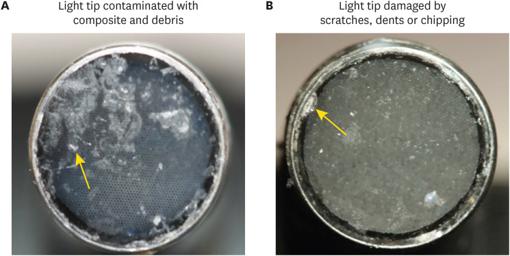
-
Abstract
PDFPubReaderePub
Objectives This study investigated the effects of physically damaged and resin-contaminated tips on radiant emittance, comparing them with new undamaged, non-contaminated tips using 3 pieces of spectrophotometric laboratory equipment.
Materials and Methods Nine tips with damage and/or resin contaminants from actual clinical situations were compared with a new tip without damage or contamination (control group). The radiant emittance was recorded using 3 spectrophotometric methods: a laboratory-grade thermopile, a laboratory-grade integrating sphere, and a portable light collector (checkMARC).
Results A significant difference between the laboratory-grade thermopile and the laboratory-grade integrating sphere was found when the radiant emittance values of the control or damaged/contaminated tips were investigated (
p < 0.05), but both methods were comparable to checkMARC (p > 0.05). Regardless of the method used to quantify the light output, the mean radiant emittance values of the damaged/contaminated tips were significantly lower than those of the control (p < 0.05). The beam profile of the damaged/contaminated tips was less homogeneous than that of the control.Conclusions Damaged/contaminated tips can reduce the radiant emittance output and the homogeneity of the beam, which may affect the energy delivered to composite restorations. The checkMARC spectrophotometer device can be used in dental offices, as it provided values close to those produced by a laboratory-grade integrated sphere spectrophotometer. Dentists should assess the radiant emittance of their light-curing units to ensure optimal curing in photoactivated, resin-based materials.
-
Citations
Citations to this article as recorded by- Effect of damage or contamination to the tips of 200 light-curing units
Abdulrahman A. Balhaddad, Afnan O. Al-Zain, Hassan A. Alyami, Husain A. Almakrami, Osama A. Alsulaiman, Eman H. Ismail, Richard B. Price, Ahmed A. Alsulaiman
BMC Oral Health.2025;[Epub] CrossRef - The Performance of Light-curing Units Used in Different Clinics at Aseer Region, Saudi Arabia: A Cross-sectional Study
Mohammed M Al Moaleem, Ghadeer S Alwadai, Nada A Alamoudi, Naif N Abogazalah, Saleh A Alqahtani, Faisal H Alshehri, Wafa H Alaajam, Mohammad A Alamri, Amjad Y Alhaydan
The Journal of Contemporary Dental Practice.2025; 26(8): 784. CrossRef - Evaluation of Radiant Power of the Light Curing Units Used in Clinics at Governmental and Privates Dental Faculties
Sami Ali Hasan, Ibrahim Al-Shami, Mohsen Al-Hamzi, Ghadeer Alwadai, Nada Alamoudi, Saleh Alqahtani, Arwa Daghrery, Wafa Alaajam, Mansoor Shariff, Hussain Kinani, Mohammed Al Moaleem
Medical Devices: Evidence and Research.2024; Volume 17: 301. CrossRef - Evaluation of the information provided in the instruction manuals of dental light‐curing units
Afnan O. Al‐Zain, Eman H. Ismail, Abdulrahman A. Balhaddad, Osamah Toras, Yousif Alharthy, Rafa Alsultan, Abeer Alrossais, Richard B. Price
Journal of Esthetic and Restorative Dentistry.2024; 36(10): 1466. CrossRef - Utilizing Light Cure Units: A Concise Narrative Review
Fatin A. Hasanain, Hani M. Nassar
Polymers.2021; 13(10): 1596. CrossRef - Improper Light Curing of Bulkfill Composite Drives Surface Changes and Increases S. mutans Biofilm Growth as a Pathway for Higher Risk of Recurrent Caries around Restorations
Haifa Maktabi, Maria Salem Ibrahim, Abdulrahman A. Balhaddad, Qoot Alkhubaizi, Isadora Martini Garcia, Fabrício Mezzomo Collares, Howard Strassler, Ana Paula P. Fugolin, Carmem S. Pfeifer, Mary Anne S. Melo
Dentistry Journal.2021; 9(8): 83. CrossRef
- Effect of damage or contamination to the tips of 200 light-curing units
- 2,544 View
- 10 Download
- 6 Crossref
-
Incorporation of amoxicillin-loaded microspheres in mineral trioxide aggregate cement: an
in vitro study - Fábio Rocha Bohns, Vicente Castelo Branco Leitune, Isadora Martini Garcia, Bruna Genari, Nélio Bairros Dornelles, Silvia Stanisçuaski Guterres, Fabrício Aulo Ogliari, Mary Anne Sampaio de Melo, Fabrício Mezzomo Collares
- Restor Dent Endod 2020;45(4):e50. Published online October 7, 2020
- DOI: https://doi.org/10.5395/rde.2020.45.e50
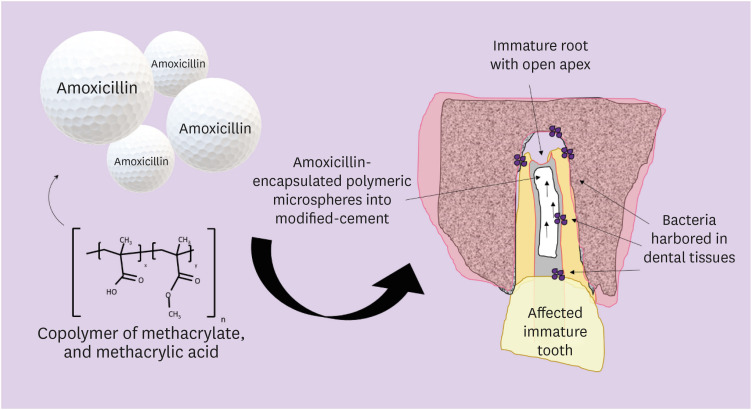
-
Abstract
PDFPubReaderePub
Objectives In this study, we investigated the potential of amoxicillin-loaded polymeric microspheres to be delivered to tooth root infection sites via a bioactive reparative cement.
Materials and Methods Amoxicillin-loaded microspheres were synthesized by a spray-dray method and incorporated at 2.5% and 5% into a mineral trioxide aggregate cement clinically used to induce a mineralized barrier at the root tip of young permanent teeth with incomplete root development and necrotic pulp. The formulations were modified in liquid:powder ratios and in composition by the microspheres. The optimized formulations were evaluated
in vitro for physical and mechanical eligibility. The morphology of microspheres was observed under scanning electron microscopy.Results The optimized cement formulation containing microspheres at 5% exhibited a delayed-release response and maintained its fundamental functional properties. When mixed with amoxicillin-loaded microspheres, the setting times of both test materials significantly increased. The diametral tensile strength of cement containing microspheres at 5% was similar to control. However, phytic acid had no effect on this outcome (
p > 0.05). When mixed with modified liquid:powder ratio, the setting time was significantly longer than that original liquid:powder ratio (p < 0.05).Conclusions Lack of optimal concentrations of antibiotics at anatomical sites of the dental tissues is a hallmark of recurrent endodontic infections. Therefore, targeting the controlled release of broad-spectrum antibiotics may improve the therapeutic outcomes of current treatments. Overall, these results indicate that the carry of amoxicillin by microspheres could provide an alternative strategy for the local delivery of antibiotics for the management of tooth infections.
-
Citations
Citations to this article as recorded by- Functionalized calcium carbonate microparticles in ethyl cellulose films: A vehicle for sustained amoxicillin release for medical applications
Petru Niga, Simone Sala, Jenny Rissler, Lina Nyström, Anna Fureby, Ulla Elofsson, Joachim Schoelkopf, Roger Roth, Patrick Gane, Mehnath Sivaraj
PLOS One.2026; 21(4): e0320280. CrossRef - Local drug delivery for regeneration and disinfection in endodontics: A narrative review
Anu Elsa Swaroop, Sylvia Mathew, P. Harshini, Shruthi Nagaraja
Journal of Conservative Dentistry and Endodontics.2025; 28(2): 119. CrossRef - Modified Mineral Trioxide Aggregate—A Versatile Dental Material: An Insight on Applications and Newer Advancements
C. Pushpalatha, Vismaya Dhareshwar, S. V. Sowmya, Dominic Augustine, Thilla Sekar Vinothkumar, Apathsakayan Renugalakshmi, Amal Shaiban, Ateet Kakti, Shilpa H. Bhandi, Alok Dubey, Amulya V. Rai, Shankargouda Patil
Frontiers in Bioengineering and Biotechnology.2022;[Epub] CrossRef - Local Drug Delivery Systems for Vital Pulp Therapy: A New Hope
Ardavan Parhizkar, Saeed Asgary, Carlo Galli
International Journal of Biomaterials.2021; 2021: 1. CrossRef
- Functionalized calcium carbonate microparticles in ethyl cellulose films: A vehicle for sustained amoxicillin release for medical applications
- 2,679 View
- 17 Download
- 4 Crossref
- Reference values for pulp oxygen saturation as a diagnostic tool in endodontics: a systematic review and meta-analysis
- Paula Lambert, Sergio Augusto Quevedo Miguens, Caroline Solda, Juliana Tomaz Sganzerla, Leandro Azambuja Reichert, Carlos Estrela, Fernando Branco Barletta
- Restor Dent Endod 2020;45(4):e48. Published online October 5, 2020
- DOI: https://doi.org/10.5395/rde.2020.45.e48
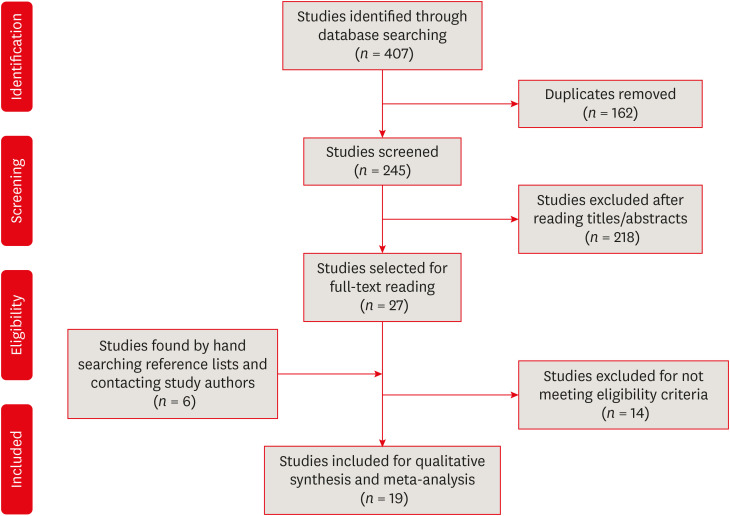
-
Abstract
PDFPubReaderePub
Objectives This systematic review aimed to identify mean oxygen saturation values (SpO2) using pulse oximetry in permanent maxillary anterior teeth.
Materials and Methods The MEDLINE, Scientific Electronic Library Online, Cochrane Central Register of Controlled Trials, EMBASE, and Literatura Latino Americana em Ciências da Saúde electronic databases were searched. Combinations and variations of “oximetry” AND “dental pulp test” were used as search terms. Studies reporting means and standard deviations of SpO2 values were included. Two reviewers independently extracted data following the Preferred Reporting Items for Systematic Reviews and Meta-Analyses checklist. Heterogeneity was assessed using the
I 2 statistic, and all analyses were performed using R software. Study quality was assessed using the Quality Assessment of Diagnostic Accuracy Studies-2 tool and the Newcastle-Ottawa scale.Results Of the 251 studies identified, 19 met the eligibility criteria and were included (total sample, 4,541 teeth). In the meta-analysis, the mean SpO2 values were 84.94% (95% confidence interval [CI], 84.85%–85.04%) for the central incisors, 89.29% (95% CI, 89.22%–89.35%) for the lateral incisors, and 89.20% (95% CI, 89.05%–89.34%) for the canines. The studies were predominantly low-quality due to the high risk of bias associated with the index test, unclear risk regarding patient selection, and concerns about outcome assessment.
Conclusions Although most studies were low-quality, the oxygen saturation levels in normal pulp could be established (minimum saturation, 77.52%). Despite the risk of bias of the included studies, the reference values reported herein are clinically relevant for assessments of changes in pulp status.
Trial Registration International Prospective Register of Systematic Reviews Identifier:
CRD42018085598 -
Citations
Citations to this article as recorded by- Lower dental pulp oxygen saturation in children with molar incisor hypomineralization: a cross-sectional study
Jade de Souza CAVALCANTE, Carlos ESTRELA, Fabrício Kitazono de CARVALHO, Francisco Wanderley Garcia de PAULA-SILVA, Manoel Damião de SOUSA-NETO, Kelly Fernanda MOLENA, Alexandra Mussolino de QUEIROZ
Brazilian Oral Research.2026;[Epub] CrossRef - Reference Values for Pulse Oximetry Testing in Permanent Teeth: A Systematic Review and Meta‐Analysis
Lilian Tietz, Theodoro Weissheimer, Cassiano Kuchenbecker Rösing, Marcus Vinicius Reis Só
International Endodontic Journal.2026;[Epub] CrossRef - Clinical Validation of Smartphone-Enabled Pulse Oximetry for Objective Pulp Vitality Assessment: A Diagnostic Accuracy Study
Celso Luiz Caldeira, Stephanie Isabel Diaz Zamalloa, Claudia Regina Guimaro Sakitani, Fernando Branco Barletta, Marinella Holzhausen
Journal of Endodontics.2025; 51(12): 1752. CrossRef - Laser Doppler Flowmetry and Continuous Tissue Oxygenation Monitoring: Best of Vitality Tests?
Herman J. J. Roeykens, Rani D’haese, Wolfgang Jacquet, Roeland J. G. De Moor, Stefan Vandeweghe
Oral.2025; 5(4): 83. CrossRef - Diagnostic accuracy of Transmitted-light plethysmography for the assessment of pulpal circulation in traumatized young permanent incisors
Satoko Kakino, Hiroaki Ohki, Kaori Kohi, Yuko Matsumura, Tsutomu Iwamoto
Scientific Reports.2025;[Epub] CrossRef - Future trends in endodontics
Foo Suanhow, Tawil Bill
Journal of Applied Biotechnology & Bioengineering.2024; 11(1): 1. CrossRef - Assessment of Pulpal Oxygen Saturation in Caries-free and Carious Maxillary Primary Central Incisors Using a Customized Dental Pulse Oximeter
Kranthi Reddy Kanumuru, Nancy Solomon, Hemalatha Ramkumar, Shankar Paulindraraj, Trophimus Gnanabagyan Jayakaran, Senthil Dakshinamoorthy
International Journal of Clinical Pediatric Dentistry.2023; 16(4): 560. CrossRef - Age-Related Variation of Pulpal Oxygen Saturation in Healthy Primary and Permanent Teeth in Children: A Clinical Study
Andreea Igna, Darian Rusu, Emilia Ogodescu, Ștefania Dinu, Marius Boariu, Adrian Voicu, Ștefan-Ioan Stratul
Journal of Clinical Medicine.2022; 12(1): 170. CrossRef - Pulp oxygen saturation measurement as a diagnostic tool for assessing pulp status in primary teeth: A systematic review and meta-analysis
Kanamarlapudi Venkata Saikiran, Deepa Gurunathan, Sainath Reddy Elicherla, Sreekanth Kumar Mallineni, Sivakumar Nuvvula
Journal of Indian Society of Pedodontics and Preventive Dentistry.2022; 40(4): 349. CrossRef - Diagnostic Value of Serum Chitinase‐3‐Like Protein 1 for Liver Fibrosis: A Meta‐analysis
Xiaoting Huang, Jialing Zhuang, Yongqiang Yang, Jiaxin Jian, Wen Ai, Chunyong Liu, Wenzhi Tang, Changyu Jiang, Yongshen He, Lesheng Huang, Se Peng, Jin Shui Pan
BioMed Research International.2022;[Epub] CrossRef - Assessment of Pulpal Status in Primary Teeth Following Direct Pulp Capping in an Experimental Canine Model
Andreea Igna, Cornel Igna, Mariana Ioana Miron, Larisa Schuszler, Roxana Dascălu, Mihaela Moldovan, Adrian Aristide Voicu, Carmen Darinca Todea, Marius Boariu, Maria-Alexandra Mârțu, Ștefan-Ioan Stratul
Diagnostics.2022; 12(8): 2022. CrossRef
- Lower dental pulp oxygen saturation in children with molar incisor hypomineralization: a cross-sectional study
- 3,465 View
- 47 Download
- 11 Crossref
- Shade reproduction and the ability of lithium disilicate ceramics to mask dark substrates
- Maryam Iravani, Sayna Shamszadeh, Narges Panahandeh, Seyedeh Mahsa Sheikh-Al-Eslamian, Hassan Torabzadeh
- Restor Dent Endod 2020;45(3):e41. Published online July 16, 2020
- DOI: https://doi.org/10.5395/rde.2020.45.e41
-
Abstract
PDFPubReaderePub
Objectives This study aimed to evaluate the ability of lithium disilicate ceramics to reproduce the A2 shade and to mask A4 substrates.
Materials and Methods Twenty-four discs (8 mm in diameter, shade A2) of high translucency (groups 1–3) and low translucency (groups 4–6) of IPS e.max ceramic with different thicknesses (0.5, 0.75, and 1 mm) were fabricated as monolithic structures. In addition, discs of medium opacity (group 7–8) with different core/veneer combinations (0.3 mm/0.7 mm and 0.5 mm/0.5 mm) were fabricated as bilayer structures. Specimens were superimposed on an A4 substrate (complex). The color changes of the complex were measured using a spectrophotometer on a black background, and the ΔE values of the complex were compared with either the A4 substrate or the A2 shade tab. One-way analysis of variance, the Tukey honest significant difference test, and the Fisher test were used to analyze the data (
p < 0.05).Results Significant between-group differences were found for comparisons to both the A4 substrate and the A2 shade (
p < 0.05). When compared with the A4 substrate, the ΔE values in all groups were in the non-acceptable range. When compared with the A2 shade, the ΔE values in all groups, except groups 2 and 3, were in the clinically acceptable range.Conclusions All translucencies and thicknesses masked the underlying dark substrate. However, the low-translucency IPS e.max Press better reproduced the A2 shade.
-
Citations
Citations to this article as recorded by- The effects of material thicknesses and substrates on the translucency and color masking ability of additively manufactured definitive crown materials
Ting Wang, Yun‐Ju Wang, Chao‐Chieh Yang, John A. Levon, Tien‐Min G. Chu, Wei‐Shao Lin
Journal of Prosthodontics.2026;[Epub] CrossRef - The effect of thickness, cement type, and substrate color on the optical properties of three-dimensional-printed composite resin
Elif Önal, Zeynep Şahin, Lale Karaağaçlioğlu
The Journal of Indian Prosthodontic Society.2026; 26(2): 150. CrossRef - The Impact of Surface Treatments and 3D Printing Machines on the Biaxial Flexural Strength of 3D-Printed Composite Resins
Mohammed K. Fahmi
The Open Dentistry Journal.2025;[Epub] CrossRef - Masking Ability of the Combined Application of Opaque Resin Composite and High‐Translucency Zirconia on Discolored Substrates
Shuping Chen, Lei Jiang, Run Chen
Journal of Esthetic and Restorative Dentistry.2025; 37(10): 2298. CrossRef - Comparative Evaluation of the Translucency of Polyetheretherketone (PEEK) Veneered With Two Different Materials: An In Vitro Study
M P Chinmayi, Gautam Shetty, S M Kedar, Lokesh B Kanchan, Rohit S Kundu, Krishna Kumar U, Maria Jenifer
Cureus.2025;[Epub] CrossRef - Masking capacity of minimally invasive lithium disilicate restorations on discolored teeth—The impact of ceramic thickness, the material's translucency, and the cement color
Kevser Pala, Eva Maria Reinshagen, Thomas Attin, Jürg Hüsler, Ronald E. Jung, Alexis Ioannidis
Journal of Esthetic and Restorative Dentistry.2024; 36(1): 107. CrossRef - Comparing the color match of monolithic CAD-CAM dental ceramics with the VITA Classical shade guide
Mohammadjavad Shirani, Maryam Emami, Ramin Mosharraf, Omid Savabi, Mehrdad Akhavankhaleghi, Kamran Azadbakht
The Journal of Prosthetic Dentistry.2024; 132(3): 605. CrossRef - Quantitative examination of factors influencing the colour reproduction ability of lithium disilicate glass-ceramics
József Saláta, Ferenc Szabó, Péter Csuti, Melinda Antal, Péter Márton, Péter Hermann, Judit Borbély, Emese Ábrám
BMC Oral Health.2024;[Epub] CrossRef - Aesthetic restoration using liner-treated lithium disilicate laminate veneers in discolored teeth after endodontic treatment : A case report
Ji-Hyun Kim, Min-Soo Bae, Yeon-Hee Park, Jung-Jin Lee, Tae-Sung Bae3, Jae-Min Seo
Korean Journal of Dental Materials.2023; 50(2): 91. CrossRef - Can we use the translucency parameter to predict the CAD/CAM ceramic restoration aesthetic?
Jie Wang, Jiawei Yang, Kaige Lv, Hongming Zhang, Hui Huang, Xinquan Jiang
Dental Materials.2023; 39(3): e1. CrossRef - Final Color of CAD-CAM Produced Thin Lithium Disilicate Ceramics Cemented with Different Colored Resin Cements on Darker Backgrounds
Merve BANKOĞLU GÜNGÖR
ADO Klinik Bilimler Dergisi.2023; 12(2): 234. CrossRef - Masking Ability of Monolithic and Layered Zirconia Crowns on Discolored Substrates
Cristina Gasparik, Manuela Maria Manziuc, Alexandru Victor Burde, Javier Ruiz-López, Smaranda Buduru, Diana Dudea
Materials.2022; 15(6): 2233. CrossRef - Effects of background color and thickness on the optical properties of CAD-CAM resin-matrix ceramics
Afnan F. Alfouzan, Sarah M. Alnafaiy, Lama S. Alsaleh, Noor H. Bawazir, Hanan N. Al-Otaibi, Sara M. Al Taweel, Huda A. Alshehri, Nawaf Labban
The Journal of Prosthetic Dentistry.2022; 128(3): 497.e1. CrossRef - Effect of CAD/CAM Ceramic Thickness on Shade Masking Ability of Discolored Teeth: In Vitro Study
Passent Ellakany, Marwa Madi, Nourhan M. Aly, Zainb S. Al-Aql, Maher AlGhamdi, Abdulrahman AlJeraisy, Adel S. Alagl
International Journal of Environmental Research and Public Health.2021; 18(24): 13359. CrossRef
- The effects of material thicknesses and substrates on the translucency and color masking ability of additively manufactured definitive crown materials
- 2,765 View
- 25 Download
- 14 Crossref
 First
First Prev
Prev

