
Search
- Page Path
- HOME > Search
Research Articles
- Microleakage and characteristics of resin-tooth tissues interface of a self-etch and an etch-and-rinse adhesive systems
- Xuan Vinh Tran, Khanh Quang Tran
- Restor Dent Endod 2021;46(2):e30. Published online May 18, 2021
- DOI: https://doi.org/10.5395/rde.2021.46.e30
-
 Abstract
 PDF
PDF PubReader
 ePub
ePub Objectives This study was conducted to compare the microleakage and characteristics of the resin-tooth tissue interface between self-etch and etch-and-rinse adhesive systems after 48 hours and 3 months.
Materials and Methods 40 extracted premolar teeth were randomly divided into 2 groups: 1-step self-etch adhesive system – Optibond™ All-In-One, and 2-step etch-and-rinse adhesive system - Adper™ Single Bond 2. Both groups were subjected to 500 thermocycles (5°C–55°C) before scanning electron microscope (SEM) analysis or microleakage trial at 48-hour and 3-month time periods.
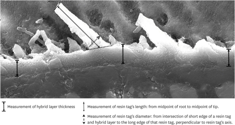
Results SEM images showed the hybrid layer thickness, diameter, and length of resin tags of the self-etch adhesive (0.42 ± 0.14 µm; 1.49 ± 0.45 µm; 16.35 ± 14.26 µm) were smaller than those of the etch-and-rinse adhesive (4.39 ± 1.52 µm; 3.49 ± 1 µm; 52.81 ± 35.81 µm). In dentin, the microleakage scores of the 2 adhesives were not different in both time periods (48 hours/3 months). However, the microleakage score of etch-and-rinse adhesive increased significantly after 3 months (0.8 ± 0.63 and 1.9 ± 0.88,
p < 0.05).Conclusions The self-etch adhesive exhibited better long-term sealing ability in dentin when compared to that of the etch-and-rinse adhesive. The greater hybrid layer thickness and dimensions of resin tags did not guarantee reliable, long-lasting sealing in the bonding area.
-
Citations
Citations to this article as recorded by
- A systematic review of shear bond strength of sixth- and fourth-generation adhesives in primary teeth
Maryam Hajiahmadi, Najmeh Akhlaghi, Hamid Mosleh, Ehsan Samani, Sheida Bagheri, Zohreh Salehi
Dental Research Journal.2026;[Epub] CrossRef - Comparative evaluation of shear bond strength of nano-hybrid composites to dentin: influence of universal adhesives versus two-step etch-and-rinse adhesives
Soodabeh Shoale, Mahsa Kowkabi, Artam Enayat, Hamed Manafi
Discover Applied Sciences.2026;[Epub] CrossRef - Effect of aging on microshearing bond strength of different adhesive systems
Cansu Dağdelen Ahısha, Mine Betül Üçtaşlı
BMC Oral Health.2026;[Epub] CrossRef - Efficacy of different adhesive systems in bonding direct resin composite restorations: a systematic review and meta-analysis
Ravinder S. Saini, Rajesh Vyas, Sunil Kumar Vaddamanu, Syed Altafuddin Quadri, Seyed Ali Mosaddad, Artak Heboyan
Evidence-Based Dentistry.2025; 26(2): 115. CrossRef - Characterisation of universal adhesive bonded resin-dentin interface after focused ultrasound smear layer conditioning
Cheryl Fu, Peta L. Clode, Amr S. Fawzy
International Journal of Adhesion and Adhesives.2025; 142: 104115. CrossRef - Effect of Dentin Pretreatment With Dimethyl Sulfoxide Solution on Interfacial Fracture Toughness of Composite Resin to Wet and Dry Dentin
Fatemeh Molaei, Mehrsima Ghavami-Lahiji, Seyedeh Maryam Tavangar, Hannah Wesley
International Journal of Dentistry.2025;[Epub] CrossRef - Resin tags formation by modified Renewal MI formulations in a carious dentine model
Nabih Alkhouri, Wendy Xia, Paul Ashley, Anne Young
Frontiers in Oral Health.2024;[Epub] CrossRef - Effect of propolis added to single‐bottle adhesives on water permeation through the hybrid layer
Lucineide Silva da Rocha, Daniela Ferreira de Oliveira, Cinthya Luna Veloso de Lima, Ticiano Gomes do Nascimento, Johnnatan Duarte de Freitas, Jeniffer Mclaine Duarte de Freitas, Isabel Cristina Celerino de Moraes Porto
European Journal of Oral Sciences.2024;[Epub] CrossRef - Exploration and preliminary clinical investigation of an adhesive approach for primary tooth restoration
Xiangqin Xu, Jiansheng Zhu, May Lei Mei, Huaying Wu, Kaipeng Xie, Shoulin Wang, Yaming Chen
The Journal of Biomedical Research.2023; 37(2): 138. CrossRef - Adhesion to enamel and dentine: an update
Rana Alkattan
Primary Dental Journal.2023; 12(3): 33. CrossRef - Effects of carbodiimide combined with ethanol–wet bonding pretreatment on dentin bonding properties: an in vitro study
Xiaoxiao You, Long Chen, Jie Xu, Sihui Li, Zhenghao Zhang, Ling Guo
PeerJ.2022; 10: e14238. CrossRef - The effects of amalgam contamination and different surface modifications on microleakage of dentin bonded to bulk fill composite when using different adhesive protocols
Nojoud Alshehri, Abdullah Aljamhan, Mohammed Bin-Shuwaish
BMC Oral Health.2022;[Epub] CrossRef - Development of low-shrinkage dental adhesives via blending with spiroorthocarbonate expanding monomer and unsaturated epoxy resin monomer
Zonghua Wang, Xiaoran Zhang, Shuo Yao, Jiaxin Zhao, Chuanjian Zhou, Junling Wu
Journal of the Mechanical Behavior of Biomedical Materials.2022; 133: 105308. CrossRef - Influence of silver nanoparticles on the resin-dentin bond strength and antibacterial activity of a self-etch adhesive system
Jia Wang, Wei Jiang, Jingping Liang, Shujun Ran
The Journal of Prosthetic Dentistry.2022; 128(6): 1363.e1. CrossRef
- A systematic review of shear bond strength of sixth- and fourth-generation adhesives in primary teeth
- 3,491 View
- 59 Download
- 13 Web of Science
- 14 Crossref

-
In vitro evaluation of a newly produced resin-based endodontic sealer - Yoo-Seok Song, Yoorina Choi, Myung-Jin Lim, Mi-Kyung Yu, Chan-Ui Hong, Kwang-Won Lee, Kyung-San Min
- Restor Dent Endod 2016;41(3):189-195. Published online July 26, 2016
- DOI: https://doi.org/10.5395/rde.2016.41.3.189
-
Abstract
PDFPubReaderePub
Objectives A variety of root canal sealers were recently launched to the market. This study evaluated physicochemical properties, biocompatibility, and sealing ability of a newly launched resin-based sealer (Dia-Proseal, Diadent) compared to the existing root canal sealers (AHplus, Dentsply DeTrey and ADseal, Metabiomed).
Materials and Methods The physicochemical properties of the tested sealers including pH, solubility, dimensional change, and radiopacity were evaluated. Biocompatibility was measured using the 3-(4,5-dimethylthiazol-2-yl)-2,5-diphenyltetrazolium bromide (MTT) assay. For microleakage test, single-rooted teeth were instrumented, and obturated with gutta-percha and one of the sealers (
n = 10). After immersion in 1% methylene blue solution for 2 weeks, the specimens were split longitudinally. Then, the maximum length of staining was measured. Statistical analysis was performed by one-way analysis of variance followed by Tukey test (p = 0.05).Results Dia-Proseal showed the highest pH value among the tested sealers (
p < 0.05). ADseal showed higher dimensional change compared to AHplus and Dia-Proseal (p < 0.05). The solubility values of AHplus and Dia-Proseal were similar, whereas ADseal had the lowest solubility value (p < 0.05). The flow values of sealer in increasing order were AHplus, DiaProseal, and ADseal (p < 0.05). The radiopacity of AHplus was higher than those of ADseal and Dia-Proseal (p < 0.05). The cell viability of the tested materials was statistically similar throughout the experimental period. There were no significant differences in microleakage values among the tested samples.Conclusions The present study indicates that Dia-Proseal has acceptable physicochemical properties, biocompatibility, and sealing ability.
-
Citations
Citations to this article as recorded by- Comparative analysis between resin-based root canal sealer and recent bioceramic-based root canal sealers using MicroCT, film thickness, and solubility
Amira Galal Ismail, Manar M. Galal, Tamer M. Hamdy
Journal of Oral Biology and Craniofacial Research.2026; 16(2): 101400. CrossRef - Comparison of Apical Sealing Ability of Different Endodontic Sealers – An In Vitro Study
Supriya Patil, Rahul Singh, B Jyothi Lekshmi, Sameer Ahmed Khan, H Shalini, Prashanth Kumar Katta
Journal of Pharmacy and Bioallied Sciences.2025; 17(Suppl 1): S513. CrossRef - Comparative evaluation of ICON resin infiltration and bioactive glass adhesive for managing initial caries lesions using quantitative light-induced fluorescence: a randomized clinical trial
Zakereyya S.M. Albashaireh, Susan N. Al-Khateeb, Malak K. Altallaq
Journal of Dentistry.2025; 159: 105853. CrossRef - An In Vitro Comparison of Epoxy Resin Sealer Removal During Endodontic Retreatment
Prashant A Bondarde, Aditi S Patkar, Aishwarya R Pawar, Rukmini Pande, Akshata Deshpande, Rachana S Agrawal, Seema Gupta
Cureus.2025;[Epub] CrossRef - Stereomicroscopic evaluation of sealing ability of four different root canal sealers: an in-vitro study
Sonam Sah, Panna Mangat, Ajay Kumar, Neha Sah, Ganiga Channaiah Shivakumar, Marco Di Blasio, Gabriele Cervino, Giuseppe Minervini
BMC Oral Health.2024;[Epub] CrossRef - Physicochemical properties of AH plus bioceramic sealer, Bio-C Sealer, and ADseal root canal sealer
Tamer M. Hamdy, Manar M. Galal, Amira Galal Ismail, Shehabeldin Saber
Head & Face Medicine.2024;[Epub] CrossRef - Biological investigation of resinous endodontic sealers containing calcium hydroxide
Carlos Roberto Emerenciano Bueno, Francine Benetti, Marina Tolomei Sandoval Cury, Ana Maria Veiga Vasques, Leopoldo Cosme-Silva, Índia Olinta de Azevedo Queiroz, Ana Cláudia Rodrigues da Silva, Rogério de Castilho Jacinto, Luciano Tavares Angelo Cintra, E
PLOS ONE.2023; 18(7): e0287890. CrossRef - Comparison of the apical seal obtained by Adseal, Proseal, and AH26 sealers in root canal obturation with lateral compaction technique
Akam Saeidi, Romina Hajipour, Elham Mahmoudi, Farideh Feizi, Soraya Khafri
Dental Research Journal.2023;[Epub] CrossRef - Evaluation of Cytotoxicity of Calcium Silicate-based Mineral Trioxide Aggregate Sealers: A Systematic Review of In Vitro Studies
Nezar Boreak, Mazen Ahmed Qadi, Faisal Hadi Khormi, Luay Mutaen Faqiri, Sadeem Omar Zaylai, Yaser Ali Jad, Bassam Ali Hamdi, Asayil Juraybi
The Journal of Contemporary Dental Practice.2023; 24(8): 610. CrossRef - Comparative evaluation of push-out bond strength of bioceramic and epoxy sealers after using various final irrigants: An in vitro study
Chandrasekhar Veeramachaneni, Swathi Aravelli, Sreeja Dundigalla
Journal of Conservative Dentistry.2022; 25(2): 145. CrossRef - Comparative Evaluation of Root Reinforcement Using MTA-based, Epoxy Resin-based, and Silicone-based Endodontic Sealers in Canals Instrumented with Single-file Rotary System: An In Vitro Study
Reshma Rajasekhar, Varsha Maria Sebastian, Farhat Nasreen, Pramod Junjanna, Azeem Hassan, Venkidesh Hari Maratt
The Journal of Contemporary Dental Practice.2022; 22(10): 1098. CrossRef - The Short-Term Antibacterial Activity of Three Selected Endodontic Sealers against Enterococcus faecalis Bacterial Culture
Matej Rosa, Yuliya Morozova, Roman Moštěk, Pavel Holík, Lucia Somolová, Barbora Novotná, Soňa Zábojníková, Kateřina Bogdanová, Kateřina Langová, Iva Voborná, Lenka Pospíšilová, Josef Paul Kovařík
Life.2022; 12(2): 158. CrossRef - Antimicrobial potential of AH Plus supplemented with bismuth lipophilic nanoparticles on E. faecalis isolated from clinical isolates
Jesús Alejandro Torres-Betancourt, Rene Hernandez-Delgadillo, Jorge Jaime Flores-Treviño, Juan Manuel Solís-Soto, Nayely Pineda-Aguilar, Maria Argelia Akemi Nakagoshi-Cepeda, Rosa Isela Sánchez-Nájera, Shankararaman Chellam, Claudio Cabral-Romero
Journal of Applied Biomaterials & Functional Materials.2022;[Epub] CrossRef - A micro-computed tomographic study using a novel test model to assess the filling ability and volumetric changes of bioceramic root repair materials
Fernanda Ferrari Esteves Torres, Jader Camilo Pinto, Gabriella Oliveira Figueira, Juliane Maria Guerreiro-Tanomaru, Mario Tanomaru-Filho
Restorative Dentistry & Endodontics.2021;[Epub] CrossRef - Energy-Dispersive X-Ray Spectrometry Analysis and Radiopacity of Five Different Root Canal Sealers
Gözde Kandemir Demirci, Mehmet Emin Kaval, Seniha Miçooğulları Kurt, Burcu Serefoglu, Pelin Güneri, Michael Hülsmann, Mehmet Kemal Caliskan
Brazilian Dental Journal.2021; 32(5): 1. CrossRef - Ultrasonic vibration and thermo‐hydrodynamic technique for filling root canals: Technical overview and a case series
Yong‐Sik Cho
International Endodontic Journal.2021; 54(9): 1668. CrossRef - Physicochemical Properties of Two Generations of MTA-Based Root Canal Sealers
Sawsan Abu Zeid, Hadeel Yaseen Edrees, Abeer Abdulaziz Mokeem Saleh, Osama S. Alothmani
Materials.2021; 14(20): 5911. CrossRef - Micro-computed tomographic evaluation of a new system for root canal filling using calcium silicate-based root canal sealers
Mario Tanomaru-Filho, Fernanda Ferrari Esteves Torres, Jader Camilo Pinto, Airton Oliveira Santos-Junior, Karina Ines Medina Carita Tavares, Juliane Maria Guerreiro-Tanomaru
Restorative Dentistry & Endodontics.2020;[Epub] CrossRef - Radiopacity of endodontic materials using two models for conversion to millimeters of aluminum
Victor Manuel OCHOA-RODRÍGUEZ, Jorge Homero WILCHES-VISBAL, Barbara ROMA, Hernán COAGUILA-LLERENA, Mário TANOMARU-FILHO, Andréa GONÇALVES, Rubens SPIN-NETO, Gisele FARIA
Brazilian Oral Research.2020;[Epub] CrossRef - Flow characteristics and alkalinity of novel bioceramic root canal sealers
Anastasios Katakidis, Konstantinos Sidiropoulos, Elisabeth Koulaouzidou, Christos Gogos, Nikolaos Economides
Restorative Dentistry & Endodontics.2020;[Epub] CrossRef - Micro-computed tomographic evaluation of the flow and filling ability of endodontic materials using different test models
Fernanda Ferrari Esteves Torres, Juliane Maria Guerreiro-Tanomaru, Gisselle Moraima Chavez-Andrade, Jader Camilo Pinto, Fábio Luiz Camargo Villela Berbert, Mario Tanomaru-Filho
Restorative Dentistry & Endodontics.2020;[Epub] CrossRef - SELECTED PROPERTIES OF CONTEMPORARY ENDODONTIC SEALERS: PART 1
M Rosa, Y Morozova, R Moštěk, A Jusku, V Kováčová, L Somolová, I Voborná, T Kovalský
Česká stomatologie a praktické zubní lékařství.2020; 120(4): 107. CrossRef - Calcium phosphates as fillers for methacrylate-based sealer
Flávia Veronezi Rostirolla, Vicente Castelo Branco Leitune, Fabio Rocha Bohns, Fernando Freitas Portella, Susana Maria Werner Samuel, Fabrício Mezzomo Collares
Clinical Oral Investigations.2019; 23(12): 4417. CrossRef - Do in vitro solubility studies on endodontic sealers demonstrate a high level of evidence? A systematic review
Ankur Razdan, Ana Raquel Benetti, Lars Bjørndal
Acta Odontologica Scandinavica.2019; 77(4): 253. CrossRef - Physicochemical properties of two epoxy resin-based sealants: Topseal® and Adseal™. a comparative study
Julio César Cardona-Hidalgo, José Manuel González-Carreño, Julio César Avendaño-Rueda
Revista Facultad de Odontología.2019;[Epub] CrossRef - In Vitro Comparison of Biocompatibility of Calcium Silicate-Based Root Canal Sealers
Ju Kyung Lee, Sunil Kim, Sukjoon Lee, Hyeon-Cheol Kim, Euiseong Kim
Materials.2019; 12(15): 2411. CrossRef - Physicochemical Properties of Epoxy Resin-Based and Bioceramic-Based Root Canal Sealers
Ju Kyung Lee, Sang Won Kwak, Jung-Hong Ha, WooCheol Lee, Hyeon-Cheol Kim
Bioinorganic Chemistry and Applications.2017; 2017: 1. CrossRef
- Comparative analysis between resin-based root canal sealer and recent bioceramic-based root canal sealers using MicroCT, film thickness, and solubility
- 2,579 View
- 21 Download
- 27 Crossref
- Marginal microleakage of cervical composite resin restorations bonded using etch-and-rinse and self-etch adhesives: two dimensional vs. three dimensional methods
- Maryam Khoroushi, Ailin Ehteshami
- Restor Dent Endod 2016;41(2):83-90. Published online April 18, 2016
- DOI: https://doi.org/10.5395/rde.2016.41.2.83
-
Abstract
PDFPubReaderePub
Objectives This study was evaluated the marginal microleakage of two different adhesive systems before and after aging with two different dye penetration techniques.
Materials and Methods Class V cavities were prepared on the buccal and lingual surfaces of 48 human molars. Clearfil SE Bond and Single Bond (self-etching and etch-and-rinse systems, respectively) were applied, each to half of the prepared cavities, which were restored with composite resin. Half of the specimens in each group underwent 10,000 cycles of thermocycling. Microleakage was evaluated using two dimensional (2D) and three dimensional (3D) dye penetration techniques separately for each half of each specimen. Data were analyzed with SPSS 11.5 (SPSS Inc.), using the Kruskal-Wallis and Mann-Whitney U tests (α = 0.05).
Results The difference between the 2D and 3D microleakage evaluation techniques was significant at the occlusal margins of Single bond groups (
p = 0.002). The differences between 2D and 3D microleakage evaluation techniques were significant at both the occlusal and cervical margins of Clearfil SE Bond groups (p = 0.017 andp = 0.002, respectively). The difference between the 2D and 3D techniques was significant at the occlusal margins of non-aged groups (p = 0.003). The difference between these two techniques was significant at the occlusal margins of the aged groups (p = 0.001). The Mann-Whitney test showed significant differences between the two techniques only at the occlusal margins in all specimens.Conclusions Under the limitations of the present study, it can be concluded that the 3D technique has the capacity to detect occlusal microleakage more precisely than the 2D technique.
-
Citations
Citations to this article as recorded by- Post‐Gel Polymerization Shrinkage Strain and Marginal Integrity of Repeatedly Preheated Thermo‐Viscous and Matrix‐Modified Bulk‐Fill Resin Composite (Pre‐Clinical Study)
Ahmed Amir, Rasha Zaghlool, Mona Riad
Journal of Esthetic and Restorative Dentistry.2026; 38(1): 97. CrossRef - The current advancements in chitosan nanoparticles in the management of non-surgical periodontitis treatment
Mehrnaz Sadighi Shamami, Mohammad Ekhlaspour, Jameel M. A. Sulaiman, Radhwan Abdul Kareem, Nahed Mahmood Ahmed Alsultany, Kamyar Nasiri, Naghmeh Shenasa
Nanotoxicology.2025; 19(3): 290. CrossRef - Effect of different types of adhesive systems on the bond strength and marginal integrity of composite restorations in cavities prepared with the erbium laser—a systematic review
Deepti Dua, Ankur Dua, Eugenia Anagnostaki, Riccardo Poli, Steven Parker
Lasers in Medical Science.2022; 37(1): 19. CrossRef - Comparing the Ability of Various Resin-Based Composites and Techniques to Seal Margins in Class-II Cavities
Abdullah Saleh Aljamhan, Sultan Ali Alhazzaa, Abdulrahman Hamoud Albakr, Syed Rashid Habib, Muhammad Sohail Zafar
Polymers.2021; 13(17): 2921. CrossRef - Comparison of the Ability of Two Brands of CBCT with That of SEM to Detect the Marginal Leakage of Class V Composite Resin Restorations
Mitra Karbasi Kheir, Leili Khayam, Mehrbakhsh Nilashi
The Scientific World Journal.2021; 2021: 1. CrossRef - Analysis of microleakage and marginal gap presented by new polymeric systems in class V restorations: An in vitro study
Jefferson Ricardo Pereira, Hugo Alberto Vidotti, Lindomar Corrêa Júnior, Alef Vermudt, Mauro de Souza Almeida, Saulo Pamato
The Saudi Dental Journal.2021; 33(3): 156. CrossRef - Hydrolysis-resistant and stress-buffering bifunctional polyurethane adhesive for durable dental composite restoration
Jiahui Zhang, Xiaowei Guo, Xiaomeng Zhang, Huimin Wang, Jiufu Zhu, Zuosen Shi, Song Zhu, Zhanchen Cui
Royal Society Open Science.2020; 7(7): 200457. CrossRef - A comparison of the marginal and internal fit of porcelain laminate veneers fabricated by pressing and CAD-CAM milling and cemented with 2 different resin cements
Ziad N. Al-Dwairi, Rana M. Alkhatatbeh, Nadim Z. Baba, Charles J. Goodacre
The Journal of Prosthetic Dentistry.2019; 121(3): 470. CrossRef - Microleakage in class V cavities prepared using conventional method versus Er:YAG laser restored with glass ionomer cement or resin composite
Sertac Peker, Figen Eren Giray, Basak Durmus, Nural Bekiroglu, Betül Kargül, Mutlu Özcan
Journal of Adhesion Science and Technology.2017; 31(5): 509. CrossRef
- Post‐Gel Polymerization Shrinkage Strain and Marginal Integrity of Repeatedly Preheated Thermo‐Viscous and Matrix‐Modified Bulk‐Fill Resin Composite (Pre‐Clinical Study)
- 2,193 View
- 10 Download
- 9 Crossref
- Effect of 38% carbamide peroxide on the microleakage of silorane-based versus methacrylate-based composite restorations
- Sedighe Sadat Hashemi Kamangar, Maryam Ghavam, Nazanin Mahinfar, Seyed Jalal Pourhashemi
- Restor Dent Endod 2014;39(3):172-179. Published online May 13, 2014
- DOI: https://doi.org/10.5395/rde.2014.39.3.172
-
Abstract
PDFPubReaderePub
Objectives This study aimed to assess the effect of 38% carbamide peroxide on the microleakage of class V cavities restored with either a silorane-based composite or two methacrylate-based composites.
Materials and Methods A total of 96 class V cavities were prepared on the buccal surface of extracted human teeth with both enamel and dentin margins and were randomly assigned into three groups of Filtek P90 (3M-ESPE) + P90 system adhesive (3M-ESPE)(group A), Filtek Z250 (3M-ESPE) + Adper Prompt L-Pop (3M-ESPE)(group B) and Filtek Z350XT (3M-ESPE) + Adper Prompt L-Pop (group C). Half of the teeth were randomly underwent bleaching (38% carbamide peroxide, Day White, Discus Dental, applying for 15 min, twice a day for 14 day) while the remaining half (control) were not bleached. Dye penetration was measured following immersion in basic fuchsine. Data were statistically analyzed using Kruskal-Wallis and Mann-Whitney U tests at a level of 0.05.
Results No significant differences were found between composites in the control groups in enamel (
p = 0.171) or dentin (p = 0.094) margins. After bleaching, microleakage of Z250 (in enamel [p = 0.867] or dentin [p = 0.590] margins) and Z350 (in enamel [p = 0.445] or dentin [p = 0.591] margins) did not change significantly, but the microleakage of P90 significantly increased in both enamel (p = 0.042) and dentin (p = 0.002) margins.Conclusions No significant differences were noted between the bleached and control subgroups of two methacrylate-based composites in enamel or dentin margins. Microleakage of silorane-based composite significantly increased after bleaching.
- 1,570 View
- 3 Download
- Micro-CT evaluation of internal adaptation in resin fillings with different dentin adhesives
- Seung-Hoon Han, Sung-Ho Park
- Restor Dent Endod 2014;39(1):24-31. Published online January 20, 2014
- DOI: https://doi.org/10.5395/rde.2014.39.1.24
-
Abstract
PDFPubReaderePub
Objectives The purpose of present study was to evaluate the internal adaptation of composite restorations using different adhesive systems.
Materials and Methods Typical class I cavities were prepared in 32 human third molars. The teeth were divided into the following four groups: 3-step etch-and-rinse, 2-step etch-and-rinse, 2-step self-etch and 1-step self-etch system were used. After the dentin adhesives were applied, composite resins were filled and light-cured in two layers. Then, silver nitrate solution was infiltrated, and all of the samples were scanned by micro-CT before and after thermo-mechanical load cycling. For each image, the length to which silver nitrate infiltrated, as a percentage of the whole pulpal floor length, was calculated (%SP). To evaluate the internal adaptation using conventional method, the samples were cut into 3 pieces by two sectioning at an interval of 1 mm in the middle of the cavity and they were dyed with Rhodamine-B. The cross sections of the specimens were examined by stereomicroscope. The lengths of the parts where actual leakage was shown were measured and calculated as a percentage of real leakage (%RP). The values for %SP and %RP were compared.
Results After thermo-mechanical loading, all specimens showed significantly increased %SP compared to before thermo-mechanical loading and 1-step self-etch system had the highest %SP (
p < 0.05). There was a tendency for %SP and %RP to show similar microleakage percentage depending on its sectioning.Conclusions After thermo-mechanical load cycling, there were differences in internal adaptation among the groups using different adhesive systems.
-
Citations
Citations to this article as recorded by- Effect of different factors on microleakage and fracture strength of CAD‐CAM produced inlays
Meryem Gülce Subaşı, Gürel Pekkan, Meral Arslan Malkoç, Hilal Ekşi Özsoy
Journal of Prosthodontics.2025;[Epub] CrossRef - Non-Destructive In Vitro Evaluation of an Internal Adaptation of Recent Pulp-Capping Materials in Permanent Teeth Using OCT and Micro-CT
Ahmed Y. Alzahrani, Amani A. Al Tuwirqi, Nada O. Bamashmous, Turki A. Bakhsh, Eman A. El Ashiry
Children.2023; 10(8): 1318. CrossRef - Internal Adaptation of Cusp-weakened Class I Preparations Restored with Bulk-fill, Bi-layered, and Incremental Restorative Techniques: A Micro-CT Analysis
DH Floriani, RN Rached, SA Ignácio, EM Souza
Operative Dentistry.2022; 47(5): 527. CrossRef - An in vitro micro-CT assessment of bioactive restorative materials interfacial adaptation to dentin
Priyanka Angadala, Jyothi Mandava, Ravichandra Ravi, KoteswarRao Hanumanthu, Prasanthi Penmatsa, Hema Pulidindi
Dental Research Journal.2022; 19(1): 56. CrossRef - Tomographic Evaluation of the Internal Adaptation for Recent Calcium Silicate‐Based Pulp Capping Materials in Primary Teeth
A. A. Al Tuwirqi, E. A. El Ashiry, A. Y. Alzahrani, N. Bamashmous, T. A. Bakhsh, Iole Vozza
BioMed Research International.2021;[Epub] CrossRef - Micro-computed tomography in preventive and restorative dental research: A review
Mehrsima Ghavami-Lahiji, Reza Tayefeh Davalloo, Gelareh Tajziehchi, Paria Shams
Imaging Science in Dentistry.2021; 51(4): 341. CrossRef - Validation of a method of quantifying 3D leakage in dental restorations
Fabio A.P. Rizzante, Rana A.F. Sedky, Adilson Y. Furuse, Sorin Teich, Sérgio K. Ishikiriama, Gustavo Mendonça
The Journal of Prosthetic Dentistry.2020; 123(6): 839. CrossRef - Comparison of micro-CT and conventional dye penetration for microleakage assessment after different aging conditions
Rayssa Ferreira Zanatta, Annette Wiegand, Christian Dullin, Alessandra Bühler Borges, Carlos Rocha Gomes Torres, Marta Rizk
International Journal of Adhesion and Adhesives.2019; 89: 161. CrossRef - Comparison of Internal Adaptation of Bulk-fill and Increment-fill Resin Composite Materials
FS Alqudaihi, NB Cook, KE Diefenderfer, MC Bottino, JA Platt
Operative Dentistry.2019; 44(1): E32. CrossRef - Effects of occlusal cavity configuration on 3D shrinkage vectors in a flowable composite
Dalia Kaisarly, Moataz El Gezawi, Guangyun Lai, Jian Jin, Peter Rösch, Karl-Heinz Kunzelmann
Clinical Oral Investigations.2018; 22(5): 2047. CrossRef - Bonding Strategies of Resin Cement to Er, Cr:YSGG Lased Dentin: Micro-CT Evaluation and Microshear Bond Strength Testing
Gökçe Meriç, Simge Taşar, Kaan Orhan
The International Journal of Artificial Organs.2016; 39(2): 72. CrossRef - Calcium hypochlorite as a dentin deproteinization agent: Microleakage, scanning electron microscopy and elemental analysis
Michele Bortoluzzi de Conto Ferreira, Bruno Carlini Júnior, Daniel Galafassi, Delton Luiz Gobbi
Microscopy Research and Technique.2015; 78(8): 676. CrossRef
- Effect of different factors on microleakage and fracture strength of CAD‐CAM produced inlays
- 1,965 View
- 7 Download
- 12 Crossref
- Effect of different air-drying time on the microleakage of single-step self-etch adhesives
- Horieh Moosavi, Maryam Forghani, Esmatsadat Managhebi
- Restor Dent Endod 2013;38(2):73-78. Published online May 28, 2013
- DOI: https://doi.org/10.5395/rde.2013.38.2.73
-
Abstract
PDFPubReaderePub
Objectives This study evaluated the effect of three different air-drying times on microleakage of three self-etch adhesive systems.
Materials and Methods Class I cavities were prepared for 108 extracted sound human premolars. The teeth were divided into three main groups based on three different adhesives: Opti Bond All in One (OBAO), Clearfil S3 Bond (CSB), Bond Force (BF). Each main group divided into three subgroups regarding the air-drying time: without application of air stream, following the manufacturer's instruction, for 10 sec more than manufacturer's instruction. After completion of restorations, specimens were thermocycled and then connected to a fluid filtration system to evaluate microleakage. The data were statistically analyzed using two-way ANOVA and Tukey-test (α = 0.05).
Results The microleakage of all adhesives decreased when the air-drying time increased from 0 sec to manufacturer's instruction (
p < 0.001). The microleakage of BF reached its lowest values after increasing the drying time to 10 sec more than the manufacturer's instruction (p < 0.001). Microleakage of OBAO and CSB was significantly lower compared to BF in all three drying time (p < 0.001).Conclusions Increasing in air-drying time of adhesive layer in one-step self-etch adhesives caused reduction of microleakage, but the amount of this reduction may be dependent on the adhesive components of self-etch adhesives.
-
Citations
Citations to this article as recorded by- Species profile of volatile organic compounds emission and health risk assessment from typical indoor events in daycare centers
Hailin Zheng, Júlia Csemezová, Marcel Loomans, Shalika Walker, Florent Gauvin, Wim Zeiler
Science of The Total Environment.2024; 918: 170734. CrossRef - Development of Drying Process for Removal of Residual Moisture from Biomass Pretreated with Ethanol and Its Kinetic and Thermodynamic Analysis
Seo-Young Park, Jin-Hyun Kim
Biotechnology and Bioprocess Engineering.2021; 26(5): 814. CrossRef - Effect of 9.3 μm CO2 and 2.94 μm Er:YAG Laser vs. Bur Preparations on Marginal Adaptation in Enamel and Dentin of Mixed Class V Cavities Restored With Different Restorative Systems
Clara Isabel Anton y Otero, Enrico Di Bella, Ivo Krejci, Tissiana Bortolotto
Frontiers in Dental Medicine.2021;[Epub] CrossRef - Development of Drying Process for Removal of Residual Solvent from Crystalline Vancomycin and Kinetic and Thermodynamic Analysis Thereof
Tae-Hun Yoon, Jin-Hyun Kim
Biotechnology and Bioprocess Engineering.2020; 25(5): 777. CrossRef - Effect of adhesive air-drying time on bond strength to dentin: A systematic review and meta-analysis
Mohamed M. Awad, Ali Alrahlah, Jukka P. Matinlinna, Hamdi Hosni Hamama
International Journal of Adhesion and Adhesives.2019; 90: 154. CrossRef - Optical Evaluation of Enamel Microleakage with One-Step Self-Etch Adhesives
Alaa Turkistani, Maha Almutairi, Nouf Banakhar, Reem Rubehan, Sulafa Mugharbil, Ahmed Jamleh, Adnan Nasir, Turki Bakhsh
Photomedicine and Laser Surgery.2018; 36(11): 589. CrossRef - Improved drying method for removal of residual solvents from paclitaxel by pre-treatment with ethanol and water
Chung-Gi Lee, Jin-Hyun Kim
Process Biochemistry.2015; 50(6): 1031. CrossRef
- Species profile of volatile organic compounds emission and health risk assessment from typical indoor events in daycare centers
- 2,417 View
- 3 Download
- 7 Crossref
- Effective application duration of sodium ascorbate antioxidant in reducing microleakage of bonded composite restoration in intracoronally-bleached teeth
- Jae-Young Park, Tae-Yub Kwon, Young-Kyung Kim
- Restor Dent Endod 2013;38(1):43-47. Published online February 26, 2013
- DOI: https://doi.org/10.5395/rde.2013.38.1.43
-
Abstract
PDFPubReaderePub
Objectives The aim of this study was to determine an appropriate application duration of sodium ascorbate (SA) antioxidant gel in reducing microleakage of bonded composite restoration in intracoronally-bleached teeth.
Materials and Methods Eighty endodontically-treated human incisors were randomly divided into eight groups: control, no bleaching; IB and DB, immediate and delayed bonding after bleaching, respectively; S10m, S60m, S24h, S3d and S7d, bleaching + SA gel for 10 min, 60 min, 24 hr, 3 day and 7 day, respectively. For bleaching, a mixture of 30% hydrogen peroxide and sodium perborate was applied for 7 day. All access cavities were restored using One-Step adhesive (Bisco Inc.) and then Aelite LS Packable composite (Bisco Inc.). The bonded specimens were subjected to 500 thermal cycles, immersed in 1% methylene blue for 8 hr, and longitudinally sectioned. Microleakage was assessed with a 0 - 4 scoring system and analyzed using nonparametric statistical methods (α = 0.05).
Results Group IB showed a significantly higher microleakge than the control group (
p = 0.006) and group DB a statistically similar score to the control group (p > 0.999). Although groups S10m, S60m, and S24h exhibited significantly higher scores than group DB (p < 0.05), the microleakage in groups S3d and S7d was statistically similar to that in group DB (p = 0.771,p > 0.999).Conclusions Application of SA gel for 3 day after nonvital bleaching was effective in reducing microleakage of composite restoration in intracoronally-bleached teeth.
-
Citations
Citations to this article as recorded by-
Effect of Herbal Antioxidant on Push-out Bond Strength of Resin-based Composite to Dentin after Intracoronal Bleaching: An
in vitro
Study
Parinitha MS, Akshay G, Vidya G. Doddawad, Ashwini Tumkur Shivakumar, Sowmya Halasabalu Kalgeri
Journal of Pharmacology and Pharmacotherapeutics.2025; 16(4): 439. CrossRef - Evaluation of the effect of the application of Quercus cerris extract and the use of fluoride bonding material on the bonding strength of orthodontic brackets after tooth bleaching with hydrogen peroxide
Ezgi Ay, Derya Dursun
PeerJ.2025; 13: e19335. CrossRef - Photon-Induced Photoacoustic Streaming Activation of the Postbleaching Antioxidant Application Rapidly Improves Bonding to Pulp Chamber Dentin
Nasibe Aycan Yilmaz, Hicran Dönmez Özkan
Photobiomodulation, Photomedicine, and Laser Surgery.2021; 39(4): 289. CrossRef - Hypericum perforatum L.: A Potent Antioxidant Source for the Treatment of Oxidized Dentin: An Experimental In Vitro Study
Nasibe Aycan Yilmaz, Rukiye Yavaser, Arife Alev Karagozler
Journal of Advanced Oral Research.2021; 12(1): 57. CrossRef - Influence of a short‐time antioxidant application on the dentin bond strength after intracoronal bleaching
Muhammet Karadas, Sezer Demirbuga
Microscopy Research and Technique.2019; 82(10): 1720. CrossRef - Composite resin shear bond strength on bleached dentin increased by 35% sodium ascorbate application
Tunjung Nugraheni, N Nuryono, Siti Sunarintyas, Ema Mulyawati
Dental Journal (Majalah Kedokteran Gigi).2017; 50(4): 178. CrossRef - Antioxidant therapy enhances pulpal healing in bleached teeth
Adriano Fonseca Lima, Marcelo Rocha Marques, Diana Gabriela Soares, Josimeri Hebling, Giselle Maria Marchi, Carlos Alberto de Souza Costa
Restorative Dentistry & Endodontics.2016; 41(1): 44. CrossRef - Influence of Ethanol Pretreatment on the Bonding of Resin Composite to Bleached Dentin
Ga-Eun Son, Tae-Yub Kwon, Young Kyung Kim
Korean Journal of Dental Materials.2015; 42(4): 279. CrossRef - Effect of 35% Sodium Ascorbate Treatment on Microtensile Bond Strength after Nonvital Bleaching
Jason R. Hansen, Kenneth J. Frick, Mary P. Walker
Journal of Endodontics.2014; 40(10): 1668. CrossRef - Pull-out bond strength of a self-adhesive resin cement to NaOCl-treated root dentin: effect of antioxidizing agents
Maryam Khoroushi, Marzieh Kachuei
Restorative Dentistry & Endodontics.2014; 39(2): 95. CrossRef
-
Effect of Herbal Antioxidant on Push-out Bond Strength of Resin-based Composite to Dentin after Intracoronal Bleaching: An
in vitro
Study
- 2,199 View
- 13 Download
- 10 Crossref
- Coronal microleakage of four temporary restorative materials in Class II-type endodontic access preparations
- Sang-Mi Yun, Lorena Karanxha, Hee-Jin Kim, Sung-Ho Jung, Su-Jung Park, Kyung-San Min
- Restor Dent Endod 2012;37(1):29-33. Published online March 2, 2012
- DOI: https://doi.org/10.5395/rde.2012.37.1.29
-
Abstract
PDFPubReaderePub
Objectives The purpose of this study was to evaluate the microleakage of 4 temporary materials in teeth with Class II-type endodontic access preparations by using a glucose penetration model.
Materials and Methods Glucose reaction test was performed to rule out the presence of any reaction between glucose and temporary material. Class II-type endodontic access preparations were made in extracted human premolars with a single root (
n = 10). Each experimental group was restored with Caviton (GC), Spacer (Vericom), IRM (Dentsply-Caulk), or Fuji II(GC). Microleakage of four materials used as temporary restorative materials was evaluated by using a glucose penetration model. Data were analyzed by the one-way analysis of variance followed by a multiple-comparison Tukey test. The interface between materials and tooth were examined under a scanning electron microscope (SEM).Results There was no significant reaction between glucose and temporary materials used in this study. Microleakage was significantly lower for Caviton and Spacer than for Fuji II and IRM. SEM observation showed more intimate adaptation of tooth-restoration interfaces in Caviton and Spacer than in IRM and Fuji II.
Conclusions Compared to IRM and Fuji II, Caviton and Spacer can be considered better temporary sealing materials in Class II-type endodontic access cavities.
-
Citations
Citations to this article as recorded by- Impact of spacers and thermocycling on porosity and gaps in class II endodontic temporary restorations evaluated by microcomputed tomography
Fahda N. Algahtani, Manal Alkadi, Hiba R. Talic, Sarah S. AlShalawi, Lujain M. Alqarni, Reem M. Barakat, Rasha Haridy, Sara M. ElKhateeb, Rahaf A. Almohareb
Scientific Reports.2025;[Epub] CrossRef - Comparative Evaluation of Sealing Ability, Water Absorption, and Solubility of Three Temporary Restorative Materials: An in vitro Study
AR Prabhakar, N Shantha Rani
International Journal of Clinical Pediatric Dentistry.2017; 10(2): 136. CrossRef - Sealing Ability of Three Different Materials Used as Retrograde Filling
Ji-Hoon Park, Seung-Bok Kang, Yong-Hoon Choi, Ji-Hyun Bae
Journal of Korean Dental Science.2012; 5(2): 60. CrossRef
- Impact of spacers and thermocycling on porosity and gaps in class II endodontic temporary restorations evaluated by microcomputed tomography
- 2,579 View
- 12 Download
- 3 Crossref
Basic Researchs
- The effects of total-etch, wet-bonding, and light-curing of adhesive on the apical seal of a resin-based root canal filling system
- Won-Il Ryu, Won-Jun Shon, Seung-Ho Baek, In-Han Lee, Byeong-Hoon Cho
- J Korean Acad Conserv Dent 2011;36(5):385-396. Published online September 30, 2011
- DOI: https://doi.org/10.5395/JKACD.2011.36.5.385
-
Abstract
PDFPubReaderePub
Objectives This study evaluated the effects of adhesion variables such as the priming concepts of canal wall and the curing modes of adhesives on the sealing ability of a resin-based root canal filling system.
Materials and Methods Apical microleakage of the Resilon-RealSeal systems filled with 3 different combinations of adhesion variables was compared with the conventional gutta-percha filling using a dye penetration method. Experimental groups were SEDC, Resilon (Resilon Research LLC) filling with self-etch RealSeal (SybronEndo) primer and dual-cure RealSeal sealer; NELC, Resilon filling with no etching, Scotchbond Multi-Purpose (3M ESPE) primer application and light-curing adhesive; and TELC, Resilon filling with Scotchbond Multi-Purpose primer and adhesive used under total etch / wet bonding and light-cure protocols. GPCS, gutta-percha filling with conventional AH26 plus sealer, was the control group.
Results The median longitudinal dye penetration length of TELC was significantly shorter than those of GPCS and SEDC (Kruskal-Wallis test,
p < 0.05). In the cross-sectional microleakage scores, TELC showed significant differences from other groups at 2 to 5 mm from the apical foramen (Kruskal-Wallis test,p < 0.05).Conclusions When a resin-based root canal filling material was used, compared to the self-etching primer and the dual-cure sealer, the total etch/wet-bonding with primer and light-curing of adhesive showed improved apical sealing and was highly recommended.
- 1,674 View
- 1 Download
- Influence of rebonding procedures on microleakage of composite resin restorations
- Mi-Ae Lee, Duck-Kyu Seo, Ho-Hyun Son, Byeong-Hoon Cho
- J Korean Acad Conserv Dent 2010;35(3):164-172. Published online May 31, 2010
- DOI: https://doi.org/10.5395/JKACD.2010.35.3.164
-
Abstract
PDFPubReaderePub
During a composite resin restoration, an anticipating contraction gap is usually tried to seal with low-viscosity resin after successive polishing, etching, rinsing and drying steps, which as a whole is called rebonding procedure. However, the gap might already have been filled with water or debris before applying the sealing resin. We hypothesized that microleakage would decrease if the rebonding agent was applied before the polishing step, i.e., immediately after curing composite resin. On the buccal and lingual surfaces of 35 extracted human molar teeth, class V cavities were prepared withthe occlusal margin in enamel and the gingival margin in dentin. They were restored with a hybrid composite resin Z250 (3M ESPE, USA) using an adhesive AdperTM Single Bond 2 (3M ESPE). As rebonding agents, BisCover LV (Bisco, USA), ScotchBond Multi-Purpose adhesive (3M ESPE) and an experimental adhesive were applied on the restoration margins before polishing step or after successive polishing and etching steps. The infiltration depth of 2% methylene blue into the margin was measured using an optical stereomicroscope. The correlation between viscosity of rebonding agents and mciroleakage was also evaluated. There were no statistically significant differences in the microleakage within the rebonding procedures, within the rebonding agents, and within the margins. However, when the restorations were not rebonded, the microleakage at gingival margin was significantly higher than those groups rebonded with 3 agents (p < 0.05). The difference was not observed at the occlusal margin. No significant correlation was found between viscosity of rebonding agents and microleakage, except very weak correlation in case of rebonding after polishing and etching at gingival margin (r = -0.326, p = 0.041).
-
Citations
Citations to this article as recorded by- Antibacterial effect of self-etching adhesive systems onStreptococcus mutans
Seung-Ryong Kim, Dong-Hoon Shin
Restorative Dentistry & Endodontics.2014; 39(1): 32. CrossRef
- Antibacterial effect of self-etching adhesive systems onStreptococcus mutans
- 1,691 View
- 7 Download
- 1 Crossref
Original Articles
- Comparison of marginal microleakage between low and high flowable resins in class V cavity
- Sang-Bae Bae, Young-Gon Cho, Myeong-Seon Lee
- J Korean Acad Conserv Dent 2009;34(6):477-483. Published online November 30, 2009
- DOI: https://doi.org/10.5395/JKACD.2009.34.6.477
-
Abstract
PDFPubReaderePub
The purpose of this study was to compare the microleakage of low and high viscosity flowable resins in class V cavities applied with 1-step adhesives.
Forty class V cavities were prepared on the cervices of buccal and lingual surfaces of extracted molar teeth and divided into four groups (n=8). Cavities were restored with AQ Bond Plus/Metafil Flo α, G-Bond/UniFil LoFlo Plus (Low flow groups), AQ Bond Plus/Metafil Flo and G-Bond/UniFil Flow (High flow group), respectively.
Specimens were immersed in a 2% methylene blue solution for 24 hours, and bisected longitudinally. They were observed microleakages at the enamel and dentinal margins.
In conclusion, the low viscosity flowable resins showed lower marginal microleakage than do the high viscosity flowable resins in class V cavities.
- 1,219 View
- 7 Download
- Apical microleakage of MTA with 4-META/MMA & TBB resin as a root-end filling material
- Jin-Cheol Kim, Mi-Ri Kim, Hyun-Jung Ko, Won-Kyung Yang
- J Korean Acad Conserv Dent 2009;34(4):371-376. Published online July 31, 2009
- DOI: https://doi.org/10.5395/JKACD.2009.34.4.371
-
Abstract
PDFPubReaderePub
We evaluated
in vitro microleakage of Mineral Trioxide Aggregate (MTA) powder with 4-methacryloxyethyl trimellitate anhydride (4-META) / methyl methacrylate (MMA) & tri-n-butylborane (TBB) resin as a retrograde filling material by using methylene blue dye method.Fifty-two single rooted, extracted teeth were instrumented and obturated with gutta percha and AH plus sealer. The apical 3mm of each root was resected and 3mm deep ultrasonic root end preparation was done. External surface of roots was coated with nail varnish. Prepared teeth were randomly divided into five groups; Negative control: completely covered with nail varnish; Positive control: coated with nail varnish except for apical foramen; Group 1 (retrofilled with Portland cement); Group 2 (retrofilled with MTA); Group 3 (retrofilled with MTA powder mixed with 4-META/MMA & TBB resin). Immediately after completion of root-end filling, all specimens were submerged in methylene blue dye for 72 hours in 37℃ incubator. The roots were longitudinally sectioned and measured for extent of dye penetration by three different examiners under microscope (×10). The results were statistically analyzed using one way ANOVA and Turkey's HSD test. No leakage was evident in negative control and complete leakage in positive control group. Group 3 showed significantly less leakage than group 1 and 2 (p < 0.01). There was no significant difference between group 1 and 2 (p > 0.01).
It was concluded that MTA powder with 4-META/MMA & TBB resin was excellent in reducing initial apical microleakage.
-
Citations
Citations to this article as recorded by- Characteristics of novel root-end filling material using epoxy resin and Portland cement
Sang-Jin Lee, Jin Chung, Hee-Sam Na, Eun-Joo Park, Hyo-Jin Jeon, Hyeon-Cheol Kim
Clinical Oral Investigations.2013; 17(3): 1009. CrossRef - Sealing Ability of Three Different Materials Used as Retrograde Filling
Ji-Hoon Park, Seung-Bok Kang, Yong-Hoon Choi, Ji-Hyun Bae
Journal of Korean Dental Science.2012; 5(2): 60. CrossRef - Physical properties of novel composite using Portland cement for retro-filling material
Sang-Jin Lee, Ok-In Cho, Jiwan Yum, Jeong-Kil Park, Bock Hur, Hyeon-Cheol Kim
Journal of Korean Academy of Conservative Dentistry.2010; 35(6): 445. CrossRef
- Characteristics of novel root-end filling material using epoxy resin and Portland cement
- 1,625 View
- 1 Download
- 3 Crossref
- Microleakage of the experimental composite resin with three component photoinitiator systems
- Ji-Hoon Kim, Dong-Hoon Shin
- J Korean Acad Conserv Dent 2009;34(4):333-339. Published online July 31, 2009
- DOI: https://doi.org/10.5395/JKACD.2009.34.4.333
-
Abstract
PDFPubReaderePub
This study was done to determine if there is any difference in microleakage between experimental composite resins, in which various proportions of three component photoinitiators (Camphoroquinone, OPPI, Amine) were included.
Four kinds of experimental composite resin were made by mixing 3.2% silanated barium glass (78 wt.%, average size; 1 µm) with each monomer system including variously proportioned photoinitiator systems used for photoinitiating BisGMA/BisEMA/TEGDMA monomer blend (37.5:37.5:25 wt.%). The weight percentage of each component were as follows (in sequence Camphoroquinone, OPPI, Amine): Group A - 0.5%, 0%, 1% / Group B - 2%, 0.2%, 2% / Group C - 0.2%, 1%, 0.2% / Group D - 1%, 1%, 2%.
Each composite resin was used as a filling material for round class V cavities (diameter: 2/3 of mesiodistal width; depth: 1.5 mm) made on extracted human premolars and they were polymerized using curing light unit (XL 2500, 3M ESPE) for 40 s with an intensity of 600 mW/cm2. Teeth were thermocycled five-hundred times between 50℃ and 550℃ for 30s at each temperature.
Electrical conductivity (µA) was recorded two times (just after thermocycling and after three-month storage in saline solution) by electrochemical method.
Microleakage scores of each group according to evaluation time were as follows [Group: at first record / at second record; unit (µA)]: A: 3.80 (0.69) / 13.22 (4.48), B: 3.42 (1.33) / 18.84 (5.53), C: 4.18 (2.55) / 28.08 (7.75), D: 4.12 (1.86) / 7.41 (3.41).
Just after thermocycling, there was no difference in microleakage between groups, however, group C showed the largest score after three-month storage. Although there seems to be no difference in microleakage between groups just after thermocycling, composite resin with highly concentrated initiation system or classical design (Camphoroquinone and Amine system) would be more desirable for minimizing microleakage after three-month storage.
-
Citations
Citations to this article as recorded by- Comparison of polymerization shrinkage of dual-cure core build-up resin according to shade and curing mode
Yoorina Choi, Karl Lee, Hoon-Sang Chang
Oral Biology Research.2019; 43(4): 243. CrossRef - Optimal combination of 3-component photoinitiation system to increase the degree of conversion of resin monomers
Chang-Gyu Kim, Ho-Jin Moon, Dong-Hoon Shin
Journal of Korean Academy of Conservative Dentistry.2011; 36(4): 313. CrossRef
- Comparison of polymerization shrinkage of dual-cure core build-up resin according to shade and curing mode
- 1,350 View
- 1 Download
- 2 Crossref
- Microleakage of resilon by methacrylate-based sealer and self-adhesive resin cement
- Sun-Young Ham, Jin-Woo Kim, Hye-Jin Shin, Kyung-Mo Cho, Se-Hee Park
- J Korean Acad Conserv Dent 2008;33(3):204-212. Published online May 31, 2008
- DOI: https://doi.org/10.5395/JKACD.2008.33.3.204
-
Abstract
PDFPubReaderePub
The purpose of this study was to compare the apical microleakage in root canal filled with Resilon by methacrylate-based root canal sealer or 2 different self-adhesive resin cements. Seventy single-rooted extracted human teeth were sectioned at the CEJ perpendicular to the long axis of the roots with diamond disk. Canal preparation was performed with crown-down technique using Profile NiTi rotary instruments and GG drill. Each canal was prepared to ISO size 40, .04 taper and 1 mm short from the apex. The prepared roots were randomly divided into 4 experimental groups of 15 roots each and 5 roots each for positive and negative control group. The root canals were filled by lateral condensation as follows. Group 1: Guttapercha with AH-26, Group 2: Resilon with RealSeal primer & sealer, Group 3: Resilon with Rely-X Unicem, Group 4: Resilon with BisCem. After stored in 37℃, 100% humidity chamber for 7 days, the roots were coated with 2 layers of nail varnish except apical 3 mm. The roots were then immersed in 1% methylene blue dye for 7 days. Apical microleakage was measured by a maximum length of linear dye penetration after roots were separated longitudinally. One way ANOVA and Scheffe's post-hoc test were performed for statistical analysis. Group 1 showed the least apical leakage and there was no statistical significance between Group 2, 3, 4. According to the results, the self adhesive resin cement is possible to use as sealer instead of primer & sealant when root canal filled by Resilon.
- 1,148 View
- 1 Download
- Microleakage of endodontic temporary restorative materials under dynamic loading
- Dong-Ho Jung, Young-Sin Noh, Hae-Doo Lee, Hoon-Sang Chang, Hyun-Wook Ryu, Kyung-San Min
- J Korean Acad Conserv Dent 2008;33(3):198-203. Published online May 31, 2008
- DOI: https://doi.org/10.5395/JKACD.2008.33.3.198
-
Abstract
PDFPubReaderePub
The purpose of this study was to compare the sealing abilities of four endodontic temporary restorative materials using a methylene blue dye penetration test under dynamic loading. Standardized access cavities were prepared in forty-four intact human permanent molar teeth, and the cavities were restored with Caviton, MD-Temp, IRM, or ZOE. After thermocycling, an intermittent load of 98 N at 1 Hz was applied for 1,000 cycles to the long axis of the functional cusp of each of the teeth, which were immersed in a 1% methylene blue solution. The teeth were split in half, and the linear depth of dye penetration was evaluated according to the criteria. The results were analyzed using one-way ANOVA (p = 0.05) and Duncan's multiple range test. The results demonstrated that Caviton and MD-Temp showed significantly lower microleakage than IRM and ZOE. It was concluded that Caviton and MD-Temp exhibited better sealing ability than IRM and ZOE under dynamic loading.
-
Citations
Citations to this article as recorded by- Coronal microleakage of four temporary restorative materials in Class II-type endodontic access preparations
Sang-Mi Yun, Lorena Karanxha, Hee-Jin Kim, Sung-Ho Jung, Su-Jung Park, Kyung-San Min
Restorative Dentistry & Endodontics.2012; 37(1): 29. CrossRef
- Coronal microleakage of four temporary restorative materials in Class II-type endodontic access preparations
- 3,165 View
- 24 Download
- 1 Crossref
- Microleakage of resilon: Effects of several self-etching primer
- Jong-Hyeon O, Se-Hee Park, Hye-Jin Shin, Kyung-Mo Cho, Jin-Woo Kim
- J Korean Acad Conserv Dent 2008;33(2):133-140. Published online March 31, 2008
- DOI: https://doi.org/10.5395/JKACD.2008.33.2.133
-
Abstract
PDFPubReaderePub
The purpose of this study was to compare the apical microleakage in root canal filled with Resilon by several self-etching primers and methacrylate-based root canal sealer. Seventy single-rooted human teeth were used in this study. The canals were instrumented by a crown-down manner with Gate-Glidden drills and .04 Taper Profile to ISO #40. The teeth were randomly divided into four experimental groups of 15 teeth each according to root canal filling material and self-etching primers and two control groups (positive and negative) of 5 teeth each as follows: group 1 - gutta percha and AH26® sealer; group 2 - Resilon, RealSeal™ primer and RealSeal™ sealer; group 3 - Resilon, Clearfil SE Bond® primer and RealSeal™ sealer group 4 - Resilon, AdheSe® primer and RealSeal™ sealer. Apical leakage was measured by a maximum length of linear dye penetration of roots sectioned longitudinally by diamond disk. Statistical analysis was performed using the One-way ANOVA followed by Scheffe's test. There were no statistical differences in the mean apical dye penetration among the groups 2, 3 and 4 of self-etching primers. And group 1, 2 and 3 had also no statistical difference in apical dye penetration. But, there was statistical difference between group 1 and 4 (p < 0.05). The group 1 showed the least dye penetration. According to the results of this study, Resilon with self-etching primer was not sealed root canal better than gutta precha with AH26® at sealing root canals. And there was no significant difference in apical leakage among the three self-etching primers.
- 1,103 View
- 1 Download
- Polymerization shrinkage, hygroscopic expansion and microleakage of resin-based temporary filling materials
- Nak Yeon Cho, In-Bog Lee
- J Korean Acad Conserv Dent 2008;33(2):115-124. Published online March 31, 2008
- DOI: https://doi.org/10.5395/JKACD.2008.33.2.115
-
Abstract
PDFPubReaderePub
The purpose of this study was to measure the polymerization shrinkage and hygroscopic expansion of resin-based temporary filling materials and to evaluate microleakage at the interface between the materials and cavity wall.
Five resin-based temporary filing materials were investigated: Fermit (Vivadent), Quicks (Dentkist), Provifil (Promedica), Spacer (Vericom), Clip (Voco). Caviton (GC) was also included for comparison. Polymerization shrinkage of five resin-based temporary filling materials was measured using the bonded disc method. For the measurement of hygroscopic expansion, the discs of six cured temporary filling materials were immersed in saline and a LVDT displacement sensor was used to measure the expansion for 7 days. For estimating of microleakage, Class I cavities were prepared on 120 extracted human molars and randomly assigned to 6 groups of 20 each. The cavities in each group were filled with six temporary filling materials. All specimens were submitted to 1000 thermo-cycles, with temperature varying from 5℃/55℃. Microleakage was determined using a dye penetration test.
The results were as follows:
Fermit had significantly less polymerization shrinkage than the other resin-based temporary filling materials. Fermit (0.22 %) < Spacer (0.38 %) < Quicks (0.64 %), Provifil (0.67 %), Clip (0.67 %)
Resin-based temporary filling materials showed 0.43 - 1.1 % expansion in 7 days.
Fermit showed the greatest leakage, while Quicks exhibited the least leakage.
There are no correlation between polymerization shrinkage or hygroscopic expansion and microleakage of resin-based temporary filling materials.
-
Citations
Citations to this article as recorded by- Comparison of color stability, gloss, mechanical and physical properties according to dental temporary filling materials type
Ji-Won Choi, You-Young Shin, Song-Yi Yang
Korean Journal of Dental Materials.2022; 49(3): 97. CrossRef - Comparative analysis of strain according to two wavelengths of light source and constant temperature bath deposition in ultraviolet-curing resin for dental three-dimensional printing
Dong-Yeon Kim, Gwang-Young Lee, Hoo-Won Kang, Cheon-Seung Yang
Journal of Korean Acedemy of Dental Technology.2020; 42(3): 208. CrossRef - Effect of cavity disinfectants on antibacterial activity and microtensile bond strength in class I cavity
Bo-Ram KIM, Man-Hwan OH, Dong-Hoon SHIN
Dental Materials Journal.2017; 36(3): 368. CrossRef - Shear bond strength of a self-adhesive resin cement to resin-coated dentin
Jee-Youn Hong, Cheol-Woo Park, Jeong-Uk Heo, Min-Ki Bang, Jae-Jun Ryu
The Journal of Korean Academy of Prosthodontics.2013; 51(1): 27. CrossRef - Coronal microleakage of four temporary restorative materials in Class II-type endodontic access preparations
Sang-Mi Yun, Lorena Karanxha, Hee-Jin Kim, Sung-Ho Jung, Su-Jung Park, Kyung-San Min
Restorative Dentistry & Endodontics.2012; 37(1): 29. CrossRef - Microtensile bond strength of resin inlay bonded to dentin treated with various temporary filling materials
Tae-Woo Kim, Bin-Na Lee, Young-Jung Choi, So-Young Yang, Hoon-Sang Chang, Yun-Chan Hwang, In-Nam Hwang, Won-Mann Oh
Journal of Korean Academy of Conservative Dentistry.2011; 36(5): 419. CrossRef - The Effect of Temporary Filling Materials on The Adhesion between Dentin Adhesive-coated Surface and Resin Inlay
Tae-Gun Kim, Kwang-Won Lee, Mi-Kyung Yu
Journal of Korean Academy of Conservative Dentistry.2008; 33(6): 553. CrossRef
- Comparison of color stability, gloss, mechanical and physical properties according to dental temporary filling materials type
- 1,837 View
- 12 Download
- 7 Crossref
- Effect of a new resin monomer on the microleakage of composite resin restorations
- JH Bae, YK Kim, PY Yoon, MA Lee, BH Cho
- J Korean Acad Conserv Dent 2007;32(5):469-475. Published online September 30, 2007
- DOI: https://doi.org/10.5395/JKACD.2007.32.5.469
-
Abstract
PDFPubReaderePub
The purpose of this study was to evaluate the effect of a new resin monomer on the microleakage of composite resin restorations. By adding new methoxylated Bis-GMA (Bis-M-GMA, 2,2-bis[4-(2-methoxy-3-methacryloyloxy propoxy) phenyl] propane) having low viscosity, the content of TEGDMA which has adverse effects on polymerization shrinkage might be decreased. As a result, microleakage might be improved.
2 mm × 2 mm × 2 mm cavities with occlusal margins in enamel and gingival margins in dentin were prepared on buccal and lingual surfaces of 40 extracted human premolars. Prepared teeth were randomly divided into four groups and restored with Clearfil SE bond (Kuraray, Japan) and one of experimental composite resins; EX1, Experimental composite resin1 (Bis-M-GMA/TEGDMA = 95/5 wt%, 40 nm nanofillers); EX2, Experimental composite resin2 (Bis-M-GMA/TEGDMA = 95/5 wt%, 20 nm nanofillers); EX3, Experimental composite resin3 (Bis-GMA/TEGDMA = 70/30 wt%, 40 nm nanofillers); and Filtek Z250 (3M ESPE, USA) was filled as a control group. The restored teeth were thermocycled, and immersed in 2% methylene blue solution for 24 hours. The teeth were sectioned buccolingually with a low speed diamond saw and evaluated for microleakage under stereomicroscope. The data were statistically analyzed by Pearson Chi-Square test and Fisher Exact test (p = 0.05).
The microleakage scores seen at the enamel margin were significantly lower than those of dentin margin (p = 0.007). There were no significant differences among the composite resins in the microleakage scores within each margin (p > 0.05). Bis-M-GMA, a new resin monomer having low viscosity, might in part replace high viscous Bis-GMA and might improve the quality of composite resin.
-
Citations
Citations to this article as recorded by- Sealing Ability of Three Different Materials Used as Retrograde Filling
Ji-Hoon Park, Seung-Bok Kang, Yong-Hoon Choi, Ji-Hyun Bae
Journal of Korean Dental Science.2012; 5(2): 60. CrossRef - Surface roughness of experimental composite resins using confocal laser scanning microscope
JH Bae, MA Lee, BH Cho
Journal of Korean Academy of Conservative Dentistry.2008; 33(1): 1. CrossRef
- Sealing Ability of Three Different Materials Used as Retrograde Filling
- 1,668 View
- 4 Download
- 2 Crossref
- Microleakage of 2-step adhesive systems in diamond-prepared cavity
- Myung-Goo Lee, Kwon-Hwan Cho, Young-Gon Cho
- J Korean Acad Conserv Dent 2007;32(5):437-444. Published online September 30, 2007
- DOI: https://doi.org/10.5395/JKACD.2007.32.5.437
-
Abstract
PDFPubReaderePub
The purpose of this study was to compare the marginal microleakage of different 2-step adhesive systems in Class V cavities prepared with different diamond points.
Forty Class V cavities were prepared with two different (coarse or fine) diamond points on cervical third of extracted molars. The occlusal and gingival margin of cavities was located in enamel and dentin, respectively. They were divided into one of four equal groups (n = 10) and ; Group 1-prepared with coarse diamond point (EX-41), restored with Single Bond and Z 250, Group 2-prepared with fine diamond piont (TF-21F), restored with Single Bond and Z 250, Group 3-prepared with coarse diamond point (EX-41), restored with Clearfil SE Bond and Clearfil AP-X, Group 4-prepared with fine diamond point (TF-21F), restored with Clearfil SE Bond and Clearfil AP-X.
Specimens were thermocycled, immersed in a 2% methylene blue solution for 24 hours, and bisected longitudinally. They were observed leakages at enamel and dentinal margins. Data were analyzed using Mann-Whitney and Wilcoxon signed ranked test.
In this study, marginal microleakage of Single Bond was not affected by type of diamond points. But Clearfil SE Bond showed higher marginal microleakage at both enamel and dentinal margin when Class V cavity was prepared with coarse diamond point.
-
Citations
Citations to this article as recorded by- Microshear bond strength of a self-etching primer adhesive to enamel according to the type of bur
Jin-Ho Jeong, Young-Gon Cho, Myung-Seon Lee
Journal of Korean Academy of Conservative Dentistry.2011; 36(6): 477. CrossRef - Effect of cutting instruments on the dentin bond strength of a self-etch adhesive
Young-Gon Lee, So-Ra Moon, Young-Gon Cho
Journal of Korean Academy of Conservative Dentistry.2010; 35(1): 13. CrossRef
- Microshear bond strength of a self-etching primer adhesive to enamel according to the type of bur
- 1,404 View
- 3 Download
- 2 Crossref
- Microleakage of composite resin restoration according to the number of thermocycling
- Chang-Youn Kim, Dong-Hoon Shin
- J Korean Acad Conserv Dent 2007;32(4):377-384. Published online July 31, 2007
- DOI: https://doi.org/10.5395/JKACD.2007.32.4.377
-
Abstract
PDFPubReaderePub
Present tooth bonding system can be categorized into total etching bonding system (TE) and self-etching boding system (SE) based on their way of smear layer treatment. The purposes of this study were to compare the effectiveness between these two systems and to evaluate the effect of number of themocycling on microleakage of class V composite resin restorations.
Total forty class V cavities were prepared on the single-rooted bovine teeth and were randomly divided into four experimental groups: two kinds of bonding system and another two kinds of thermocycling groups. Half of the cavities were filled with Z250 follwing the use of TE system, Single Bond and another twenty cavities were filled with Metafil and AQ Bond, SE system. All composite restoratives were cured using light curing unit (XL2500, 3M ESPE, St. Paul, MN, USA) for 40 seconds with a light intensity of 600 mW/cm2.
Teeth were stored in distilled water for one day at room temperature and were finished and polished with Sof-Lex system. Half of teeth were thermocycled 500 times and the other half were thermocycled 5,000 times between 5℃ and 55℃ for 30 second at each temperature.
Teeth were isolated with two layers of nail varnish except the restoration surface and 1 mm surrounding margins. Electrical conductivity (µA) was recorded in distilled water by electrochemical method. Microleakage scores were compared and analyzed using two-way ANOVA at 95% level.
From this study, following results were obtained: There was no interaction between variables of bonding system and number of thermocycling (p = 0.485). Microleakage was not affected by the number of thermocycling either (p = 0.814). However, Composite restoration of Metafil and AQ Bond, SE bond system showed less microleakage than composite restoration of Z250 and Single Bond, TE bond system (p = 0.005).
-
Citations
Citations to this article as recorded by- Evaluation of Shear Bond Strength and Microleakage of Bulk-fill Resin Composites
Hanbyeol Lee, Hyunwoo Seo, Juhyun Lee, Howon Park
THE JOURNAL OF THE KOREAN ACADEMY OF PEDTATRIC DENTISTRY.2015; 42(4): 281. CrossRef - Effect of Er:YAG lasing on the dentin bonding strength of two-step adhesives
Byeong-Choon Song, Young-Gon Cho, Myung-Seon Lee
Journal of Korean Academy of Conservative Dentistry.2011; 36(5): 409. CrossRef - Microleakage of the experimental composite resin with three component photoinitiator systems
Ji-Hoon Kim, Dong-Hoon Shin
Journal of Korean Academy of Conservative Dentistry.2009; 34(4): 333. CrossRef
- Evaluation of Shear Bond Strength and Microleakage of Bulk-fill Resin Composites
- 1,445 View
- 2 Download
- 3 Crossref
- Effect of microleakage of a self-etching primer adhesive according to types of cutting instruments
- Yong-Hee Kim, Jae-Gu Park, Young-Gon Cho
- J Korean Acad Conserv Dent 2007;32(4):327-334. Published online July 31, 2007
- DOI: https://doi.org/10.5395/JKACD.2007.32.4.327
-
Abstract
PDFPubReaderePub
The purpose of this study was to evaluate the effect of burs on microleakage of Class V resin restorations when a self-etching primer adhesive was used.
Forty Class V cavities were prepared with four different cutting burs on extracted third molars, and divided into one of four equal groups (n = 10); Group 1-plain cut carbide bur (no. 245), Group 2-cross cut carbide bur (no. 557), Group 3-fine diamond bur (TF-21F), Group 4-standard diamond bur (EX-41).
The occlusal and gingival margin of cavities was located in enamel and dentin, respectively. Cavities were treated with Clearfil SE Bond and restored with Clearfil AP-X. Specimens were thermocycled, immersed in a 2% methylene blue solution for 24 hours, and bisected longitudinally. They were observed leakages at enamel and dentinal margins. Data were analyzed using Mann-Whitney and Wilcoxon signed ranked test.
The results of this study were as follows;
1. At enamel margin, microleakage of group 4 was statistically higher than those of group 1, 2 and 3 (p < 0.01).
2. At dentinal margin, microleakage of group 4 was statistically higher than group 3 (p < 0.01), but group 1 and 2 were not statistically different with group 3 and 4.
3. Enamel microleakage was statistically higher than dentinal microleakage in group 1, 2 and 3 (p < 0.05), but statistical difference between the microleakage of enamel and dentinal margin was not in group 4.
In conclusion, the use of coarse diamond bur showed high microleakage at both enamel and dentinal margin when Clearfil SE Bond was used in class V cavity.
-
Citations
Citations to this article as recorded by- Microshear bond strength of a self-etching primer adhesive to enamel according to the type of bur
Jin-Ho Jeong, Young-Gon Cho, Myung-Seon Lee
Journal of Korean Academy of Conservative Dentistry.2011; 36(6): 477. CrossRef - Effect of cutting instruments on the dentin bond strength of a self-etch adhesive
Young-Gon Lee, So-Ra Moon, Young-Gon Cho
Journal of Korean Academy of Conservative Dentistry.2010; 35(1): 13. CrossRef
- Microshear bond strength of a self-etching primer adhesive to enamel according to the type of bur
- 1,460 View
- 1 Download
- 2 Crossref
- The effect of C-factor and volume on microleakage of composite resin restorations with enamel margins
- Bong-Joo Koo, Dong-Hoon Shin
- J Korean Acad Conserv Dent 2006;31(6):452-459. Published online November 30, 2006
- DOI: https://doi.org/10.5395/JKACD.2006.31.6.452
-
Abstract
PDFPubReaderePub
Competition will usually develop between the opposing walls as the restorative resin shrinks during polymerization. Magnitude of this phenomenon may be depended upon cavity configuration and volume.
The purpose of this sturdy was to evaluate the effect of cavity configuration and volume on microleakage of composite resin restoration that has margins on the enamel site only.
The labial enamel of forty bovine teeth was ground using a model trimmer to expose a flat enamel surface. Four groups with cylindrical cavities were defined, according to volume and configuration factor (Depth × Diameter / C-factor) - Group I: 1.5 mm × 2.0 mm / 4.0, Group II: 1.5 mm × 6.0 mm / 2.0, Group III: 2.0 mm × 1.72 mm / 5.62, Group IV: 2.0 mm × 5.23 mm / 2.54.
After treating with fifth-generation one-bottle adhesive - BC Plus™ (Vericom, AnYang, Korea), cavities were bulk filled with microhybrid composite resin - Denfill™ (Vericom). Teeth were stored in distilled water for one day at room temperature and were finished and polished with Sof-Lex system. Specimens were thermocycled 500 times between 5℃ and 55℃ for 30 second at each temperature.
Teeth were isolated with two layers of nail varnish except the restoration surface and 1 mm surrounding margins. Electrical conductivity (µA) was recorded in distilled water by electrochemical method. Microleakage scores were compared and analyzed using two-way ANOVA at 95% level.
The results were as follows:
1. Small cavity volume showed lower microleakage score than large one, however, there was no statistically significant difference.
2. There was no relationship between cavity configuration and microleakage.
Factors of cavity configuration and volume did not affect on microleakage of resin restorations with enamel margins only.
-
Citations
Citations to this article as recorded by- Influence of rebonding procedures on microleakage of composite resin restorations
Mi-Ae Lee, Duck-Kyu Seo, Ho-Hyun Son, Byeong-Hoon Cho
Journal of Korean Academy of Conservative Dentistry.2010; 35(3): 164. CrossRef - Microleakage of the experimental composite resin with three component photoinitiator systems
Ji-Hoon Kim, Dong-Hoon Shin
Journal of Korean Academy of Conservative Dentistry.2009; 34(4): 333. CrossRef - A survey on the use of composite resin in Class II restoration in Korea
Dong-Ho Shin, Se-Eun Park, In-Seok Yang, Juhea Chang, In-Bog Lee, Byeong-Hoon Cho, Ho-Hyun Son
Journal of Korean Academy of Conservative Dentistry.2009; 34(2): 87. CrossRef - Difference in bond strength according to filling techniques and cavity walls in box-type occlusal composite resin restoration
Eun-Joo Ko, Dong-Hoon Shin
Journal of Korean Academy of Conservative Dentistry.2009; 34(4): 350. CrossRef
- Influence of rebonding procedures on microleakage of composite resin restorations
- 1,845 View
- 8 Download
- 4 Crossref
- Estimation of relation between techniques of dye penetration for microleakage and SEM evaluation for marginal adaptation of the restoration
- Soon-Joo Hwang, Dong-Hoon Shin
- J Korean Acad Conserv Dent 2006;31(5):337-343. Published online September 30, 2006
- DOI: https://doi.org/10.5395/JKACD.2006.31.5.337
-
Abstract
PDFPubReaderePub
The purpose of this study was to estimate the relation between techniques used for microleakage from dye penetration and for marginal adaptation from SEM evaluation of the restoration.
Using high speed #330 bur, class V cavities (4 × 3 × 1.5 mm around CEJ) were prepared on the buccal surface of 20 extracted human molars. Six dimples as reference points for SEM and dye penetration evaluation were made with 1/2 round bur. Cavity was bulk filled with microhybrid composite resin (Esthet X) and all-in-one adhesive (Xeno III). Teeth were stored in saline solution for one day, after then, they were finished and polished using Sof-Lex system.
Fifty percent silver nitrate dye solution was used for the evaluation of microleakage and resin replica was used for marginal adaptation. All of these were done after 1000 times thermocycling between 5 and 55℃.
Vertical sections were made through three dimples of restoration to obtain samples for the evaluation of dye penetration and inner marginal adaptation. Outer adaptational estimation was done with an intact restoration before sectioning. Dye penetration was determined in three degrees and percentage of outer and inner leaky margin was estimated from SEM image.
The data were analysed statistically: Spearman's rho test were used to check relationships between two methods.
The result were as follows:
There were significant relationships between degree of dye penetration and inner and outer marginal adaptations each (p < 0.01).
However, there was no significant relationship between the results of inner and outer marginal adaptation.
Within the results of this study, relationship between the percentage of marginal adaptation and microleakage shows significant relationship. However, inner and outer marginal adaptation did not show any significant relationship mutually.
-
Citations
Citations to this article as recorded by- Evaluation of Shear Bond Strength and Microleakage of Bulk-fill Resin Composites
Hanbyeol Lee, Hyunwoo Seo, Juhyun Lee, Howon Park
THE JOURNAL OF THE KOREAN ACADEMY OF PEDTATRIC DENTISTRY.2015; 42(4): 281. CrossRef - Comparison of marginal microleakage between low and high flowable resins in class V cavity
Sang-Bae Bae, Young-Gon Cho, Myeong-Seon Lee
Journal of Korean Academy of Conservative Dentistry.2009; 34(6): 477. CrossRef
- Evaluation of Shear Bond Strength and Microleakage of Bulk-fill Resin Composites
- 1,574 View
- 10 Download
- 2 Crossref
- Effect of resin sealants on the reduction of microleakage in composite restorations
- Young-Gon Cho, Mun-Hong Kim, Myung-Goo Lee
- J Korean Acad Conserv Dent 2006;31(4):282-289. Published online July 31, 2006
- DOI: https://doi.org/10.5395/JKACD.2006.31.4.282
-
Abstract
PDFPubReaderePub
The purpose of this study was to compare the ability of three resin surface sealants to prevent microleakage in Class V composite resin restorations. Forty Class V cavities with the occlusal margin in enamel and gingival margin in dentin were prepared on the buccal surfaces of sound extracted molars, and restored with composite resin. Restorations were randomly assigned into one of four equal groups (n = 10): a control group, without resin sealing, and three experimental groups in which margins were sealed with Fortify Plus, Biscover and Permaseal, respectively. Specimens were thermocycled, immersed in a 2% methylene blue solution for 4 hours, sectioned longitudinally, and observed the leakage at the occlusal and gingival margins. The result was analyzed using Kruskal-Wallis test, Mann-Whitney test and Wilcoxon signed rank test.
In conclusion, the ability to reduce microleakage at occlusal margins was similar in all of three sealants. However at gingival margin, it depended on the type of used resin surface sealant. At gingival margin, control and Fortify Plus group showed statistically higher microleakage than PermaSeal group, and Fortify Plus group also showed higher microleakage than BisCover group (p < 0.05).
-
Citations
Citations to this article as recorded by- Influence of rebonding procedures on microleakage of composite resin restorations
Mi-Ae Lee, Duck-Kyu Seo, Ho-Hyun Son, Byeong-Hoon Cho
Journal of Korean Academy of Conservative Dentistry.2010; 35(3): 164. CrossRef - A survey on the use of composite resin in Class II restoration in Korea
Dong-Ho Shin, Se-Eun Park, In-Seok Yang, Juhea Chang, In-Bog Lee, Byeong-Hoon Cho, Ho-Hyun Son
Journal of Korean Academy of Conservative Dentistry.2009; 34(2): 87. CrossRef - Microleakage of 2-step adhesive systems in diamond-prepared cavity
Myung-Goo Lee, Kwon-Hwan Cho, Young-Gon Cho
Journal of Korean Academy of Conservative Dentistry.2007; 32(5): 437. CrossRef
- Influence of rebonding procedures on microleakage of composite resin restorations
- 1,516 View
- 0 Download
- 3 Crossref
- The effect of marginal microleakge according to thickness of flowable resin
- Gi-Gang Song, Young-Gon Cho
- J Korean Acad Conserv Dent 2005;30(5):363-371. Published online September 30, 2005
- DOI: https://doi.org/10.5395/JKACD.2005.30.5.363
-
Abstract
PDFPubReaderePub
This study investigated the effect of thickness of flowable resin lining on marginal leakage in class II composite restorations. 80 experimental teeth were prepared with class II preparations with enamel margin or dentin margin. Each group was devided into four groups according to flowable resin lining thickness ; Control group - no flowable resin lining, Group 1 - 0.5 mm flowable resin lining, Group 2 - 1 mm flowable resin lining, Group 3 - 2 mm flowable resin lining. The cavities were restored using Scotchbond Multi-Purpose adhesive system, Filtek Flow and Filtek Z 250 composite resin.
Following one day storage in distilled water, the restored teeth were thermocycled for 500 cycles and immersed in 2% methylene blue for 24 hours.
The results of this study were as follows:
1. Ranking of mean microleakage scores at the enamel margins was Group 1 < Control = Group 2 < Group 3. The microleakage of Group 3 was significantly higher than that of Control, Group 1 and Group 2 (p < 0.05).
2. Ranking of mean microleakage scores at the dentin margins was Group 1 < Group 2 < Control < Group 3. The microleakage of Group 3 was significantly higher than that of Control, Group 1 (p < 0.05).
3. Compared with microleakage between the enamel and dentin margins, enamel margin group were significantly lower than dentin margin group.
- 951 View
- 0 Download
- Effect of biscover on the marginal microleakage of composite resin restoration
- Young-Gon Cho, Hee-Young Choi
- J Korean Acad Conserv Dent 2005;30(5):355-362. Published online September 30, 2005
- DOI: https://doi.org/10.5395/JKACD.2005.30.5.355
-
Abstract
PDFPubReaderePub
The purpose of this study was to compare the effect on marginal leakage of a resin surface sealant (Biscover) applied before or after polymerization of composite resin to unsealed composite restorations. Thirty Class V cavities with the occlusal margin in enamel and cervical margin in dentin or cementum were prepared on the buccal surfaces of sound extracted molars and restored with a microfilled light-cured composite resin (Micronew). Restorations were randomly assigned into one of three equal groups (n = 10): a control group - no surface sealing, group 1 - applied Biscover after polymerization of the composite resin, and group 2 - applied Biscover before polymerization of the composite resin. Specimens were thermocycled, immersed in a 2% methylene blue solution for 4 hours, sectioned longitudinally, and analyzed for leakage at the occlusal and gingival margins. The results of this study were as follows;
1. In sealed group, group 2 showed higher microleakage than group 1 at both occlusal and gingival margins, but there was no significant difference between two groups (p > 0.05).
2. Unsealed control group showed a little higher microleakage than sealed group at occlusal margins, and a little higher or similar microleakage than sealed group at gingival margins (p > 0.05).
3. Control group and group 2 showed significantly less microleakage at the occlusal margins, but group 1 showed no significantly difference between microleakage at the occlusal and gingival margins.
-
Citations
Citations to this article as recorded by- The role of surface-sealant application on bond effectiveness of all-in-one self-etch adhesives
Safa Tuncer, Neslihan Tekçe, Mustafa Demirci, Canan Baydemir
Journal of Adhesion Science and Technology.2017; 31(6): 677. CrossRef - Influence of rebonding procedures on microleakage of composite resin restorations
Mi-Ae Lee, Duck-Kyu Seo, Ho-Hyun Son, Byeong-Hoon Cho
Journal of Korean Academy of Conservative Dentistry.2010; 35(3): 164. CrossRef - The Effect of Three Surface Sealants on Microleakage of Class V Composite Resin Restorations
Won Cheol Lee, Jae-Jun Ryu
The Journal of Korean Academy of Prosthodontics.2009; 47(2): 182. CrossRef - Evaluation on the abrasion resistance of a surface sealant
Soo-Mee Kim, Sae-Hee Han, Young-Gon Cho
Journal of Korean Academy of Conservative Dentistry.2007; 32(3): 180. CrossRef
- The role of surface-sealant application on bond effectiveness of all-in-one self-etch adhesives
- 1,466 View
- 2 Download
- 4 Crossref
- Microleakage of the class V cavity according to restoration site and cavity size using SEM and three-dimensional reconstruction techniques
- In-Seo Yang, Dong-Hoon Shin
- J Korean Acad Conserv Dent 2005;30(2):112-120. Published online March 31, 2005
- DOI: https://doi.org/10.5395/JKACD.2005.30.2.112
-
Abstract
PDFPubReaderePub
This study was done to evaluate whether there were any differences in microleakage of class V composite restorations according to restoration site and cavity size.
Total sixty-four restorations were made in molar teeth using Esthet-X. Small (2 × 2 × 1.5 mm) and large (4 × 2 × 1.5 mm) restorations were made at the buccal/lingual surface and the proximal surface each. After 1,000 times of thermocycling (5℃ - 55℃), resin replica was made and the percentage of marginal gap to the whole periphery of the restoration was estimated from SEM evaluation.
Thermocycled tooth was dye penetrated with 50% silver nitrate solution. After imbedding in an auto-curing resin, it was serially ground with a thickness of 0.25 mm. Volumetric microleakage was estimated after reconstructing three dimensionally.
Two-way ANOVA and independent T-test for dye volume, Mann-Whitney U test for the percentage of marginal gap, Spearman's rho test for the relationship between two techniques were used.
The results were as follows:
1. The site and size of the restoration affected on the microleakage of restoration. Namely, much more leakage was seen in the proximal and the large restorations rather than the buccal/lingual and the small restorations.
2. Close relationship was found between two techniques (Correlation coefficient = 0.614 / P = 0.000).
Within the limits of this study, it was noted that proximal and the large restorations leaked more than buccal/lingual and the small restorations. Therefore, it should be strictly recommended large exposure of margins should be avoided by reducing unnecessary tooth reduction.
- 1,309 View
- 5 Download
- Surface roughness and microleakage of class V composite restorations : Effect of surface sealing
- Min-Jeong Kim, Mi-Jeong Lee, Mi-Kyung Yu, Soo-Joung Park, Kwang-Won Lee
- J Korean Acad Conserv Dent 2005;30(1):22-30. Published online January 31, 2005
- DOI: https://doi.org/10.5395/JKACD.2005.30.1.022
-
Abstract
PDFPubReaderePub
The purpose of this study was to compare the effect of surface sealing materials on microleakage and surface roughness in Class V composite restorations.
Twenty five standardized Class V cavity preparations were made on the facial surface of human premolars and were randomly assigned to 5 groups. The teeth were restored with Z-250 after applying Single Bond. Following 7 days storage in distilled water at 37℃, the restorations were sealed as following systems : No sealing ; Single Bond Adhesive ; Biscover ; Fortify ; Optiguard. Then, toothbrush abrasion test was conducted using a wear testing machine.
Surface roughness was measured by means of profilometer before and after toothbrushing and the results were statistically analysed by using a paired t-test and ANOVA. The bonded interfaces and the changes of surface roughness were examined by SEM.
For microleakage test, specimens were stained in a 2% methylene blue solution, then longitudinally sectioned and analyzed for leakage at occlusal and cervical interfaces using stereomicroscope. The results were statistically analysed by using a Kruskal-Wallis and Mann-Whitney U test.
Surface roughness was increasing in all groups after toothbrushing, but no statistically significant differences. In SEM observation, surface sealant was partially retained and partially detached in bonded interfaces. Especially, microgap was identified in cervical margins. In microleakage test, there was better seal in the enamel region and a significant difference between groups at occlusal margin. Control group and Single Bond group had significantly better marginal seal at enamel margin than cervical margin.
-
Citations
Citations to this article as recorded by- Microleakage of 2-step adhesive systems in diamond-prepared cavity
Myung-Goo Lee, Kwon-Hwan Cho, Young-Gon Cho
Journal of Korean Academy of Conservative Dentistry.2007; 32(5): 437. CrossRef
- Microleakage of 2-step adhesive systems in diamond-prepared cavity
- 1,395 View
- 1 Download
- 1 Crossref
- COMPARISON OF APICAL SEAL WITH OR WITHOUT THE USE OF DENTIN ADHESIVE SYSTEM
- Min-Jo Lee, Sang-Hyuk Park, Gi-Woon Choi
- J Korean Acad Conserv Dent 2005;30(1):7-15. Published online January 14, 2005
- DOI: https://doi.org/10.5395/JKACD.2005.30.1.007
-
Abstract
PDFPubReaderePub
ABSTRACT The purpose of this study was to compare the sealing ability of root canal obturation with or without the use of dentin adhesive system. Forty extracted human teeth with one canal were selected and decoronated. The teeth were divided into two Groups. The obturation procedure of Group 1 was the same as that of Group 2 with the exception of dentin adhesive system. Group 2 were obturated with dentin adhesive system, AH-26, and gutta-percha.
After obturation, the teeth were immersed in methylene blue solution for 84 hours. The teeth were sectioned horizontally at 1.5 mm (Level 1), 2.0 mm (Level 2), 2.5 mm (Level 3) from the root apex using a low speed microtome. Distance of dye-penetrated surface and total dentinal surface were measured using SigmaScan Pro 5.0, and the ratio of dye-penetrated distance to the total dentinal distance was analyzed statistically by Mann-Whitney U-test.
In both groups, the mean leakage ratio was decreased cervically.
At level 1, there was no significant difference between group 1 and grpup 2 (p > 0.05).
At level 2 and 3, group 1 showed significantly higher mean leakage ratio than group 2 (p < 0.05). The results suggest that using dentin adhesive system in root canal obturation procedure reduces the apical microleakage.
-
Citations
Citations to this article as recorded by- Effect of Application over Time for Each Type of Blending Tea on Bovine Tooth Coloration
Se-Won Bae, Im-Hee Jung, Min-Ha Hong, Eun-Jin Kwon, Ji-Hyeon Kim, Ji-Hyeon Lee, Hee-Jung Lim, Do-Seon Lim
Journal of Dental Hygiene Science.2022; 22(1): 57. CrossRef - Microleakage of resilon: Effects of several self-etching primer
Jong-Hyeon O, Se-Hee Park, Hye-Jin Shin, Kyung-Mo Cho, Jin-Woo Kim
Journal of Korean Academy of Conservative Dentistry.2008; 33(2): 133. CrossRef
- Effect of Application over Time for Each Type of Blending Tea on Bovine Tooth Coloration
- 1,528 View
- 1 Download
- 2 Crossref
- New quantitative measuring technique for microleakage of the restored tooth through 3D reconstruction
- Sang-Yoon Ha, Dong-Hoon Shin
- J Korean Acad Conserv Dent 2004;29(5):413-422. Published online September 30, 2004
- DOI: https://doi.org/10.5395/JKACD.2004.29.5.413
-
Abstract
PDFPubReaderePub
Established microleakage tests have their own disadvantages. In this study, 3D reconstruction method was tried to overcome these disadvantages.
Four types of microleakage tests were used and relationships among them were estimated: penetrated dye volume; marginal adaptability; degree of dye penetration and relative penetrated length to cavity wall.
Twenty-four Class V cavities were bulk filled with composite (Esthet X) following surface treatments: N group (no treatment); E group (etching only); T group (etching + Prime & Bond NT). 50% silver nitrate was used as a dye solution after thermocycling (5℃ & 55℃, 1,000 times). Teeth were serially ground with a thickness of 0.2 mm. Volume of dye penetration was estimated from a three-dimensionally reconstructed image with a software (3D-DOCTOR). Percentage of margin without gap was estimated from SEM and degree of dye penetration and the relative length of dye penetration to overall cavity wall were also estimated.
ANOVA and Scheffe test for dye volume, Kruskal-Wallis and Mann-Whitney test for marginal quality, Spearman's rho test for checking of relationships among methods were used.
The results were as follows:
1. Dye penetration could be seen from several directions, furthermore, its volumetric estimation was possible.
2. Reverse relationship was found between dye volume and marginal quality (r = -0.881 / p = 0.004).
3. Very low relationship was seen between dye volume and two-dimensional tests (degree of dye penetration and relative length). However, 2D evaluation methods showed high relationship (p = 0.002-0.054) each other.
4. Three times vertical section could be recommended as a 2D test.
-
Citations
Citations to this article as recorded by- Estimation of relation between techniques of dye penetration for microleakage and SEM evaluation for marginal adaptation of the restoration
Soon-Joo Hwang, Dong-Hoon Shin
Journal of Korean Academy of Conservative Dentistry.2006; 31(5): 337. CrossRef - Microleakage of the class V cavity according to restoration site and cavity size using SEM and three-dimensional reconstruction techniques
In-Seo Yang, Dong-Hoon Shin
Journal of Korean Academy of Conservative Dentistry.2005; 30(2): 112. CrossRef
- Estimation of relation between techniques of dye penetration for microleakage and SEM evaluation for marginal adaptation of the restoration
- 1,566 View
- 0 Download
- 2 Crossref
- Effect of wetness on the enamel bonding
- Keun-Ho Ko, Young-Gon Cho, Cheul-Hee Jin, Sang-Hoon Yoo, Jong-Uk Kim, Byung-Cheul Park, Young-Jae Ki, Hee-Young Choi, Jong-Jin Kim
- J Korean Acad Conserv Dent 2004;29(3):205-211. Published online May 31, 2004
- DOI: https://doi.org/10.5395/JKACD.2004.29.3.205
-
Abstract
PDFPubReaderePub
This study evaluated the microleakage and interfacial gap between enamel and composite resin under the dry and wet condition of the enamel surface. V shaped class 5 cavities were prepared on the occlusal portion of extracted human molars. Samples were divided into three groups: D group (air dry for 10-15 s), BD group (blot dry with moist cotton pellet), and DR group (air dry for 10-15 s and rewet with Aqua-Prep F for 20 s).
Cavities were filled using Aelitefil composite resin after applied One-Step. Microleakage was tested by 2% methylene blue dye solution and the data were statistically analysed by Kruskal-Wallis test and Mann-Whitney test. Also Enamel-resin interface was observed under SEM. Group BD showed statistically lower microleakage than group D (p < 0.05), but there was no statistically significant difference between group BD and DR (p > 0.05). At the enamel-resin interface, group D showed the gap of 2 µm thickness, but group BD and DR showed close adaptation.
In conclusion, the use of blot dry and rewetting agent (Aqua-Prep F) resulted in decreased microleakage and improved adhesion between enamel and resin when using One-Step.
-
Citations
Citations to this article as recorded by- Effect of an intermediate bonding resin and flowable resin on the compatibility of two-step total etching adhesives with a self-curing composite resin
Sook-Kyung Choi, Ji-Wan Yum, Hyeon-Cheol Kim, Bock Hur, Jeong-Kil Park
Journal of Korean Academy of Conservative Dentistry.2009; 34(5): 397. CrossRef
- Effect of an intermediate bonding resin and flowable resin on the compatibility of two-step total etching adhesives with a self-curing composite resin
- 1,472 View
- 2 Download
- 1 Crossref
- A study on the material properties of various composite resins for core build-up
- Soo-Il Shin, Dong-Hoon Shin
- J Korean Acad Conserv Dent 2004;29(2):191-199. Published online March 31, 2004
- DOI: https://doi.org/10.5395/JKACD.2004.29.2.191
-
Abstract
PDFPubReaderePub
The purposes of this study were to estimate the material properties of the recently developed domestic composite resins for core filling material (Chemical, Dual A, Dual B; Vericom, Korea) and to compare them with other marketed foreign products (CorePaste, Den-Mat, USA; Ti-Core, Essential Dental Systems, USA; Support, SCI-Pharm, USA). Six assessments were made; working time, setting time, depth of polymerization, flexural strength, bonding strength, and marginal leakage. All items were compared to ISO standards.
All domestic products satisfied the minimum requirements from ISO standards (working time: above 90 seconds, setting time: within 5 minutes), and showed significantly higher flexural strength than Core Paste. Dual A and B could, especially, reduce the setting time to 60 seconds when cured with 600 mW/cm2 light intensity. All experimental materials showed 6 mm depth of polymerization.
Bond strengths of Ti-Core and Dual B materials were significantly higher than the other materials. Furthermore, three domestic products and Ti-Core could reduce the microleakage effectively.
-
Citations
Citations to this article as recorded by- Effect of surface treatments of fiber posts on bond strength to composite resin cores
Hye-Jo Keum, Hyun-Mi Yoo
Journal of Korean Academy of Conservative Dentistry.2010; 35(3): 173. CrossRef
- Effect of surface treatments of fiber posts on bond strength to composite resin cores
- 1,637 View
- 3 Download
- 1 Crossref
- Marginal microleakage of single step adhesives
- Young-Gon Cho, Jin-Ho Jeong, Young-Jae Ki, Hee-Young Choi, Cheul-Hee Jin, Sang-Hoon Yoo, Jong-Uk Kim, Byung-Cheul Park
- J Korean Acad Conserv Dent 2004;29(2):162-169. Published online March 31, 2004
- DOI: https://doi.org/10.5395/JKACD.2004.29.2.162
-
Abstract
PDFPubReaderePub
This study evaluated the marginal microleakage of five single step adhesives. Class V cavity preparations with occlusal margins in enamel and gingival margins in dentin were prepared on both buccal and lingual surfaces of extracted human molar teeth. Prepared teeth were randomly divided into five groups and restored using one of the single step adhesives and composite resins: Prompt L-Pop/Filtek Z-250 (Group 1), AQ Bond/Metafil CX (Group 2), One-Up Bond F/Palfique Toughwell (Group 3), Futurabond/Admira (Group 4), Xeno III/Spectrum TPH (Group 5).
The restored teeth were thermocycled. Microleakage was assessed by dye penetration using 2% methylene blue dye solution. The teeth were bisected buccolingually and evaluated for microleakage under steromicroscope. The data were statistically analysed by Kruskal-Wallis test and Mann-Whitney tests.
The results of this study were as follows;
Microleakage of enamel margins in group 3 was statistically higher than that in groups 1, 2, 4, 5 (p < 0.05).
Microleakage of dentin margins in group 1 was statistically higher than that in groups 2, 5, and that in group 3 was statistically higher than that in groups 2, 4, 5 (p < 0.05).
Dentin marginal microleakage was higher than enamel marginal microleakage in all experimental groups.
In conclusion, Prompt L-Pop showed the least leakage at enamel margin, and AQ Bond showed at dentin margin in this study. Marginal miroleakage in dentin was higher than that in enamel.
-
Citations
Citations to this article as recorded by- Microtensile bond strength of single step adhesives to dentin
Young-Gon Cho, Young-Jae Kee
Journal of Korean Academy of Conservative Dentistry.2005; 30(4): 312. CrossRef - Comparative bond strength of single step adhesives to different dentinal depths
Young-Gon Cho, Cheol-Hee Jin, Jung-Bum Min
Journal of Korean Academy of Conservative Dentistry.2005; 30(4): 319. CrossRef
- Microtensile bond strength of single step adhesives to dentin
- 1,472 View
- 1 Download
- 2 Crossref
- A quantitative analysis about microleakage of all-in-one adhesives
- Yong-Hee Kang, Soo-Il Shin, Dong-Hoon Shin
- J Korean Acad Conserv Dent 2004;29(1):66-72. Published online January 31, 2004
- DOI: https://doi.org/10.5395/JKACD.2004.29.1.066
-
Abstract
PDFPubReaderePub
All-in-one adhesives were recently developed for reducing the techique sensitivity and chair time, but lots of concerns were made on bondability, longevity, and microleakage.
The object of this study was to evaluate microleakage and marginal quality of all-in-one adhesives using electrochemical method and SEM analysis quantitatively.
After making Class V cavities, they were bulk filled with Heliomolar(#A1) after surface treatment with three adhesives: Adper Prompt (Group A), One up bond F (Group O), Xeno III (Group X). Electrical conductivity (microamphere, µA) was checked two times: before and after cavity filling.
Percentage of leaky margin was estimated from SEM image (×1,000).
The data were statistically analysed: ANOVA and Paired T test for electrical conductivity, Kruskal-Wallis and Mann-Whitney test for marginal quality, Spearman's rho test for checking of relationships between 2 methods.
The result were as follows:
1. There was no difference in microleakage between adhesive systems and every specimen showed some of microleakage after filling.
2. Microleakage was reduced about 70% with composite resin filling.
3. Marginal quality was the best in group A, decreasing among groups in the following order: group O, followed by group X. There were significant differences between group A and group X (p=0.015), and between group O and group X (p=0.019).
4. There was no relationship between the microleakage measured by electrochemical method and marginal quality measured by SEM analysis.
Within the results of this study, there was no difference in microleakage among groups by electrical conductivity. However, significant difference in marginal quality was seen among groups. It was believed that these dissimilar results might be induced because of their own characteristics. Analysis of microleakage needs various methods for accuracy.
- 1,273 View
- 1 Download
- Effect of each light curing units on the microhardness and microleakage of composite resin
- Eu-Jin Jung, Hee-Joo Lee, Bock Hur
- J Korean Acad Conserv Dent 2004;29(1):58-65. Published online January 31, 2004
- DOI: https://doi.org/10.5395/JKACD.2004.29.1.058
-
Abstract
PDFPubReaderePub
The objectives of this study was to evaluate current visible light curing units regarding microhardness and microleakage. Fourty samples of composite resin(Z-250, 3M) were cured by different light curing units(Flipo, LOKKI; Credi II, 3M; XL 3000, 3M; Optilux 500,Demetron) in acrylic blocks. Microhardness was measured using a calibrated Vickers indenter on both top and bottom surfaces after 24 hours of storage in air at room temperature. Class V cavities were prepared on buccal and lingual surfaces of fourty extracted human molars. Each margin was on enamel and dentin/cementum. Composite resin(Z-250, 3M) was filled in cavities and cured by four different light curing units(Flipo, LOKKI; Credi II, 3M; XL 3000, 3M; Optilux 500, Demetron).
The results of this syudy were as follows:
Microhardness
1. Flipo showed low microhardness compared to Optilux 500, Credi II significantly in upper surface. Flipo didn't show a significant difference compared to XL 3000.
2. The microhardness resulting from curing with Flipo was lower than that of others on lower surfaces.
Microleakage
1. Dentin margin showed significantly high dye penetration rate than enamel margin in all groups(p<0.05).
2. No significant differences were found on both enamel and dentin margin regarding curing units.
-
Citations
Citations to this article as recorded by- Comparison of Surface Microhardness of the Flowable Bulk-Fill Resin and the Packable Bulk-Fill Resin according to Light Curing Time and Distance
Hyung-Min Kim, Moon-Jin Jeong, Hee-Jung Lim, Do-Seon Lim
Journal of Dental Hygiene Science.2023; 23(2): 123. CrossRef
- Comparison of Surface Microhardness of the Flowable Bulk-Fill Resin and the Packable Bulk-Fill Resin according to Light Curing Time and Distance
- 1,586 View
- 3 Download
- 1 Crossref
- The effect of additional enamel etching on microleakage of the adhesion of self-etching primer system
- Jung-Jin Yoon, Kyung-San Min, Chan-Ui Hong
- J Korean Acad Conserv Dent 2003;28(5):363-368. Published online September 30, 2003
- DOI: https://doi.org/10.5395/JKACD.2003.28.5.363
-
Abstract
PDFPubReaderePub
The purpose of this study is to evaluate the effect of additional enamel etching with phosphoric acid on the microleakage of the adhesion of self-etching primer system.
Class V cavity(4 mm×3 mm×1.5 mm) preparations with all margins in enamel were prepared on buccal surface of 42 extracted human upper central incisor teeth. Prepared teeth were randomly divided into 3 groups.
Group 1 : no additional pretreatment with 37% phosphoric acid (NE).
Group 2 : additional pretreatment with 37% phosphoric acid for 10 seconds (E10s).
Group 3 : additional pretreatment with 37% phosphoric acid for 20 seconds (E20s).
The adhesives(Clearfil SE Bond®, Kuraray, Osaka, Japan) and composite resins(Clearfil AP-X®, Osaka, Kuraray, Japan) were applied following the manufacturer's instructions. All the specimens were finished with the polishing disc(3M dental product, St Paul, MN, USA), thermocycled for 500 cycles between 5℃ and 55℃ and resected apical 3-mm root. 0.028 stainless steel wire was inserted apically into the pulp chamber of each tooth and sealed into position with sticky wax. Surrounding tooth surface was covered with a nail varnish 2 times except areas 1 mm far from all the margins. After drying for one day, soaked the samples in the distilled water. Microleakage was assessed by electrochemical method(System 6514, Electrometer®, Keithley, USA) in the distilled water.
In this study, the microleakage was the lowest in group 1(NE) and the highest in group 3(E20s)(NE<E10s<E20s). But no statistically significant differences were found(p=0.5).
On the basis of findings from this experiment, it can be concluded that additional enamel etching has no influence on the microleakage of the adhesion of self-etching primer system.
- 998 View
- 1 Download
- Mechanical properties and microleakage of composite resin materials cured by variable light intensities
- Seung-Ryul Han, Kyung-San Min, Dong-Hoon Shin
- J Korean Acad Conserv Dent 2003;28(2):134-145. Published online March 31, 2003
- DOI: https://doi.org/10.5395/JKACD.2003.28.2.134
-
Abstract
PDFPubReaderePub
Mechanical properties and microleakage of two composites [conventional hybrid type DenFil (VERICOM Co., Anyang, Korea) / micro matrix hybrid type Esthet X (Dentsply Caulk, Milford, DE, U.S.A.)] were evaluated to assess whether variable light intensity curing is better than conventional curing technique.
Curing was done for 40 seconds in two ways of 2 step soft-start technique and 5 step ramping technique. Three kinds of light intensities of 50, 100, 200 mW/cm2 were initially used for 10, 20, 30 seconds each and the maximum intensity of 600 mW/cm2 was used for the rest of curing time in a soft-start curing technique. In a ramping technique, curing was done with the same initial intensities and the light intensity was increased 5 times with the same rate to the maximum intensity of 600 mW/cm2.
After determining conditions that showed no different mechanical properties with conventional technique, Esthet X composite was filled in a class V cavity, which dimension was 4×3×1.5 mm and cured under those conditions.
Microleakage was evaluated in two ways of dye penetration and maximum gap estimation through SEM observation. ANOVA and Spearman's rho test were used to confirm any statistical significance among groups.
The results were as follows:
Several curing conditions of variable light intensities resulted in the similar mechanical properties with a conventional continuous curing technique, except conditions that start curing with an initial light intensity of 50 mW/cm2,
Conventional and ramping techniques were better than soft-start technique in mechanical properties of microhardness and compressive strength.
Soft-start group that started curing with an initial light intensity of 100 mW/cm2 for 10 seconds showed the least dye penetration. Soft-start group that started curing with an initial light intensity of 200 mW/cm2 for 10 seconds showed the smallest marginal gap, if there was no difference among groups.
Soft-start technique resulted in better dye-proof margin than conventional technique (p=0.014) and ramping technique(p=0.002).
There was a very low relationship(p=0.157) between the methods of dye penetration and marginal gap determination through SEM evaluation.
From the results of this study, it was revealed that ramping technique would be better than conventional technique in mechanical properties, however, soft-start technique might be better than conventional one in microleakage.
It was concluded that much endeavor should be made to find out the curing conditions, which have advantages of both aspects or to solve these kinds of problems through a novel idea of polymerization.
- 1,240 View
- 2 Download
- Microleakage of various composite resin systems
- In-Soo Kim, Kyung-San Min, Dong-Hoon Shin
- J Korean Acad Conserv Dent 2003;28(2):127-133. Published online March 31, 2003
- DOI: https://doi.org/10.5395/JKACD.2003.28.2.127
-
Abstract
PDFPubReaderePub
The object of this study was to compare the microleakage between various composite resin systems of multistep, one-bottle, and self-etching systems using electrical conductivity.
After making class V cavities (4×3×1.5 mm around CEJ), they were bulk filled with three kinds of resins of A3. Teeth were storaged in a saline solution for one day, after then, they were finished and polished using Sof-Lex system. Another stress of thermocycling was made for 500 times from 5° to 55℃ with each dwelling time of 10 seconds. Electrical conductivity (microamphere, µA) was checked four times: before and after cavity preparation, after filling, after thermocycling.
One-way ANOVA and 95% Scheffe Post Hoc test was used for checking any statistical difference among groups. Another 95% Paired Samples T-test was also used for estimating any significant difference within group after cavity filling or thermocycling.
The results were as follows:
Every specimen showed various range of microleakage after filling.
There was, however, no difference between composite resin systems.
All composite resin systems showed marked increase in microleakage with a thermocycling (p<0.05), there was, however, no difference between composite resin systems.
Although there was no significant difference between groups (p=0.078), one-bottle and self-etching systems seemed to be unstable than multistep system.
Within the limits of this study, it was concluded that much more consideration should be needed when using thermally unstable one-bottle and self-etching systems that have multi-advantages from simplified step. More studies will be needed to solve these kinds of problems.
-
Citations
Citations to this article as recorded by- Effect of a new resin monomer on the microleakage of composite resin restorations
JH Bae, YK Kim, PY Yoon, MA Lee, BH Cho
Journal of Korean Academy of Conservative Dentistry.2007; 32(5): 469. CrossRef - Microhardness and microleakage of composite resin cured by visible light with various band of wavelength
Soo-Man Park, Jae-Yong Lee, Seung-Ryul Han, Sang-Yoon Ha, Dong-Hoon Shin
Journal of Korean Academy of Conservative Dentistry.2002; 27(4): 403. CrossRef
- Effect of a new resin monomer on the microleakage of composite resin restorations
- 1,495 View
- 0 Download
- 2 Crossref
- The comparison of microleakage on intracoronal restoration after non-vital bleaching
- Jae-Eun Jung, Hee-Joo Lee, Bock Hur
- J Korean Acad Conserv Dent 2003;28(1):57-63. Published online January 31, 2003
- DOI: https://doi.org/10.5395/JKACD.2003.28.1.057
-
Abstract
PDFPubReaderePub
The purpose of the present study were to evaluate microleakage of a fourth generation dentin-bonding agent following a walking bleaching treatment, to determine the effect of temporary postbleaching dressing with calcium hydroxide on microleakage and to investigate the effect of delayed intracoronal restoration on microleakage.
The results of this study were as follows:
1. Bleached groups showed more microleakage than unbleached group.
2. Immediately restored group following bleaching procedure showed the highest microleakage score.
3. One-week delayed restorations showed less microleakage but there were no statistically significant difference between group II and III.
4. Provisional dressing with calcium hydroxide had no influence on microleakage.
It is necessary to know the time that has elapsed from the bleaching treatment to the restoration procedure to achieve optimal seal, as well as to reduce the risk of microleakage in adhesive restoration.
- 1,157 View
- 3 Download
- The effects of EDTA and pulsed Nd:YAG laser on apical leakage of canal obturation
- Jin-Soo Kwon, Hee-Joo Lee, Bock Hur
- J Korean Acad Conserv Dent 2003;28(1):50-56. Published online January 31, 2003
- DOI: https://doi.org/10.5395/JKACD.2003.28.1.050
-
Abstract
PDFPubReaderePub
The purpose of this study was to evaluate the effects of EDTA and pulsed Nd:YAG laser on apical leakage of canal obturation. Forty-eight single-rooted teeth were used in this study. The teeth were instrumented up to a size 40 K-file and irrigated with 2.5% NaOCl between each file size. And the teeth were divided into 4 groups. In group A, the root canals were irrigated with a final flush of 5ml 2.5% NaOCl as a control group. The teeth in group B were irrigated with a final flush of 5ml 17% EDTA. The teeth in group C and D were irradiated by pulsed Nd:YAG laser(laser parameters were set at 1W, 100mJ, 10Hz, and 2W, 100mJ, 20Hz respectively).
The results were as follows:
1. Apical leakage was observed in 50% of samples in group A, 30% of samples in group B, 20% of samples in group C, and 10% of samples in group D.
2. The teeth in group B had less leakage than group A, but there was no statistically significant differences(p>0.05).
3. The teeth in group C, D had less leakage than group A, and there was statistically significant differences(p<0.05).
4. The teeth in group C, D had less leakage than group B, but there was no statistically significant differences(p>0.05).
5. There was no significant differences in apical leakage between group C and group D(p>0.05).
- 1,121 View
- 1 Download
- Effect of a rewetting agent on dentin adhesion
- Young-Gon Cho, Yil-Yoon Park, Young-Gon Lee
- J Korean Acad Conserv Dent 2003;28(1):11-22. Published online January 31, 2003
- DOI: https://doi.org/10.5395/JKACD.2003.28.1.011
-
Abstract
PDFPubReaderePub
This study compared the dentin adhesion to composite resin according to air-dry, blot dry, application of rewetting agent on air-dry or blot dry dentin surface by microleakag test and SEM observation.
For microleakage test, class V cavity preparations with dentinal margins were prepared on both buccal and lingual surfaces of 40 extracted human molars. For SEM observation, occlusal dentin of 20 extracted human molars were exposed.
After etched the dentin, prepared teeth were randomly divided into four groups;
D group: air dry for 10-15 sec., B group: blot dry with moist cotton pellet, D-R group: air dry and rewet with Aqua-Prep F for 20 sec., B-R group: blot dry and rewet with Aqua-Prep F for 20 sec.
Treated cavities and surfaces were filled or constructed using One-Step adhesives and Aelitefil composite resins. Specimens were stored in distilled water for 24 hours. For microleakage test, the specimens were thermocycled and soaked into 2% methylene blue. The specimens were sectioned longitudinally and evaluated for microleakage under steromicroscope. The data were statistically analysed by Kruskal-Wallis Test, Mann-Whitney and Wilcoxon signed ranked tests. For SEM observation, the specimens were bisectioned mesiodiatally. After decalcified and deproteinized, specimens were observed under SEM.
The results of this study were as follows;
1. The microleakges on dentinal margin were the highest in D group compared with B group, D-R group, and B-R group(p<0.05). But there was no significant difference between B group, D-R group and B-R group.
2. D group showed gap and a few resin tags between dentin and composite resin.
3. B group, D-R group, and B-R groups showed close adaptation between dentin and composite resin. It showed that resin rags in B group were numerous and long, in D-R group were few and short, in B-R group were numerous and short or long.
4. Adhesive layer showed in D-R group (10 µm) and B-R group (3 µm)
In conclusion, use of rewetting agent to dry dentin was efficient to dentin adhesion, also it did not provide reverse effect on blot dry dentin.
-
Citations
Citations to this article as recorded by- Effect of rewetting agent on dentinal microtensile bond strength
Hee-Young Kang, Young-Gon Cho, Jong-Uk Kim, Byung-Cheul Park, Sang-Hoon Yoo, Cheul-Hee Jin, Hee-Young Choi, Young-Jae Ki
Journal of Korean Academy of Conservative Dentistry.2004; 29(2): 153. CrossRef
- Effect of rewetting agent on dentinal microtensile bond strength
- 3,783 View
- 5 Download
- 1 Crossref
- The influence of IRM temporary restorations on marginal microleakage of dentin adhesives
- Young-Gon Cho, Hyun-Kyung Kim, Young-Gon Lee
- J Korean Acad Conserv Dent 2003;28(1):1-10. Published online January 31, 2003
- DOI: https://doi.org/10.5395/JKACD.2003.28.1.001
-
Abstract
PDFPubReaderePub
This study investigated the influence of IRM on marginal microleakage of 5th generation adhesives. Class V cavities with gingival margins in dentin were prepared on both buccal and lingual surfaces of 60 extracted human molar teeth. Prepared teeth were randomly divided into six groups. Group 1 and 4 received no temporary restoration with IRM. Group 2 and 5 were covered with IRM mixed at P/L ratio(10g/1g). Group 3 and 6 were covered with IRM mixed at P/L ratio(10g/2g). The temporary restorations were removed mechanically with an ultrasonic scaler after one-week storage in distilled water. The cavities were restored using one of two adhesives and composites; Single Bond/Filtek Z 250(Group 1, 2 and 3), UniFil Bond/UniFil F(Group 4, 5 and 6).
Following one day storage in distilled water, the restored teeth were thermocycled for 500 cycles(between 5℃ and 55℃) and immersed in 2% methylene blue for dye penetration testing. The results were analysed using Kruskal-Wallis Test, Mann-Whitney and Wilcoxon signed ranked test at a significance level of 0.05.
The results of this study were as follows:
1. Ranking of mean microleakage scores at the enamel margins was Group 1<Group 3<Group 2<Group 4<Group 5<Group 6. The microleakage of Group 6 was significantly higher than that of Groups 1, 2 and 3(p<0.05).
2. At the enamel margins, without regard to pretreatment with IRM, the microleakage of Single Bond was lower than that of UniFil Bond.
3. Ranking of mean microleakage scores at the dentin margins was Group 4<Group 1<Group 5<Group 6<Group 3<Group 2. But there were no significant difference among microleakages of each group(p>0.05).
4. At the dentin margins, the microleakage of the group not pretreated with IRM was lower than that of the group pretreated with IRM. And the microleakage of UniFil Bond was lower than that of Single Bond.
5. Compared with microleakages between the enamel and dentin margins of each groups, Group 1, 2, 3, 4, 5 and 6 at dentin margin were higher microleakage than those at enamel margin. There were significant difference between enamel and dentin microleakage of Group 2 and 3(p<0.05).
-
Citations
Citations to this article as recorded by- Effect of microleakage of a self-etching primer adhesive according to types of cutting instruments
Yong-Hee Kim, Jae-Gu Park, Young-Gon Cho
Journal of Korean Academy of Conservative Dentistry.2007; 32(4): 327. CrossRef
- Effect of microleakage of a self-etching primer adhesive according to types of cutting instruments
- 1,555 View
- 1 Download
- 1 Crossref
- Marginal microleakage of self-etching primer adhesives and a self-etching adhesive
- Young-Gon Cho, Kong-Chul Cho
- J Korean Acad Conserv Dent 2002;27(5):493-501. Published online September 30, 2002
- DOI: https://doi.org/10.5395/JKACD.2002.27.5.493
-
Abstract
PDFPubReaderePub
This study evaluated the microleakage performance of four self-etcing primer adhesives(Clearfil SE Bond, Clearfil Liner Bond 2, UniFil Bond, and FL Bond) and one self-etching adhesive(Prompt L-Pop). Class V cavity preparations with occlusal margins in enamel and gingival margins in dentin were prepared on both buccal and lingual surfaces of 50 extracted human molar teeth. Prepared teeth were randomly divided into five groups and restored using one of five adhesives and composite resins: Prompt L-Pop/Filtek Z 250(Group 1), Clearfil SE Bond/Clearfil AP-X(Group 2), Clearfil Liner Bond 2/Clearfil AP-X(Group 3), UniFil Bond/UniFil F(Group 4), and FL Bond/Filtek Z 250(Group 5).
Following one day storage in room temperature water, the restored teeth were thermocycled for 500 cycles between 5℃ and 55℃. Marginal microleakage was assessed by dye penetration using 2% methylene blue dye. After 24 hours, the teeth were sectioned longitudinally and evaluated for microleakage under steromicroscope. The data were statistically analysed by Kruskal-Wallis Test, Mann-Whitney and Wilcoxon signed ranked tests.
The results of this study were as follows;
1. The microleakges at both enamel and dentinal margins were the lowest in group 4, increasing among groups in the following order: group 2, follwed by group 5, follwed by group 1, and the highest in group 3.
2. At the enamel margins, the microleakage of group 3 was significantly higher than those of groups 2, 4 and 5(p<0.05), and also the microleakage of group 1 was statistically higher than those of groups 2 and 5(p<0.05).
3. At the dentinal margins, microleakage of group 3 was significantly higher than microleakages of groups 1, 2, 4 and 5(p<0.05).
4. Compared with microleakages between the enamel and dentinal margins of each group, groups 1, 4 and 5 at enamel margin and group 2 and group 3 at dentinal margin were higher microleakage. But there was no significant difference between enamel and dentinal microleakages of each group(p>0.05).
-
Citations
Citations to this article as recorded by- The Impact of Dentin Primers on Collagen Secondary Structure: An Infrared Spectroscopic Analysis Using Amide III Deconvolution
Konstantinos Anastasiadis, Oikonomou Michael
Journal of Esthetic and Restorative Dentistry.2026; 38(7): 1458. CrossRef - Effect of cutting instruments on the dentin bond strength of a self-etch adhesive
Young-Gon Lee, So-Ra Moon, Young-Gon Cho
Journal of Korean Academy of Conservative Dentistry.2010; 35(1): 13. CrossRef - Comparison of bond strength of a fiber post cemented with various resin cements
Hyun-A Lee, Young-Gon Cho
Journal of Korean Academy of Conservative Dentistry.2008; 33(6): 499. CrossRef - Enamel adhesion of light- and chemical-cured composites coupled by two step self-etch adhesives
Sae-Hee Han, Eun-Soung Kim, Young-Gon Cho
Journal of Korean Academy of Conservative Dentistry.2007; 32(3): 169. CrossRef - Effect of microleakage of a self-etching primer adhesive according to types of cutting instruments
Yong-Hee Kim, Jae-Gu Park, Young-Gon Cho
Journal of Korean Academy of Conservative Dentistry.2007; 32(4): 327. CrossRef - Influence of appication time of self-etching primers on dentinal microtensile bond strength
Young-Gon Cho, Young-Gon Lee, Jong-Uk Kim, Byung-Cheul Park, Jong-Jin Kim, Hee-Young Choi, Cheul-Hee Jin, Sang-Hoon Yoo
Journal of Korean Academy of Conservative Dentistry.2004; 29(5): 430. CrossRef - Morphological patterns of self-etching primers and self-etching adhesive bonded to tooth structure
Young-Gon Cho, Seok-Jong Lee, Jin-Ho Jeong, Young-Gon Lee, Soo-Mee Kim
Journal of Korean Academy of Conservative Dentistry.2003; 28(1): 23. CrossRef - Shear bond strength of self-etching adhesives to dentin and sem analysis
Young-Gon Cho, Kee-Sun Roh, Soo-Mee Kim, Young-Gon Lee, Jin-Ho Jeong, Young-Jae Ki
Journal of Korean Academy of Conservative Dentistry.2003; 28(3): 222. CrossRef - The effect of additional enamel etching on microleakage of the adhesion of self-etching primer system
Jung-Jin Yoon, Kyung-San Min, Chan-Ui Hong
Journal of Korean Academy of Conservative Dentistry.2003; 28(5): 363. CrossRef
- The Impact of Dentin Primers on Collagen Secondary Structure: An Infrared Spectroscopic Analysis Using Amide III Deconvolution
- 1,477 View
- 2 Download
- 9 Crossref
- Microleakage of posterior packable composite resin at the gingival margins of class II cavities
- Su-Jin Choi, Mi-Ja Kim, Hyuk-Choon Kwon
- J Korean Acad Conserv Dent 2002;27(3):249-256. Published online May 31, 2002
- DOI: https://doi.org/10.5395/JKACD.2002.27.3.249
-
Abstract
PDFPubReaderePub
The use of flowable composite resins as liners in class II packable composite restoration has been suggested by some manufacturers. However, the contributions of this technique are unproven. The purpose of this study was to compare the gingival microleakage in class II packable composite restorations with or without the use of flowable composite resins as liners.
Slot cavities were prepared on both proximals of 80 extracted human molars and randomly assigned to 8 groups of 20 each. The gingival margins were located at 1mm above CEJ in 80 cavities (group1-4) and 1mm below CEJ in 80 cavities (group5-8). The prepared teeth were mounted in the customized tray with adjacent teeth to simulate clinical conditions and metallic matrix band (Sectional matrix) and wooden wedges were applied. After acid etching and application of Single Bond, each group was restored with the following materials using incremental placement technique: Group 1,5 (Filtek P60), group 2, 3, 4 and group 6, 7, 8 (AeliteFlo, TetricFlow, Revolution/Filtek P60). All specimens were thermocycled 500 times between 5℃ and 55℃ with 1 mimute dwell time, immersed 2% methylene blue dye for 24 hours and then rinsed with tab water. The specimens were embedded in clear resin and sectioned longitudinally through the center of restoration with a low speed diamond saw. Dye penetration at gingival margin was viewed at 20 magnification and analyzed on a scale of 0 to 4. Kruscal-Wallis One way analysis and Mann-Whitney Rank sum test were used to analyze the results.
The results of this study were as follows.
1. The leakage values seen at the enamel margin were significantly lower than those seen at the dentin margin(P<0.05).
2. On the enamel margin, packable composite resins with flowable liners showed lower leakage than those without flowable liners, but there were no significant differences among the four groups(P>0.05).
3. On the dentin margin, four groups demonstrated moderate to severe leakage, and there were no significant differences in leakage values(P>0.05).
-
Citations
Citations to this article as recorded by- Self-adhesion of low-viscosity composites to dentin surface
Tae-Hee Cho, Kyoung-Kyu Choi, Sang-Hyuk Park, Sang-Jin Park
Journal of Korean Academy of Conservative Dentistry.2003; 28(3): 209. CrossRef
- Self-adhesion of low-viscosity composites to dentin surface
- 1,662 View
- 8 Download
- 1 Crossref
- Microleakage of microfill and flowable composite resins in class V cavity after load cycling
- Suk-Ho Kang, Oh-young Kim, Myung-Hwan Oh, Byeong-Hoon Cho, Chung-Moon Um, Hyuk-Choon Kwon, Ho-Hyun Son
- J Korean Acad Conserv Dent 2002;27(2):142-149. Published online March 31, 2002
- DOI: https://doi.org/10.5395/JKACD.2002.27.2.142
-
Abstract
PDFPubReaderePub
Low-viscosity composite resins may produce better sealed margins than stiffer compositions (Kemp-Scholte and Davidson, 1988; Crim, 1989). Flowable composites have been recommended for use in Class V cavities but it is also controversial because of its high rates of shrinkage. On the other hand, in the study comparing elastic moduli and leakage, the microfill had the least leakage (Rundle et al. 1997). Furthermore, in the 1996 survey of the Reality Editorial Team, microfills were the clear choice for abfraction lesions.
The purpose of this study was to evaluate the microleakage of 6 compostite resins (2 hybrids, 2 microfills, and 2 flowable composites) with and without load cycling.
Notch-shaped Class V cavities were prepared on buccal surface of 180 extracted human upper premolars on cementum margin. The teeth were randomly divided into non-load cycling group (group 1) and load cycling group (group 2) of 90 teeth each. The experimental teeth of each group were randomly divided into 6 subgroups of 15 samples. All preparations were etched, and Single bond was applied. Preparations were restored with the following materials (n=15): hybrid composite resin [Z250(3M Dental Products Inc. St.Paul, USA), Denfil(Vericom, Ahnyang, Korea)], microfill [Heliomolar RO(Vivadent, Schaan, Liechtenstein), Micronew(Bisco Inc. Schaumburg, IL, USA)], and flowable composite [AeliteFlo(Bisco Inc. Schaumburg, IL, USA), Revolution(Kerr Corp. Orange, CA, USA)]. Teeth of group 2 were subjected to occlusal load (100N for 50,000 cycles) using chewing simulator(MTS 858 Mini Bionix II system, MTS Systems Corp., Minn. USA). All samples were coated with nail polish 1mm short of the restoration, placed in 2% methylene blue for 24 hours, and sectioned with a diamond wheel. Enamel and dentin/cementum margins were analyzed for microleakage on a sclale of 0 (no leakage) to 3 (3/3 of wall). Results were statistically analyzed by Kruscal-Wallis One way analysis, Mann-Whitney U-test, and Student-Newmann-Keuls method. (p=0.05)
Results 1. There was significantly less microleage in enamel margins than dentinal margins of all groups. (p<0.05)
2. There was no significant difference between six composite resin in enamel margin of group 1.
3. In dentin margin of group 1, flowable composite had more microleakage than others but not of significant differences.
4. There was no significant difference between six composite resin in enamel margin of group 2.
5. In dentin margin of group 2, the microleakage were R>A=H=M>D>Z. But there was no significant differences.
6. In enamel margins, load cycling did not affect the marginal microleakage in significant degree.
7. In dentin margins, load cycling did affect the marginal microleakage only in Revolution. (p<0.05)
-
Citations
Citations to this article as recorded by- Effect of a new resin monomer on the microleakage of composite resin restorations
JH Bae, YK Kim, PY Yoon, MA Lee, BH Cho
Journal of Korean Academy of Conservative Dentistry.2007; 32(5): 469. CrossRef
- Effect of a new resin monomer on the microleakage of composite resin restorations
- 1,605 View
- 2 Download
- 1 Crossref
- MICROHARDNESS AND MICROLEAKAGE OF COMPOSITE RESIN CURED BY VISIBLE LIGHT WITH VARIOUS BAND OF WAVELENGTH
- Soo-Man Park, Jae-Yong Lee, Seung-Ryul Han, Sang-Yoon Ha, Dong-Hoon Shin
- J Korean Acad Conserv Dent 2002;27(4):403-410. Published online January 14, 2002
- DOI: https://doi.org/10.5395/JKACD.2002.27.4.403
-
Abstract
PDFPubReaderePub
ABSTRACT Several ways of curing are being tried to improve material’s properties and reduce marginal gap. However, all are considering about the pattern of light intensity. It was noted from the preliminary study the change of light wavelength from filter changing may give an impact on material’s property and microleakage.
The object of this study was to verify the effect of filters with various wavelength width on the microhardness and microleakage of composite resin; hybrid type of DenFil and submicron hybrid type of Esthet X. Composite resins were cured using 3 kinds of filter; narrow-banded(465-475 nm), mid-banded(430-470 nm), wide-banded(400-500 nm). After the estimation of microhardness, degree of dye penetration and the maximum gap from SEM evaluation were done between 4 groups that showed no difference in microhardness value of the lower surface.
The results were as follows:
Adequate microhardness could not be gained with a narrow-banded filter irrespective of curing time. At the upper surface, DenFil should be polymerized with middle or wide-banded filter for 20 seconds at least, while Esthet X be cured with middle or wide-banded filter for 30 seconds at least to get similar hardness value to control group.
There was little dye penetration in enamel margin, but all dentin margins showed much more dye penetration irrespective of curing conditions. Although there was no statistical difference, groups cured with mid-banded filter for 40 seconds and with wide-width filter for 20 seconds showed relatively less dye penetration.
It was revealed from the SEM examination that group cured with wide-banded filter had the smallest gap without statistical significance. Spearman’s rho test showed that the correlation between the results of dye penetration and SEM examination was very low.
From these results, it could be concluded that curing with wide-width filter would be better than the other techniques, even though the curing technique using mid-width filter seems to have its own unique advantage.
-
Citations
Citations to this article as recorded by- The Effect of Two Different Light-Curing Units and Curing Times on Bulk-Fill Restorative Materials
Gokcen Deniz Bayrak, Elif Yaman-Dosdogru, Senem Selvi-Kuvvetli
Polymers.2022; 14(9): 1885. CrossRef
- The Effect of Two Different Light-Curing Units and Curing Times on Bulk-Fill Restorative Materials
- 1,538 View
- 4 Download
- 1 Crossref
- Influence of flowable composite lining on microleakage at the gingival dentin margin
- Jung Min Lee, Young Kyung Kim, Jeong-won Park
- J Korean Acad Conserv Dent 2002;27(4):394-402. Published online January 14, 2002
- DOI: https://doi.org/10.5395/JKACD.2002.27.4.394
- 1,221 View
- 0 Download
 First
First Prev
Prev

