
Search
- Page Path
- HOME > Search
Case Report
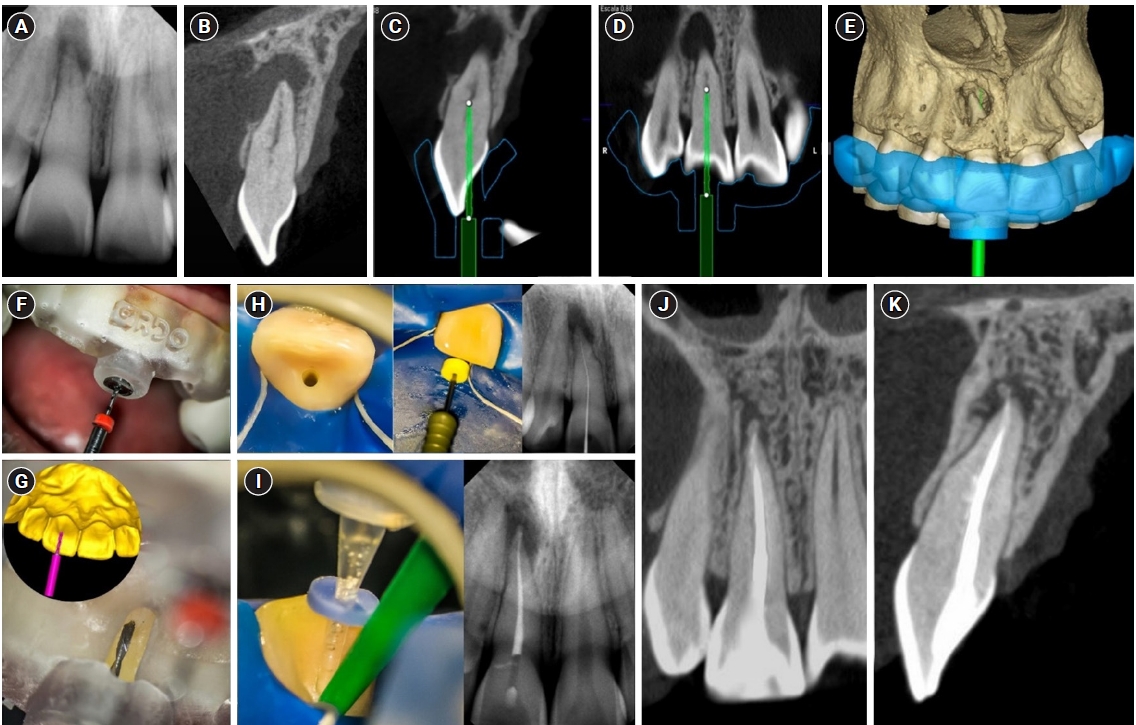
- Endodontic treatment of a molar-incisor malformation of the maxillary first molar: a case report
- Woo-Lim Kim, Se-Hee Park, Kyung-Mo Cho, Jin-Woo Kim
- Restor Dent Endod 2026;51(2):e27. Published online May 21, 2026
- DOI: https://doi.org/10.5395/rde.2026.51.e27
-
 Abstract
 PDF
PDF PubReader
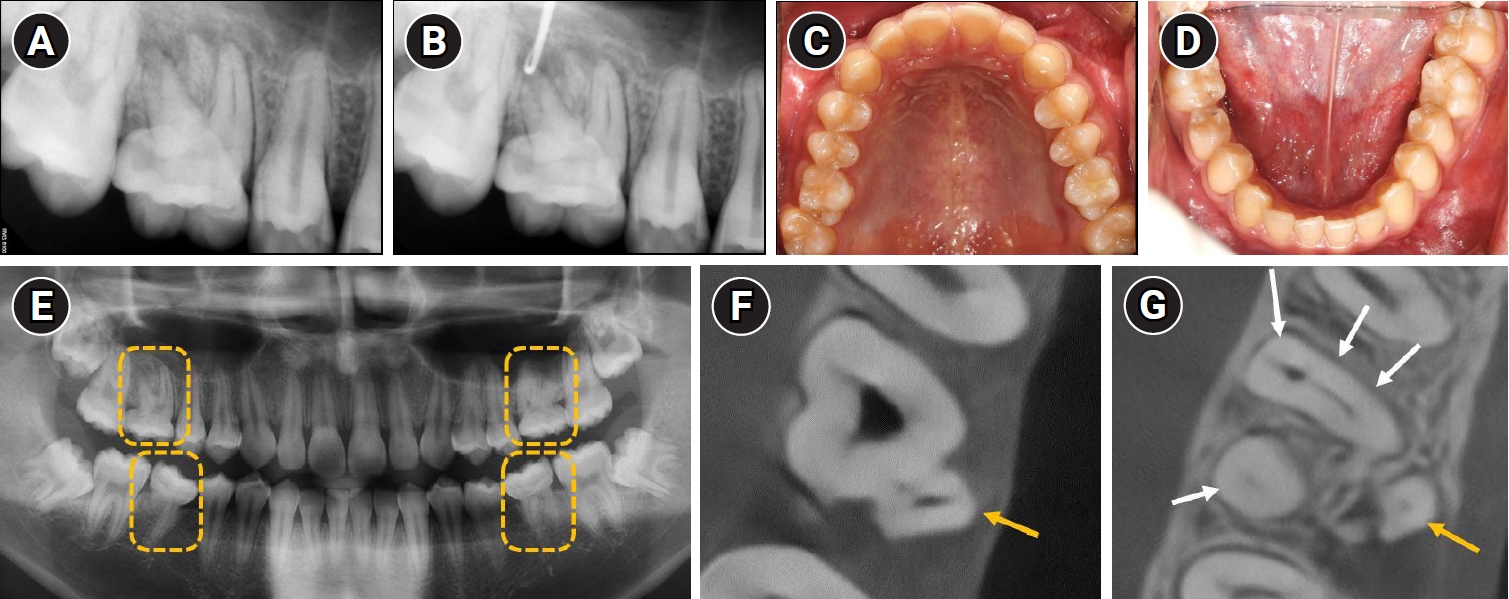
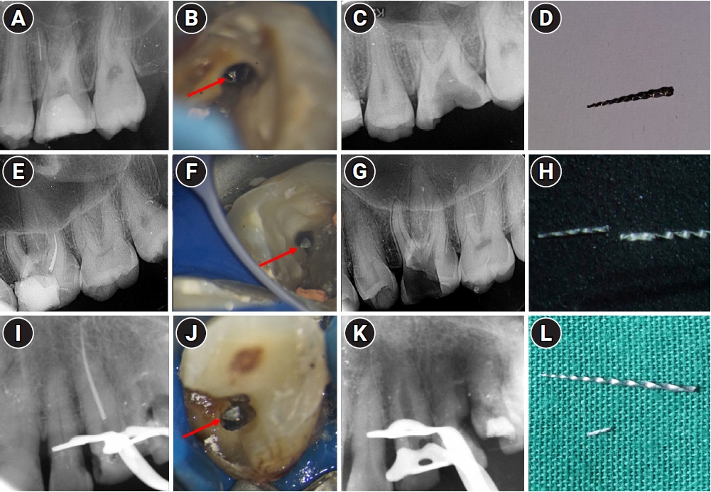
 - Molar-incisor malformation (MIM) is a developmental dental anomaly primarily affecting permanent first molars, often accompanied by structural irregularities such as cervical mineralized diaphragms (CMDs) and furcal channels. These anatomical complexities present significant challenges for endodontic treatment. This case report presents the endodontic management of a maxillary first molar diagnosed with MIM—a condition for which root canal treatment has rarely been reported. The affected tooth exhibited characteristic features of MIM, including underdeveloped roots, CMD, and an open furcal channel. Initial canal negotiation revealed four buccal canals, but the palatal canal could not be located via conventional access. A separate access approach enabled successful identification, disinfection, and obturation of the palatal canal. Follow-up imaging showed healing of the periapical lesion and favorable clinical outcomes. This case highlights the diagnostic and technical challenges in managing MIM-affected teeth and underscores the importance of advanced imaging, tailored access strategies, and careful material selection to achieve successful endodontic outcomes.
- 301 View
- 25 Download

Research Articles
- In vitro assessment of geometric characteristics in canal preparation using nickel-titanium files used for minimal invasiveness: an experimental study
- EunJin Jang, Hyeon-Cheol Kim, WooCheol Lee
- Restor Dent Endod 2026;51(2):e26. Published online May 13, 2026
- DOI: https://doi.org/10.5395/rde.2026.51.e26
-
Abstract
PDFPubReader
 ePub
ePub - Objectives
This study aimed to assess geometric characteristics in canal preparation using nickel-titanium (NiTi) files used for minimal invasiveness.
Methods
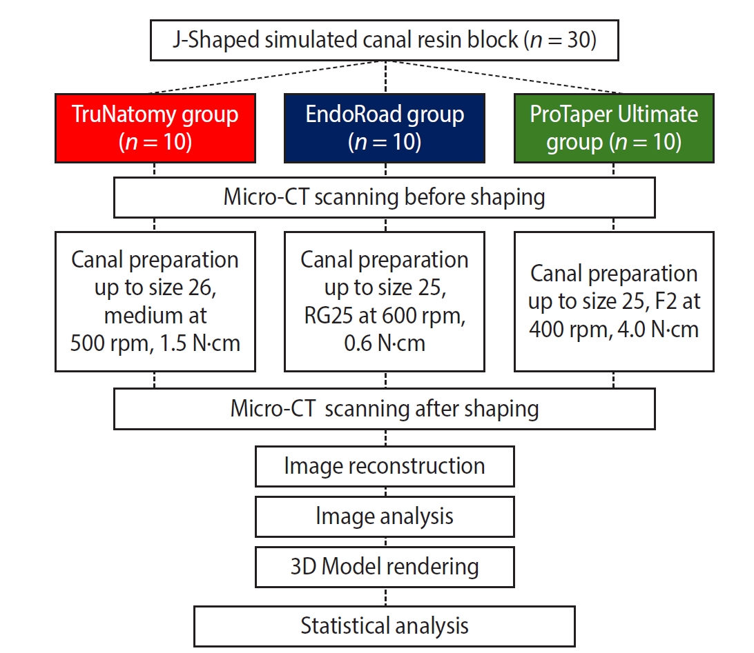
Thirty J-shaped simulated canals in resin blocks were instrumented with either TruNatomy (TR; Dentsply Sirona), EndoRoad (ER; Maruchi), or ProTaper Ultimate (PTU; Dentsply Sirona). The simulated canal blocks were scanned using microcomputed tomography before and after instrumentation. The scanned images were reconstructed, and the canal surface area was measured from 0.5 to 6.5 mm from the apex. Three-dimensional representative models of each group were rendered. The data were statistically analyzed using one-way analysis of variance and Kruskal-Wallis test at 95% significance level.
Results
TR showed a superior ability to maintain the canal’s center. TR demonstrated comparable apical preparation to PTU. ER showed a smaller and limited apical preparation than other systems, with a tendency for canal preparation toward the inner side of the curvature. PTU featured the largest prepared apical size among the file groups and tended to straighten the curvature by preparing the canal more towards the outward side. The surface area instrumented using each NiTi file showed statistically significant differences among the three groups at all levels except 0.5, 2.0, and 3.5 mm from the apex (p < 0.05). There was no statistically significant difference between TR and PTU at a level of 0.5 mm from the apex (p > 0.05).
Conclusions
While PTU is suitable for general canal preparation to facilitate irrigation and intracanal medication, TR and ER excel in preserving canal centering with minimal concern for canal transportation by minimally invasive preparation.
- 338 View
- 16 Download
- Fracture resistance of regenerated immature teeth in different simulated stages of root development: an in vitro cyclic loading study
- Kyveli-Artemis Polydora, Konstantinos Kodonas, Anastasia Fardi, Christos Gogos
- Restor Dent Endod 2026;51(2):e21. Published online April 28, 2026
- DOI: https://doi.org/10.5395/rde.2026.51.e21

-
Abstract
PDF
 Supplementary MaterialPubReaderePub
 - Objectives
This in vitro study aimed to assess the fracture resistance of simulated stages of root maturation following regenerative endodontic treatment using a cyclic loading method.
Methods
Ninety extracted maxillary central incisors were randomly allocated into three experimental groups representing different stages of root development, following revitalization: Group A for completely immature teeth immediately after treatment; Group B for teeth with apical closure, and Group C for teeth with apical closure and wall thickening. Two control groups were also included: Group D for intact teeth and Group E for simulated immature teeth without the bioceramic material. Following simulation of immature apices and treatment with a bioceramic material, all specimens were subjected to cyclic loading using a step-stress fatigue protocol until failure. The number of cycles to fracture and the peak load were recorded and statistically analyzed.
Results
Statistically significant differences in loading forces were observed between the negative control group (Group D) and Groups A, B, and E (p < 0.05). However, no statistically significant differences were detected among the experimental groups. These results indicate that apical closure and dentinal wall thickening alone did not substantially improve mechanical reinforcement under cyclic loading conditions.
Conclusions
Although intact teeth exhibited superior mechanical performance, apical closure and wall thickening alone were insufficient to enhance reinforcement under cyclic loading.
- 435 View
- 34 Download
- Initial attachment, viability, proliferation, and migration of osteoblast-like SaOS-2 cells on two resorbable xenogeneic membranes for guided tissue regeneration: ab in vitro experimental study
- Rafael Fernández-Grisales, Giovanna García-Suárez, Ximena Guerrero-Rodríguez, Carolina Berruecos-Orozco, Marco Calle-Jaramillo, Wilder Javier Rojas, Vanessa Esmeralda Duque, Daniela Serna-Guisao, Néstor Ríos-Osorio
- Restor Dent Endod 2026;51(2):e20. Published online April 13, 2026
- DOI: https://doi.org/10.5395/rde.2026.51.e20
-
Abstract
PDFPubReaderePub
- Objectives
This study evaluated the biocompatibility of a new xenogeneic resorbable membrane derived from porcine esophagus membrane (Quirumatrix, Cells Tech Co.) and compared it with a porcine pericardium membrane (Straumann Jason, Straumann Holding AG.) traditionally used for guided tissue regeneration. The parameters investigated were the viability, migration, and adhesion of SaOS-2 osteoblast-like cells derived from osteosarcoma on both membranes.
Methods
The cells were cultured in 100 mm plates in RPMI 1640 medium (40 mL), supplemented. They were incubated at 37°C in a humidified atmosphere with 95% air and 5% to 10% CO2. Cell morphology and adhesion were evaluated using phase contrast optical microscopy and scanning electron microscope. Cell viability and proliferation were evaluated using a fluorometric resazurin reduction assay, with fluorescence intensity measured at 48, 72, and 96 hours. Cell migration was evaluated using staining with Alexa Fluor 555 Phalloidin (Cell Signaling Technology) and DAPI, with a reference line. Cell migration was analyzed by measuring displacement within the delineated area using an Axio Imager M2 fluorescence microscope (Carl Zeiss). Each membrane was photographed. The statistical analysis was performed using GraphPad Prism ver. 10.2.3 (GraphPad Software). A p-value <0.05 was considered significant between experimental groups.
Results
Both membranes were shown to be biocompatible. The porcine pericardium membrane showed greater cell adhesion and proliferation compared to the porcine esophagus membrane. Cell migration was significantly greater in the Jason membrane.
Conclusions
The results revealed that both evaluated membranes are biocompatible and non-cytotoxic; further research is needed to understand their long-term behavior, interactions with other types of cells, and performance in specific therapeutic situations.
- 801 View
- 64 Download
Review Article
- Effectiveness of silver diamine fluoride in managing hypersensitivity of molar-incisor hypomineralization affected molars: a scoping review
- Vo Truong Nhu Ngoc, Do Trong Hieu, Tran Anh Tuan, Vo Nhat Minh, Trinh Khanh Linh
- Restor Dent Endod 2026;51(2):e19. Published online April 13, 2026
- DOI: https://doi.org/10.5395/rde.2026.51.e19
-
Abstract
PDFPubReader
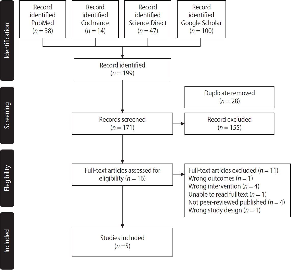
- This study aimed to evaluate the efficacy of dentinal hypersensitivity treatment with silver diamine fluoride (SDF) in molar-incisor hypomineralization (MIH)-affected molars. This scoping review was designed and structured according to the guidelines of the Preferred Reporting Items for Systematic Reviews and Meta-Analyses and its extension for scoping reviews. A search strategy was conducted across PubMed, The Cochrane Library, ScienceDirect, and Google Scholar to identify articles related to the topic. Two authors screened titles, abstracts, and full texts for review. Five studies met the eligibility criteria, comprising four randomized controlled trials and one case report, with sample sizes ranging from four to 200 participants. All included studies reported improvements in clinical outcomes, including reduced hypersensitivity following SDF application, as indicated by lower Schiff cold air sensitivity scale scores. SDF is a promising treatment strategy for reducing hypersensitivity in MIH-affected molars; however, further research using SDF alone is needed to evaluate its exact effectiveness.
- 1,686 View
- 182 Download
Research Articles
- The recovery effect of dentin biomodifiers on microtensile bond strength and sealer-penetration depth of coronal and radicular dentin: an in vitro experimental study
- Mona Rizk Aboelwafa, Yasmin Tawfik Mohamed Sobh
- Restor Dent Endod 2026;51(2):e15. Published online April 7, 2026
- DOI: https://doi.org/10.5395/rde.2026.51.e15
-
Abstract
PDFPubReaderePub
- Objectives
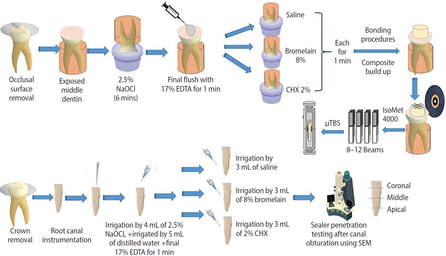
This study aimed to assess the outcomes of bromelain enzyme and chlorhexidine (CHX) following endodontic irrigation by evaluating coronal dentin microtensile bond strength (μTBS) and radicular dentin sealer penetration depth.
Methods
Fifty-one human molars with flat mid-dentin surfaces were soaked in sodium hypochlorite, then randomly assigned to three groups relying on the biomodification approach (n = 17): group 1, saline; group 2, 8% bromelain; and group 3, 2% CHX. After bonding and resin composite build-ups, the μTBS, failure mode, and bond interface were evaluated. Forty-two root canals of human molars were mechanically prepared and randomly distributed among three groups (n = 14), similar to the coronal-dentin biomodification protocol. The sealer-penetration depth was measured utilizing the scanning electron microscope. One- and two-way analyses of variance and the pairwise t- and chi-square tests were utilized.
Results
The bromelain group showed the highest statistically significant resin-dentin μTBS values, followed by the CHX and control groups. For sealer-penetration assessment, the bromelain group showed the highest penetration at the middle and apical root levels, whereas CHX demonstrated the highest penetration at the coronal level.
Conclusions
Bromelain biomodification positively influenced the resin-dentin bond strength and the sealer-penetration depth in apical and middle levels.
- 441 View
- 58 Download
- Influence of adjacent restorative material and distance on the accuracy of inlay cavity impressions with intraoral scanner: an in vitro study
- So-Yeon Lee, Sung-Ae Son, Jae-Hoon Kim, Deog-Gyu Seo, Jeong-Kil Park
- Restor Dent Endod 2026;51(1):e6. Published online January 23, 2026
- DOI: https://doi.org/10.5395/rde.2026.51.e6
-
Abstract
PDFPubReaderePub
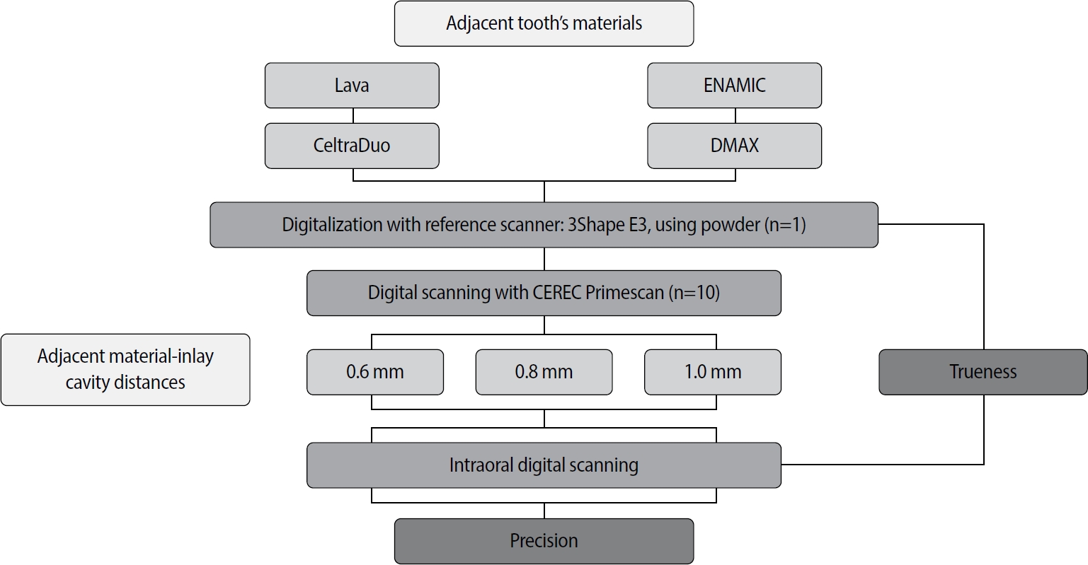
- Objectives
This study aimed to evaluate the influence of adjacent restorative material and interproximal distance on the accuracy of digital impressions of inlay cavities obtained using an intraoral scanner.
Methods
A disto-occlusal inlay cavity was prepared on a mandibular right first molar model, and digital scans were performed using a CEREC Primescan (Dentsply Sirona). The adjacent restorative materials used were Lava (3M ESPE), ENAMIC (VITA Zahnfabrik), Celtra Duo (Dentsply Sirona), and DMAX (DMAX), and the interproximal distances were set to 0.6 mm, 0.8 mm, and 1.0 mm. The obtained scan data were analyzed using GOM Inspect software (GOM GmbH).
Results
Trueness, maximum positive and negative deviations, and precision were significantly influenced by both the adjacent restorative material and the interproximal distance, while their interaction showed a significant effect only on precision. Celtra Duo demonstrated the highest trueness, with mean deviation values decreasing from 7.8 μm at a 0.6 mm interproximal distance to 7.3 μm at 1.0 mm. ENAMIC showed the best precision, presenting mean deviations of 2.6 μm at 0.6 mm, 2.9 μm at 0.8 mm, and 2.4 μm at 1.0 mm. A narrow interproximal distance of 0.6 mm resulted in lower trueness, measured at 8.3 μm, and the highest precision deviation of 3.4 μm. In contrast, an interproximal distance of 1.0 mm yielded improved scan accuracy, with increased trueness and reduced precision variation.
Conclusions
Digital impression accuracy of inlay cavities was influenced by adjacent restorative material and interproximal distance, suggesting clinical consideration is needed in CAD/CAM workflows to optimize restoration fit. -
Citations
Citations to this article as recorded by
- 3D-SCANNING IN PROSTHETIC DENTISTRY: ADVANTAGES, DISADVANTAGES, AND DEVELOPMENT PROSPECTS
V. S. Kuz, O. I. Teslenko, H. M. Kuz, H. M. Balia, Yu. S. Lunkova, O. V. Shemetov, I. M. Martynenko
Bulletin of Problems Biology and Medicine.2026; 1(1): 98. CrossRef
- 3D-SCANNING IN PROSTHETIC DENTISTRY: ADVANTAGES, DISADVANTAGES, AND DEVELOPMENT PROSPECTS
- 1,749 View
- 104 Download
- 1 Crossref
- Analysis of the reciprocating kinematics of the VDW Silver Reciproc, E-Connect Pro, Ecom, and Endopen endodontic motors: an in vitro experimental study
- Cristielly França, Juliana D. Bronzato, Dieimes Braambati, Adriana de-Jesus-Soares, Carla C. R. B. Félix, Michelle A. N. S. Ferreira, Marcos Frozoni
- Restor Dent Endod 2026;51(1):e5. Published online January 20, 2026
- DOI: https://doi.org/10.5395/rde.2026.51.e5
-
Abstract
PDFPubReaderePub
- Objectives
This study aimed to evaluate the actual parameters of four endodontic motors, each adjusted for reciprocating motion, and compare them to the manufacturers’ declared values.
Methods
The motors used were the VDW Silver Reciproc (VDW GmbH), E-Connect Pro (MK Life), Ecom (Woodpecker), and Endopen (Schuster Woodpecker). A custom optical target was attached to the motor contra-angle, the movements were recorded with a high-resolution camera, and the images were analyzed. Engagement, disengagement, net angles, and speed for each operation cycle, duration of clockwise (CW) and counter-clockwise (CCW) movement, duration of standstill after CW and CCW movement, and the number of cycles to complete a full rotation were analyzed. The data were statistically analyzed at a significance level of 5%. The replicability of all reciprocal parameters analyzed was statistically different from that reported by the manufacturers.
Results
There was no statistically significant difference between the VDW Silver Reciproc, Ecom, and Endopen for the engagement angle. The E-Connect Pro was the least reliable at the 150°/30° settings for both angle parameters. There was no significant difference between the set and actual cycle net angles for the VDW Silver Reciproc (p = 0.493). While the actual values for the Ecom and E-Connect Pro were significantly higher than the set (p < 0.001), the actual values for the Endopen were significantly lower than the set (p < 0.001).
Conclusions
Experiments on four commercially available reciprocating endodontic motors revealed that the actual motor values differed significantly from the set values.
- 1,552 View
- 77 Download
- Effect of moisture and pH on setting time and microhardness of three premixed calcium silicate-based root canal sealers: an in vitro experimental study
- Sooyoun Kim
- Restor Dent Endod 2025;50(4):e41. Published online November 28, 2025
- DOI: https://doi.org/10.5395/rde.2025.50.e41
-
Abstract
PDFPubReaderePub
- Objectives
The study aimed to investigate how environmental conditions impact the setting time and microhardness of premixed calcium silicate-based sealers.
Methods
The setting time and microhardness of three sealers (Endoseal MTA [MARUCHI], One-Fil [MEDICLUS], and Well-Root ST [VERICOM]) were evaluated under four environmental conditions: unsoaked, distilled water-soaked, phosphate-buffered saline-soaked, and pH 5-soaked gypsum molds (n = 12/group/condition). The setting time was measured with Gilmore needles, and microhardness was assessed using a Vickers tester after 3 days. Welch’s analysis of variance and Games-Howell post hoc tests were used for statistical analysis.
Results
The sealer type and environmental conditions significantly influenced setting time and microhardness (p < 0.001). The initial and final setting times were the shortest in the unsoaked samples. For Endoseal MTA and One-Fil, the unsoaked condition exhibited significantly shorter setting times than the soaked conditions. Well-Root ST exhibited significantly longer setting times in acidic conditions. Surface microhardness was highest in the unsoaked group (p < 0.001). Among the soaked groups, the phosphate-buffered saline-soaked group had the lowest hardness for Endoseal MTA, whereas the pH 5-soaked group exhibited the lowest hardness for One-Fil and Well-Root ST. Endoseal MTA consistently demonstrated a lower microhardness than the other sealers (p < 0.001).
Conclusions
Moisture, pH, and solution chemistry influenced the setting time and microhardness of premixed calcium silicate sealers. Although acidic conditions generally prolong the setting time and reduce hardness, the effects vary based on the sealers used and the setting environment. -
Citations
Citations to this article as recorded by- Setting Characteristics, Solubility, Bioactivity and Interaction with Dentin of Four Calcium Silicate-Based Endodontic Sealers
Areti Dimitra Vrochari, Anastasia Agrafioti, Maria Dimitriadi, George Eliades
Journal of Functional Biomaterials.2026; 17(4): 192. CrossRef
- Setting Characteristics, Solubility, Bioactivity and Interaction with Dentin of Four Calcium Silicate-Based Endodontic Sealers
- 1,972 View
- 97 Download
- 1 Web of Science
- 1 Crossref
- Resolvin E1 incorporated carboxymethyl chitosan scaffold accelerates repair of dental pulp stem cells under inflammatory conditions: a laboratory investigation
- Hemalatha P Balasubramanian, Nandini Suresh, Vishnupriya Koteeswaran, Velmurugan Natanasabapathy
- Restor Dent Endod 2025;50(4):e40. Published online November 28, 2025
- DOI: https://doi.org/10.5395/rde.2025.50.e40
-
Abstract
PDFSupplementary MaterialPubReaderePub
- Objectives
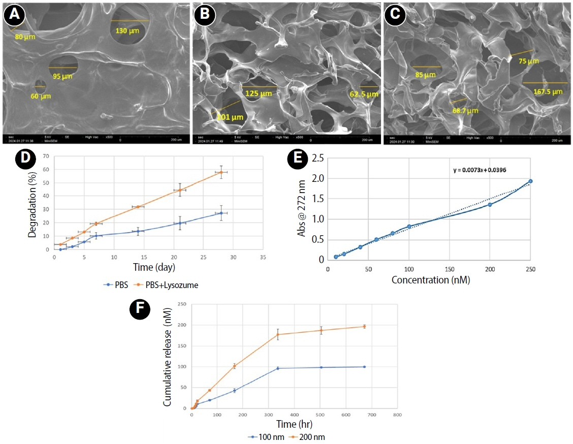
This study fabricated and characterized a resolvin E1 (RvE1)-loaded carboxymethyl chitosan (CMC) scaffold and determined its cytotoxicity and mineralization potential on inflamed human dental pulp stem cells (hDPSCs).
Methods
CMC scaffold incorporated with two concentrations of RvE1 (100 and 200 nM) was fabricated and characterized. The scaffolds’ porosity, drug release kinetics, and degradation were assessed. The impact of RvE1 on inflamed hDPSCs proliferation, proinflammatory gene expression (tumor necrosis factor alpha [TNF-α]), alkaline phosphatase activity, and alizarin red S staining was evaluated.
Results
Scanning electron microscopy analysis demonstrated a highly porous interconnected microstructure. Release kinetics showed gradual RvE1 release peaking at day 14. Cumulative degradation of the CMC scaffold at 28 days was 57.35%. Inflamed hDPSCs exposed to 200 nM RvE1-CMC scaffold exhibited significantly improved viability compared to 100 nM. Both RvE1-CMC scaffolds significantly suppressed the expression of TNF-α at 7 days. Alkaline phosphatase activity was enhanced by both RvE1 concentrations on days 7 and 14. Alizarin red staining revealed superior mineralization potential of 200 nM RvE1 on days 14 and 21.
Conclusions
This study concludes 200 nM RvE1-CMC scaffold is a promising therapy for inflamed pulp conditions, enhancing cell proliferation and biomineralization potential in inflamed hDPSCs.
- 1,260 View
- 55 Download
- Evaluation of platelet concentrates in regenerative endodontics: a systematic review and meta-analysis
- Anna Tsiolaki, Dimitrios Theocharis, Nikolaos Tsitsipas, Anastasia Fardi, Konstantinos Kodonas
- Restor Dent Endod 2025;50(4):e38. Published online November 28, 2025
- DOI: https://doi.org/10.5395/rde.2025.50.e38
-
Abstract
PDFSupplementary MaterialPubReaderePub
- Objectives
The aim of this systematic review is to compare the effectiveness of advanced platelet concentrates as regenerative endodontic therapeutic alternatives to blood clot (BC) revascularization in immature permanent necrotic teeth.
Methods
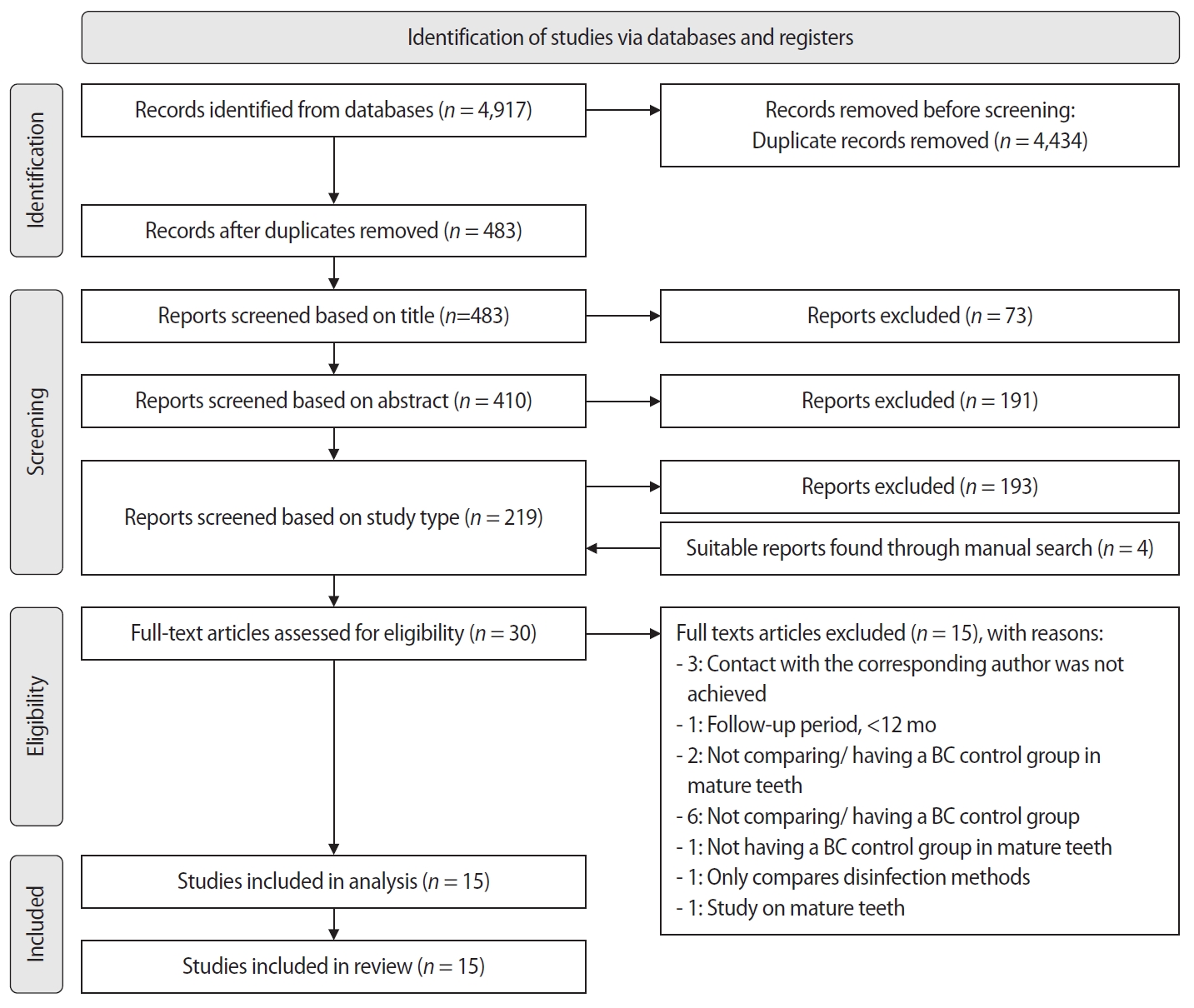
Randomized controlled trials (RCTs) comparing regenerative endodontic therapies using platelet-rich plasma (PRP), platelet-rich fibrin (PRF), or platelet pellet (PP) with the BC revascularization approach in immature permanent necrotic teeth were systematically searched in PubMed, Scopus, Cochrane Library, and Web of Science until May 2025. Data was extracted and analyzed both qualitatively and quantitatively. Study quality was assessed using the Cochrane Risk of Bias tool. A meta-analysis was conducted using IBM SPSS software (version 29.0), with success rates expressed as risk ratios and 95% confidence intervals (CIs).
Results
The initial search yielded 4,917 studies. After removing duplicates and applying eligibility criteria, 15 RCTs were included. Meta-analysis indicated no significant difference in the risk ratio (RR), as the BC method has similar success rates with PRP (10 studies; RR = 1.01; 95% CI, 0.94–1.09; p = 0.76) and PRF (8 studies; RR = 0.98; 95% CI, 0.89–1.08; p = 0.65) at 12 months. The primary outcomes evaluated were based on clinical and radiographic success.
Conclusions
Current evidence suggests PRP, PRF, and BC are all effective in treating immature permanent necrotic teeth with similar success rates. However, further research is needed to assess long-term outcomes. -
Citations
Citations to this article as recorded by- Longitudinal periapical radiographic evaluation of apexification, vital pulpotomy, and revascularization in immature permanent teeth: a retrospective comparative study
Xiaona Sun, Kailing Zhu
Frontiers in Bioengineering and Biotechnology.2026;[Epub] CrossRef
- Longitudinal periapical radiographic evaluation of apexification, vital pulpotomy, and revascularization in immature permanent teeth: a retrospective comparative study
- 2,123 View
- 109 Download
- 1 Web of Science
- 1 Crossref
- Phase transformation temperatures influence the reduction ratio of fatigue resistance of nickel-titanium reciprocating files at body temperature: an in vitro experimental study
- Walid Nehme, Alfred Naaman, Lola Pedèches, Sylvie Lê, Marie Georgelin-Gurgel, Sang Won Kwak, Hyeon-Cheol Kim, Franck Diemer
- Restor Dent Endod 2025;50(4):e35. Published online November 5, 2025
- DOI: https://doi.org/10.5395/rde.2025.50.e35
-
Abstract
PDFPubReaderePub
- Objectives
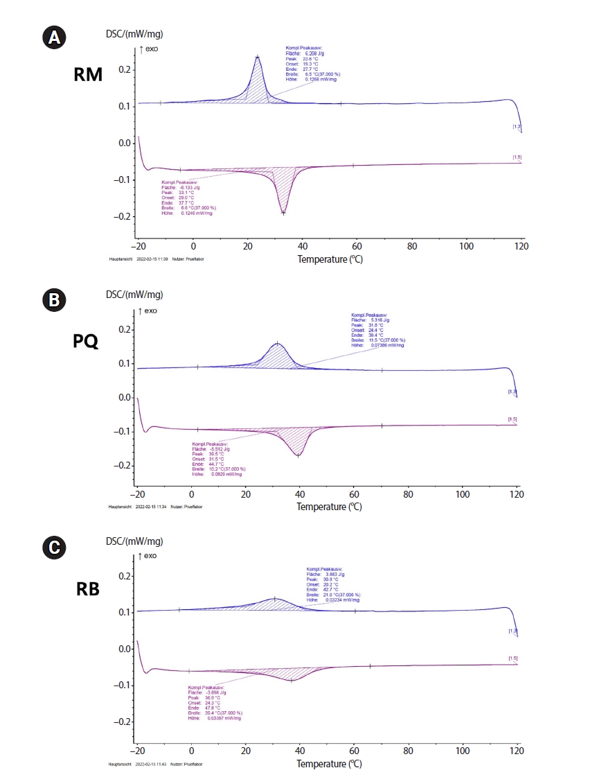
The objective of this study was to evaluate the effects of transformational temperatures on the cyclic fatigue resistance at body temperature of reciprocating file systems: R motion (RM), Procodile Q (PQ), and Reciproc Blue.
Methods
Resistance test was done in a custom-made device at room (20°C ± 1°C) and body (37°C ± 1°C) temperatures within a 60° angle of curvature and 5 mm radius of the artificial canal. The time to fracture (TTF) was recorded. The scanning electron microscope observation and differential scanning calorimetry analyses were performed. Two-way analysis of variance and Tukey post-hoc comparison were applied at a significance level of 0.05.
Results
The results showed a significant influence of temperature on instrumental breakage, regardless of the file systems (p < 0.05). The TTF is significantly decreased at body temperature (p < 0.05). PQ showed the longest TTF in both temperature conditions (p < 0.05). RM demonstrated a significantly higher TTF reduction ratio compared to the other files (p < 0.05).
Conclusions
Within the limitations of this study, the heat-treated files with reciprocating kinetics may have different reduction ratios of the fatigue resistance of the file systems under different temperature conditions. This characteristic is an important point of consideration when clinicians select the file system to reduce potential file fracture.
- 1,981 View
- 72 Download
- Calcium silicate-based sealers remnants in isthmuses of mesial roots of mandibular molars: an in vitro evaluation
- David Saldanha de Brito Alencar, Ana Cristina Padilha Janini, Lauter Eston Pelepenko, Brenda Fornazaro Moraes, Francisco Haiter Neto, Marco Antonio Hungaro Duarte, Marina Angélica Marciano
- Restor Dent Endod 2025;50(3):e25. Published online July 15, 2025
- DOI: https://doi.org/10.5395/rde.2025.50.e25
-
Abstract
PDFPubReaderePub
- Objectives
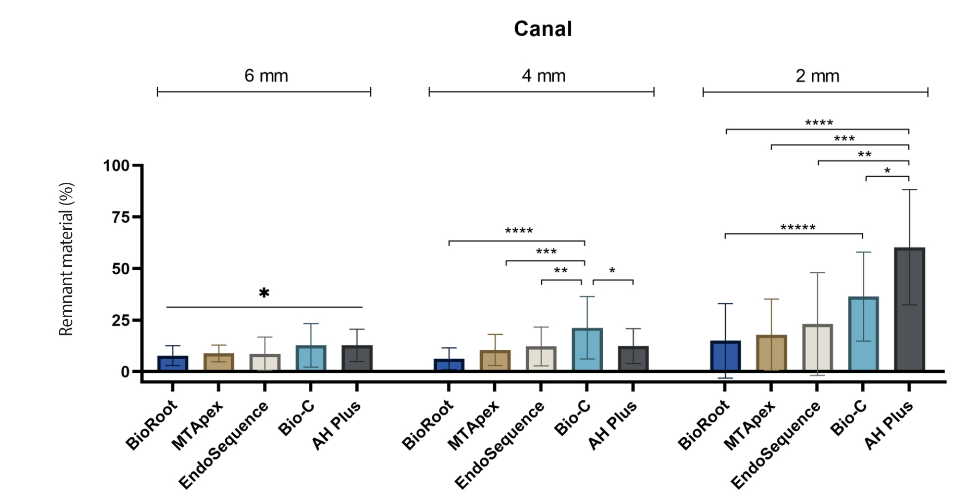
Endodontic retreatment aims to address treatment failure through the removal of root canal filling materials. This in vitro study evaluated the presence of filling material remnants in the mesial root canals, specifically focusing on the isthmuses, of mandibular molars after retreatment.
Methods
One hundred extracted mandibular molar mesial roots with isthmuses were prepared with an R25 file, obturated with one of five calcium silicate-based sealers (BioRoot RCS [Septodont], MTApex [Ultradent Products Inc.], EndoSequence BC Sealer HiFlow [Brasseler USA], Bio-C Sealer [Angelus]) or an epoxy resin-based sealer (AH Plus Jet [Dentsply Maillefer]), all stained with rhodamine B, and stored at 37ºC for 30 days to allow for setting. Retreatment was subsequently performed using R40 and XP-endo Finisher R instruments (FKG Dentaire) with 2.5% sodium hypochlorite irrigation. The presence of remaining filling material was then assessed using confocal microscopy, and setting times were tested per ISO 6876:2012.
Results
AH Plus Jet showed the most remnants at 2 mm and the longest retreatment time. Calcium silicate-based sealers exhibited prolonged setting times under dry conditions, with EndoSequence BC Sealer HiFlow showing a particularly extended setting period.
Conclusions
Despite retreatment, residues remained in all canals and isthmus regions, particularly Bio-C Sealer and AH Plus Jet in apical areas, emphasizing the difficulty of complete removal and the persistence of filling material. -
Citations
Citations to this article as recorded by- Bonding effects of mechanical removal of bioceramic sealer residues using glycine or glass microparticles abrasion
Jesus Aranda, Julia de Freitas Ceccato, Eduardo Fernández Godoy, João Felipe Besegato, Joissi Ferrari Zaniboni, Regina Guenka Palma-Dibb, Milton Carlos Kuga
International Journal of Adhesion and Adhesives.2026; 148: 104289. CrossRef
- Bonding effects of mechanical removal of bioceramic sealer residues using glycine or glass microparticles abrasion
- 2,574 View
- 123 Download
- 1 Web of Science
- 1 Crossref
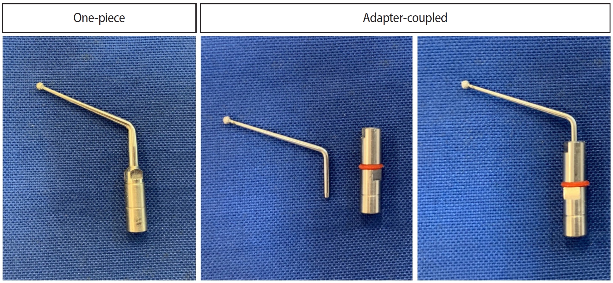
- Analysis of thermal profiles on tooth structure and insert during one-piece or adapter-coupled ultrasonic insert use: an in vitro experimental study
- Gabriela Loewen Brotto, Bruno Monguilhott Crozeta, Bruno Marques-da-Silva, Alysson Nunes Diógenes, Emmanuel João Nogueira Leal da Silva, Flávia Sens Fagundes Tomazinho
- Restor Dent Endod 2025;50(3):e24. Published online July 11, 2025
- DOI: https://doi.org/10.5395/rde.2025.50.e24
-
Abstract
PDFPubReaderePub
- Objectives
This in vitro study aimed to evaluate temperature variation on the external surface of mandibular molars and within ultrasonic inserts when using adapter-coupled versus one-piece inserts.
Methods
Twenty-four extracted human mandibular molars were divided into two groups based on the type of ultrasonic insert used: adapter-coupled and one-piece inserts. Temperature on the external surface of each tooth was measured with a thermocouple probe positioned in the furcation area, capturing data continuously. The temperature of the ultrasonic inserts was monitored in real-time using a thermal imaging camera. Measurements were taken in a controlled environment without cooling for over 120 seconds. Statistical analysis was conducted using analysis of variance (ANOVA) and two-way ANOVA with repeated measures to evaluate temperature variations between groups and over time, with significance set at 5%.
Results
In the external tooth surface temperature measurements, no significant differences were observed between the groups during the initial 15 seconds (p = 0.185) and 30 seconds (p = 0.067). However, significant differences emerged at 60 seconds (p = 0.025), 90 seconds (p = 0.024), and 120 seconds (p = 0.020), with the one-piece insert group demonstrating higher temperatures in the furcation region. Thermal imaging of the inserts revealed a significant difference at all time points (p < 0.001), with adapter-coupled inserts showing greater heating.
Conclusions
The use of ultrasonic inserts leads to a gradual rise in temperature on the external tooth surface. One-piece inserts generated higher temperatures on the tooth, while adapter-coupled inserts exhibited greater heating within the insert.
- 2,315 View
- 104 Download
- Comparative study of the effectiveness of different bleaching agents on blood-colored extracted teeth and investigation of recoloring after bleaching: an in vitro experimental study
- Gülşen Arslan, Akın Aladağ, Ayşegül Demirbaş, Murat Türkün
- Restor Dent Endod 2025;50(3):e22. Published online July 9, 2025
- DOI: https://doi.org/10.5395/rde.2025.50.e22
-
Abstract
PDFPubReaderePub
- Objectives
This study evaluated the efficacy of three distinct bleaching agents over time on blood-stained, devitalized teeth. Furthermore, the recoloring subsequent to bleaching will be monitored.
Methods
The study was conducted on 60 caries-free, unfilled, upper human incisors. The Freccia and Peters blood staining technique was employed, and four groups (n = 15) were identified: control, 35% hydrogen peroxide-treated, 37% carbamide peroxide-treated, and sodium perborate-treated groups. Color differences were measured using ΔE00, ΔWID, L*, a*, and b* values. To investigate tooth discoloration after bleaching, 10 unbleached teeth with three groups of 10 bleached teeth were compared by vine staining. The group of bleached teeth was restored immediately, another group waited one week, and the third group had sodium ascorbate applied and analyzed using one-way analysis of variance tests (p < 0.05).
Results
Among the groups, carbamide peroxide exhibited the most significant whitening during the 6-day bleaching process, followed by hydrogen peroxide and sodium perborate. Subsequent examination of the wine recoloring of post-bleaching samples demonstrated that bleached teeth exhibited a heightened propensity for recoloration in contrast to unbleached teeth. Notably, sodium ascorbate treatments for hydrogen peroxide neutralization and the wait-and-restore approach were not statistically significant in terms of preventing recoloration.
Conclusions
Sodium perborate is less effective and more time-consuming than hydrogen peroxide or carbamide peroxide for bleaching purposes. Carbamide peroxide is the most effective bleaching agent. The sodium ascorbate treatment and the wait-and-restore approach are ineffective in preventing recoloring. Bleached teeth have more discoloration than unbleached teeth. -
Citations
Citations to this article as recorded by- The Effect of Adhesive Systems on Shade Matching of Composite Veneer
Fadak Al Marar, Raghad Aljarboua, Fatimah M. Alatiyyah, Shahad AlGhamdi, Faraz Ahmed Farooqi, Lama Almuhanna, Rasha AlSheikh, Abdul Samad Khan
Dentistry Journal.2026; 14(2): 85. CrossRef
- The Effect of Adhesive Systems on Shade Matching of Composite Veneer
- 4,092 View
- 272 Download
- 1 Web of Science
- 1 Crossref
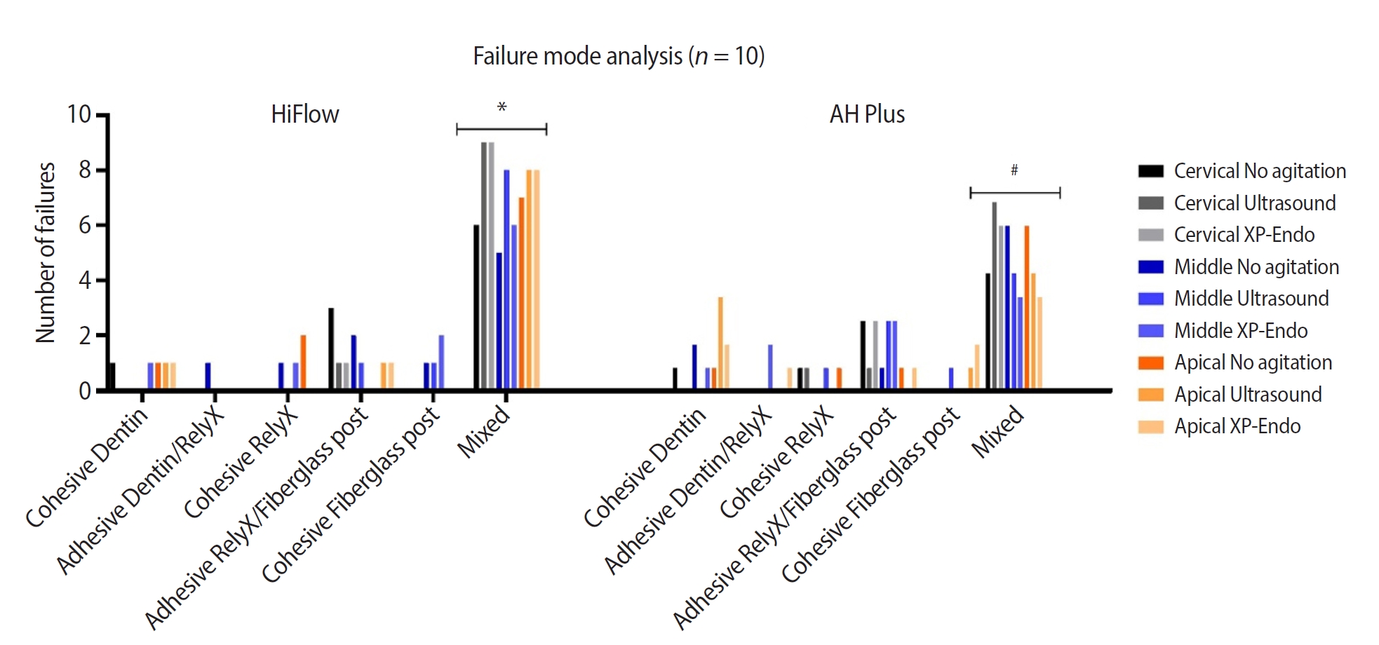
- Cleaning protocols to enhance bond strength of fiberglass posts on root canals filled with bioceramic sealer: an in vitro comparative study
- Thiago Bessa Marconato Antunes, Juliana Delatorre Bronzato, Joice Graciani, Ana Cristina Padilha Janini, Rocharles Cavalcante Fontenele, Francisco Haiter Neto, Brenda Paula Figueiredo de Almeida Gomes, Marina Angélica Marciano da Silva
- Restor Dent Endod 2025;50(2):e20. Published online May 21, 2025
- DOI: https://doi.org/10.5395/rde.2025.50.e20
-
Abstract
PDFPubReaderePub
- Objectives
This study aimed to evaluate whether the agitation protocols using ultrasonic inserts or the XP-endo Finisher R file improved the removal of two different endodontic sealer remnants and the bond strength of fiberglass posts to dentin.
Methods
Seventy-two human teeth were selected. The canals were prepared with Reciproc 50 and Easy ProDesign 30/.10 and root filled according to the endodontic sealer groups: AH Plus or EndoSequence BC Sealer HiFlow. The samples were kept at 37ºC and 95% humidity for 28 days. During the post space preparation, the obturation was removed with Largo burs, and the groups were divided according to the irrigant agitation protocols (n = 12): no agitation, agitation with R1-Clearsonic associated with E1-Irrisonic ultrasonic inserts, or agitation with XP-endo Finisher R file. The fiberglass posts were cemented with RelyX ARC. The roots were sectioned into slices and submitted to the push-out test. Micro-computed tomography analysis was used to check the effectiveness of irrigating solution agitation in the elimination of remnants.
Results
The cleaning protocols with agitation were more effective in increasing the bond strength of posts to dentin for both sealer groups compared to non-agitation (p < 0.05). There was no difference between the same cleaning protocols for the different sealers. Among the different thirds, there was no statistical difference for the same sealer in the different cleaning protocols (p > 0.05).
Conclusions
Both agitation protocols effectively clean root-filled canals sealed with resin-based and calcium silicate-based sealers during fiberglass post space preparation. These protocols result in improved bond strength compared to non-agitation methods. -
Citations
Citations to this article as recorded by- Cleaning efficacy and bond interaction of glycine-based air polishing and glass microparticles abrasion on dentin impregnated with premixed bioceramic sealer
Ândresson Aurélio Fernandes Martins, Maria Carolina Sidonio Alves, Bruno Martins Maciel, José Rodolfo Estruc Verbicário, João Felipe Besegato, Wilfredo Gustavo Escalante-Otárola, Milton Carlos Kuga
International Journal of Adhesion and Adhesives.2026; 147: 104277. CrossRef - Effect of Endodontic Sealers on the Bond Strength of Glass Fibre Posts: A Systematic Review
Thiago Bessa Marconato Antunes, Juliana D. Bronzato, Vanessa Gallego Arias Pecorari, Jennifer Santos Pereira, Talita Tartari, Adriana de Jesus Soares, Brenda P. F. A. Gomes, Marina Angélica Marciano
Australian Endodontic Journal.2026;[Epub] CrossRef
- Cleaning efficacy and bond interaction of glycine-based air polishing and glass microparticles abrasion on dentin impregnated with premixed bioceramic sealer
- 4,818 View
- 239 Download
- 2 Web of Science
- 2 Crossref
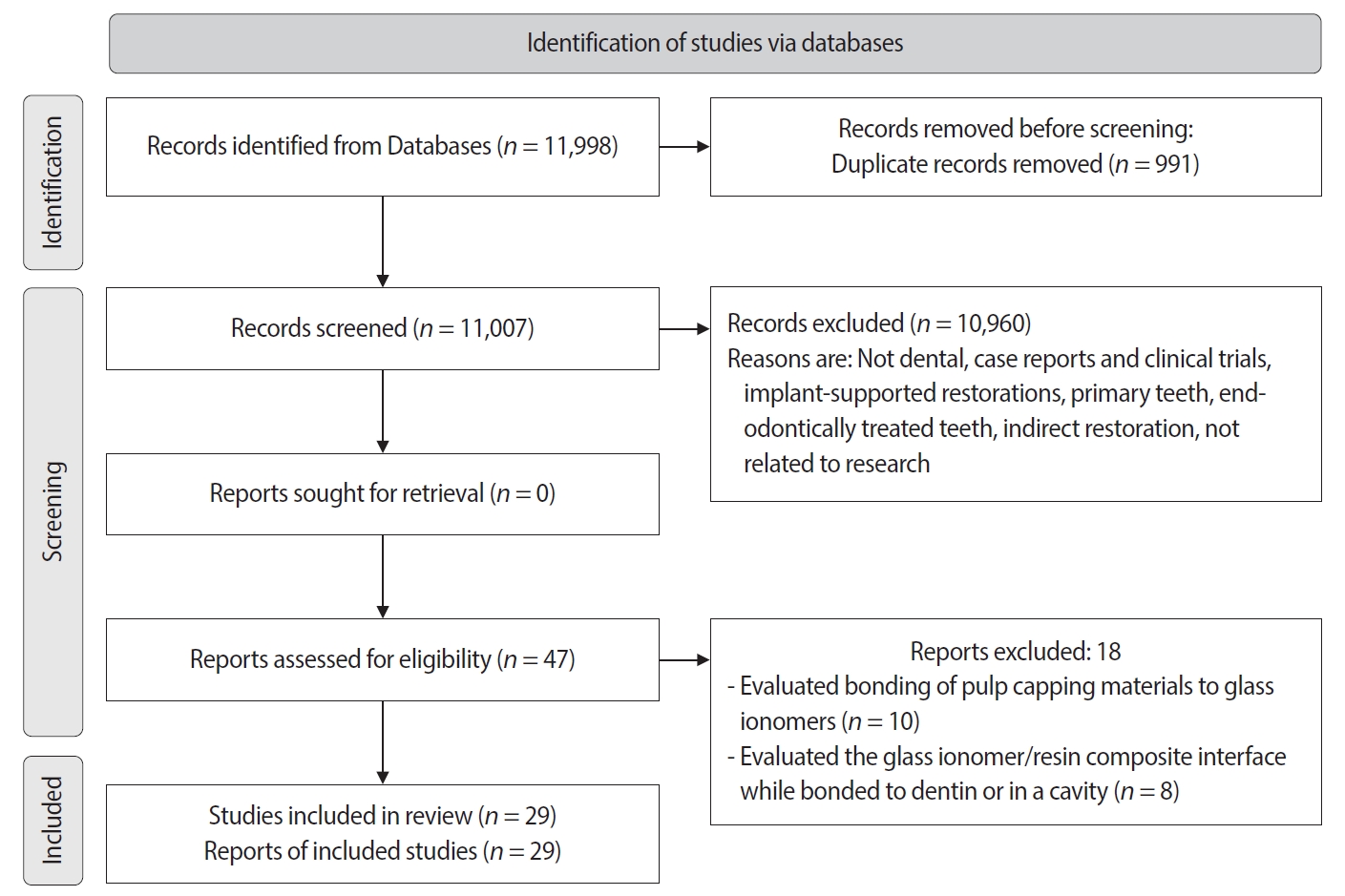
- Effect of surface treatment on glass ionomers in sandwich restorations: a systematic review and meta-analysis of laboratory studies
- Hoda S. Ismail, Ashraf Ibrahim Ali, Franklin Garcia-Godoy
- Restor Dent Endod 2025;50(2):e13. Published online April 16, 2025
- DOI: https://doi.org/10.5395/rde.2025.50.e13
-
Abstract
PDFPubReaderePub
- Objectives
This study aimed to evaluate the effect of different surface treatments on the bond strength between new or aged glass ionomers (GI) and resin composites in sandwich restorations.
Methods
A comprehensive search was conducted in three databases to identify studies focusing on the bond strength of new or aged GIs and resin composites in laboratory settings. The selected studies were assessed for potential biases based on predetermined criteria. Additionally, a meta-analysis was performed using three studies.
Results
A total of 29 studies were included, with 24 investigating the bond strength of new GIs and five focusing on GI repair. Three studies were included in the meta-analysis (with a 95% confidence interval) which revealed no significant difference in the mean MPa values of resin-modified glass ionomer (RMGI) treated with phosphoric acid or Er,Cr:YSGG laser before the application of an etch-and-rinse adhesive. Surface treatment was found to be crucial for achieving optimal bonding between GI and resin composite, regardless of the GI’s condition.
Conclusions
The combination of mechanical and chemical surface treatments does not significantly affect the bond strength between new RMGI and composite. However, for GI repair, it is recommended to use both treatments to enhance the bond strength. -
Citations
Citations to this article as recorded by- Biological Performance of Modified Glass Ionomer Cement: Antibacterial Activity, Cytocompatibility, and Rabbit Pulpal Response
Krittee Dejyong, Saowakon Indoung, Peerapon Sornying, Sareepah Manmoo, Nattapon Rotpenpian
International Dental Journal.2026; 76(3): 109491. CrossRef - The impact of alloy treatment on the dynamic cyclic fatigue resistance of triangular base cross-section NiTi endodontic instruments
Rashid El Abed, Amre R. Atmeh, Mohamed Jamal, Anas Al Jadaa, Hamza El-Faraj, Abdel Rahman Bani Amer, Taher Al Omari
Odontology.2025;[Epub] CrossRef
- Biological Performance of Modified Glass Ionomer Cement: Antibacterial Activity, Cytocompatibility, and Rabbit Pulpal Response
- 9,585 View
- 239 Download
- 1 Web of Science
- 2 Crossref
- Pattern of endodontic instrument separation and factors affecting its retrieval: a 10-year retrospective observational study in a postgraduate institute
- Velmurugan Natanasabapathy, Aswathi Varghese, Paul Kevin Abishek Karthikeyan, Srinivasan Narasimhan
- Restor Dent Endod 2025;50(1):e7. Published online February 19, 2025
- DOI: https://doi.org/10.5395/rde.2025.50.e7
-
Abstract
PDFPubReaderePub
- Objectives
This study aimed to assess the pattern of endodontic instrument separation, their retrievability, and factors affecting its retrieval, in a postgraduate institute.
Methods
Cases referred for the management of separated endodontic instruments (SEI) from 2013 to 2023 were considered for this study. Data related to demographics, tooth type, file type, and retrieval were documented in an Excel sheet. Eight prognostic factors assumed to influence the retrieval were analyzed in this study. The secondary aim was to compare the pattern of SEI and retrievability between conventional nickel-titanium files and newer generation heat-treated nickel-titanium files. Retrieval was attempted by a senior endodontist under the dental operating microscope. Various ultrasonic tips and a Broken Tool Removal loop system were used during retrieval. Simple descriptive statistics were performed. Binomial logistic regression was done to identify the effect of the eight prognostic factors on the retrieval outcome.
Results
A total of 190 SEI was reported. SEI occurred more often in posterior teeth than anterior teeth, mandibular arch than maxillary arch, and in larger files than smaller files. Separation occurred more often in the apical third compared to the other levels. Retrieval was attempted in 88 cases and successful in 70 cases (79.5%). The larger taper and apical position of the SEI negatively influenced the retrieval by 1.4 and 8.7 times, respectively.
Conclusions
Retrieval of SEI was successful in the majority of the cases. An increase in taper and apically placed SEI negatively impacted the retrieval. There was no difference in the pattern of separation nor retrievability between conventional nickel-titanium files and newer generation heat-treated nickel-titanium files. -
Citations
Citations to this article as recorded by- Risk Factors for Failure of Separated Instrument Removal: A Systematic Review and Meta‐Analysis
Le Zhao, WangYu Luo, Yue Shen, WanNing Yu, Liu Yang, Xiaolei Zhang
Australian Endodontic Journal.2026; 52(1): 331. CrossRef - Efficiency of ultrasonic retrieval for separated instruments within the middle third of root canals using modified staging platform: a comparative in-vitro study
Basim Samir Mohamed, Nihal Ezzat Sabet, Dina Ahmed Morsy
BMC Oral Health.2026;[Epub] CrossRef - Influence of the Cause of File Fracture on the Successful Removal of Fragments from Root Canals: An In Vivo Study
Ricardo Portigliatti, Eugenia Pilar Consoli Lizzi, Pablo Alejandro Rodríguez
Applied Sciences.2026; 16(8): 3832. CrossRef - Prevalence and management techniques for separated endodontic files among endodontic postgraduate students in the Qassim region: A retrospective cross-sectional study
Hanin Abdulaziz Alsalhi, Rana Rabeh Alharbi, Amnah Ameen Hawsa
Saudi Endodontic Journal.2026; 16(2): 160. CrossRef - Effectiveness of microscope-assisted root canal treatment in permanent posterior teeth: A retrospective cohort study
Ya-Ching Chang, Ting-Ya Wang
Journal of Dentistry.2025; 157: 105771. CrossRef - Deep Learning-Based Detection of Separated Root Canal Instruments in Panoramic Radiographs Using a U2-Net Architecture
Nildem İnönü, Umut Aksoy, Dilan Kırmızı, Seçil Aksoy, Nurullah Akkaya, Kaan Orhan
Diagnostics.2025; 15(14): 1744. CrossRef - MANAGEMENT OF INTRACANAL SEPARATED INSTRUMENTS: FACTORS CONTRIBUTING TO ENDODONTIC FILE SEPARATION — A NARRATIVE REVIEW
Tareq Hajaj, Paul Freiman , Serban Talpos Niculescu , Mihai Rominu , Tiberiu Hosszu , Ioana Veja
Romanian Journal of Oral Rehabilitation.2025; 17(2): 993. CrossRef
- Risk Factors for Failure of Separated Instrument Removal: A Systematic Review and Meta‐Analysis
- 8,866 View
- 418 Download
- 5 Web of Science
- 7 Crossref
- Publication rate of abstracts presented in ConsEuro meetings held between 2003 and 2019: a bibliometric analysis
- Esra Cengiz-Yanardag, Ayse Tugba Erturk-Avunduk, Izgen Karakaya
- Restor Dent Endod 2025;50(1):e10. Published online February 19, 2025
- DOI: https://doi.org/10.5395/rde.2025.50.e10
-
Abstract
PDFPubReaderePub
- Objectives
This study aimed to assess the publication rates of abstracts presented at the ConsEuro Congress using a web-scraping method and to analyze factors correlated with these publication rates.
Methods
Abstracts presented at eight ConsEuro meetings held between 2003 and 2019 were evaluated for subspecialty, study design, number of authors, and the country of the principal investigator’s institution. For abstracts confirmed as subsequently published using a web-scraping method, the following data were recorded: time to publication, the journal of publication, impact factor, quartile, index status, Scientific Journal Ranking of the journal at the year of publication, and changes in the number of authors after publication.
Results
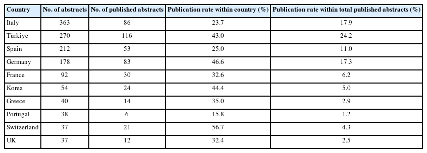
Out of 1,426 abstracts presented, 478 were published in peer-reviewed journals, yielding a publication rate of 33.5%. The median time to publication was 12 months. The leading journal in terms of publication rate was Clinical Oral Investigations. There was no statistically significant difference in publication rates across years. Abstracts related to laser therapy, caries, and dental materials had significantly higher publication rates compared to other subspecialties. Animal, basic, and clinical research studies were more likely to be published. Both study design and subspecialty influenced publication rates, which decreased over time.
Conclusions
A considerable amount of scientific data and preliminary results presented at conferences, which could contribute to scientific knowledge, are overlooked due to low publication rates. The findings of this study may encourage ConsEuro participants to submit well-planned and rigorous studies that are more likely to complete the full publication process.
- 2,907 View
- 70 Download
- Shaping ability and cyclic fatigue resistance between Genius ProFlex, ZenFlex, and TruNatomy rotary systems: an experimental study
- Raimundo Sales de Oliveira Neto, Murilo Priori Alcalde, Pedro Cesar Gomes Titato, Pedro Henrique Souza Calefi, Carlos Alberto Spironelli Ramos, Guilherme Ferreira da Silva, Rodrigo Ricci Vivan, Marco Antonio Hungaro Duarte
- Restor Dent Endod 2025;50(1):e9. Published online February 13, 2025
- DOI: https://doi.org/10.5395/rde.2025.50.e9
-
Abstract
PDFPubReaderePub
- Objectives
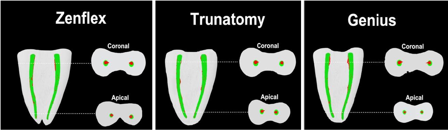
The aim of this study was to investigate the efficacy of three newly introduced rotary endodontic systems: Genius ProFlex (Medidenta), TruNatomy (Dentsply Maillefer), and ZenFlex (Kerr).
Methods
Forty-five mandibular molars with root canal curvatures <5° were utilized. Micro-computed tomography scans were performed pre- and post-preparation to assess apical transportation, centralization, percentage of dentin wear, and canal volume alterations. Eight instruments of each diameter underwent cyclic fatigue testing.
Results
The percentage of dentin wear on mesial and distal walls showed no significant differences among ZenFlex, TruNatomy, and Genius ProFlex at 1, 2, 3, and 4 mm from the apical foramen and root canal orifice (p > 0.05). Centering ability varied in the mesiolingual canal (p < 0.05). No notable differences were observed in transportation (p > 0.05). Genius ProFlex demonstrated lower volumetric changes (p < 0.05). There were significant differences in cyclic fatigue, with higher values for Genius ProFlex and lower values for TruNatomy (p < 0.05).
Conclusions
The three nickel-titanium rotary instruments are safe and efficient for root canal preparation, with Genius ProFlex exhibiting superior cyclic fatigue resistance. -
Citations
Citations to this article as recorded by- Comparison of Shaping Ability and Apical Debris Extrusion Using 4 Different Nickel–Titanium Single‐File Systems
Siyu Li, Mengzhen Tang, Xi Wang, Jian Yang, Hyun-Do Jung
International Journal of Biomaterials.2025;[Epub] CrossRef
- Comparison of Shaping Ability and Apical Debris Extrusion Using 4 Different Nickel–Titanium Single‐File Systems
- 3,776 View
- 183 Download
- 1 Crossref
- Concentrated growth factor scaffold-based pulpotomy of permanent molars with symptomatic irreversible pulpitis
- Arthi K. Harith, Vishnupriya Koteeswaran, Dinesh Kowsky, Natanasabapathy Velmurugan, Suresh Nandini
- Restor Dent Endod 2025;50(1):e1. Published online January 17, 2025
- DOI: https://doi.org/10.5395/rde.2025.50.e1
-
Abstract
PDFPubReaderePub
- Objectives
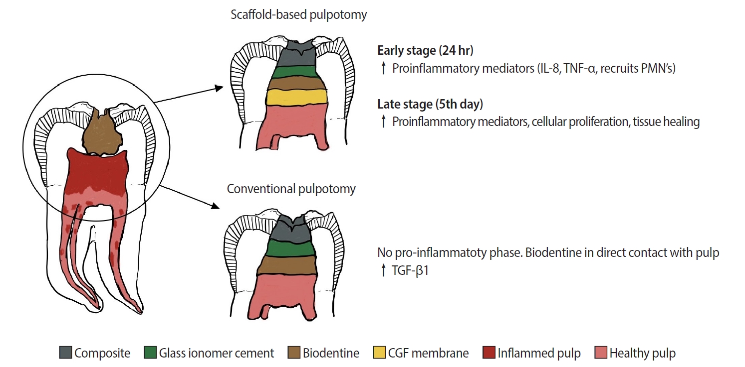
Pulpotomy is a minimally invasive procedure that aims to retain the vitality of the radicular pulp by removing the inflamed coronal pulp tissue. This case series presents the successful management of symptomatic irreversible pulpitis by pulpotomy with concentrated growth factor (CGF) scaffolds.
Methods
Six permanent mandibular molars with a diagnosis of symptomatic irreversible pulpitis were included. Under Local anesthesia and rubber dam isolation, caries were excavated using high-speed bur under coolant. Full coronal pulpotomy was done and hemostasis was achieved. CGF membrane was prepared and placed over the radicular pulp and layered with Biodentine (Septodont). Final restoration of type IX glass ionomer cement and bulk fill composite resin was placed. Patients were assessed for various clinical and radiographic parameters at intervals of 1 week and 3, 6, and 12 months. Five patients fulfilled the success criteria at the end of 1 year.
Results
Pulpotomy is considered an alternative treatment modality for root canal treatment in symptomatic irreversible pulpitis aiming at alleviating symptoms and maintaining vitality. CGF scaffold when used as a capping material acts as a reservoir for growth factors with anti-inflammatory properties and enhances healing.
Conclusions
Scaffold-based pulpotomy can be considered a biological approach to healing inflamed pulp.
- 5,128 View
- 498 Download
Case Report
- Guided endodontics, precision and predictability: a case series of mineralized anterior teeth with follow-up cone-beam computed tomography
- Rafael Fernández-Grisales, Wilder Javier Rojas-Gutierrez, Pamela Mejía, Carolina Berruecos-Orozco, Néstor Ríos-Osorio
- Restor Dent Endod 2025;50(1):e4. Published online January 6, 2025
- DOI: https://doi.org/10.5395/rde.2025.50.e4
-
Abstract
PDFPubReaderePub
- Pulp chamber and root canal obliteration (PCO/RCO) presents a challenge for clinicians when nonsurgical endodontic treatment is indicated. Guided endodontics (GE) aims to precisely locate the root canal (RC) system while preserving as much pericervical dentin as possible. GE involves integrating cone-beam computed tomography (CBCT) of the affected tooth with a digital impression of the maxillary/mandibular arch, allowing for careful planning of the drilling path to the RC system through a three-dimensional (3D) static guide. This article reports four cases of teeth with PCO/RCO, accompanied by additional diagnoses of internal and external root resorption and horizontal tooth fracture, all successfully treated with GE. These cases highlight the clinical and radiographic success of GE treatments using CBCT, establishing this technique as a predictable approach for managing mineralized teeth.
-
Citations
Citations to this article as recorded by- Static Guided Endodontics in Primary Endodontic Treatment of Anterior Teeth: A Narrative Review
Monika Kuczmaja, Wiesława Puchalska, Agata Żółtowska
Dentistry Journal.2026; 14(4): 195. CrossRef - Effect of different rotary instrument designs (protaper ultimate and protaper gold) on postoperative pain and bacterial reduction: a randomized clinical trial
Khaled Hassan Abed, Ahmed Abdel Rahman Hashem, Reem Ahmed Lutfy, Somaia Abdellatif Eissa, Dina Ahmed Morsy
BMC Oral Health.2026;[Epub] CrossRef
- Static Guided Endodontics in Primary Endodontic Treatment of Anterior Teeth: A Narrative Review
- 4,959 View
- 371 Download
- 2 Web of Science
- 2 Crossref
Review Article
-
Influence of disinfecting solutions on the surface topography of gutta-percha cones: a systematic review of
in vitro studies - Lora Mishra, Gathani Dash, Naomi Ranjan Singh, Manoj Kumar, Saurav Panda, Franck Diemer, Monika Lukomska-Szymanska, Barbara Lapinska, Abdul Samad Khan
- Restor Dent Endod 2024;49(4):e42. Published online November 1, 2024
- DOI: https://doi.org/10.5395/rde.2024.49.e42
-
Abstract
PDFSupplementary MaterialPubReaderePub
The surface integrity of gutta-percha cones is a crucial factor in the success of endodontic procedures. Disinfecting solutions play a pivotal role in sterilizing gutta-percha cones, but their influence on gutta-percha surface topography remains a subject of concern. This systematic review aimed to present a qualitative synthesis of available laboratory studies assessing the influence of disinfecting solutions on the surface topography of gutta-percha and offers insights into the implications for clinical practice. The present review followed PRISMA 2020 guidelines. An advanced database search was performed in PubMed, Google Scholar, Embase, Scopus, LILAC, non-indexed citations and reference lists of eligible studies in May 2024. Laboratory studies, in English language, were considered for inclusion. The quality (risk of bias) of the included studies was assessed using parameters for
in vitro studies. A total of 28 studies were included in the qualitative synthesis. Based on the included in vitro studies, surface deposits and alterations in the physical properties of gutta-percha cones were observed after the disinfection protocol. A comprehensive review of the available literature indicates that the choice of disinfecting solution, its concentration, and immersion time significantly affect the surface topography of gutta-percha cones.-
Citations
Citations to this article as recorded by- In Vitro Evaluation of Disinfectants on Gutta-Percha Cones: Antimicrobial Efficacy Against Enterococcus faecalis and Candida albicans
Tringa Kelmendi, Donika Bajrami Shabani, Aida Meto, Hani Ounsi
Journal of Clinical Medicine.2025; 14(19): 6846. CrossRef
- In Vitro Evaluation of Disinfectants on Gutta-Percha Cones: Antimicrobial Efficacy Against Enterococcus faecalis and Candida albicans
- 4,896 View
- 224 Download
- 1 Web of Science
- 1 Crossref
Research Articles
- Histological evaluation of pulp response to alendronate and Biodentine as pulp capping agents: an animal study
- Thangavel Boopathi, Sekar Manimaran, Joseline Charles Kerena, Mathew Sebeena, Kumaravadivel Karthick, Natesan Thangaraj Deepa
- Restor Dent Endod 2024;49(4):e39. Published online October 29, 2024
- DOI: https://doi.org/10.5395/rde.2024.49.e39
-
Abstract
PDFPubReaderePub
Objectives This study aimed to comparatively assess the histological response of the pulp toward alendronate and Biodentine in a direct pulp capping procedure.
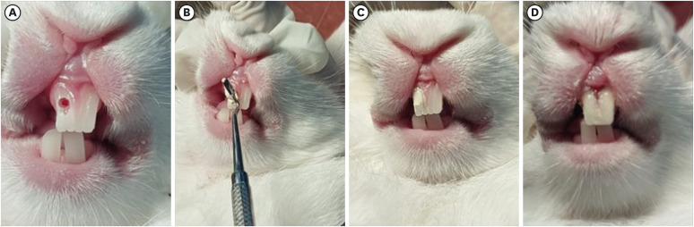
Materials and Methods Twenty-four anterior teeth from 6 New Zealand rabbits were used in this study. Firstly, all rabbits were anesthetized according to their weight. Class V cavities were prepared on the buccal surfaces of anterior teeth. A pin-point exposure of the pulp was then made using a small, sterile round carbide bur and bleeding was arrested with a saline-soaked, sterile cotton pellet. The teeth under study were divided into 2 groups (
n = 12). The intentionally exposed pulp was capped with alendronate (Group 1) and Biodentine (Group 2), correspondingly. After 30 days, all rabbits were euthanized; the teeth under study were extracted and taken up for histological analysis.Results Biodentine showed an intact, very dense dentin bridge formation with a uniform odontoblast (OD) layer pattern and mild or absent inflammatory response whereas specimens capped with alendronate demonstrated a dense dentin bridge formation with non-uniform OD layer pattern and mild to moderate inflammatory response.
Conclusions Biodentine showed more biocompatibility than alendronate. However, alendronate can initiate reparative dentin formation and may be used as an alternative pulp capping agent.
-
Citations
Citations to this article as recorded by- In Vivo Evaluation of NF-κB and TGFβ-1 Modulation by Anadara granosa Shell-Derived Calcium Carbonate Bioceramic in Rat Model
Randy Nugraha Pratama, Nurhayati Natsir, Kezia Rachellea Mustakim, Juni Jekti Nugroho
European Journal of General Dentistry.2026;[Epub] CrossRef
- In Vivo Evaluation of NF-κB and TGFβ-1 Modulation by Anadara granosa Shell-Derived Calcium Carbonate Bioceramic in Rat Model
- 4,078 View
- 143 Download
- 1 Crossref
-
Fracture resistance after root canal filling removal using ProTaper Next, ProTaper Universal Retreatment or hybrid instrumentation: an
ex vivo study - Hadeel Hassan Hanafy, Marwa Mahmoud Bedier, Suzan Abdul Wanees Amin
- Restor Dent Endod 2024;49(4):e38. Published online October 11, 2024
- DOI: https://doi.org/10.5395/rde.2024.49.e38
-
Abstract
PDFPubReaderePub
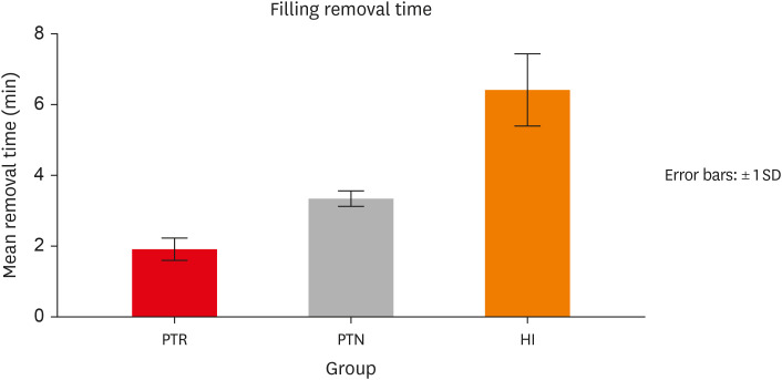
Objectives This study evaluated the effect of ProTaper Next (PTN), ProTaper Universal Retreatment (PTR) and hybrid instrumentation (HI) for canal filling removal on the fracture resistance (FR), mode of failure (MoF), and filling removal time.
Materials and Methods Ninety-six, mandibular premolars were decoronated and randomly divided into 6 groups (
n = 16), as follows: sound (S), untreated canals; prepared teeth (P), canals only prepared to ProTaper Universal finishing instrument (F4); endodontically-treated (ET), prepared and obturated canals using the single-cone technique; and groups PTN, PTR, and HI where filling was removed using PTN, PTR, or HI respectively. FR under vertical loading; MoF and time were assessed. Data were analyzed (Significance level [α] = 0.05).Results There was a significant difference in FR among all groups (
p < 0.001) (HI < P < PTN < S < ET < PTR). HI showed lower FR than S, ET and PTR, and P showed lower FR than PTR (p < 0.05). For experimental groups, there was a significant difference between every group pair (p < 0.05) No significant difference was found regarding MoF distribution (p > 0.05). HI required the highest filling removal time, while PTR required the least (p < 0.05 between every group pair).Conclusions The effect of filling removal on FR may depend on the filling removal technique/system used. PTR could be faster and protect against fracture followed by PTN; HI could adversely affect FR. FR may be associated with filling removal time.
- 3,664 View
- 136 Download
Case Report
- Straightforward replication of digital wax-up design into direct composite resin restorations in adolescents using a custom 3-dimensionally printed index
- Ra’fat Ibrahim Farah, Sanaa Najeh Al-Haj Ali, Abdullah Alharbi, Bandar Alresheedi
- Restor Dent Endod 2024;49(4):e36. Published online October 10, 2024
- DOI: https://doi.org/10.5395/rde.2024.49.e36
-
Abstract
PDFPubReaderePub
This case report introduces a straightforward, noninvasive approach for the esthetic rehabilitation of malformed anterior teeth in adolescents using direct composite restorations. The universal composite resin restorations are applied within a transparent 3-dimensionally printed rigid-resin index, which is individually customized from a digital wax-up. Compared to other methods, this technique streamlines the restoration process, significantly reducing chairside time while enhancing the predictability, accuracy, and patient acceptance of the aesthetic outcome.
-
Citations
Citations to this article as recorded by- Diastema closure and esthetic rehabilitation with peg-shaped laterals: A case series
Afsana Ansari, Dipika Yadav
The Saint's International Dental Journal.2024; 8(2): 48. CrossRef
- Diastema closure and esthetic rehabilitation with peg-shaped laterals: A case series
- 5,512 View
- 267 Download
- 1 Crossref
Research Articles
- Effects of different curing methods on the color stability of composite resins
- Massimo Pisano, Alfredo Iandolo, Dina Abdellatif, Andrea Chiacchio, Marzio Galdi, Stefano Martina
- Restor Dent Endod 2024;49(4):e33. Published online September 5, 2024
- DOI: https://doi.org/10.5395/rde.2024.49.e33
-
Abstract
PDFPubReaderePub
Objectives The aim of this study was to compare the effects of different polymerization strategies and the effectiveness of finishing and polishing procedures of composite resins on color stability.
Materials and Methods The samples were divided into 4 main groups according to the polymerization strategy, and all groups except the control group received surface treatment. Each group was subsequently divided into 3 subgroups respectively: Kuraray Clearfil Majesty ES-2 Classic, Premium and Universal. Approximately 24 hours after preparation of the samples, they were immersed for 7 days in a coffee solution. A first color measurement was performed after the preparation of the samples, the second measurement was performed after 7 days in the coffee solution. All measurements were carried out using a dental spectrophotometer to assess the CIE
L *a *b * color parameters.Results There was a statistically significant difference between ΔE values for different procedures (
p = 0.003); in particular, the differences were found only between the groups that received surface treatment and the control group. In addition, a statistically significant difference was observed between the values of ΔE for different composites in the different procedure groups.Conclusions Spectrophotometric analysis showed that the additional photopolymerization and oxygen inhibition procedures did not yield better results in relation to color stability. In addition, finishing and polishing provided better color stability compared to not performing these procedures.
-
Citations
Citations to this article as recorded by- Color Stability Under Challenge: Effects of Thermo-Aging and Mouthrinse Exposure on Anterior Teeth and Esthetic Composites
Gökçe Keçeci, Zehra Güner, Süleyman Ziya Şenyurt, Kamile Erciyas
European Journal of Therapeutics.2026; 32(1): 94. CrossRef - Color stability and degree of conversion of conventional, amine-free, and self-adhesive resin cements polymerized under different conditions
Su Young Lee, Yasushi Shimada, Jaeyoung Edwin Han, Seung-Hoon Han
Dental Materials.2026;[Epub] CrossRef - Abrasiveness and Bleaching Level of Toothpastes on Composite Resins: A Quantitative Analysis Using a Novel Brushing Simulator
Simge Meseli, Elif Alkan, Bora Korkut, Ozlem Kanar, Dilek Tagtekin
Applied Sciences.2025; 15(5): 2314. CrossRef - Comparative Evaluation of Direct and Indirect Composite Restorations in Class II Tooth Preparations - An In vivo Study
Akshun Gupta, Garima Arora, Aprajita Mehta, Satish Sane, Siddhi Nevrekar, Apurva Nagrale
Advances in Human Biology.2025; 15(4): 550. CrossRef - Micro- and Nanoplastics and the Oral Cavity: Implications for Oral and Systemic Health, Dental Practice, and the Environment—A Narrative Review
Federica Di Spirito, Veronica Folliero, Maria Pia Di Palo, Giuseppina De Benedetto, Leonardo Aulisio, Stefano Martina, Luca Rinaldi, Gianluigi Franci
Journal of Functional Biomaterials.2025; 16(9): 332. CrossRef
- Color Stability Under Challenge: Effects of Thermo-Aging and Mouthrinse Exposure on Anterior Teeth and Esthetic Composites
- 7,187 View
- 374 Download
- 4 Web of Science
- 5 Crossref
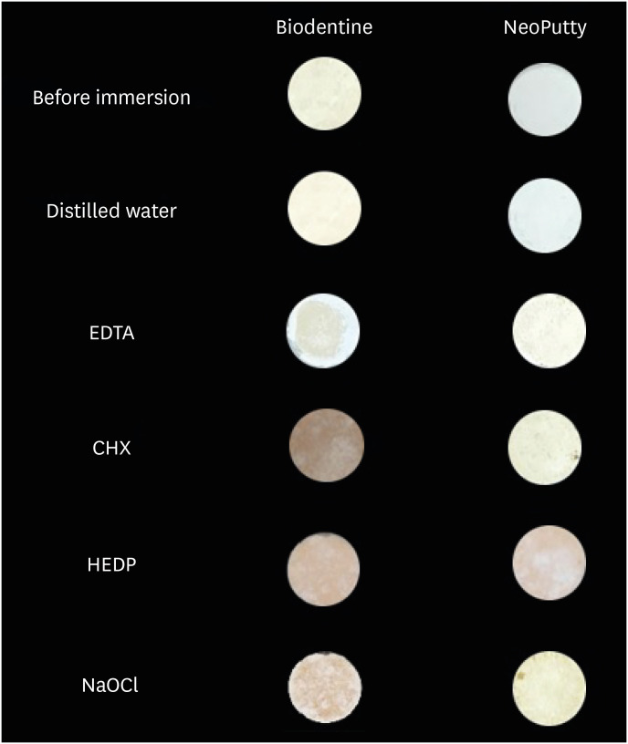
- Color stability and solubility of Biodentine and NeoPutty in contact with different irrigation solutions
- Sıla Nur Usta, Cangül Keskin
- Restor Dent Endod 2024;49(3):e25. Published online June 19, 2024
- DOI: https://doi.org/10.5395/rde.2024.49.e25
-
Abstract
PDFPubReaderePub
Objectives This study aimed to evaluate the color stability and solubility of Biodentine and NeoPutty in contact with different irrigation solutions.
Materials and Methods Biodentine and NeoPutty were set in cylindrical molds with 7 mm diameter and 1.5 mm high and immersed in distilled water, 17% ethylenediaminetetraacetic acid (EDTA), 2% chlorhexidine (CHX), 9% 1-hydroxyethylidene 1,1-diphosphonate (HEDP), and 5% sodium hypochlorite (NaOCl) solutions for 24 hours. The color change was measured with a spectrophotometer. The solubility values were calculated as the mass loss was expressed as a percentage of the original mass using an analytical balance with 10−4 g accuracy. Data were analyzed with Kruskal-Wallis followed by Mann-Whitney
U tests, and 2-way analysis of variance test followed by Bonferroni corrections for pairwise comparisons for solubility and color stability with a 5% significance threshold, respectively.Results Biodentine exhibited higher color changes compared to the NeoPutty contact with all solutions except distilled water (
p < 0.05). Both hydraulic cements (HCs) showed higher discoloration values immersion in CHX followed by NaOCl. No statistically significant difference was found between Biodentine and NeoPutty regardless of irrigation solution in terms of solubility (p > 0.05). Solubility values were lower in the distilled water group compared to EDTA and CHX (p < 0.05).Conclusions Tested HCs showed solubility and color changes at various rates. NeoPutty could be an appropriate material in aesthetic areas. The usage of HEDP as an irrigant solution can be considered suitable for various endodontic treatments due to its relatively lower solubility and discoloration values.
-
Citations
Citations to this article as recorded by- Sealing ability of Biodentine, zirconia reinforced glass ionomer cement and Mineral Trioxide Aggregate as furcation perforation repair materials: an in vitro analysis
Sumita Panwar, Yajuvender Singh Hada
Biomaterial Investigations in Dentistry.2026; 13: 21. CrossRef - Effect of calcium silicate-based materials on tooth discolouration in repairing root perforations of lower molars: an in-vitro study
Sevil Zırhlı, Davut Celık, Tugba Kosar
Journal of the Australian Ceramic Society.2026;[Epub] CrossRef - Influence of endodontic irrigants on hydraulic cements: solubility, color alteration and surface changes
Sıla Usta, Cangül Keskin, Ayşe Oktay, Emmanuel João Nogueira Leal Silva
European Oral Research.2026; 60(1): 230. CrossRef
- Sealing ability of Biodentine, zirconia reinforced glass ionomer cement and Mineral Trioxide Aggregate as furcation perforation repair materials: an in vitro analysis
- 3,479 View
- 171 Download
- 2 Web of Science
- 3 Crossref
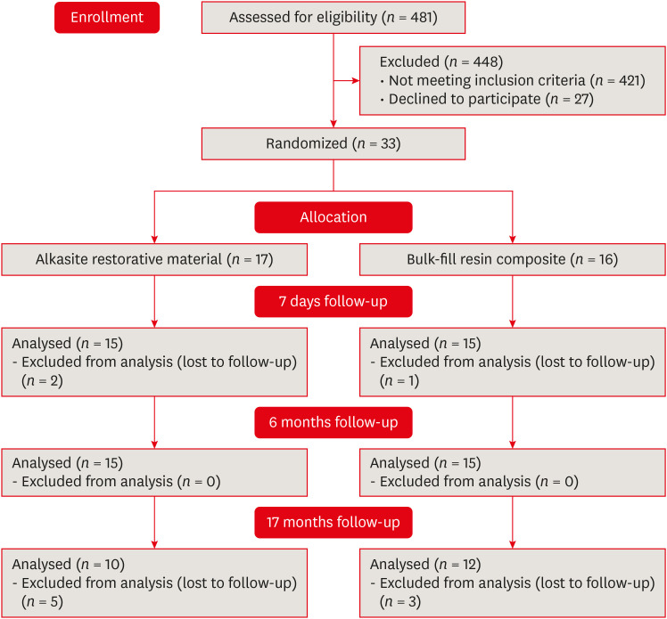
- Alkasite restorative material for endodontically treated teeth: a randomized controlled pilot study
- Davi Ariel Nobuo Bepu, Renata Siqueira Scatolin, Natalia Saud Junqueira Franco, Luiza Pejon Sanchez, Aline Evangelista Souza-Gabriel, Silmara Aparecida Milori Corona
- Restor Dent Endod 2024;49(3):e24. Published online June 11, 2024
- DOI: https://doi.org/10.5395/rde.2024.49.e24
-
Abstract
PDFPubReaderePub
Objectives This study aimed to evaluate the clinical performance of an alkasite restorative material in molars that had undergone root canal treatment.
Materials and Methods The research was registered in Brazilian Registry of Clinical Trials. The randomized clinical trial involved 33 patients, each with at least 1 mandibular molar requiring restoration after receiving endodontic treatment. Patients were randomly assigned to receive either bulk-fill resin composite (Tetric N Ceram Bulk Fill, Ivoclar Vivadent) or the alkasite restorative material (Cention N, Ivoclar Vivadent). Upon completion of the restorations, 3 calibrated professionals utilized the United States Public Health Service criteria to assess various factors, including retention, secondary caries, marginal adaptation, restoration color, marginal pigmentation, and anatomical form. Evaluations were conducted at intervals of 7 days, 6 months, and 17 months. Additionally, the assessment encompassed the presence of radiolucent lines adjacent to the restoration, material deficiencies or excess, contact points, and caries recurrence. The data underwent analysis using the Friedman and Mann-Whitney tests (α = 0.05).
Results After 17 months, the results revealed that the alkasite restorative material exhibited greater wear of anatomical shape compared to the bulk-fill resin composite (
p = 0.0189). Furthermore, the alkasite restorative material significantly differed from the natural tooth color in most cases (p = 0.0000). However, no other criteria displayed significant differences between the materials or over time (p > 0.05).Conclusions The alkasite restorative material (Cention N) emerges as a viable option for restoring endodontically treated teeth, displaying clinically acceptable alterations after a 17-month evaluation period.
Trial Registration Brazilian Registry of Clinical Trials (ReBEC) Identifier:
RBR-97kx5jv -
Citations
Citations to this article as recorded by- The Effect of Intraorifice Barrier Materials on the Fracture Resistance of Endodontically Treated Teeth: A Systematic Review and Network Meta-Analysis
Sevilay Karahan, Zeynep Buket Dağ, Emel Uzunoğlu Özyürek
Journal of Endodontics.2026; 52(5): 696. CrossRef - A Systematic Review and Meta-Analysis on the Clinical Performance and Longevity of Bioactive Composite Resin Restorations
Ahmed A. Holiel, Mounir M. Al Nakouzi, Rim Bourgi, Carlos Enrique Cuevas-Suárez, Iván Olivares Acosta, Louis Hardan, Naji Kharouf, Youssef Haikel
Journal of Composites Science.2026; 10(1): 39. CrossRef - Evaluation of Clinical Performance of Alkasite Restorative Materials: A Systematic Review and Meta-Analysis
Chloé Laporte, Rim Bourgi, Carlos Enrique Cuevas-Suárez, Naji Kharouf, Louis Hardan, Miguel Ángel Fernández-Barrera, Anh Tuan Dang, Youssef Haikel, Abigailt Flores-Ledesma
Journal of Functional Biomaterials.2026; 17(2): 93. CrossRef - 48-month clinical performance of an Alkasite restorative material versus resin composite in class II restorations: a randomized controlled trial
Ece Meral, Betül Kesim, Fatma Dilşad Öz, Sevil Gürgan
Journal of Dentistry.2026; 173: 106792. CrossRef - Alkasites in restorative dentistry: a review of their performance and properties
Alexander Bonchev, Ralitsa Bogovska-Gigova
Journal of Dentistry.2025; 160: 105916. CrossRef - Comparative Analysis of Flexural and Compressive Strengths of Bioactive Alkasite Compared to Other Ion-Releasing Restorative Materials
Hanin E. Yeslam, Fatin A. Hasanain
Biomimetics.2025; 10(11): 751. CrossRef
- The Effect of Intraorifice Barrier Materials on the Fracture Resistance of Endodontically Treated Teeth: A Systematic Review and Network Meta-Analysis
- 5,003 View
- 152 Download
- 5 Web of Science
- 6 Crossref
- Can discolored dental composites be bleached in depth?
- Luca Giachetti, Daniele Scaminaci Russo, Michele Nieri, Francesca Cinelli
- Restor Dent Endod 2024;49(3):e23. Published online June 11, 2024
- DOI: https://doi.org/10.5395/rde.2024.49.e23
-
Abstract
PDFPubReaderePub
Objectives Previous
in vitro studies determined the whitening effects of bleaching products on stained resin composite surfaces. Thisin vitro study aimed to verify the effectiveness of a whitening system on composite resin previously subjected to pigmentation, specifically examining the depth of whitening effectiveness within the material structure.Materials and Methods A commercially available nano-filled composite resin was used. Specimens were stained using a coffee-based solution and a 10% carbamide peroxide-based gel was employed as the whitening agent. The pigment’s penetration and the effect of the bleaching gel were evaluated by measuring color (CieLab values) from the outer edge to the inner part of the specimens. Color measurements were taken at 14 points, starting from 0.1 mm from the external perimeter up to 3.0 mm.
Results Analysis of variance tests showed a statistically significant difference between the Control Group (CG), Pigmentation Group, and Whitening Group. The whitening agent was effective up to 1.5 mm in depth, with Whiteness index (W) values not statistically different from those of CG up to 0.5 mm in depth.
Conclusions Whitening agents on nano-filled resin composite previously pigmented appear effective in restoring the W to values similar to the original, particularly in the superficial layers of the sample.
-
Citations
Citations to this article as recorded by- Color Stability of Tooth-Colored Restorative Materials After Exposure to Arabic Coffee and Black Tea: A Systematic Review
Abdulrhman Y Alenezi, Abdulwahab M AlEyada, Yousef H Aldhafiri, Mohammed S Alsubaie, Mohammed S Alshahrani, Mahesh Shenoy
Cureus.2025;[Epub] CrossRef - Comparative evaluation to composite resin bleaching using ozone-enhanced low-concentration hydrogen peroxide
Mahmoud K. AL-Omiri, Dania Sa’ed Hussam Abuherra, Khaled M. AL-Omiri, Ali Y. Alsaeed, Mohammad Alamri, Ali M. Alqahtani, Saleh Ali Alqahtani, Ghadeer Saleh Alwadai, Naif Abogazalah, Edward Lynch
Scientific Reports.2025;[Epub] CrossRef - The effects of mechanical and chemical degradation on the surface roughness, gloss, and color stability of bulk-fill resin composites
Merve Nezir, Hanife Altınışık, Esra Özyurt, Naz Bayar, Mediha Büyükgöze Dindar
BMC Oral Health.2025;[Epub] CrossRef
- Color Stability of Tooth-Colored Restorative Materials After Exposure to Arabic Coffee and Black Tea: A Systematic Review
- 4,602 View
- 148 Download
- 2 Web of Science
- 3 Crossref
- Pulp stones: any relevance with the levels of serum calcium, parathyroid hormone, vitamin D and uric acid
- Ceyda Gürhan, Ercan Saruhan
- Restor Dent Endod 2024;49(2):e17. Published online March 26, 2024
- DOI: https://doi.org/10.5395/rde.2024.49.e17
-
Abstract
PDFPubReaderePub
Objectives This study evaluated the effect of serum calcium, parathyroid hormone (PTH), vitamin D, and uric acid levels on pulp stone formation.
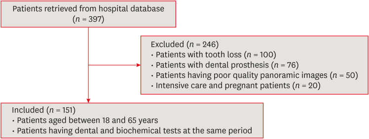
Materials and Methods Patients who were admitted to the Muğla Sıtkı Koçman University, Faculty of Dentistry, Department of Oral and Maxillofacial Radiology for dental complaints were registered. Among these patients, individuals who had routine biochemical tests at the same period in the Outpatient Clinics of Muğla Sıtkı Koçman University Training and Research Hospital were included in the study. The patients with at least 1 pulp stone on panoramic radiographs recorded as the “pulp stone group” while patients without any pulp stones were the “control group”. Demographic data and serum levels of calcium, PTH, vitamin D, and uric acid were retrospectively evaluated in both groups. Student
t -test or Mann-WhitneyU test was used to evaluate the differences between the groups.Results Among 151 patients, dental pulp stone was detected in 53.6% of patients, and 82.7% of these patients were female. Female sex and pulp stone formation were significantly associated (
p = 0.001). The mean age of the pulp stone group was 43.9, while it was 39.9 in the control group, without any significant correlation between age and pulp stone (p > 0.05). Similarly, there were no significant differences in serum levels of PTH, vitamin D, uric acid and calcium between groups (p > 0.05).Conclusions According to the present study, the effect of dental factors rather than systemic factors should be considered primarily in pulp stone formation.
-
Citations
Citations to this article as recorded by- A novel deep learning-based pipeline architecture for pulp stone detection on panoramic radiographs
Ceyda Gürhan, Hasan Yiğit, Selim Yılmaz, Cihat Çetinkaya
Oral Radiology.2025; 41(2): 285. CrossRef - Vitamin D deficiency and oral health: a systematic review of literature
Saida Ziada, Aws Wishahe, Najet Mabrouk, Souad Sahtout
BMC Oral Health.2025;[Epub] CrossRef - Association between pulp stones and systemic diseases: a retrospective study using digital panoramic radiographs in a Turkish population
Buket Beytaş Alğan, Mustafa Murat Koçak, Sibel Koçak, Baran Can Sağlam
BMC Oral Health.2025;[Epub] CrossRef
- A novel deep learning-based pipeline architecture for pulp stone detection on panoramic radiographs
- 4,504 View
- 116 Download
- 4 Web of Science
- 3 Crossref
Case Report
- Garre’s osteomyelitis of the mandible managed by nonsurgical re-endodontic treatment
- Heegyun Kim, Jiyoung Kwon, Hyun-Jung Kim, Soram Oh, Duck-Su Kim, Ji-Hyun Jang
- Restor Dent Endod 2024;49(2):e13. Published online March 18, 2024
- DOI: https://doi.org/10.5395/rde.2024.49.e13
-
Abstract
PDFPubReaderePub
Chronic osteomyelitis with proliferative periostitis, known as Garre’s osteomyelitis, is a type of osteomyelitis characterized by a distinctive gross thickening of the periosteum of bones. Peripheral reactive bone formation can be caused by mild irritation or infection. Garre’s osteomyelitis is usually diagnosed in children and young adults, and the mandible is more affected than the maxilla. The following is a case report of a 12-year-old female patient with Garre’s osteomyelitis of the mandible due to an infection of a root canal-treated tooth. Without surgical intervention, the patient’s symptoms were relieved through nonsurgical root canal re-treatment with long-term calcium hydroxide placement. A cone-beam computed tomography image obtained 6 months after treatment completion displayed complete healing of the periapical lesion and resolution of the peripheral reactive buccal bone. Due to the clinical features of Garre's osteomyelitis, which is characterized by thickening of the periosteum, it can be mistaken for other diseases such as fibrous dysplasia. It is important to correctly diagnose Garre's osteomyelitis based on its distinctive clinical features to avoid unnecessary surgical intervention, and it can lead to minimally invasive treatment options.
-
Citations
Citations to this article as recorded by- Endodontic Intervention in Chronic Osteomyelitis With Proliferative Periostitis: A Rare Case Report and Scoping Review
Gabriel Lima Braz, Ana Paula Neutzling Gomes, Lisandrea Rocha Schardosim, Nadia de Souza Ferreira, Jose Francisco Gomez-Sosa
Case Reports in Dentistry.2026;[Epub] CrossRef - How Clinical and Radiological Findings in Chronic Mandibular Osteomyelitis Do Not Always Correlate: Diagnostic Dilemmas in Dental-Related Bone Inflammations
Kamil Nelke, Ömer Uranbey, Ece Gülbağ, Büşra Ekinci, Burcu Gürsoytrak, Angela Rosa Caso, Michał Gontarz, Maciej Janeczek, Piotr Kuropka, Maciej Dobrzyński
Diagnostics.2026; 16(10): 1427. CrossRef - Focal osteomyelitis with proliferative periostitis
Zarah Yakoob
South African Dental Journal.2025; 79(09): 508. CrossRef - Garré’s osteomyelitis of the mandible in an adolescent: a case report
Wiem Feki, Imen Haddar, Marwa Bahloul, Zeineb Mnif, Thouraya Kammoun, Ines Maaloul
Journal of Medical Case Reports.2025;[Epub] CrossRef - Garré’s Chronic Sclerosing Osteomyelitis: An Overview of Clinical and Radiologic Features
Mohamed Fadil, Ayman Farouki, Rachida Saouab, Hassan En-nouali, Jamal El Fenni, Zakariya Toufga
Oxford Medical Case Reports.2025;[Epub] CrossRef
- Endodontic Intervention in Chronic Osteomyelitis With Proliferative Periostitis: A Rare Case Report and Scoping Review
- 9,904 View
- 220 Download
- 4 Web of Science
- 5 Crossref
Research Articles
- Color discrepancy of single-shade composites at different distances from the interface measured using cell phone images
- Márcia Luciana Carregosa Santana, Gabriella de Jesus Santos Livi, André Luis Faria-e-Silva
- Restor Dent Endod 2024;49(1):e7. Published online January 24, 2024
- DOI: https://doi.org/10.5395/rde.2024.49.e7
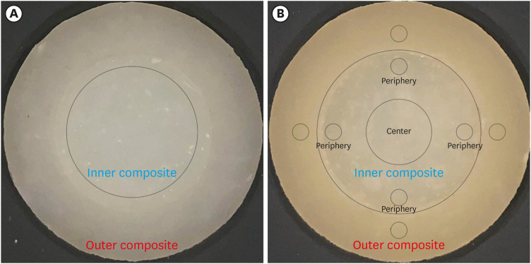
-
Abstract
PDFPubReaderePub
Objectives This study aimed to evaluate the impact of substrate color and interface distance on the color adjustment of 2 single-shade composites, Vittra APS Unique and Charisma Diamond One.
Materials and Methods Dual disc-shaped specimens were created using Vittra APS Unique or Charisma Diamond One as the center composite, surrounded by shaded composites (A1 or A3). Color measurements were taken with a spectrophotometer against a gray background, recording the color coordinates in the CIELAB color space. Illumination with a light-correcting device and image acquisition using a polarizing filter-equipped cell phone were performed on specimens over the same background. Image processing software was used to measure the color coordinates in the center and periphery of the inner composite and in the outer composite. The color data were then converted to CIELAB coordinates and adjusted using data from the spectrophotometer. Color differences (ΔE00) between the center/periphery of single-shade and outer composites were calculated, along with color changes in single-shade composites caused by different outer composites. Color differences for the inner composites surrounded by A1 and A3 were also calculated. Data were analyzed using repeated-measures analysis of variance (α = 0.05).
Results The results showed that color discrepancies were lowest near the interface and when the outer composite was whiter (A1). Additionally, Charisma Diamond One exhibited better color adjustment ability than Vittra APS Unique.
Conclusions Color discrepancies between the investigated single-shade composites diminished towards the interface with the surrounding composite, particularly when the latter exhibited a lighter shade.
-
Citations
Citations to this article as recorded by- Evaluation of color stability in single-shade composite resins using spectrophotometer and cross-polarized mobile photography
Hatice Tepe, Ozge Celiksoz, Batu Can Yaman
BMC Oral Health.2025;[Epub] CrossRef - Comparative Evaluation of the Staining Resistance of Two Single-Shade Composites in Coffee and Chlorhexidine: A Spectrophotometric Analysis
Unmesh Khanvilkar, Shrinath D Kulkarni, Siddhesh Bandekar, Ved M Talathi, Oshin Baghel, Priyanka Razdan, Seema Gupta
Cureus.2025;[Epub] CrossRef - Clinical Implications of Color Adjustment in Single-Shade Resins Post-Dental Bleaching: A Systematic Review
Samille Biasi Miranda, Caroline de Farias Charamba Leal, Rodrigo Barros Esteves Lins, Marcos Antonio Japiassu Resende Montes
Journal of Clinical Medicine.2025; 14(9): 3194. CrossRef - Accuracy and Reliability of Smartphone Versus Mirrorless Camera Images-Assisted Digital Shade Guides: An In Vitro Study
Soo Teng Chew, Suet Yeo Soo, Mohd Zulkifli Kassim, Khai Yin Lim, In Meei Tew
Applied Sciences.2025; 15(14): 8070. CrossRef
- Evaluation of color stability in single-shade composite resins using spectrophotometer and cross-polarized mobile photography
- 2,996 View
- 83 Download
- 3 Web of Science
- 4 Crossref
- The status of clinical trials regarding root canal sealers
- Ahmad AL Malak, Yasmina EL Masri, Mira Al Ziab, Nancy Zrara, Tarek Baroud, Pascale Salameh
- Restor Dent Endod 2024;49(1):e5. Published online January 15, 2024
- DOI: https://doi.org/10.5395/rde.2024.49.e5
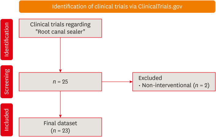
-
Abstract
PDFPubReaderePub
Objectives This study aimed to present the results and analyses of clinical trials, including updates on the different functions of root canal sealers.
Materials and Methods In June 2023, we performed a comprehensive search of ClinicalTrials.gov to identify interventional clinical trials pertaining to root canal sealers. In total, 23 clinical trials conducted up to June 2023 were included in this study.
Results Approximately half of the trials (11 out of 23) were completed, while none were terminated or withdrawn. Each included trial had a minimum of 10 participants, with 11 trials having more than 100 participants. None of the assessed trials provided outcomes, and the majority (17 out of 23) lacked associated publications. In terms of geographic distribution, the USA and Canada did not contribute to any root canal sealer trials.
Conclusions This study highlights the lack of diversity in trial locations, the absence of reported results, and a scarcity of clinical trials examining the physicochemical properties of different sealers. Most published trials primarily focused on assessing the post-operative pain effect of these sealers, but no significant difference was found regarding post-operative pain control.
- 4,554 View
- 68 Download
- A scientometric, bibliometric, and thematic map analysis of hydraulic calcium silicate root canal sealers
- Anastasios Katakidis, Konstantinos Kodonas, Anastasia Fardi, Christos Gogos
- Restor Dent Endod 2023;48(4):e41. Published online November 13, 2023
- DOI: https://doi.org/10.5395/rde.2023.48.e41
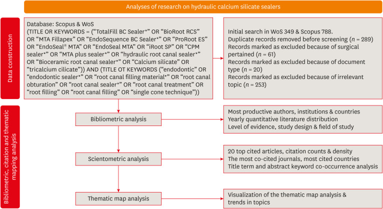
-
Abstract
PDFPubReaderePub
Objectives This scientometric and bibliometric analysis explored scientific publications related to hydraulic calcium silicate-based (HCSB) sealers used in endodontology, aiming to describe basic bibliometric indicators and analyze current research trends.
Materials and Methods A comprehensive search was conducted in Web of Science and Scopus using specific HCSB sealer and general endodontic-related terms. Basic research parameters were collected, including publication year, authorship, countries, institutions, journals, level of evidence, study design and topic of interest, title terms, author keywords, citation counts, and density.
Results In total, 498 articles published in 136 journals were retrieved for the period 2008–2023. Brazil was the leading country, and the universities of Bologna in Italy and Sao Paolo in Brazil were represented equally as leading institutions. The most frequently occurring keywords were “calcium silicate,” “root canal sealer MTA-Fillapex,” and “biocompatibility,” while title terms such as “calcium,” “sealers,” “root,” “canal,” “silicate based,” and “endodontic” occurred most often. According to the thematic map analysis, “solubility” appeared as a basic theme of concentrated research interest, and “single-cone technique” was identified as an emerging, inadequately developed theme. The co-occurrence analysis revealed 4 major clusters centered on sealers’ biological and physicochemical properties, obturation techniques, retreatability, and adhesion.
Conclusions This analysis presents bibliographic features and outlines changing trends in HCSB sealer research. The research output is dominated by basic science articles scrutinizing the biological and specific physicochemical properties of commonly used HCSB sealers. Future research needs to be guided by studies with a high level of evidence that utilize innovative, sophisticated technologies.
-
Citations
Citations to this article as recorded by- Top 100 Most Cited Articles on Antibiotics in Endodontics: A Bibliometric Analysis
Hajar Albanyan, Mohammed Asseery, Haitham Alahmari, Ikram Ul Haq, Ali Alaqla
Journal of Endodontics.2026; 52(3): 345. CrossRef - Top-cited articles on vital pulp therapy for irreversible pulpitis in permanent teeth: a bibliometric analysis
İkbal Sena Çelebi Keskin, Ferda Karabay
Odontology.2026;[Epub] CrossRef - Agri-Food Sector: Contemporary Trends, Possible Gaps, and Prospective Directions
José Roberto Herrera Cantorani, Meire Ramalho de Oliveira, Luiz Alberto Pilatti, Thales Botelho de Sousa
Metrics.2025; 2(1): 3. CrossRef - Scientific mapping of experimental research on solar cookers: Global trends, evolution, and future directions
Flavio Odoi-Yorke, Bismark Baah, Richard Opoku
Solar Energy Advances.2025; 5: 100093. CrossRef - Bibliometric analysis of the GentleWave system: trends, collaborations, and research gaps
Raimundo Sales de Oliveira Neto, Thais de Moraes Souza, João Vitor Oliveira de Amorim, Thaine Oliveira Lima, Guilherme Ferreira da Silva, Rodrigo Ricci Vivan, Murilo Priori Alcalde, Marco Antonio Hungaro Duarte
Restorative Dentistry & Endodontics.2025; 50(2): e17. CrossRef - A Scientometric Review of Practical Applications in Quantum Natural Language Processing (QNLP): Trends, Gaps, and Research Opportunities
Victor R. Silva, Fábio R. Barbosa, Jasson C. Silva, Francisco J. Santos, Ricardo A. L. Rabelo, Joel J. P. C. Rodrigues
IEEE Access.2025; 13: 210169. CrossRef - A bibliometric analysis of global research trend and progress on Dy doped materials
Sangeeta Kadyan, Manju Nain, Ashima Makhija, Poonam Punia, Anil Ohlan, Sajjan Dahiya, R. Punia, A.S. Maan
Journal of Alloys and Compounds Communications.2024; 3: 100006. CrossRef - Comparative bioactivity and immunomodulatory potential of the new Bioroot Flow and AH Plus Bioceramic sealer: An in vitro study on hPDLSCs
José Luis Sanz, Sergio López-García, David García-Bernal, Francisco Javier Rodríguez-Lozano, Leopoldo Forner, Adrián Lozano, Laura Murcia
Clinical Oral Investigations.2024;[Epub] CrossRef - Analyzing collaboration and impact: A bibliometric review of four highly published authors’ research profiles on collaborative maps
Willy Chou, Julie Chi Chow
Medicine.2024; 103(28): e38686. CrossRef
- Top 100 Most Cited Articles on Antibiotics in Endodontics: A Bibliometric Analysis
- 3,599 View
- 51 Download
- 7 Web of Science
- 9 Crossref
- Investigation of fracture prevalence of instruments used in root canal treatments at a faculty of dentistry: a prospective study
- Mehmet Eskibağlar, Merve Yeniçeri Özata, Mevlüt Sinan Ocak, Faruk Öztekin
- Restor Dent Endod 2023;48(4):e38. Published online November 1, 2023
- DOI: https://doi.org/10.5395/rde.2023.48.e38
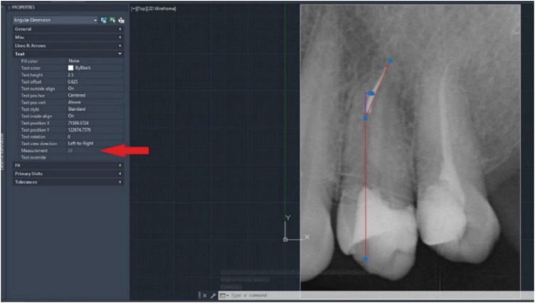
-
Abstract
PDFPubReaderePub
Objectives The aim of this study was to examine the use of hand or rotary files by pre-graduation (fourth- and fifth-year) and postgraduate students in endodontic treatments and to determine the incidence of file fracture and the management of cases with broken instruments.
Materials and Methods A total of 2,168 teeth undergoing primary endodontic treatment were included in this study. It was determined that 79 of these teeth resulted in broken tools. In the case of broken tools, the education level of the treating clinician, the tooth that was being treated, the canal and fracture level, the curvature of the tooth and the management of the broken instrument were recorded. Periapical radiographs of the patients were used to calculate curvature following the Schneider method.
Results There was no significant difference in the incidence of broken tools according to education level (
p > 0.05). The incidence of file fracture in molar teeth (73.4%) was higher than in other teeth (p < 0.05). More files were broken in the mandibular molar MB canal (20.25%) and in the apical third of the canals (72.1%). The risk of instrument fracture was high in teeth with moderate (44.3%) and severe (38%) curvature canals. The management of apically broken (80%) files mostly involved lefting (p < 0.05).Conclusions There was no statistically significant difference between fourth-year students, fifth-year students and postgraduate students in terms of instrument fracture.
-
Citations
Citations to this article as recorded by- Assessment of Techniques of Surpassing Ledges in Curved Canals and Its Effect on File: An In Vitro Study
Nezar Boreak, Abdulrahman Ali Sawadi, Yahya Yaqoup Hazazi, Sameer J Oqayshi, Wed AY Ayash, Essa Ibrahim Al Essa, Abdulaziz Ismail Shafei, Amjad Hassan Khawaji
World Journal of Dentistry.2026; 16(11): 1032. CrossRef - Secondary retrieval and complication management for failed separated instrument removal in the apical third in curved root canals: a case series
Mei Fu, Jia Guo, Fengting Wang, Benxiang Hou, Chen Zhang
BMC Oral Health.2026;[Epub] CrossRef - Prevalence and management techniques for separated endodontic files among endodontic postgraduate students in the Qassim region: A retrospective cross-sectional study
Hanin Abdulaziz Alsalhi, Rana Rabeh Alharbi, Amnah Ameen Hawsa
Saudi Endodontic Journal.2026; 16(2): 160. CrossRef - Case Study of a Broken Instrument in a Primary Tooth and Literature Review
Masashi Nakano, Tatsuya Akitomo, Masashi Ogawa, Mariko Kametani, Momoko Usuda, Satoru Kusaka, Chieko Mitsuhata, Ryota Nomura
Children.2025; 12(2): 149. CrossRef - Neodymium-Doped Yttrium Aluminum Perovskite (Nd:YAP) Laser in the Elimination of Endodontic Nickel-Titanium Files Fractured in Rooted Canals (Part 2: Teeth With Significant Root Curvature)
Amaury Namour, Marwan El Mobadder, Clément Cerfontaine, Patrick Matamba, Lucia Misoaga, Delphine Magnin , Praveen Arany, Samir Nammour
Cureus.2025;[Epub] CrossRef - Pattern of endodontic instrument separation and factors affecting its retrieval: a 10-year retrospective observational study in a postgraduate institute
Velmurugan Natanasabapathy, Aswathi Varghese, Paul Kevin Abishek Karthikeyan, Srinivasan Narasimhan
Restorative Dentistry & Endodontics.2025; 50(1): e7. CrossRef - Remoção de instrumentos fraturados nos canais radiculares: Desafios, estratégias e perspectivas clínicas
João Victor da Fonseca Barbosa, Eduardo Kitto Miranda Teixeira , Laura Rodrigues Barbosa, Martinelle Ferreira da Rocha Taranto, Jáder Camilo Pinto
Research, Society and Development.2025; 14(10): e98141049749. CrossRef - Comparative evaluation of curvature severity of mesiobuccal canals in permanent maxillary 1st molar using multiple complexity risk criteria: A cone-beam computed tomography-based cross-sectional study of central Indian subpopulation
Mahima Mathur, Suparna Ganguly Saha, Rolly S. Agarwal, Shakti Singh, Manasi Kewlani, Shaurya Sahu
Endodontology.2025; 37(4): 393. CrossRef - Methods for preventing fragmentation of endodontic instruments: a systematic review
A. V. Mitronin, D. A. Ostanina, K. A. Archakov, Yu. A. Mitronin
Endodontics Today.2025; 23(4): 672. CrossRef - Perception of Dental Interns About Intracanal Fracture of Endodontic Instruments in the Central Region of Saudi Arabia: A Cross-Sectional Study
Abdullah Ahmad A. Aloyouni, Muhammad Atif Saleem Agwan, Saleh Suliman S. Almuzaini, Faris Saleh A. Alqazlan, Abdulaziz Abdulrhman A. Alshumaym, Khalid Abdullah G. Alfuryah
Journal of Pharmacy and Bioallied Sciences.2024; 16(Suppl 4): S3890. CrossRef - Predictive factors in the retrieval of endodontic instruments: the relationship between the fragment length and location
Ricardo Portigliatti, Eugenia Pilar Consoli Lizzi, Pablo Alejandro Rodríguez
Restorative Dentistry & Endodontics.2024;[Epub] CrossRef - Causes and prevention of endodontic file fractures: a review of the literature
Erkal Damla, Er Kürşat
Acta Stomatologica Marisiensis Journal.2024; 7(2): 33. CrossRef - PREVALENCE AND ENDODONTIC MANAGEMENT OF SEPARATED INSTRUMENTS INSIDE THE ROOT CANAL
Cristina Coralia Nistor, Ana Maria Țâncu , Elena Claudia Coculescu , Albu Cristina Crenguta , Stefan Milicescu , Bogdan Dimitriu
Romanian Journal of Oral Rehabilitation.2024; 16(1): 96. CrossRef
- Assessment of Techniques of Surpassing Ledges in Curved Canals and Its Effect on File: An In Vitro Study
- 4,070 View
- 106 Download
- 5 Web of Science
- 13 Crossref
Review Article
-
Does minimally invasive canal preparation provide higher fracture resistance of endodontically treated teeth? A systematic review of
in vitro studies - Sıla Nur Usta, Emmanuel João Nogueira Leal Silva, Seda Falakaloğlu, Mustafa Gündoğar
- Restor Dent Endod 2023;48(4):e34. Published online October 17, 2023
- DOI: https://doi.org/10.5395/rde.2023.48.e34
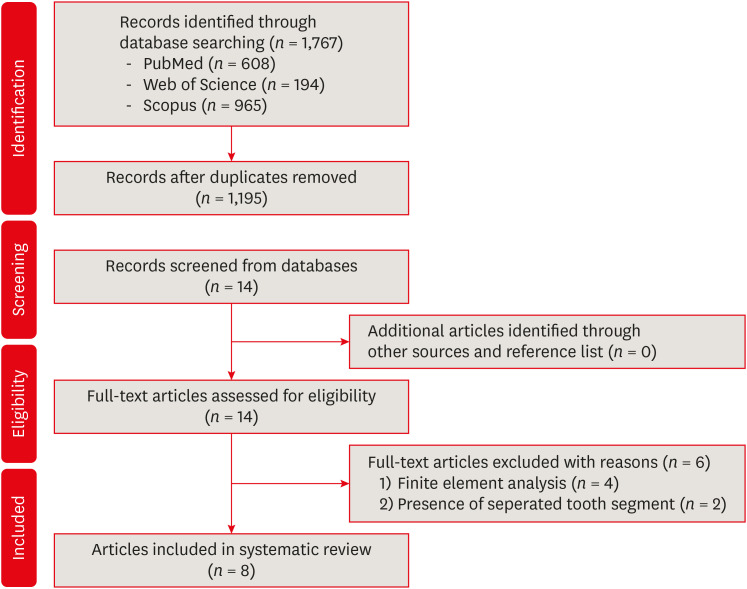
-
Abstract
PDFPubReaderePub
This systematic review aimed to investigate whether minimally invasive root canal preparation ensures higher fracture resistance compared to conventional root canal preparation in endodontically treated teeth (ETT). A comprehensive search strategy was conducted on the “PubMed, Web of Science, and Scopus” databases, alongside reference and hand searches, with language restrictions applied. Two independent reviews selected pertinent laboratory studies that explored the effect of minimally invasive root canal preparation on fracture resistance, in comparison to larger preparation counterparts. The quality of the studies was assessed, and the risk of bias was categorized as low, moderate, or high. The electronic search yielded a total of 1,767 articles. After applying eligibility criteria, 8 studies were included. Given the low methodological quality of these studies and the large variability of fracture resistance values, the impact of reduced apical size and/or taper on the fracture resistance of the ETT can be considered uncertain. This systematic review could not reveal sufficient evidence regarding the effect of minimally invasive preparation on increasing fracture resistance of ETT, primarily due to the inherent limitations of the studies and the moderate risk of bias.
-
Citations
Citations to this article as recorded by- Combined Effects of Instrumentation Geometry and Sealer Type on Mandibular Premolar Root Fracture Resistance
Funda Yilmaz, Berkuk Sayar, Berna Aslan, Mustafa A Badi
Cureus.2026;[Epub] CrossRef - Impact of conservative versus conventional instrumentation on the release of inflammatory mediators and post‐operative pain in mandibular molars with asymptomatic irreversible pulpitis: A randomized clinical trial
Sıla Nur Usta, Ana Arias, Emre Avcı, Emmanuel João Nogueira Leal Silva
International Endodontic Journal.2025; 58(6): 862. CrossRef - Mapping risk of bias criteria in systematic reviews of in vitro endodontic studies: an umbrella review
Rafaella Rodrigues da Gama, Lucas Peixoto de Araújo, Evandro Piva, Leandro Perello Duro, Adriana Fernandes da Silva, Wellington Luiz de Oliveira da Rosa
Evidence-Based Dentistry.2025; 26(4): 179. CrossRef - Micro‐computed tomography evaluation of minimally invasive root canal preparation in 3D‐printed C‐shaped canal
Nutcha Supavititpattana, Siriwan Suebnukarn, Panupat Phumpatrakom, Kamon Budsaba
Australian Endodontic Journal.2024; 50(3): 621. CrossRef - Ex vivo investigation on the effect of minimally invasive endodontic treatment on vertical root fracture resistance and crack formation
Andreas Rathke, Henry Frehse, Maria Bechtold
Scientific Reports.2024;[Epub] CrossRef
- Combined Effects of Instrumentation Geometry and Sealer Type on Mandibular Premolar Root Fracture Resistance
- 5,305 View
- 145 Download
- 4 Web of Science
- 5 Crossref
Research Articles
-
Effect of cryotherapy duration on experimentally induced connective tissue inflammation
in vivo - Jorge Vera, Mayra Alejandra Castro-Nuñez, María Fernanda Troncoso-Cibrian, Ana Gabriela Carrillo-Varguez, Edgar Ramiro Méndez Sánchez, Viviana Sarmiento, Lourdes Lanzagorta-Rebollo, Prasanna Neelakantan, Monica Romero, Ana Arias
- Restor Dent Endod 2023;48(3):e29. Published online August 2, 2023
- DOI: https://doi.org/10.5395/rde.2023.48.e29
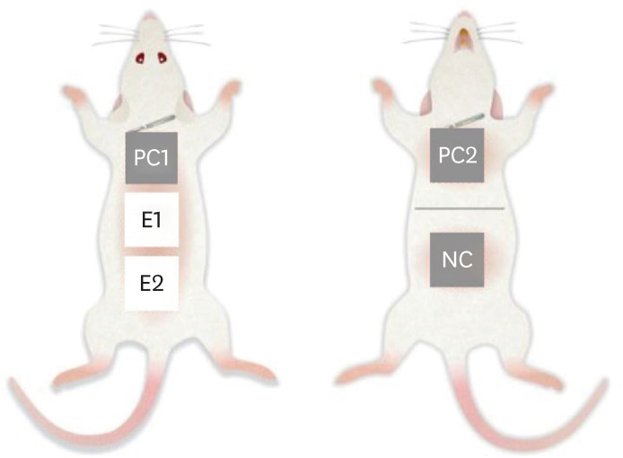
-
Abstract
PDFPubReaderePub
Objectives This study tested the hypothesis that cryotherapy duration influences lipopolysaccharide (LPS)-induced inflammation in a rat model.
Materials and Methods Six Wistar rats (
Rattus norvegicus albinus ) were used. Five sites were selected per animal and divided into 5 groups: a negative control group (NC), 2 positive control groups (PC1 and PC2), and 2 experimental groups (E1 and E2). Cryotherapy was applied for 1 minute (E1) or 5 minutes (E2). An acute inflammatory response was induced in the PC and E groups via subcutaneous administration of 0.5 mL/kg. In the PC2 group, a catheter was inserted without additional treatment. For the E1 and E2 groups, 2.5°C saline solution was administered through the implanted catheters for 1 and 5 minutes, respectively. The rats were sacrificed, and samples were obtained and processed for histological analysis, specifically examining the presence of polymorphonuclear neutrophils and hemorrhage. The χ2 test was used to compare the presence of acute inflammation across groups. Dependent variables were compared using the linear-by-linear association test.Results Inflammation and hemorrhage varied significantly among the groups (
p = 0.001). A significantly higher degree of acute inflammation was detected (p = 0.0002) in the PC and E1 samples than in the E2 group, in which cryotherapy was administered for 5 minutes. The PC and E1 groups also exhibited significantly greater numbers of neutrophils (p = 0.007), which were essentially absent in both the NC and E2 groups.Conclusions Cryotherapy administration for 5 minutes reduced the acute inflammation associated with LPS and catheter implantation.
-
Citations
Citations to this article as recorded by- The impact of using cold irrigation on postoperative endodontic pain and substance P level: a randomized clinical trial
Reem Mohammed Amr Sharaf, Tariq Yehia Abdelrahman, Maram Farouk Obeid
Odontology.2026; 114(2): 527. CrossRef - Cryotherapy as a supplementary aid to inferior alveolar nerve block in patients with symptomatic irreversible pulpitis: A randomized controlled trial
Setu Katyal, Poonam Bogra, Rajinder Bansal, Vishakha Grover, Saurabh Gupta, Saru Gupta
Medicine International.2025; 5(5): 1. CrossRef - Determining Efficacy of Intracanal Cryotherapy on Post Endodontic Pain in Irreversible Pulpitis
Anam Fayyaz Bashir, Ussamah Waheed Jatala, Moeen ud din Ahmad, Muhammad Talha Khan, Saima Razzaq Khan, Aisha Arshad Butt
Pakistan Journal of Health Sciences.2024; : 68. CrossRef
- The impact of using cold irrigation on postoperative endodontic pain and substance P level: a randomized clinical trial
- 4,390 View
- 61 Download
- 2 Web of Science
- 3 Crossref
- Tip and taper compatibility of accessory gutta-percha points with rotary and reciprocating instruments
- Júlia Niero Zanatta Streck, Sabrina Arcaro, Renan Antônio Ceretta, Eduardo Antunes Bortoluzzi, Lucas da Fonseca Roberti Garcia, Josiane de Almeida, Patrícia Maria Poli Kopper, Anarela Vassen Bernardi
- Restor Dent Endod 2023;48(3):e22. Published online June 8, 2023
- DOI: https://doi.org/10.5395/rde.2023.48.e22
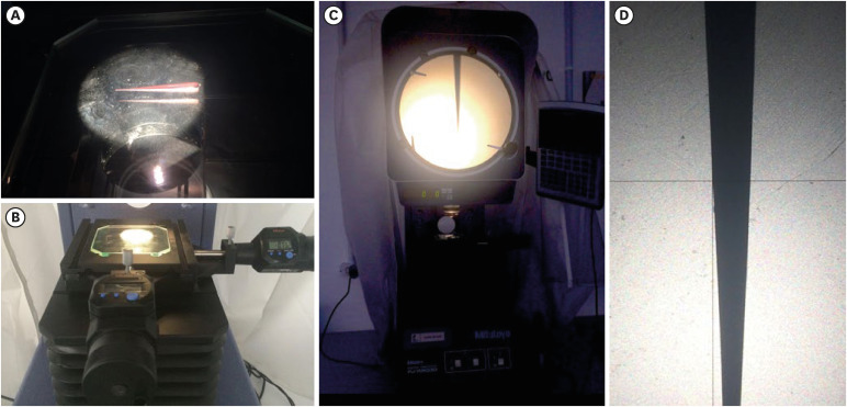
-
Abstract
PDFPubReaderePub
Objectives This study was conducted to evaluate and compare the tip and taper compatibility of accessory gutta-percha points (AGPs) with various rotary and reciprocating instruments.
Materials and Methods Using a profile analyzer, tip and taper measurements were taken of 10 AGPs of each of the 14 models available from Odous de Deus and the 4 models available from Dentsply-Maillefer. Diameter measurements were taken at 1-mm intervals, from 3 mm from the tip (D3) to 16 mm.
Results Based on the mean values obtained, 3-dimensional (3D) models of the AGPs were drawn in Autodesk Fusion 360 and superimposed on 3D models of each instrument selected (Mtwo, Reciproc, RaCe, K3, and ProDesign Logic) to determine the compatibility between the instrument and the AGP. Data corresponding to the tips and tapers of the various AGPs, as well as the tip and taper differences between the AGPs and the instruments, were analyzed using descriptive statistics. The tapers of the AGPs were subject to the American National Standards Institute/American Dental Association No. 57 standard. The Odous de Deus extra-long medium and extra-long extra-medium AGPs were shown to be compatible with Mtwo, K3, and ProDesign Logic instruments with taper 0.06 and tip sizes 25 and 30, while the Dentsply fine and fine medium cones were compatible with Mtwo, RaCe, and K3 instruments with conicity of 0.04 and tip sizes 35 and 40.
Conclusions Both the Odous de Deus and Dentsply commercial brands included 2 AGP models with tip (D3) and taper compatibility with Mtwo, RaCe, K3, and/or Prodesign Logic instruments.
- 2,962 View
- 46 Download
Review Article
- Stem cell-derived exosomes for dentin-pulp complex regeneration: a mini-review
- Dina A. Hammouda, Alaa M Mansour, Mahmoud A. Saeed, Ahmed R. Zaher, Mohammed E. Grawish
- Restor Dent Endod 2023;48(2):e20. Published online May 3, 2023
- DOI: https://doi.org/10.5395/rde.2023.48.e20
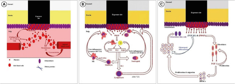
-
Abstract
PDFPubReaderePub
This mini-review was conducted to present an overview of the use of exosomes in regenerating the dentin-pulp complex (DPC). The PubMed and Scopus databases were searched for relevant articles published between January 1, 2013 and January 1, 2023. The findings of basic
in vitro studies indicated that exosomes enhance the proliferation and migration of mesenchymal cells, as human dental pulp stem cells, via mitogen-activated protein kinases and Wingless-Int signaling pathways. In addition, they possess proangiogenic potential and contribute to neovascularization and capillary tube formation by promoting endothelial cell proliferation and migration of human umbilical vein endothelial cells. Likewise, they regulate the migration and differentiation of Schwann cells, facilitate the conversion of M1 pro-inflammatory macrophages to M2 anti-inflammatory phenotypes, and mediate immune suppression as they promote regulatory T cell conversion. Basicin vivo studies have indicated that exosomes triggered the regeneration of dentin-pulp–like tissue, and exosomes isolated under odontogenic circumstances are particularly strong inducers of tissue regeneration and stem cell differentiation. Exosomes are a promising regenerative tool for DPC in cases of small pulp exposure or for whole-pulp tissue regeneration.-
Citations
Citations to this article as recorded by- Extracellular vesicles derived from dental mesenchymal stem cells for regenerative medicine: a scoping review
Maria Emília Mota, Márcia Martins Marques, Thaís Gimenez, Suely Kunimi Kubo Ariga, Tiago Góss dos Santos, Fábio Abreu Alves, Maria Stella Moreira
Molecular Biology Reports.2026;[Epub] CrossRef - Stem cell-derived exosomes in tissue regeneration of oral and maxillofacial region: A systematic review
Qianlei Dai, Qianfen Dai
Medicine.2026; 105(1): e46948. CrossRef - Stem Cell-Derived Exosomes in Wound Healing and Skin Regeneration: Emerging Therapeutic Strategies and Mechanisms
Nithin Vidiyala, Pavani Sunkishala, Prashanth Reddy Parupathi, Dinesh Nyavanandi
Cells.2026; 15(10): 872. CrossRef - Cell Homing Strategies in Regenerative Endodontic Therapy
David Kim, Sahng G. Kim
Cells.2025; 14(3): 201. CrossRef - Impact of dental pulp cells-derived small extracellular vesicles on the properties and behavior of dental pulp cells: an in-vitro study
Dina A. Hammouda, Alaa M. Mansour, Ahmed R. Zaher, Mohammed E. Grawish
BMC Oral Health.2025;[Epub] CrossRef - Methodological Approaches for Economic Comparison of Mesenchymal Stem Cell and Exosome-based Therapies with Conventional Endodontic Treatments in Regenerative Endodontics
Madina A. Kurmanalina Kurmanalina, Nadiar M. Mussin, Aigul M. Sumanova, Violetta R. Detochkina, Maryam Mardani, Nader Tanideh, Amin Tamadon
West Kazakhstan Medical Journal.2025; 67(2): 188. CrossRef - Exosomal circ_0003057 promotes osteo/odontogenic differentiation of hDPSCs by binding with EIF4A3 through upregulated parental gene ANKH
Bingtao Wang, Yuanyuan Kong, Huixian Dong, Feng Lai, Zixin Guo, Liecong Lin, Jingyi Xu, Jingkun Zhang, Yiguo Jiang, Qianzhou Jiang
International Endodontic Journal.2025; 58(9): 1433. CrossRef - Mechanistic insights into dental stem cells‐derived exosomes in regenerative endodontics
Paras Ahmad, Nathan Estrin, Nima Farshidfar, Yufeng Zhang, Richard J. Miron
International Endodontic Journal.2025; 58(9): 1384. CrossRef - Development and characterization of an exosome-loaded biomimetic hydroxyapatite/gelatin scaffold for enhanced dental pulp regeneration
Yuen-Shan Tsai, Shih-Jung Cheng, Tsao-Li Chuang, Shu-Fang Chang, Feng-Huei Lin, Chun-Pin Lin
Journal of Dental Sciences.2025;[Epub] CrossRef - Exosomes as Promising Therapeutic Tools for Regenerative Endodontic Therapy
Qingyue Kong, Yujie Wang, Nan Jiang, Yifan Wang, Rui Wang, Xiaohan Hu, Jing Mao, Xin Shi
Biomolecules.2024; 14(3): 330. CrossRef - Role and Molecular Mechanism of miR-586 in the Differentiation of Dental Pulp Stem Cells into Odontoblast-like Cells
Gang Pan, Qianwen Zhou, Chenhua Pan, Yingxue Zhang
Cell Biochemistry and Biophysics.2024; 83(1): 507. CrossRef
- Extracellular vesicles derived from dental mesenchymal stem cells for regenerative medicine: a scoping review
- 6,448 View
- 135 Download
- 9 Web of Science
- 11 Crossref
Research Articles
- The clinical success of ART restorations and Hall technique in primary molars: a randomized 18-month follow-up study
- Esra Oz, Zuhal Kırzıoglu, Canan Kale
- Restor Dent Endod 2023;48(2):e19. Published online May 1, 2023
- DOI: https://doi.org/10.5395/rde.2023.48.e19
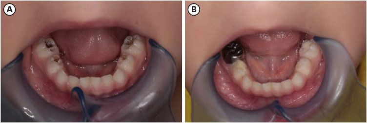
-
Abstract
PDFPubReaderePub
Objectives The aim of the study was to evaluate the clinical and radiographic success of the Hall technique (HT) and atraumatic restorative treatment (ART) restorations using high-viscosity glass-ionomer cement for the management of occlusal carious lesions in primary molars.
Materials and Methods This randomized clinical study observed 40 children (aged 5–6 years). For each child, one tooth was treated with HT and one with ART. The primary outcome measures for HT restorations were successful, minor, and major failure rates. Clinical evaluations of ART restorations were performed according to the modified United States Public Health Service criteria during 18-month follow-up. McNemar test was used for statistical analysis.
Results Thirty of 40 (75%) participants returned for 18 months of follow-up. In the clinical evaluations of teeth that were treated with HT, the patients did not have complaints of pain or other symptoms, all crowns remained in the oral cavity, the gums were healthy, and the teeth were functional in all evaluations. At the end of the 18-month follow-up, the surface texture and marginal integrity criteria of ART restorations were recorded as 26.7% and 33.3%, respectively. In the radiographic evaluation of 30 patients treated with ART and HT, all restorations were considered successful.
Conclusions The 18-month clinical and radiographic results after treatments applied to single-surface cavities in anxious children showed that both treatment methods were successful.
-
Citations
Citations to this article as recorded by- Two-year outcomes of hall technique and modified hall technique in deep carious lesions of primary molars: a randomized clinical trial
Sumeyye Konukman Turker, Ayse Isil Cihan
BMC Oral Health.2026;[Epub] CrossRef - Success rate of Hall Technique for restoring carious primary molars - systematic review and meta-analysis
Tamara Kerber Tedesco, Nicola Patricia Innes, Claudia Lopez Gallegos, Gabriela Seabra Silva, Thais Gimenez, Mariana Minatel Braga, Mariana Pinheiro Araujo, Jayakumar Jayaraman, Waraf Al-yaseen, Daniela Prócida Raggio
Evidence-Based Dentistry.2025; 26(1): 65. CrossRef - Effectiveness of Dental Restorative Materials in the Atraumatic Treatment of Carious Primary Teeth in Pediatric Dentistry: A Systematic Review
Gianna Dipalma, Angelo Michele Inchingolo, Lucia Casamassima, Paola Nardelli, Danilo Ciccarese, Paolo De Sena, Francesco Inchingolo, Andrea Palermo, Marco Severino, Cinzia Maria Norma Maspero, Alessio Danilo Inchingolo
Children.2025; 12(4): 511. CrossRef - Clinical, radiographic, and microhardness evaluation of caries in primary molars managed with modified Hall technique
Eman El Sayed El Bedewy, Nahed A.A. Abohamila, Shereen A. M. Ali, Shimaa M.M. Hadwa
Tanta Dental Journal.2025; 22(1): 133. CrossRef - Pain Perception During Minimally Invasive Caries Removal in Children: A Randomized Clinical Trial Comparing Chemo-Mechanical Caries Removal
Dhirja Goel, Neha Awasthi, Yanina Singh, Sukhdeep Singh, Nenung Yirang
Journal of International Society of Preventive and Community Dentistry.2025; 15(4): 348. CrossRef - Clinical and histological evaluation of hall technique with and without silver diamine fluoride in the treatment of carious primary molars
Marwa M.A.Z. Abd-Elhaleium, Fatma A.-A. El-Hendawy, Lamis A. El-Ghareb, Sara Y. AboAli
Tanta Dental Journal.2025; 22(2): 351. CrossRef - Comparative success of minimally invasive treatments for cavitated caries in primary teeth: a network meta-analysis
Rasoul Sahebalam, Mahsa Ghorbani, Alireza Sarraf Shirazi, Motahareh Khosrojerdi, Mana Mowji
BMC Oral Health.2025;[Epub] CrossRef
- Two-year outcomes of hall technique and modified hall technique in deep carious lesions of primary molars: a randomized clinical trial
- 7,581 View
- 134 Download
- 6 Web of Science
- 7 Crossref
- Effects of surrounding and underlying shades on the color adjustment potential of a single-shade composite used in a thin layer
- Mariana Silva Barros, Paula Fernanda Damasceno Silva, Márcia Luciana Carregosa Santana, Rafaella Mariana Fontes Bragança, André Luis Faria-e-Silva
- Restor Dent Endod 2023;48(1):e7. Published online December 29, 2022
- DOI: https://doi.org/10.5395/rde.2023.48.e7
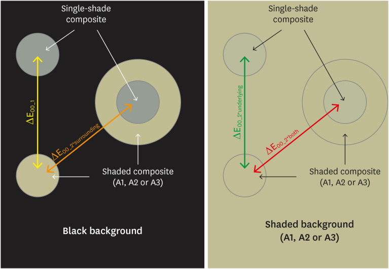
-
Abstract
PDFPubReaderePub
Objectives This study aimed to evaluate the surrounding and underlying shades’ effect on the color adjustment potential (CAP) of a single-shade composite used in a thin layer.
Materials and Methods Cylinder specimens (1.0 mm thick) were built with the Vittra APS Unique composite, surrounded (dual specimens) or not (simple specimens) by a control composite (shade A1, A2, or A3). Simple specimens were also built only with the control composites. Each specimen’s color was measured against white and black backgrounds or the simple control specimens with a spectrophotometer (CIELAB system). The whiteness index for dentistry (WID) and translucency parameters (TP00) were calculated for simple specimens. Differences (ΔE00) in color between the simple/dual specimens and the controls were calculated. The CAP was calculated based on the ratios between data from simple and dual specimens.
Results The Vittra APS Unique composite showed higher WID and TP00 values than the controls. The highest values of ΔE00 were observed among simple specimens. The color measurements of Vittra APS Unique (simple or dual) against the control specimens presented the lowest color differences. Only surrounding the single-shade composite with a shaded composite barely impacted the ΔE00. The highest CAP values were obtained using a shaded composite under simple or dual specimens.
Conclusions The CAP of Vittra APS Unique was strongly affected by the underlying shade, while surrounding this composite with a shaded one barely affected its color adjustment.
-
Citations
Citations to this article as recorded by- Impact of water absorption on the translucency of single-shade and conventional resin composites: an in vitro comparative study
Ceyda Sari, Elifnur Aydemir Aydın
Odontology.2026;[Epub] CrossRef - Baseline color-matching in anterior non-carious cervical lesions of patients of two single-shade resin composites: a randomized clinical trial
Ayşe Nur Doğan, Soley Arslan
Odontology.2026;[Epub] CrossRef - Evaluation of color blending effect of a single-shade resin composite with hybrid ceramics: an in vitro study
Jongchan Lee, Jinsoo Ahn, Sun-Young Kim
BMC Oral Health.2026;[Epub] CrossRef - At‐Home and In‐Office Bleaching Protocols on the Color Match of Restorations Made With Single‐Shade Composites
Luciana Vasconcelos Ramos, Dayana Fernandes Rocha Aparicio, André Luis Faria‐e‐Silva, Maíra do Prado, Andréa Vaz Braga Pintor, Marcela Baraúna Magno
Journal of Esthetic and Restorative Dentistry.2025; 37(6): 1567. CrossRef - Evaluation of color matching of three single-shade composites employing simulated 3D printed cavities with different thicknesses using CIELAB and CIEDE2000 color difference formulae
Engin Kariper, Aylin Cilingir
REVIEWS ON ADVANCED MATERIALS SCIENCE.2025;[Epub] CrossRef - Impact of kombucha, coffee, and turmeric beverages on the color stability of a single-shade versus a multi-shade resin-based composite
Hanin E. Yeslam, Abdulaziz F. Bakhsh
PeerJ.2025; 13: e19759. CrossRef - Comparative Study of Esthetic Outcome of Pedo Shades of Composite Resin—A Randomized Controlled Trial: In Vivo and In Vitro Study
Priyanka Raj, Shikha Choubey, Divya Doneria, Diksha Bhat, Shivani Mathur, Shailja Sinha
International Journal of Clinical Pediatric Dentistry.2025; 18(S1): S22. CrossRef - Influence of cavity wall thickness on the color adjustment potential of single-shade resin composites
Fabrício Luscino Alves de Castro, Letícia Brandão Durand
The Journal of the American Dental Association.2024; 155(7): 605. CrossRef - Assessing color mismatch in single-shade composite resins for enamel replacement
Rafaella Mariana Fontes de Bragança, Diana Leyva Del Rio, Luiz Alves Oliveira-Neto, William Michael Johnston
The Journal of Prosthetic Dentistry.2024; 132(3): 613.e1. CrossRef - Color discrepancy of single-shade composites at different distances from the interface measured using cell phone images
Márcia Luciana Carregosa Santana, Gabriella de Jesus Santos Livi, André Luis Faria-e-Silva
Restorative Dentistry & Endodontics.2024;[Epub] CrossRef - Is It Possible for Single-shade Composites to Mimic the Color, Lightness, Chroma, and Hue of Other Single-shade Composites? An In Vitro Study
M Buldur, G Ayan
Operative Dentistry.2024; 49(6): 691. CrossRef - Color evaluation of a one-shade used for restoration of non-carious cervical lesions: an equivalence randomized clinical trial
Michael Willian Favoreto, Amanda de Oliveira de Miranda, Thalita P. Matos, Andrea dos Santos de Castro, Mylena de Abreu Cardoso, Julia Beatriz, Jenny Collantes-Acuña, Alessandra Reis, Alessandro Dourado Loguercio
BMC Oral Health.2024;[Epub] CrossRef - Influence of Thickness on the Translucency Parameter and Whiteness Index of Single-Shade Resin Composites
Ö Yağcı, M Fidan
Operative Dentistry.2024; 49(2): 189. CrossRef - A Comparative Study of the Sensitivity and Specificity of the Ishihara Test With Various Displays
Thomas Klinke, Wolfgang Hannak, Klaus Böning, Holger Jakstat
International Dental Journal.2024; 74(4): 892. CrossRef - Color match evaluation using instrumental method for three single-shade resin composites before and after in-office bleaching
Aylin Cilingir, Engin Kariper
REVIEWS ON ADVANCED MATERIALS SCIENCE.2023;[Epub] CrossRef - The role of interface distance and underlying substrate on the color adjustment potential of single‐shade composites
Gabriella Jesus Santos de Livi, Tauan Rosa Santana, Rafaella Mariana Fontes Bragança, Rosa Maria Viana de Bragança Garcez, André Luis Faria‐e‐Silva
Journal of Esthetic and Restorative Dentistry.2023; 35(8): 1279. CrossRef
- Impact of water absorption on the translucency of single-shade and conventional resin composites: an in vitro comparative study
- 5,473 View
- 121 Download
- 16 Web of Science
- 16 Crossref
Review Article
- Effect of endodontic sealer on postoperative pain: a network meta-analysis
- Cynthia Maria Chaves Monteiro, Ana Cristina Rodrigues Martins, Alessandra Reis, Juliana Larocca de Geus
- Restor Dent Endod 2023;48(1):e5. Published online December 29, 2022
- DOI: https://doi.org/10.5395/rde.2023.48.e5
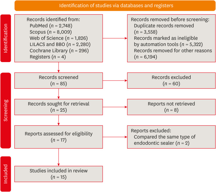
-
Abstract
PDFPubReaderePub
This systematic review and network meta-analysis aimed to answer the following focused research question: “Does the type of endodontic sealer affect the postoperative pain in patients who received endodontic treatment?” Different databases and grey literature were surveyed. Only one randomized controlled trial were included. The risk of bias in the studies was evaluated by using the Cochrane Collaboration’s tool. A random-effects meta-analysis was conducted to compare the risk and intensity of postoperative pain. The quality of the body of evidence was assessed using the Grading of Recommendations Assessment, Development, and Evaluation approach. Out of 11,601 studies, 15 remained for qualitative analyses and 12 for meta-analysis. Seven studies were classified at high risk of bias, and 8 studies raised some concerns. No significant differences between the endodontic materials were observed in the direct comparisons, both in risk and in intensity of postoperative pain (pairwise comparisons with 2 studies: I2 = 0%;
p > 0.05 and 8 studies: I2 = 23%;p > 0.05, respectively). The certainty of the evidence was graded as low or moderate. There was no difference in the risk and intensity of postoperative pain after filling with different endodontic sealers. Further systematic reviews should be conducted.Trial Registration PROSPERO Identifier:
CRD42020215314 -
Citations
Citations to this article as recorded by- Does the Use of a Bioceramic Sealer Reduce Postoperative Pain Compared With an Epoxy Resin‐Based Sealer After Primary Root Canal Treatment and Retreatment?—An Umbrella Review
Lokhasudhan Govindaraju, Rajeswari Kalaiselvam, Mathan Rajan Rajendran, Aleksandar Jakovljevic, Jelena Jacimovic, Henry F. Duncan, Venkateshbabu Nagendrababu
International Endodontic Journal.2026; 59(3): 341. CrossRef - Evidence synthesis of postoperative pain with bioceramic vs. epoxy resin sealers: umbrella review of randomized trials within existing systematic reviews
Mrunali Dahikar, Ashish Mandwe, Kulvinder Singh Banga, Alexander Maniangat Luke, Suraj Arora, Unmesh Khanvilkar, Ajinkya M. Pawar
Frontiers in Dental Medicine.2026;[Epub] CrossRef - Factors Influencing Apical Extrusion of 2 Types of Endodontic Sealers with Different Delivery Systems
Shumaila Iqbal, Nicholas S. Adams, Josette Camilleri
Journal of Endodontics.2026; 52(5): 806. CrossRef - Evaluation of Postoperative Pain Frequency in Single‐Session Endodontic Treatments With Patency and Foraminal Enlargement
Viviane Barbosa Godoy, Ana Grasiela Limoeiro, Vanessa Sandini, Vini Mehta, Wayne Martins Nascimento, Marilia Fagury Videira Marceliano‐Alves, Marcos Frozoni
Clinical and Experimental Dental Research.2026;[Epub] CrossRef - Silicone vs. Silicon/Silica in Intraoral Healing: A Systematic Review
David Parker, Aditi Bopardikar, Georgios E. Romanos
Materials.2026; 19(7): 1425. CrossRef - Effect of occlusal reduction on post-operative pain of symptomatic and asymptomatic molar teeth
Aysenur Kamacı Esen, Fatma Furuncuoğlu, Fatima Betul Basturk, Muhammet Nuri Taşcıoğlu, Masoud Parirokh
Acta Odontologica Scandinavica.2025; 84: 371. CrossRef - An Observational Study on Pain Occurrence After Root Canal Treatment: Role of Operator Experience When Using a Bioceramic Sealer
Mihai Merfea, Ioana Sofia Pop-Ciutrila, Mindra Eugenia Badea, Ada Gabriela Delean, Oana Cimponeriu, Razvan Corneliu Pop, Maria Peter, Iulia Clara Badea, Sanda Ileana Cimpean
Journal of Clinical Medicine.2025; 14(13): 4558. CrossRef - Assessment of Postoperative Pain After Single‐ or Multiple‐Visit Endodontic Therapy and Its Molecular Aspects: A Randomised Controlled Study
Larissa Nunes Rosa Bedene, Denise Piotto Leonardi, Joana Santana Couto, Bruno Marques‐da‐Silva, Marilisa Carneiro Leão Gabardo, João Arnando Brancher, Flávia Sens Fagundes Tomazinho
Australian Endodontic Journal.2025; 51(3): 668. CrossRef - Clinical and Radiographic Outcomes of Root Canal Obturation with Hydraulic Condensation and Tricalcium Silicate Bioceramic Sealer: A 12-Month Observational Study on Periapical Healing
Kostadin Zhekov, Vesela Stefanova
Journal of Functional Biomaterials.2025; 16(11): 412. CrossRef - Comparative evaluation of postoperative pain and periapical healing after root canal treatment using three different endodontic sealers: A randomized controlled clinical trial
Ruchika Pandey, Nitin Kararia, Deepak Kumar Sharma, Vishal Rathod, Anand Vilas Bansod, Dhaval Desai
Journal of Conservative Dentistry and Endodontics.2024; 27(9): 962. CrossRef - Effect of bioceramic-based and resin-based sealers on postoperative discomfort following root canal therapy: a systematic review and meta-analysis
Mansi Supare, Ajinkya M. Pawar, Kashmira Sawant, Dian Agustin Wahjuningrum, Suraj Arora, Firas Elmsmari, Mohmed Isaqali Karobari, Bhagyashree Thakur
PeerJ.2024; 12: e18198. CrossRef - Comparative Evaluation of Incidences of Post Operative Pain in Patient Treated in Single Visit Root Canal Treatment by Using Different Sealers: - An in-Vivo Study
Sadashiv Daokar, Aishwarya Ranjalkar, Kalpana Pawar, Komal Potfode, Dhanashri Padwal, Sana Khan
International Journal of Innovative Science and Research Technology (IJISRT).2024; : 2743. CrossRef
- Does the Use of a Bioceramic Sealer Reduce Postoperative Pain Compared With an Epoxy Resin‐Based Sealer After Primary Root Canal Treatment and Retreatment?—An Umbrella Review
- 7,819 View
- 148 Download
- 11 Web of Science
- 12 Crossref
Research Articles
- Comparison of shaping ability of the Reciproc Blue and One Curve with or without glide path in simulated S-shaped root canals
- Vincenzo Biasillo, Raffaella Castagnola, Mauro Colangeli, Claudia Panzetta, Irene Minciacchi, Gianluca Plotino, Simone Staffoli, Luca Marigo, Nicola Maria Grande
- Restor Dent Endod 2022;47(1):e3. Published online December 28, 2022
- DOI: https://doi.org/10.5395/rde.2022.47.e3
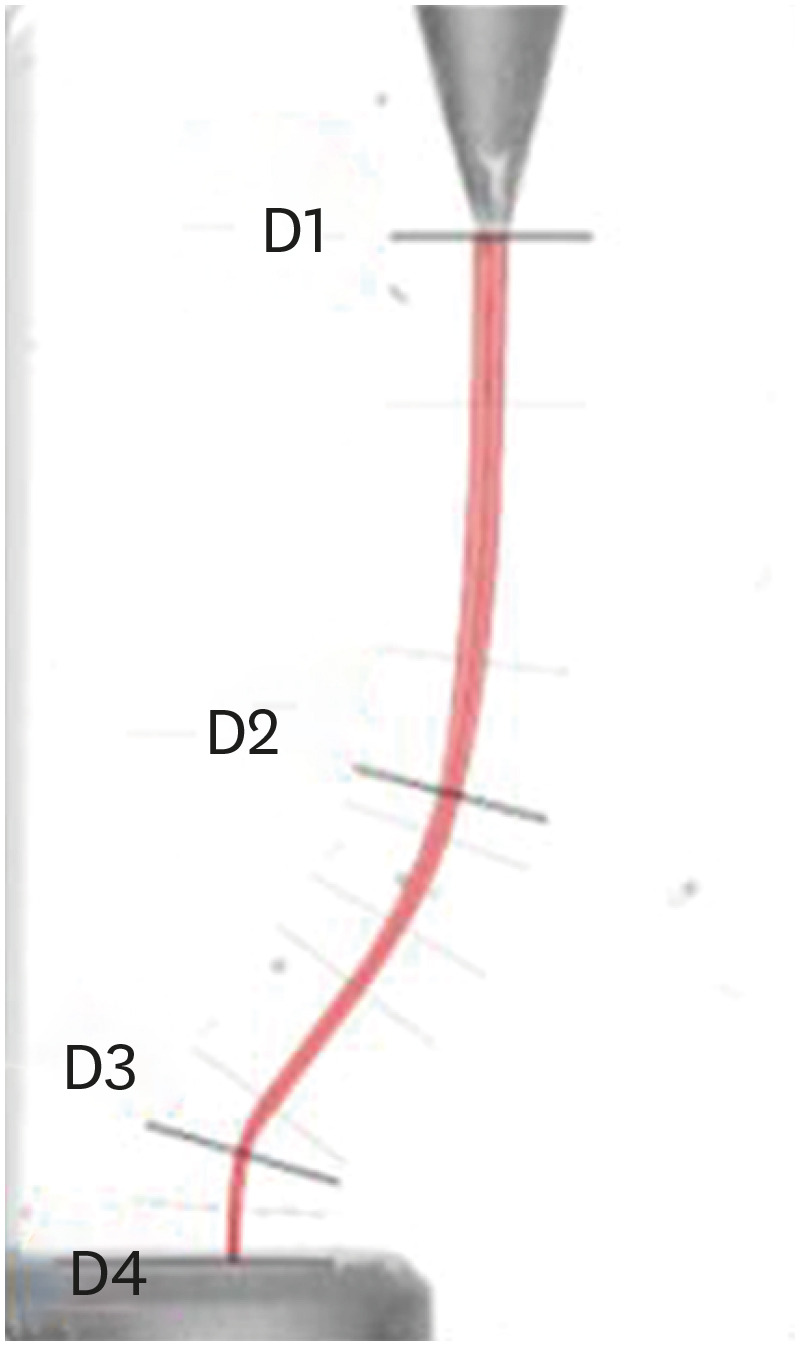
-
Abstract
PDFPubReaderePub
Objectives This study aimed to assess the impact of a glide-path on the shaping ability of 2 single-file instruments and to compare the centering ability, maintenance of original canal curvatures and area of instrumentation in simulated S-shaped root canals.
Materials and Methods Forty simulated S-shaped root canals were used and were prepared with One Curve (group OC), One G and OC (group GOC), Reciproc Blue (group RB) and R-Pilot and RB (group PRB) and scanned before and after instrumentation. The images were analyzed using AutoCAD. After superimposing the samples, 4 levels (D1, D2, D3, and D4) and 2 angles (Δ1 and Δ2) were established to evaluate the centering ability and modification of the canal curvatures. Then, the area of instrumentation (ΔA) was measured. The data were analyzed using 2-way analysis of variance and Tukey's test for multiple comparisons (
p < 0.05).Results Regarding the centering ability in the apical part (D3, D4), the use of the glide-path yielded better results than the single-file groups. Among the groups at D4, OC showed the worst results (
p < 0.05). The OC system removed less material (ΔA) than the RB system, and for Δ1, OC yielded a worse result than RB (p < 0.05).Conclusions The glide-path improved the centering ability in the apical part of the simulated S-shaped canals. The RB system showed a better centering ability in the apical part and major respect of the canal curvatures compared with OC system.
-
Citations
Citations to this article as recorded by- Evaluation of Apical Debris Extrusion and the Remaining Canal Material during Retreatment of a Bioceramic Sealer by the XP-endo Finisher File System, Followed by Various Supplementary Methods: An in Vitro Study
Paras Mull Gehlot, Parvathi Sudeep, Annapoorna B Mariswamy
World Journal of Dentistry.2025; 15(10): 837. CrossRef - Shaping Ability of Rotary NiTi Systems in S‐Shaped Root Canals of Mandibular Molars
Renata M. S. Leal, Emmanuel J. N. L. Silva, Maria C. B. P. Campos, Clarissa T. Rodrigues, Marco A. H. Duarte, Bruno C. Cavenago
Australian Endodontic Journal.2025; 51(1): 133. CrossRef - Comparison of Debris Extrusion and Preparation Time by Traverse, R‐Motion Glider C, and Other Glide Path Systems in Severely Curved Canals
Taher Al Omari, Layla Hassouneh, Khawlah Albashaireh, Alaa Dkmak, Rami Albanna, Ali Al-Mohammed, Ahmed Jamleh, Lucas da Fonseca Roberti Garcia
International Journal of Dentistry.2025;[Epub] CrossRef - Glide Path – An Ineluctable Route for Successful Endodontic Mechanics: A Literature Review
Mahima Bharat Mehta, Anupam Sharma, Aniket Jadhav, Aishwarya Handa, Abhijit Bajirao Jadhav, Ashwini A. Narayanan
Journal of the International Clinical Dental Research Organization.2024; 16(2): 101. CrossRef - Screw-in force, torque generation, and performance of glide-path files with three rotation kinetics
Jee-Yeon Woo, Ji-Hyun Jang, Seok Woo Chang, Soram Oh
Odontology.2024; 112(3): 761. CrossRef - Glide Path in Endodontics: A Literature Review of Current Knowledge
Vlad Mircea Lup, Giulia Malvicini, Carlo Gaeta, Simone Grandini, Gabriela Ciavoi
Dentistry Journal.2024; 12(8): 257. CrossRef - In Vitro Research Methods Used to Evaluate Shaping Ability of Rotary Endodontic Files—A Literature Review
Ranya F. Elemam, Ana Mano Azul, João Dias, Khaled El Sahli, Renato de Toledo Leonardo
Dentistry Journal.2024; 12(10): 334. CrossRef - Endodontic glide path - importance and performance techniques
Milica Jovanovic-Medojevic, Мiljan Stosic, Vanja Opacic-Galic, Violeta Petrovic
Srpski arhiv za celokupno lekarstvo.2023; 151(5-6): 380. CrossRef
- Evaluation of Apical Debris Extrusion and the Remaining Canal Material during Retreatment of a Bioceramic Sealer by the XP-endo Finisher File System, Followed by Various Supplementary Methods: An in Vitro Study
- 3,737 View
- 53 Download
- 8 Web of Science
- 8 Crossref
- Dentinal tubule penetration of sodium hypochlorite in root canals with and without mechanical preparation and different irrigant activation methods
- Renata Aqel de Oliveira, Theodoro Weissheimer, Gabriel Barcelos Só, Ricardo Abreu da Rosa, Matheus Albino Souza, Rodrigo Gonçalves Ribeiro, Marcus Vinicius Reis Só
- Restor Dent Endod 2023;48(1):e1. Published online December 1, 2022
- DOI: https://doi.org/10.5395/rde.2023.48.e1
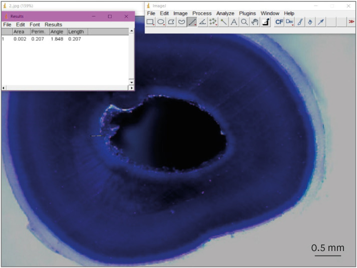
-
Abstract
PDFPubReaderePub
Objectives This study evaluated the dentinal penetration depth of 2.5% sodium hypochlorite (NaOCl) in root canals with and without preparation and different irrigant activation protocols.
Materials and Methods Sixty-three bovine mandibular incisors were randomly allocated to 6 groups (
n = 10): G1, preparation + conventional needle irrigation (CNI); G2, preparation + passive ultrasonic irrigation (PUI); G3, preparation + Odous Clean (OC); G4, no preparation + CNI; G5, no preparation + PUI; G6, no preparation + OC; and CG (negative control;n = 3). Samples were filled with crystal violet for 72 hours. Irrigant activation was performed. Samples were sectioned perpendicularly along the long axis, 3 mm and 7 mm from the apex. Images of the root thirds of each block were captured with a stereomicroscope and analyzed with an image analysis software. One-way analysis of variance, followed by the Tukeypost hoc test, and the Student’st -test were used for data analysis, with a significance level of 5%.Results The NaOCl penetration depth was similar when preparation was performed, regardless of the method of irrigation activation (
p > 0.05). In the groups without preparation, G6 showed greater NaOCl penetration depth (p < 0.05). The groups without preparation had a greater NaOCl penetration depth than those with preparation (p = 0.0019).Conclusions The NaOCl penetration depth was similar in groups with root canal preparation. Without root canal preparation, OC allowed deeper NaOCl penetration. The groups without preparation had greater NaOCl penetration than those undergoing root canal preparation.
-
Citations
Citations to this article as recorded by- Novel approaches involving curcumin in endodontic and periodontal diseases: a scoping review
Yuxi Xing, Yanbing Zhu, Yukai Shen, Yuou Xu, Ziman Xu, Mengxue Wang, Xudong Ma, Lehua Liu, Shu Chen
BMC Oral Health.2026;[Epub] CrossRef - Influence of passive ultrasonic irrigation cycles on the penetration depth of sodium hypochlorite into root dentin
Hüseyin Gündüz, Esin Özlek, Züleyha Baş
Scientific Reports.2025;[Epub] CrossRef - Evaluating the Effects of Various Antioxidants on Dentinal Tubule Penetrability of a Resin-Based Sealer: A Confocal Laser Microscopic Study
Sanjeev Srivastava, Shijita Sinha, Abhishek Singh, Aditya Singh, Pragyan Paliwal, Syed H Mehdii
Cureus.2025;[Epub] CrossRef - Impact of different activation procedures on sodium hypochlorite penetration into dentinal tubules after endodontic retreatment via confocal laser scanning microscopy
Betul Gunes, Kübra Yeşildal Yeter, Yasin Altay
BMC Oral Health.2024;[Epub] CrossRef - Debridement ability of the WaveOne Gold and TruNatomy systems in the apical third of root canals: ex vivo assessment
Sara Carvalho Avelar de Oliveira, Carlos Eduardo da Silveira Bueno, Rina Andréa Pelegrine, Carlos Eduardo Fontana, Alexandre Sigrist de Martin, Carolina Pessoa Stringheta
Brazilian Dental Journal.2024;[Epub] CrossRef - Combined effect of electrical energy and graphene oxide on Enterococcus faecalis biofilms
Myung-Jin LEE, Mi-Ah KIM, Kyung-San MIN
Dental Materials Journal.2023; 42(6): 844. CrossRef
- Novel approaches involving curcumin in endodontic and periodontal diseases: a scoping review
- 3,251 View
- 78 Download
- 5 Web of Science
- 6 Crossref
- Evaluation of blood clot, platelet-rich plasma, and platelet-rich fibrin–mediated regenerative endodontic procedures in teeth with periapical pathology: a CBCT study
- Swati Markandey, Haridas Das Adhikari
- Restor Dent Endod 2022;47(4):e41. Published online October 21, 2022
- DOI: https://doi.org/10.5395/rde.2022.47.e41
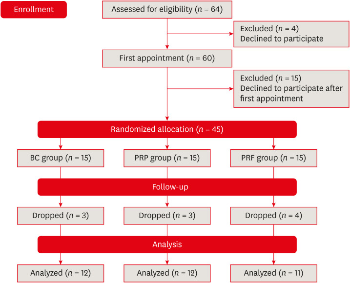
-
Abstract
PDFSupplementary MaterialPubReaderePub
Objectives This study compared the clinical and radiological outcomes of regenerative endodontic procedures (REPs) using blood clots (BCs), platelet-rich plasma (PRP), and platelet-rich fibrin (PRF) through intraoral periapical radiography (IOPAR) and cone-beam computed tomography (CBCT).
Materials and Methods Forty-five single-rooted necrotic teeth with periapical pathology were randomly allocated to receive BC, PRP, or PRF as an individual scaffold. Outcomes were evaluated in 35 teeth in 23 patients with a follow-up period of 12–24 months through qualitative IOPAR scoring and quantitative CBCT measurements. Healing of periapical lesions and in immature teeth, changes in the apical foramen diameter (AFD), root wall thickness (RWT), and root length (RL) were assessed. A
p value less than 0.05 was considered to indicate statistical significance.Results All teeth were asymptomatic except 1 in the PRP group. Periapical lesion healing was seen in all except 2 teeth in the BC group and 3 in the PRP group. Both IOPAR and CBCT revealed no significant differences in bone healing or changes in AFD, RWT, and RL among the 3 groups. A positive pulp sensibility response to the cold test was seen in 2 teeth in the BC group, but none to the electric pulp test. Intracanal calcification (ICC) was evident in more teeth in the BC group than in the PRP and PRF groups, and was also significantly higher in immature teeth.
Conclusions Our results revealed that BC, PRP, and PRF have similar potential as scaffolds in REPs, and ICC may be a concern for long-term outcomes.
-
Citations
Citations to this article as recorded by- Regenerative Endodontics and Stem Cell-Based Therapies – A Systematic Review
Wjoud Ahmed Alshamrani, Sarah Sulaiman Alzarea, Joud Khalid Alabbas, Ayah Khalid Alabbas, Mawiyah Ibrahim Aljaddua, Osama Khattak, Rakhi Issrani
Journal of Pharmacy and Bioallied Sciences.2026; 18(Suppl 1): S29. CrossRef - Collagen Scaffolds in Regenerative Endodontic Procedures: Current Evidence, Limitations, and Future Perspectives
Qiong-Ling Shi, Xiao Zhu, Chen Chen, Jing-Yi Chen, Dan-Yang Lu, Ying Shi, Yan-Qi Chen, Zhi-Fang Wu
Polymers.2026; 18(7): 894. CrossRef - Regenerative potential of concentrated growth factor compared to platelet-rich fibrin in treatment of necrotic mature teeth: a randomized clinical trial
Taghreed Salah, Wael Hussein, Heba Abdelkafy
BDJ Open.2025;[Epub] CrossRef - Clinical and radiographic outcomes of non-surgical retreatment of mature maxillary incisors using two regenerative endodontic techniques in adolescents: a 24-month randomized clinical trial
Ahmad Abdel Hamid Elheeny, Sherif Shafik EL Bahnasy, Yassmin Mohamed ElMakawi, Mohammed Turky, Eman Farouk Ahmed, Norhan Khaled Omar Wahba
BDJ Open.2025;[Epub] CrossRef - Clinical and Radiographic Outcomes of Autologous Platelet-Rich Products in Regenerative Endodontics: A Systematic Review and Meta-Analysis
Raewyn Huang, Wei Chen, Matthew Fang, Ove A. Peters, Sepanta Hosseinpour
Dentistry Journal.2025; 13(6): 236. CrossRef - Regenerative Endodontics in the Treatment of Periapical Lesions of Endodontic Origin: A Systematic Review of Randomized Controlled Trials
BelHaj Salah Kawthar, Berrima Fatma, Boukhris Hanen, Gnaba Imen, Ben Youssef Souha
The Journal of Contemporary Dental Practice.2025; 26(6): 623. CrossRef - Advanced Platelet-Rich Fibrin Plus Sealed Exclusively with Glass Ionomer Cement: Setting a New Standard for Healing, Aesthetics and Predictive Modelling in Regenerative Endodontics
Dubravka Turjanski, Dragutin Lisjak, Petra Bučević Sojčić, Jelena Valpotić, Tea Borojević Renić, Kristina Goršeta, Domagoj Glavina
Materials.2025; 18(18): 4421. CrossRef - Evaluation of different scaffolds and intracanal medications in revascularization of nonvital immature permanent teeth
Mona M. A. Sameia, Abeer M. Darrag, Neveen A. Shaheen, Dina A. Attia
Tanta Dental Journal.2025; 22(4): 693. CrossRef - Biomimetic pulp scaffolds prepared from extracellular matrix derived from stem cells from human exfoliated deciduous teeth promote pulp–dentine complex regeneration
Ning Yang, Rou Shen, Wenxiao Yang, Shengcai Zhang, Tianxing Gong, Yao Liu
International Endodontic Journal.2024; 57(9): 1279. CrossRef - Arrest and Repair of Inflammatory Root Resorption After an Endodontic Regeneration Procedure – A Hypothesis and Case Report
Arieh Y. Kaufman, Bill Kahler
Journal of Endodontics.2024; 50(12): 1743. CrossRef - Effect of Platelet Rich Plasma in Regenerative Endodontic Treatment: A Review of Clinical Trials
Hojat Rezazadeh, Mehrnaz Okhovatfard
Research Journal of Pharmacy and Technology.2023; : 5562. CrossRef
- Regenerative Endodontics and Stem Cell-Based Therapies – A Systematic Review
- 5,892 View
- 149 Download
- 7 Web of Science
- 11 Crossref
Review Article
- Influence of the root canal filling technique on the success rate of primary endodontic treatments: a systematic review
- Daniel Feijolo Marconi, Giovana Siocheta da Silva, Theodoro Weissheimer, Isadora Ames Silva, Gabriel Barcelos Só, Leonardo Thomasi Jahnke, Jovito Adiel Skupien, Marcus Vinicius Reis Só, Ricardo Abreu da Rosa
- Restor Dent Endod 2022;47(4):e40. Published online October 11, 2022
- DOI: https://doi.org/10.5395/rde.2022.47.e40
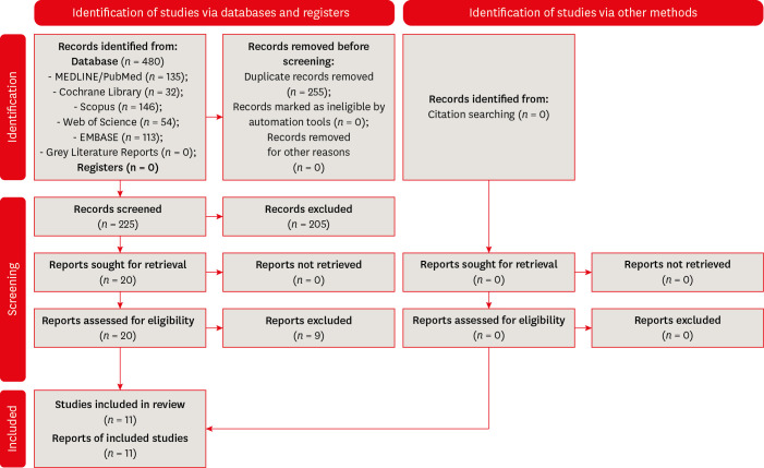
-
Abstract
PDFSupplementary MaterialPubReaderePub
Objectives This study aimed to investigate the influence of different obturation techniques compared to cold lateral compaction on the success rate of primary non-surgical endodontic treatments.
Materials and Methods Systematic searches were performed for studies published up to May 17th, 2022 in MEDLINE/PubMed, Cochrane Library, Web of Science, Scopus, EMBASE, and Grey Literature Reports. Randomized clinical trials and nonrandomized (nonrandomized clinical trials, prospective or retrospective) studies that evaluated the success rate of primary non-surgical endodontic treatments obturated with the cold lateral compaction (control) and other obturation techniques were included. The revised Cochrane risk of bias tools for randomized trials (RoB 2) and nonrandomized studies of interventions (ROBINS-I) were used to evaluate the risk of bias. The Grading of Recommendations Assessment, Development, and Evaluation (GRADE) tool was used to evaluate the certainty of evidence.
Results Eleven studies (4 randomized clinical trials (RCTs), 4 prospective, and 3 retrospectives) were included. Two RCTs were classified as having some concerns risk of bias and 2 as a low risk of bias. Two nonrandomized studies were classified as having a critical risk of bias and 5 as having a moderate risk of bias. The GRADE analysis demonstrated a very low to moderate certainty of evidence.
Conclusions This systematic review generally evidenced no differences in the success rate of primary non-surgical endodontic treatments when the cold lateral compaction technique and other obturation techniques are performed. Further well-designed studies are still necessary.
-
Citations
Citations to this article as recorded by- Assessing Sealing Ability of C-Root SP Strontium Silicate Sealer With Different Obturation Techniques: An in vitro Study
Suixin Hu, Jianshe Li, Meng Xu, Laiqing Xu, Yangming Yin, Peng Xue, Liping Dong, Lin Wang, Huixia He, Ying Liu, Qiang Luo, Fei Chen
International Dental Journal.2026; 76(1): 109283. CrossRef - Effect of root canal filling techniques and materials on endodontic treatment outcomes: a systematic review and meta-analysis
Ahmed Mushtaq, Sura Alsanafi, Firas Elmsmari, José A. González, Marc Garcia-Font, Francesc Abella Sans, Kelvin I. Afrashtehfar, Paul V. Abbott
Scientific Reports.2026;[Epub] CrossRef - Dimensional and cross-sectional compatibility of contemporary nickel–titanium files and their corresponding gutta-percha cones: A micro-computed tomography study
Cem Sarkan, Faruk Haznedaroglu
The Saudi Dental Journal.2026;[Epub] CrossRef - Comparative Analysis Of Obturation Techniques In Endodontics: Lateral Vs. Thermoplasticized. Thermoplasticized
Juan Esteban Díaz Pacheco , Rómulo Guillermo López Torres , Verónica Alejandra Salame Ortíz
Salud, Ciencia y Tecnología.2025; 5: 1626. CrossRef - Effect of ultrasonic activation of endodontic sealers on root canal filling quality during the single-cone obturation procedure: a systematic review and meta-analysis of laboratory-based studies
Shuting Feng, Weiqing Zhou, Xiaojun Chu, Shuaimei Xu, Xiongqun Zeng
Odontology.2025; 113(4): 1380. CrossRef - In Vitro and In Vivo Evaluation of a New Experimental Polydimethylsiloxane-Based Endodontic Sealer
Fabiola Cardoso Maldonado, Cesar Gaitan Fonseca, Carlos Bermudez Jimenez, Luis Alejandro Aguilera Galaviz, Margarita L. Martinez-Fierro, Lorena Troncoso Vazquez, Martha Eugenia Reyes Ortiz
Journal of Functional Biomaterials.2025; 16(11): 402. CrossRef - Evaluation of three obturation techniques in 3D-printed models of oval canals with standardized prepared morphology: a micro-CT study
Wenjun Xia, Qisheng Gu, Yingshuang Song, Yunjia Liu, Xuetao Deng, Wenhao Qian
BMC Oral Health.2025;[Epub] CrossRef - Clinical and Radiographic Failure of Nonsurgical Endodontic Treatment and Retreatment Using Single-cone Technique With Calcium Silicate-based Sealers: A Systematic Review and Meta-analysis
Mohammad A. Sabeti, Negah Karimpourtalebi, Arash Shahravan, Omid Dianat
Journal of Endodontics.2024; 50(6): 735. CrossRef - Method of microbial decontamination of endodontic absorbent paper points: a randomised experimental study
O. A. Pavlovskaya, O. A. Kachanova, V. V. Volobuev, M. N. Mitropanova, A. R. Gazarova, V. Y. Zobenko, A. G. Uvarova
Pediatric dentistry and dental prophylaxis.2024; 24(2): 157. CrossRef - The Push-Out Bond Strength, Surface Roughness, and Antimicrobial Properties of Endodontic Bioceramic Sealers Supplemented with Silver Nanoparticles
Karla Navarrete-Olvera, Nereyda Niño-Martínez, Idania De Alba-Montero, Nuria Patiño-Marín, Facundo Ruiz, Horacio Bach, Gabriel-Alejandro Martínez-Castañón
Molecules.2024; 29(18): 4422. CrossRef - Clinical outcome of non-surgical root canal treatment using different sealers and techniques of obturation in 237 patients: A retrospective study
Mateusz Radwanski, Krystyna Pietrzycka, Tan Fırat Eyüboğlu, Mutlu Özcan, Monika Lukomska-Szymanska
Clinical Oral Investigations.2024;[Epub] CrossRef
- Assessing Sealing Ability of C-Root SP Strontium Silicate Sealer With Different Obturation Techniques: An in vitro Study
- 8,279 View
- 128 Download
- 10 Web of Science
- 11 Crossref
Case Report
- Endodontic micro-resurgery and guided tissue regeneration of a periapical cyst associated to recurrent root perforation: a case report
- Fernando Córdova-Malca, Hernán Coaguila-Llerena, Lucía Garré-Arnillas, Jorge Rayo-Iparraguirre, Gisele Faria
- Restor Dent Endod 2022;47(4):e35. Published online September 3, 2022
- DOI: https://doi.org/10.5395/rde.2022.47.e35
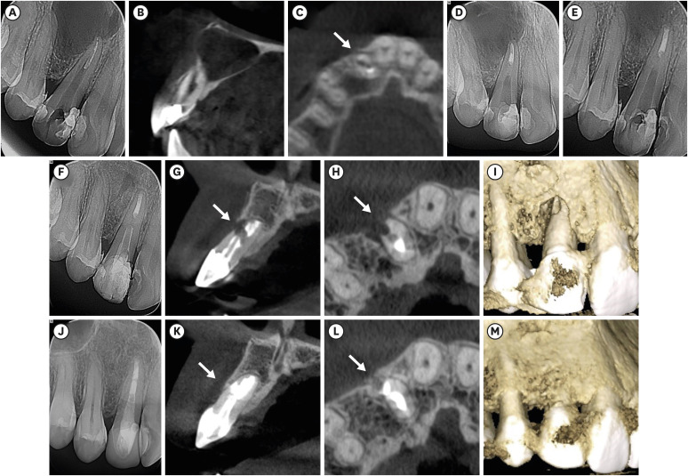
-
Abstract
PDFPubReaderePub
Although the success rates of microsurgery and micro-resurgery are very high, the influence of a recurrent perforation combined with radicular cyst remains unclear. A 21-year-old white female patient had a history of root perforation in a previously treated right maxillary lateral incisor. Analysis using cone-beam computed tomography (CBCT) revealed an extensive and well-defined periapical radiolucency, involving the buccal and palatal bone plate. The perforation was sealed with bioceramic material (Biodentine) in the pre-surgical phase. In the surgical phase, guided tissue regeneration (GTR) was performed by combining xenograft (lyophilized bovine bone) and autologous platelet-rich fibrin applied to the bone defect. The root-end preparation was done using an ultrasonic tip. The retrograde filling was performed using a bioceramic material (Biodentine). Histopathological analysis confirmed a radicular cyst. The patient returned to her referring practitioner to continue the restorative procedures. CBCT analysis after 1-year recall revealed another perforation in the same place as the first intervention, ultimately treated by micro-resurgery using the same protocol with GTR, and a bioceramic material (MTA Angelus). The 2-year recall showed healing and bone neoformation. In conclusion, endodontic micro-resurgery with GTR showed long-term favorable results when a radicular cyst and a recurrent perforation compromised the success.
-
Citations
Citations to this article as recorded by- Outcome of endodontic micro-resurgery: A systematic review
Faisal Alnassar, Riyadh Alroomy, Qamar Hashem, Abdullah Alqedairi, Nabeel Almotairy
Saudi Endodontic Journal.2025; 15(2): 112. CrossRef - Platelet-Rich Plasma and Platelet-Rich Fibrin in Endodontics: A Scoping Review
Simão Rebimbas Guerreiro, Carlos Miguel Marto, Anabela Paula, Joana Rita de Azevedo Pereira, Eunice Carrilho, Manuel Marques-Ferreira, Siri Vicente Paulo
International Journal of Molecular Sciences.2025; 26(12): 5479. CrossRef - Non-surgical Approach to a Maxillary Cyst-Like Lesion: Orthograde Endodontic Treatment With Neodymium-Doped Yttrium Aluminum Garnet (Nd:YAG) Decontamination of the Canal System
Beatrice Spaggiari, Paolo Vescovi, Silvia Pizzi, Roberta Iaria, Ilaria Giovannacci
Cureus.2025;[Epub] CrossRef - Persistent Periradicular Lesion Associated With Concurrent Root Fracture and Odontogenic Keratocyst: A Case Report
Mehdi Vatanpour, Fatemeh Rezaei
Clinical Case Reports.2025;[Epub] CrossRef - Management of Apico-marginal Defects With Endodontic Microsurgery and Guided Tissue Regeneration: A Report of Thirteen Cases
Abayomi O. Baruwa, Jorge N.R. Martins, Mariana D. Pires, Beatriz Pereira, Pedro May Cruz, António Ginjeira
Journal of Endodontics.2023; 49(9): 1207. CrossRef
- Outcome of endodontic micro-resurgery: A systematic review
- 3,831 View
- 71 Download
- 4 Web of Science
- 5 Crossref
Research Articles
- Effect of intracanal cryotherapy on postoperative pain after endodontic treatment: systematic review with meta-analysis
- Fernanda Garcias Hespanhol, Ludmila Silva Guimarães, Lívia Azeredo Alves Antunes, Leonardo Santos Antunes
- Restor Dent Endod 2022;47(3):e30. Published online July 4, 2022
- DOI: https://doi.org/10.5395/rde.2022.47.e30
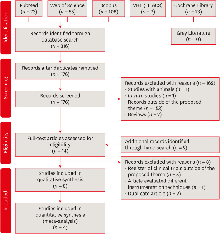
-
Abstract
PDFPubReaderePub
Objectives This study aimed to evaluate the effectiveness of final irrigation with cold saline solution after endodontic treatment compared with saline solution at room temperature against postoperative pain following endodontic treatment.
Materials and Methods A broad search was performed in the PubMed, Web of Science, Scopus, Cochrane Library, Virtual Health Library (LILACS), and Grey Literature databases. Two independent reviewers performed data extraction, risk of bias using the Cochrane methodology, and certainty of evidence using the Grading of Recommendations, Assessment, Development and Evaluations (GRADE) approach.
Results Eight studies were included in qualitative synthesis. Intracanal cryotherapy favored the reduction of postoperative pain in the systematic review. Four studies were included in meta-analyses. The meta-analysis showed that intracanal cryotherapy reduced postoperative pain in teeth with symptomatic apical periodontitis (SAP) at 24 hours. There was no association between intracanal cryotherapy and control (room temperature) groups in teeth with normal periapical tissue with respect to postoperative pain at 24 hours and 48 hours.
Conclusions Intracanal cryotherapy was effective in reducing postoperative pain after endodontic treatment in teeth with SAP.
-
Citations
Citations to this article as recorded by- Postoperative Pain After Endodontic Treatment in HIV‐Positive Patients Under HAART: A Prospective Observational Cohort Study
Marcos Felipe Iparraguirre Nuñovero, Marco Antonio Hungaro Duarte, Luciana Reis Azevedo Alanis, Bruno Cavalini Cavenago, Ulisses Xavier da Silva Neto, Everdan Carneiro
International Endodontic Journal.2026; 59(5): 788. CrossRef - Effect of low-temperature intracanal sodium hypochlorite on root surface temperature reduction and organic tissue dissolution: an in vitro study
Marcos Felipe Iparraguirre Nuñovero, Marco Antonio Hungaro Duarte, Ulisses Xavier da Silva Neto, Vânia Portela Ditzel Westphalen, Pedro Cesar Gomes Titato, Bruno Cavalini Cavenago, Everdan Carneiro
Scientific Reports.2026;[Epub] CrossRef - Effectiveness of intracanal cryotherapy in reducing post-endodontic pain in irreversible pulpitis: a systematic review and meta-analysis
Maged Mohamed, Asmaa Abdelmajeed, Muhammad Salah-Uddin Anwar Laithy, Dina Abozaid
Scientific Reports.2026;[Epub] CrossRef - Impact of intracanal cryotherapy on postoperative pain in symptomatic apical periodontitis: A systematic review and meta-analysis of randomized clinical trials
Nishtha K. Patel, Prerak Doshi, Shaily R. Dalal, Pooja R. Kesharani, Shilpa S. Shah, Mohil H. Kale
Endodontology.2025; 37(2): 101. CrossRef - Evaluation of Post‐Endodontic Pain Reduction Using Intracanal Cryotherapy in Symptomatic Apical Periodontitis
Anam Fayyaz Bashir, Ussamah Waheed Jatala, Muhammad Amber Fareed, Sheryar Sheryar, Saadia Ahmad Chattha, Saima Razaq Khan, Shahzad Ahmad, Shazia Iqbal, Muhammad Sohail Zafar, Shahzad Ali
Australian Endodontic Journal.2025; 51(3): 677. CrossRef - Comparing cryotherapy and ketorolac tromethamine against room-temperature saline irrigation using interleukin-8 levels and post-operative pain within single-visit endodontic treatment of symptomatic irreversible pulpitis superimposed by apical periodontit
Yousra Khaled Ezzat, Alaa Diab, Olfat Shaker, Sarah Abouelenien
BMC Oral Health.2025;[Epub] CrossRef - Determining Efficacy of Intracanal Cryotherapy on Post Endodontic Pain in Irreversible Pulpitis
Anam Fayyaz Bashir, Ussamah Waheed Jatala, Moeen ud din Ahmad, Muhammad Talha Khan, Saima Razzaq Khan, Aisha Arshad Butt
Pakistan Journal of Health Sciences.2024; : 68. CrossRef - The effect of intracanal cryotherapy with and without foraminal enlargement on pain prevention after endodontic treatment: a randomized clinical trial
Marcos Felipe Iparraguirre Nuñovero, Marco Antonio Hungaro Duarte, André Vinícius Kaled Segato, Ulisses Xavier da Silva Neto, Vania Portela Ditzel Westphalen, Everdan Carneiro
Scientific Reports.2024;[Epub] CrossRef - Effect of cryotherapy duration on experimentally induced connective tissue inflammationin vivo
Jorge Vera, Mayra Alejandra Castro-Nuñez, María Fernanda Troncoso-Cibrian, Ana Gabriela Carrillo-Varguez, Edgar Ramiro Méndez Sánchez, Viviana Sarmiento, Lourdes Lanzagorta-Rebollo, Prasanna Neelakantan, Monica Romero, Ana Arias
Restorative Dentistry & Endodontics.2023;[Epub] CrossRef - Evaluation of knowledge and awareness of pediatric oral health among school teachers of Hazaribag before and after oral health education.
Vipin Ahuja, Annapurna Ahuja, Nilima Thosar
F1000Research.2023; 12: 1292. CrossRef
- Postoperative Pain After Endodontic Treatment in HIV‐Positive Patients Under HAART: A Prospective Observational Cohort Study
- 4,411 View
- 98 Download
- 9 Web of Science
- 10 Crossref
- Clinical and radiographic outcomes of regenerative endodontic treatment performed by endodontic postgraduate students: a retrospective study
- Hadi Rajeh Alfahadi, Saad Al-Nazhan, Fawaz Hamad Alkazman, Nassr Al-Maflehi, Nada Al-Nazhan
- Restor Dent Endod 2022;47(2):e24. Published online May 9, 2022
- DOI: https://doi.org/10.5395/rde.2022.47.e24
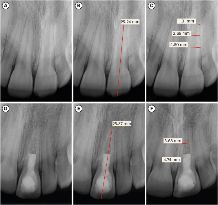
-
Abstract
PDFPubReaderePub
Objectives Regenerative endodontic treatment is a clinical procedure aimed at biologically regenerating damaged root canal tissue of immature permanent teeth. This study aimed to report the outcomes of regenerative endodontic treatment performed by endodontic postgraduate students.
Materials and Methods Clinical and radiographic data of 27 patients, aged 10–22 years, who underwent regenerative treatment of immature permanent teeth from 2015 to 2019 were followed up, wherein clinical and radiographic examinations were performed for each patient. Postoperative success rate and tooth survival were analyzed, and the postoperative radiographic root area changes were quantified.
Results A total of 23 patients attended the dental appointments, showing that all teeth survived and were asymptomatic. Specifically, 7 periapical pathosis cases were completely healed, 12 were incompletely healed, and 4 cases failed. Moreover, significant differences were found between discolored and non-discolored teeth, and between the presence or absence of periapical radiolucency. Additionally, 3 anterior teeth showed complete closure of the apical foramen, while the apical foramen width was reduced in 17 teeth and failed in 3 teeth. Root length was also found to have been increased in 7 anterior and 4 posterior teeth, and the average length ranged from 4.00–0.63 mm in the anterior teeth, 2.85–1.48 mm of the mesial root, and 2.73–2.16 mm of the molar teeth distal root. Furthermore, calcified tissue deposition was observed in 7 teeth.
Conclusions A favorable outcome of regenerative endodontic treatment of immature permanent teeth with necrotic pulp was achieved with a high survival rate.
-
Citations
Citations to this article as recorded by- Regenerative Endodontics and Stem Cell-Based Therapies – A Systematic Review
Wjoud Ahmed Alshamrani, Sarah Sulaiman Alzarea, Joud Khalid Alabbas, Ayah Khalid Alabbas, Mawiyah Ibrahim Aljaddua, Osama Khattak, Rakhi Issrani
Journal of Pharmacy and Bioallied Sciences.2026; 18(Suppl 1): S29. CrossRef - Pre‐Operative Factors on Prognosis of Regenerative Endodontic Procedures: A Systematic Review and Meta‐Analysis
Filipe Colombo Vitali, Alexandre Henrique dos Reis‐Prado, Pablo Silveira Santos, Ana Paula Portes Zeno, Patrícia de Andrade de Risso, Lucianne Cople Maia, Francine Benetti, Cleonice da Silveira da Teixeira
International Endodontic Journal.2025; 58(12): 1814. CrossRef - Clinical, radiographic, and biomarker perspectives of low-level laser therapy during regenerative endodontic procedures in necrotic immature young teeth: a randomized clinical study
Pragya Pandey, Neha Jasrasaria, Ramesh Bharti, Rakesh Kumar Yadav, Monika Kumari, Abinia Vaishnavi, Rahul Pandey
Lasers in Medical Science.2025;[Epub] CrossRef - Allogeneic Bone Marrow Mesenchymal Stromal Cell Transplantation Induces Dentin Pulp Complex-like Formation in Immature Teeth with Pulp Necrosis and Apical Periodontitis
Jose Francisco Gomez-Sosa, José E. Cardier, Olga Wittig, Dylana Díaz-Solano, Eloisa Lara, Kharelys Duque, Giselle Ramos-González
Journal of Endodontics.2024; 50(4): 483. CrossRef - Radiographic assessment of dental post and core placement at different educational levels in an undergraduate student clinic: a 4-year retrospective study
Turki Alshehri, Nourhan M. Aly, Raand Altayyar, Deena Alghamdi, Shahad Alotaibi, Passent Ellakany
F1000Research.2024; 12: 976. CrossRef - Evaluation of the efficacy of injectable platelet‐rich fibrin versus platelet‐rich plasma in the regeneration of traumatized necrotic immature maxillary anterior teeth: A randomized clinical trial
Maha Mohamed Abo‐Heikal, Jealan M. El‐Shafei, Samia A. Shouman, Nehal N. Roshdy
Dental Traumatology.2024; 40(1): 61. CrossRef - Radiographical assessment of post and core placement errors encountered by Saudi dental students at different educational levels
Turki Alshehri, Nourhan M. Aly, Raand Altayyar, Deena Alghamdi, Shahad Alotaibi, Passent Ellakany
F1000Research.2023; 12: 976. CrossRef
- Regenerative Endodontics and Stem Cell-Based Therapies – A Systematic Review
- 5,211 View
- 100 Download
- 7 Web of Science
- 7 Crossref
 First
First Prev
Prev

